REVIEW
Erosion
—diagnosis and risk factors
A. Lussi&T. Jaeggi
Received: 9 July 2007 / Accepted: 18 December 2007 / Published online: 29 January 2008
# Springer-Verlag 2007
Abstract Dental erosion is a multifactorial condition: The interplay of chemical, biological and behavioural factors is crucial and helps explain why some individuals exhibit more erosion than others. The erosive potential of erosive agents like acidic drinks or foodstuffs depends on chemical factors, e.g. pH, titratable acidity, mineral content, clearance on tooth surface and on its calcium-chelation properties. Biological factors such as saliva, acquired pellicle, tooth structure and positioning in relation to soft tissues and tongue are related to the pathogenesis of dental erosion. Furthermore, behavioural factors like eating and drinking habits, regular exercise with dehydration and decrease of salivary flow, excessive oral hygiene and, on the other side, an unhealthy lifestyle, e.g. chronic alcoholism, are predis-posing factors for dental erosion. There is some evidence that dental erosion is growing steadily. To prevent further progression, it is important to detect this condition as early as possible. Dentists have to know the clinical appearance and possible signs of progression of erosive lesions and their causes such that adequate preventive and, if necessary, therapeutic measures can be initiated. The clinical exami-nation has to be done systematically, and a comprehensive case history should be undertaken such that all risk factors will be revealed.
Keywords Erosion . Clinical appearance . Chemical . Biological and behavioural risk factors
Introduction
Erosion has, for many years, been a condition of little interest to clinicians and researchers. This has changed during the last years, and there is some evidence that the presence of dental erosion is growing steadily. In the UK, the prevalence of erosion was shown to have increased from the time of the children’s dental health survey in year 1993 compared to 1996/1997 [82]. In another UK study, the progression of erosion was investigated: 1,308 children were examined at the age of 12 years and 2 years later. In this study, 4.9% of the subjects at baseline and 13.1% 2 years later had deep-enamel or dentine lesions. Approx-imately 12% of erosion-free children at 12 years developed the condition over the subsequent 2 years. New or more advanced lesions were seen in 27% of the children over the study period [20]. The progression of erosion seems to be greater in older adults (52–56 years) compared to younger (32–36 years) and has a skewed distribution [64]. In this study, the group with high-erosion progression was found to have four or more dietary acid intakes per day, a low buffering capacity of stimulated saliva and used a hard-bristle toothbrush. Intake frequency of the same magnitude was also associated with an increased risk for erosion in children. In this investigation, the erosion group ate fruits significantly more frequently and had different drinking habits, such as swishing, sucking or holding drinks in their mouths [83].
Dental erosion is a multifactorial condition. To prevent further progression, it is important to detect this condition as early as possible. It is fundamental to diagnose the possible risk factors such that preventive measures can be initiated. This overview is aimed to give some basic aspects about the diagnosis and the risk factors of erosion. A more detailed description can be found elsewhere [67].
We declare that we have no conflict of interest. A. Lussi (*)
:
T. JaeggiDepartment of Preventive, Restorative and Pediatric Dentistry, School of Dental Medicine, University of Bern,
Freiburgstrasse 7,
CH-3010 Bern, Switzerland e-mail: [email protected]
Diagnosis
Diagnosis of early forms of erosion is difficult, as it is accompanied by few signs and fewer if any symptoms. There is no device available in routine dental practice for the specific detection of dental erosion and its progression. Therefore, clinical appearance is the most important feature for dental professionals to diagnose this condition. This is of particular importance in the early stage of dental erosion [68]. At a more advanced stage, it can be very difficult to determine if dentine is exposed or not [31]. It is possible to use disclosing agents to render dentine involvement visible. The appearance of smooth silky-glazed, sometimes dull, enamel surface with the absence of perikymata and intact enamel along the gingival margin are some typical signs of enamel erosion on facial and oral sites. It has been hypothesized that the preserved enamel band along the oral and facial gingival margin could be due to some plaque remnants, which could act as a diffusion barrier for acids. This phenomenon could also be due to an acid-neutralizing effect of the sulcular fluid [66]. In the more advanced stages, further changes in the morphology can be found. These changes result in developing a concavity in enamel, the width of which clearly exceeds its depth. Facial erosion should be distinguished from wedge-shaped defects which are located at or apical to the enamel–cementum junction. The coronal part of wedge-shaped defects ideally has a sharp margin and cuts at right angle into the enamel surface, whereas the apical part bottoms out to the root surface. Thereby, the depth of the defect exceeds its width. The initial features of erosion on occlusal and incisal surfaces are the same as described above. Further progres-sion of occlusal eroprogres-sion leads to a rounding of the cusps and restorations rising above the level of the adjacent tooth surfaces. In severe cases, the whole occlusal morphology disappears. Erosive lesions have to be distinguished from attrition and abrasion. The latter are often flat, have glossy areas with distinct margins and corresponding features at the antagonistic teeth. It is sometimes challenging to distinguish between the influences of erosion, attrition or abrasion during a clinical examination. Indeed, they may occur simultaneously with sometimes similar shape. The most commonly reported areas with this condition are occlusal surfaces [6]. Figures 1, 2,3, and 4 show typical pattern of dental erosion process.
The clinical examination should be done systematically using a simple but accurate index. This is a difficult task to achieve as an index with a too-fine grading shows a small inter- and intraexaminer reliability [60]. For a dental practitioner, the most important part is to recognize the condition and to describe its dimension and severity. It is important to search for a general pattern and not to over-interpret one single sign. For epidemiological purpose, an
index with high detection capability and reliability is most important. Whenever possible, the clinical examination should be accomplished by a thorough history taking with respect to general health, diet and habits and by the assessment of saliva flow rates and buffer capacity. People who show signs and symptoms of erosion are often not aware of this condition. Only when a comprehensive case history is undertaken will all the risk factors be revealed.
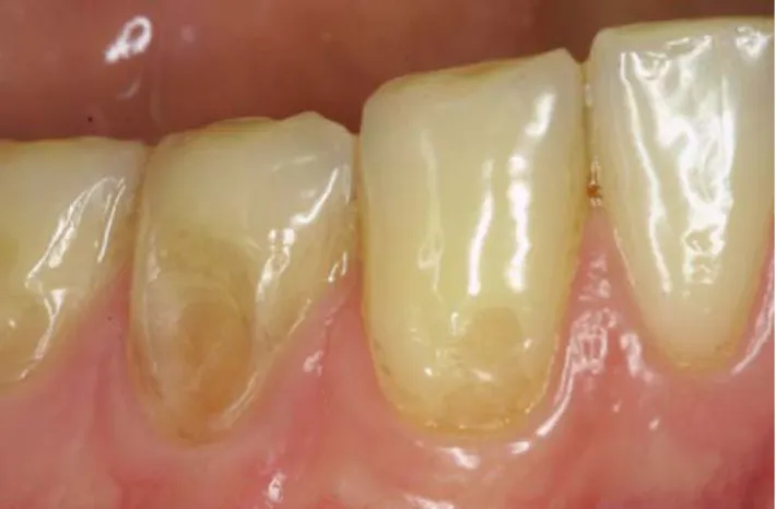
It is difficult to judge the activity and progression of dental erosion. One tool is the comparison of clinical photographs of tooth surfaces to estimate the possible substance loss over time. Thereby, the discoloration of the lesions and their sensitivity state may give some information about the activity of the tooth surface. Further, study casts as well as the ex-amination of dental radiography, especially bitewings longi-tudinally taken, can provide information about the substance loss over time. For research purpose, computed controlled mapping [13] or profilometric measurements using acid-resistant markers [6,92] are tools to monitor progression. Fig. 1 Facial erosion: The intact enamel border along the gingival margin of tooth 12 and some plaque remnants are clearly visible. Note the smooth silky-glazed appearance and the absence of perikymata on the enamel surface
Fig. 2 Advanced facial erosion of teeth 43, 44 and 45 with dentinal involvement. The width of the lesions exceeds its depth
Risk factors
When an acidic solution comes in contact with enamel, it has to diffuse first through the acquired pellicle, and only thereafter can it interact with enamel. The acquired pellicle is an organic film, free of bacteria, covering oral hard and soft tissues. It is composed of mucins, glycoproteins and proteins, including several enzymes [41]. On the surface of enamel, the hydrogen ion component of the acid will start to dissolve the enamel crystal. First, the prism sheath area and then the prism core are dissolved, leaving the well-known honey comb appearance [74]. Thereafter, fresh, unionized acid will eventually diffuse into the interprismatic areas of enamel and dissolve further mineral in the region underneath the surface [23,27,65]. This will lead to an outflow of ions (dissolution) and subsequently to a local pH rise in the tooth substance immediately below and in the liquid surface layer adjacent to the enamel surface [65]. The events in dentine are, in principle, the same but are even more complex.
There are different predisposing factors and aetiologies of the erosive condition. The interplay of chemical, biological and behavioural factors is crucial and helps explain why some individuals exhibit more erosion than others, even if they are exposed to the same acid challenge
in their diets. Comprehensive knowledge of the different risk factors is a prerequisite to initiate adequate preventive (non-interventive) and, if necessary, therapeutic (interven-tive) measures. When a restoration becomes inevitable, in all situations, the preparations have to follow the principles of minimally invasive treatment.
Figure 5 is an attempt to reveal the multifactorial pre-disposing factors and aetiologies of the erosive condition.
Chemical factors
Several in vitro and in situ studies show that the erosive potential of an acidic drink or foodstuff is not exclusively dependent on its pH value but is also strongly influenced by its mineral content, its titratable acidity (‘the buffering capacity’) and by the calcium-chelation properties [7,8,37,
44, 61, 72, 73, 76, 77, 80, 86, 88, 94, 95, 96]. The pH value, calcium, phosphate and fluoride content of a drink or foodstuff determine the degree of saturation with respect to the tooth mineral, which is the driving force for dissolution. Solutions oversaturated with respect to dental hard tissue will not dissolve it. A low degree of undersaturation with respect to enamel or dentine leads to a very initial surface demineralization which is followed by a local rise in pH and increased mineral content in the liquid surface layer adjacent to the tooth surface. This layer will then become saturated with respect to enamel (or dentine) and will not demineralize further.
Acids such as citric acid exist in water as a mixture of hydrogen ions, acid anions (e.g. citrate) and undissociated acid molecules, with the amounts of each determined by the acid-dissociation constant and the pH of the solution. The hydrogen ion directly attacks the crystal surface. Over and above the effect of the hydrogen ion, the citrate anion may complex with calcium, also removing it from the crystal surface. Each acid anion has a different strength of calcium complexation dependent on the structure of the molecule and how easily it can attract the calcium ion [28]. Consequently, acids such as citric acids have double actions
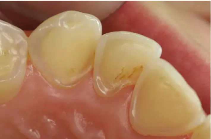
Fig. 4 a–c Typical pattern of advanced occlusal erosion of teeth 45 and 46 of three different patients: The whole occlusal morphology disappears, and extensive exposed dentinal areas are visible
Fig. 3 Erosion with involvement of dentine on the oral surface of tooth 13, 12 and 11. Intact enamel borders along the gingival margin
and may be very damaging to the tooth surface. Up to 32% of the calcium in saliva can be complexed by citrate at concentrations common in fruit juices, thus reducing the super-saturation of saliva and increasing the driving force for dissolution with respect to tooth minerals [75].
The dissolution with water of drinks containing organic acids with high buffering capacity will hardly reduce the pH but will reduce the titratable acidity. This is of some importance, as the greater the buffering capacity of the drink, the longer it will take for saliva to neutralize the acid. But dilution will also reduce concentrations of Ca and P (if present), which have a protective effect [10,61,62].
The calcium and phosphate content of a foodstuff or beverage are important factors for the erosive potential as they influence the concentration gradient within the local environ-ment of the tooth surface. The addition of calcium (and phosphate) salts to erosive drinks showed promising results. Addition of calcium to a low-pH blackcurrant juice drink has been shown to reduce the erosive effect of the drink [48]. In a follow-up study, a blackcurrant drink with added calcium was compared to a conventional orange drink in situ. Servings of 250 ml of each drink were consumed four times per day during 20 working days. Measurements of enamel loss were made by profilometry on enamel samples for up to 20 days. The experimental carbonated blackcurrant drink supplemented with calcium caused significantly less enamel loss than the conventional carbonated orange drink at all time points measured [98]. When Ca was added to a sports drink, a reduction of the erosive potential was found [46].
Today, several Ca-enriched orange juices and sports drinks are on the market which hardly soften the enamel surface. Yoghurt is another example of food with a low pH (~4.0), yet it has hardly any erosive effect due to its high calcium and phosphate content, which makes it supersaturated with respect to apatite. A yoghurt or another milk-based food may have an erosive potential when it has a low content of Ca and/or P and a low pH. It has to be kept in mind that, with the added mineral, enamel dissolution could not always be completely prevented. But the progression can be retarded which has some implications for the patient and the clinician. Theoretically, fluoride has some protective effect in a drink with a pH higher than that indicated by the saturation curve of fluorapatite at given Ca and P concentrations. Lussi et al. [61,62] and Mahoney et al. [71] found an inverse correlation of the erosive potential of different beverages with their fluoride content. It is unlikely that fluoride at the concentra-tion present in beverages alone has any great beneficial effect on erosion because the challenge is high. However, it is possible that, under conditions in which the other erosive factors are not excessive, fluoride in solution may exert some protective effect [71]. It appears that topical fluoride application can positively affect the tooth-wear process. The influence of immersion in fluoride solutions on brushing abrasion of eroded dentine slabs was investigated by Attin et al. [3]. Bovine dentine specimens were alternately stored in a demineralising and in a remineralising solution and brushed five times. Before each immersion in the reminer-alising solution, the specimens were not treated, treated with 250 ppm or treated with 2,000 ppm neutral sodium fluoride solution for 1 min. The least wear was found in the uneroded controls, whereas the greatest wear was found in the group without fluoride treatment. Significantly greater wear resis-tance was found as fluoride concentration increased. The same group evaluated the abrasion resistance of eroded enamel brushed with an acidified fluoride gel [4]. They found that treatment with a slightly acidic amine/sodium fluoride gel increased the abrasion resistance of bovine enamel compared to an unfluoridated gel. They speculated that the fluoride is incorporated into and deposited on the enamel during treatment with the acidic gel, which could be one reason for the higher resistance.
The adhesiveness and displacement of the liquid are other factors to be considered in the erosive process. There appear to be differences in the ability of beverages to adhere to enamel based on their thermodynamic properties, e.g. the thermodynamic work of adhesion [50].
In summary, the two very often-cited parameters, the pH and the titratable acidity, do not readily explain the erosive potential of food and drink. The mineral content is also an important parameter, as is the ability of any of the components to complex calcium and to remove it from the mineral surface.
Fig. 5 Interactions of the different factors for the development of dental erosion (adapted from [68])
Biological factors
Biological factors such as saliva, acquired pellicle, tooth structure and positioning in relation to soft tissues and tongue are related to dental erosion development.
A very important biological parameter is saliva. Several salivary protective mechanisms come into play during an erosive challenge: dilution and clearance of an erosive agent from the mouth, neutralisation and buffering of acids, and slowing down the rate of enamel dissolution through the common ion effect by salivary calcium and phosphate [104]. Practical experience demonstrates the importance of saliva in patients suffering from salivary-flow impairment. Studies have shown that erosion may be associated with low salivary flow or/and low buffering capacity [53, 64,89]. The dry mouth condition is usually related to aging [18, 81, 84], even though some other studies have not found this correlation [9, 43]. It is well established that patients taking medication can also present decreased saliva output [101], as well as those who have received radiation therapy for neck and head cancer [19]. Tests of the stimulated and unstimulated flow rate as well as of the buffer capacity of saliva may provide some information about the susceptibility of an individual to dental erosion. However, it has to be kept in mind that these parameters are two of a multifactorial condition. Sialomet-ric evaluations should be carried at a fixed time point or in a limited time interval in the morning, avoiding intra-individual variations due to the circadian cycle [29]. Studies have shown that sour foodstuff has a strong influence on the anticipatory salivary flow [15, 55], which can be significantly increased when compared to the normal unstimulated flow rate [25]. Hypersalivation also occurs in advance of vomiting as a response from the‘vomiting center’ of the brain [56], as frequently seen in individuals suffering from anorexia and bulimia nervosa, rumination or chronic alcoholism. It is suggested that this could minimize the erosion caused by acids of gastric origin. On the other hand, patients with symptoms of gastro-esophageal reflux disease (GERD) should not expect the salivary output to increase before the gastric juice regurgitation because this is an involuntary response not co-coordinated by the auto-nomic nervous system [42, 90]. Therefore, there may be insufficient time for saliva to act before erosion occurs. The influence of saliva on the remineralisation/rehardening of erosive damaged dental hard tissue is discussed controver-sially. There is evidence that acid-softened enamel can reharden after exposure to saliva or remineralisation solution and that dietary products and fluoride can enhance the rehardening process [2, 26, 33]. Other investigations could not find a significant rehardening effect of saliva in situ [16,32,34,58]. Some limited increase of the abrasion resistance of softened enamel was found after intraoral
exposure to saliva [5, 51]. It seems that in vitro, some rehardening could be expected when supersaturated solution or saliva with no protein added are used [23,
24], whereas in situ, this is only the case to a very small amount.
The salivary acquired pellicle is a protein-based layer which is rapidly formed on dental surfaces after its removal by tooth brushing with dentifrice, other prophylaxis, measures or chemical dissolution. This organic layer becomes detectable on dental surfaces after few minutes of exposure to the oral environment [39, 93]. Enzymatic activity is also detectable at early stages of pellicle formation [40, 41]. It is suggested that it grows until reaching an equilibrium between protein adsorption and de-sorption within 2 h [57].
The acquired pellicle may protect against erosion by acting as a diffusion barrier or a perm-selective membrane preventing the direct contact between the acids and the tooth surface [39,102,103], reducing the dissolution rate of hydroxyapatite [57].
Millward et al. [78] monitored the pH at the surface of teeth of healthy volunteers after drinking 1% citric acid. They observed that the pH recovered to above pH 5.5 within 2 min from a site adjacent to the palatal surface of the upper central incisor and within 4–5 min from another palatal surface of the upper first molar. Other observations have revealed a longer clearance time on upper incisors for patients with active erosions and normal saliva values compared to patients with no erosion (Lussi, unpublished data). These differences could be due to the anatomy of the teeth and soft tissues that may influence the retention/ clearance pattern of erosive agents. Furthermore, soft tissue movements by the tongue and buccal mucosa and swallow-ing pattern can influence clearance rate. The importance of the tongue in modifying the tooth-wear process has long been the subject of speculation. Holst and Lange [45] considered mechanical abrasion caused by tongue to be a contributing factor in erosion caused by vomiting. Obser-vations from animal studies also provide support in that beverages produced erosion mainly on the lingual surfaces of rat molar teeth in areas where the tongue contacts the teeth [96]. Dugmore and Rock [21] found that orthodontic anomaly appeared to confer a protective effect.
Behavioural factors
During and after an erosive challenge, behavioural factors play a role in modifying the extent of tooth wear. The manner that dietary acids are introduced into the mouth will affect which teeth are contacted by the erosive challenge and possibly the clearance pattern. As lifestyles have changed through the decades, the total amount and
frequency of consumption of acidic foods and drinks have also changed. Soft drink consumption in the USA increased by 300% in 20 years [11], and serving sizes increased from 185 g (6.6 oz) in the 1950s to 340 g (12 oz) in the 1960s and to 570 g (20 oz) in the late 1990s. Around the year 1995, between 56 and 85% of children at school in the USA consumed at least one soft drink daily, with the highest amounts ingested by adolescent males. Of this group, 20% consumed four or more servings daily [36]. Studies in children and adults have shown that this number of servings per day is associated with the presence and the progression of erosion when other risk factors exist [64,83].
High erosion was associated with a method of drinking whereby the drink was kept in the mouth for a longer period [54]. One study investigated a randomly selected group of Swiss adults and diagnosed the cause of tooth wear. It showed that the consumption of erosive drinks and foodstuffs was strongly associated with erosion on facial and occlusal surfaces. Severe palatal erosions were scarce and highly associated with chronic vomiting [60]. The presence of calculus (odds ratio 0.48) or eating fruit other than apples or citrus fruit (0.48) reduced the chances of erosion. High consumption of carbonated drinks increased the odds of erosion being present at 12 years by 252% and was a strong predictor of the amount of erosion found at age 14 [21]. In a sample of 987 children (2 to 5 year-olds), consumption of vitamin C supplements, carbonated drinks and fruit syrup from a feeding bottle at bed- or naptime were related to erosion [1]. Considerable risk of erosion was found when citrus fruits were eaten more than twice a day and soft drinks were drunk daily [53].
Excessive consumption of acidic candies combined with a low salivary buffering capacity may aggravate erosive lesions [17, 63]. The high intake of herbal teas, widely perceived as a healthy drink, may have an erosive potential exceeding that of orange juice [85]. An increase in agitation (e.g. when a patient is swishing his/her drink in the mouth) will enhance the dissolution process because the solution on the surface layer adjacent to tooth mineral will be readily renewed. Further, the amount of the drink in the mouth in relation to the amount of saliva present will modify the dissolution process [69]. Several authors have suggested that using a straw is beneficial, since the straw directs drinks past the anterior teeth and towards the pharynx [22,
38, 49]. However, the placing of a straw labial to the anterior teeth can be destructive [70]. Nighttime exposure to erosive agents may be particularly destructive because of the absence of salivary flow. Two examples of this are bedtime baby bottle-feeding with acidic beverages and gastroesophageal reflux with regurgitation during sleeping. Healthier lifestyle paradoxically can lead to dental health problems in the form of dental erosion, as it often involves regular exercise and what is considered healthy diets with
more fruits and vegetables. A lactovegetarian diet, which includes the consumption of acidic foods, has been associated with a higher prevalence of dental erosion [30]. The benefits of exercise are well-proven; however, exercise increases the loss of body fluids and may lead to dehydration and decreased salivary flow. A few case reports and studies have reported an association between sports activities and dental erosion. The cause could be direct acid exposure or strenuous exercise which may increase gastro-esophageal reflux. Risk groups are swimmers exercising in water with low pH and athletes consuming frequently erosive sport drinks. In a study with 25 swimmers and 20 cyclists, the latter showed significantly more tooth wear into dentine. However, no association between erosion and sports drink consumption was found [79]. Professional swimmers train several hours in the water, which should have proper pH regulation. The main disinfection techni-ques used are gas chlorination and sodium hypochlorite. In the Netherlands, where the sodium hypochlorite method is used, only 0.14% of the tested pools were found in the year 2001 to have low pH values [59]. Another case report confirmed these findings [91]. In a review by Geurtsen [35], an increased prevalence of dental erosion among intensive swimmers due to low-pH gas-chlorinated pool water was described. The recommended pH for swimming pools is between pH 7.2 and 8.0. Swimming activities in pH-adjusted pools do not harm the teeth [100]. However, erosion among competitive swimmers was found in 39% of swim-team members who trained in a pool with a pH of 2.7 which is a H concentration 100,000 times higher than that recommended for swimming pools [12]. Sports drinks are often erosive [46,47,95,97], and when consumed during strenuous activity when the person is in a state of some dehydration, the possible destructive effects may be enhanced further.
Health-conscious individuals also tend to have better than average oral hygiene. While good oral hygiene is of proven value in the prevention of periodontal disease and dental caries, frequent tooth brushing with abrasive oral hygiene products may enhance dental erosion.
At the other end of the spectrum, an unhealthy lifestyle may also be associated with dental erosion [105]. Alco-holics may be at particular risk for dental erosion and tooth wear. Robb and Smith [87] reported significantly more tooth wear in 37 alcoholic patients than in age- and sex-matched controls. Tooth wear was most pronounced in men and those with frequent alcohol consumption.
Wine has properties such as low pH and low content of P and Ca, which renders it to have an erosive potential. Professional wine tasting is very common all over the world. In some countries (e.g. Sweden, Finland), wine tasters are employed by the state to support their state-owned wine shops. Full-time Swedish wine tasters test on
average 20–50 different wines nearly 5 days a week. Wiktorsson et al. [99] investigated the prevalence and severity of tooth erosion in 19 qualified wine tasters in relation to number of years of wine tasting, salivary flow rate and buffer capacity. Salivary flow rate and buffer capacity of unstimulated and stimulated saliva were mea-sured. Data on occupational background and dental and medical histories were collected. Fourteen subjects had tooth erosion mainly on the labio-cervical surfaces of maxillary incisors and canines. The severity of the erosion tended to increase with years of occupational exposure. Caries activity in all subjects was low. It was concluded that full-time wine tasting is an occupation associated with an increased risk for tooth erosion. In a cross-sectional comparative study, the prevalence and severity of tooth-surface loss between wine-makers (exposed) and their spouses (non-exposed) was examined. There was a difference in the prevalence and severity of tooth surface loss between the two groups [14]. On the other hand, many other studies were not able to find an association between dental erosion and behavioural factors [30,52], or they found only a weak association [82]. One can only speculate about the reasons. A possible explanation is the mode of questioning the persons (orally vs written questionnaire), the statistics employed (multivar-iate vs univar(multivar-iate) and the population group under study (selected vs randomly).
Although no detrimental effects were described on a population level, one has to keep in mind that factors like sports-drink consumption and occupation can be, for some patients, a cofactor in the development of or in the increase in dental erosion when other factors are present. It is unlikely that one or two isolated factors (e.g. sports drink, dehydration) will be responsible for a multifactorial condition like erosion.
Conclusion
This overview shows the importance of early diagnosis of dental erosion and of accurate detection of possible risk factors and their interplay. These facts are prerequisites to initiate adequate preventive (and therapeutic) measures.
References
1. Al-Malik MI, Holt RD, Bedi R (2001) The relationship between erosion, caries and rampant caries and dietary habits in preschool children in Saudi Arabia. Int J Paediatr Dent 11:430–439 2. Amaechi BT, Higham SM (2001) In vitro remineralisation of
eroded enamel lesions by saliva. J Dent 29:371–376
3. Attin T, Zirkel C, Hellwig E (1998) Brushing abrasion of eroded dentin after application of sodium fluoride solutions. Caries Res 32:344–350
4. Attin T, Dreifuss H, Hellwig E (1999) Influence of acidified fluoride gel on abrasion resistance of eroded enamel. Caries Res 33:135–139
5. Attin T, Knöfel S, Buchalla W, Tütüncü R (2001) In situ evaluation of different remineralisation periods to decrease brushing abrasion of demineralized enamel. Caries Res 35:216– 222
6. Bartlett D, Blunt L, Smith BG (1997) Measurement of tooth wear in patients with palatal erosion. Br Dent J 182:179–184 7. Bartlett D (2005) The implication of laboratory research on tooth
wear and erosion. Oral Dis 11:3–6
8. Beiraghi S, Atkins S, Rosen S, Wilson S, Odom J, Beck M (1989) Effect of calcium lactate in erosion andS. mutans in rats when added to Coca-Cola. Ped Dent 11:312–315
9. Ben-Aryeh H, Shalev A, Szargel R, Laor A, Laufer D, Gutman D (1986) The salivary flow rate and composition of whole and parotid resting and stimulated saliva in young and old healthy subjects. Biochem Med Metab Biol 36:260–265
10. Cairns AM, Watson M, Creanor SL, Foye RH (2002) The pH and titratable acidity of a range of diluting drinks and their potential effect on dental erosion. J Dent 30:313–317
11. Calvadini C, Siega-Riz AM, Popkin BM (2000) US adolescent food intake trends from 1965 to 1996. Archs Dis Child 83:18–24 12. Centerwall BS, Armstrong CW, Funkhouser LS, Elzay RP (1986) Erosion of dental enamel among competitive swimmers at a gas-chlorinated swimming pool. Am J Epidemiol 123:641– 647
13. Chadwick RG, Mitchell HL, Cameron I, Hunter B, Tulley M (1997) Development of a novel system for assessing tooth and restoration wear. J Dent 25:41–47
14. Chikte UM, Naidoo S, Kolze TJ, Grobler SR (2005) Patterns of tooth surface loss among winemakers. SADJ 60:370–374 15. Christensen CM, Navazesh M (1984) Anticipatory salivary flow
to the sight of different foods. Appetite 5:307–315
16. Collys K, Cleymaet R, Coomants D, Michotte Y, Slop D (1993) Rehardening of surface softened and surface etched enamel in vitro and by intraoral exposure. Caries Res 27:15–20
17. Distler W, Bronner H, Hickel R, Petschelt A (1993) Die Säurefreisetzung beim Verzehr von zuckerfreien Fruchtbonbons in der Mundhöhle in vivo. Dtsch Zahnärztl Z 48:492–494 18. Dodds MW, Johnson DA, Yeh CK (2005) Health benefits of
saliva: a review. J Dent 33:223–233
19. Dreizen S, Brown LR, Daly TE, Drane JB (1977) Prevention of xerostomia-related dental caries in irradiated cancer patients. J Dent Res 56:99–104
20. Dugmore CR, Rock WP (2003) The progression of tooth erosion in a cohort of adolescents of mixed ethnicity. Int J Paediatr Dent 13:295–303
21. Dugmore CR, Rock WP (2004) A multifactorial analysis of factors associated with dental erosion. Br Dent J 196:283–286 22. Edwards M, Ashwood RA, Littlewood SJ, Brocklebank LM,
Fung DE (1998) A videofluoroscopic comparison of straw and cup drinking: the potential influence on dental erosion. Br Dent J 185:244–249
23. Eisenburger M, Hughes J, West NX, Shellis RP, Addy M (2001) The use of ultrasonication to study remineralisation of eroded enamel. Caries Res 35:61–66
24. Eisenburger M, Addy M, Hughes JA, Shellis RP (2001) Effect of time on the remineralisation of enamel by synthetic saliva after citric acid erosion. Caries Res 35:211–215
25. Engelen L, de Wijk RA, Prinz JF, van der Bilt A, Bosman F (2003) The relation between saliva flow after different stimula-tions and the perception of flavor and texture attributes in custard desserts. Physiol Behav 78:165–169
26. Feagin F, Koulourides T, Pigman W (1969) The characterization of enamel surface demineralization, remineralization, and
asso-ciated hardness changes in human and bovine material. Archs Oral Biol 14:1407–1417
27. Featherstone JDB, Rodgers BE (1981) Effect of acetic, lactic and other organic acids on the formation of artificial carious lesions. Caries Res 15:377–385
28. Featherstone JDB, Lussi A (2006) Understanding the chemistry of dental erosion. In: Whitford GM (ed) Monographs in Oral Science. Dental erosion: from diagnosis to therapy.. Karger, Basel, pp 66–76
29. Flink H, Tegelberg A, Lagerlof F (2005) Influence of the time of measurement of unstimulated human whole saliva on the diagnosis of hyposalivation. Arch Oral Biol 50:553–559 30. Ganss C, Schlechtriemen M, Klimek J (1999) Dental erosions in
subjects living on a raw food diet. Caries Res 33:74–80 31. Ganss C, Klimek J, Lussi A (2006) Accuracy and consistency of
the visual diagnosis of exposed dentine on worn occlusal/incisal surfaces. Caries Res 40:208–12
32. Garberoglio R, Cozzani G (1979) In vivo effect of oral environment of etched enamel: a scanning electron microscopic study. J Dent Res 58:1859–1865
33. Gedalia I, Dakuar A, Shapira L, Lewinsten I, Goultschin J, Rahamim E (1991a) Enamel softening with Coca-Cola and rehardening with milk or saliva. Am J Dent 4:120–122 34. Gedalia I, Ionat-Bendat D, Ben-Mosheh S, Shapira L (1991b)
Tooth enamel softening with a cola type drink and rehardening with hard cheese or stimulated saliva in situ. J Oral Rehabil 18:501–506
35. Geurtsen W (2000) Rapid general dental erosion by gas-chlorinated swimming pool water. Review of the literature and case report. Am J Dent 13:291–293
36. Gleason P, Suitor C (2001) Children’s diets in the mid-1990s: dietary intake and its relationship with school meal participation. US Department of Agriculture, Food and Nutrition Service, Office of Analysis, Nutrition and Evaluation, Alexandria, VA, USA
37. Grando LJ, Tames DR, Carsoso AC, Gabilan NH (1996) In vitro study of enamel erosion caused by soft drinks and lemon juice in deciduous teeth analysed by stereomicroscopy and scanning electron microscopy. Caries Res 30:373–378
38. Grobler SR, Jenkins GN, Kotze D (1985) The effects of the composition and method of drinking of soft drinks on plaque pH. Br Dent J 158:293–296
39. Hannig M (1999) Ultrastructural investigation of pellicle morphogenesis at two different intraoral sites during a 24-h period. Clin Oral Invest 3:88–95
40. Hannig M, Fiebiger M, Guntzer M, Dobert A, Zimehl R, Nekrashevych Y (2004) Protective effect of the in situ formed short-term salivary pellicle. Arch Oral Biol 49:903–910 41. Hannig C, Hannig M, Attin T (2005) Enzymes in the acquired
enamel pellicle. Eur J Oral Sci 113:2–13
42. Hara AT, Lussi A, Zero DT (2006) Extrinsic causes of erosion. Biological factors. In: Whitford GM (ed) Monographs in Oral Science. Dental erosion: from diagnosis to therapy. Karger, Basel, pp 88–99
43. Heintze U, Birkhed D, Bjorn H (1983) Secretion rate and buffer effect of resting and stimulated whole saliva as a function of age and sex. Swed Dent J 7:227–238
44. Holloway PJ, Mellanby M, Stewart RJC (1958) Fruit drinks and tooth erosion. Br Dent J 104:305–309
45. Holst JJ, Lange F (1939) Perimylolysis. A contribution towards the genesis of tooth wasting from non-mechanical causes. Acta Odontol Scand 1:36–48
46. Hooper S, West NX, Sharif N, Smith S, North M, De’Ath J, Parker DM, Roedig-Penman A, Addy M (2004) A comparison of enamel erosion by a new sports drink compared to two
proprietary products: a controlled, crossover study in situ. J Dent 32:541–545
47. Hooper S, Hughes JA, Newcombe RG, Addy M, West NX (2005) A methodology for testing the erosive potential of sports drinks. J Dent 33:343–348
48. Hughes JA, West NX, Parker DM, Newcombe RG, Addy M (1999) Development and evaluation of a low erosive black-currant juice drink. 3. Final drink and concentrate, formulae comparison in situ and overview of the concept. J Dent 27:345– 350
49. Imfeld T (1996) Prevention of progression of dental erosion by professional and individual prophylactic measures. Eur J Oral Sci 104:215–220
50. Ireland AJ, McGuinness N, Sherriff M (1995) An investigation into the ability of soft drinks to adhere to enamel. Caries Res 29:470–476
51. Jaeggi T, Lussi A (1999) Toothbrush abrasion of erosively altered enamel after intraoral exposure to saliva—an in situ study. Caries Res 33:455–461
52. Jaeggi T, Schaffner M, Bürgin W, Lussi A (1999) Erosionen und keilförmige Defekte bei Rekruten der Schweizer Armee. Schweiz Monatsschr Zahnmed 109:1171–1178
53. Jarvinen VK, Rytomaa II, Heinonen OP (1991) Risk factors in dental erosion. J Dent Res 70:942–947
54. Johansson AK, Lingström P, Birkhed D (2002) Comparison of factors potentially related to the occurrence of dental erosion in high- and low-erosion groups. Eur J Oral Sci 110:204–211 55. Lee VM, Linden RW (1992) An olfactory-submandibular
salivary reflex in humans. Exp Physiol 77:221–224
56. Lee M, Feldman M (1998) Nausea and vomiting. In: Feldman M, Scharschmidt B, Sleisenger M (eds) Sleisenger and For-dstran’s gastrointestinal and liver disease: pathophysiology, diagnosis, management, 6th ed. Saunders, Philadelphia,, pp 117–127
57. Lendenmann U, Grogan J, Oppenheim FG (2000) Saliva and dental pellicle—a review. Adv Dent Res 14:22–28
58. Lippert F, Parker DM, Jandt KD (2004) In situ remineralisation of surface softened human enamel studied with AFM nano-indentation. Surf Sci 553:105–114
59. Lokin PA, Huysmans MC (2004) Is Dutch swimming pool water erosive? Ned Tijdschr Tandheelkd 111:14–16
60. Lussi A, Schaffner M, Hotz P, Suter P (1991) Dental erosion in an adult Swiss population. Comm Dent Oral Epidemiol 19:286– 290
61. Lussi A, Jaeggi T, Schärer S (1993) The influence of different factors on in vitro enamel erosion. Caries Res 27:387–393 62. Lussi A, Jaeggi T, Jaeggi-Schärer S (1995) Prediction of the
erosive potential of some beverages. Caries Res 29:349–354 63. Lussi A, Portmann P, Burhop B (1997) Erosion on abraded
dental hard tissues by acid lozenges: an in situ study. Clin Oral Invest 1:191–194
64. Lussi A, Schaffner M (2000) Progression of and risk factors for dental erosion and wedge-shaped defects over a 6-year period. Caries Res 34:182–187
65. Lussi A, Hellwig E (2001) Erosive potential of oral care products. Caries Res 35:52–56
66. Lussi A, Jaeggi T, Zero D (2004) The role of diet in the aetiology of dental erosion. Caries Res 38(suppl 1):34–44
67. Lussi A (2006a) Dental Erosion. From Diagnosis to Therapy. In: Whitford GM (ed) Monographs in oral science. Dental erosion: from diagnosis to therapy. Karger, Basel, pp 1–219
68. Lussi A (2006b) Erosive tooth wear—a multifactorial condition of growing concern and increasing knowledge. In: Whitford GM (ed) Monographs in oral science. Dental erosion: from diagnosis to therapy. Karger, Basel, pp 1–8
69. Lussi A, Jaeggi T (2006) Extrinsic causes of erosion. Diet. Chemical factors. In: Whitford GM (ed) Monographs in Oral Science. Dental erosion: from diagnosis to therapy. Karger, Basel, pp 77–87
70. Mackie IC, Blinkhorn AS (1989) Unexplained losses of enamel on upper incisor teeth. Dent Update 16:403–404
71. Mahoney E, Beattie J, Swain M, Kilpatrick N (2003) Preliminary in vitro assessment of erosive potential using the ultra-micro-indentation system. Caries Res 37:218–224
72. Maupomé G, Diez-de-Bonilla J, Torres-Villasenor G, Andrade-Delgado L, Castano VM (1998) In vitro quantitative assessment of enamel microhardness after exposure to eroding immersion in cola drink. Caries Res 32:148–153
73. Meurman JH, Härkönen M, Näveri H, Koskinen J, Torkko H, Rytömaa I, Järvinen V, Turunen R (1990) Experimental sports drinks with minimal dental erosion effect. Scand J Dent Res 98:120–128
74. Meurman JH, Frank RM (1991) Scanning electron microscopic study of the effect of salivary pellicle on enamel erosion. Caries Res 25:1–6
75. Meurman JH, ten Cate JM (1996) Pathogenesis and modifying factors of dental erosion. Eur J Oral Sci 104:199–206
76. Miller WD (1907) Experiments and observations on the wasting of tooth tissue erroneously designated as erosion, abrasion, denudation, etc. Dent Cosmos 49:109–124
77. Miller CD (1952) Enamel erosive properties of fruits and various beverages. J Am Diet Assoc 28:319–324
78. Millward A, Shaw L, Harrington E, Smith AJ (1997) Continuous monitoring of salivary flow rate and pH at the surface of the dentition following consumption of acidic beverages. Caries Res 31:44–49
79. Milosevic A, Kelly MJ, McLean AN (1997) Sports supplement drinks and dental health in competitive swimmers and cyclists. Br Dent J 182:303–308
80. Mistry M, Grenby TH (1993) Erosion by soft drinks of rat molar teeth assessed by digital image analysis. Caries Res 27:21–25 81. Navazesh M, Mulligan RA, Kipnis V, Denny PA, Denny PC
(1992) Comparison of whole saliva flow rates and mucin concentrations in healthy Caucasian young and aged adults. J Dent Res 71:1275–1278
82. Nunn JH, Gordon PH, Morris AJ, Pine CM, Walker A (2003) Dental erosion—changing prevalence? A review of British national childrens’ surveys. Int J Paediatr Dent 13:98–105 83. O’Sullivan EA, Curzon MEJ (2000) A comparison of acidic
dietary factors in children with and without dental erosion. J Dent Child 78:186–192
84. Percival RS, Challacombe SJ, Marsh PD (1994) Flow rates of resting whole and stimulated parotid saliva in relation to age and gender. J Dent Res 73:1416–1420
85. Phelan J, Rees J (2003) The erosive potential of some herbal teas. J Dent 31:241–246
86. Restarski JS, Gortner RA, McCay CM (1945) Effect of acid beverages containing fluorides upon the teeth of rats and puppies. J Am Dent Assoc 32:668–675
87. Robb ND, Smith BG (1990) Prevalence of pathological tooth wear in patients with chronic alcoholism. Br Dent J 169:367–369 88. Rytomaa I, Meurman JH, Koskinen J, Laakso T, Gharazi L, Turunen R (1988) In vitro erosion of bovine enamel caused by acidic drinks and other foodstuffs. Scand J Dent Res 96:324–333 89. Rytomaa I, Jarvinen V, Kanerva R, Heinonen OP (1998) Bulimia
and tooth erosion. Acta Odontol Scand 56:36–40
90. Saksena R, Bartlett DW, Smith BG (1999) The role of saliva in regurgitation erosion. Eur J Prosthodont Restor Dent 7:121–124 91. Scheper WA, van Nieuw Amerongen A, Eijkman MA (2005) Oral conditions in swimmers. Ned Tijdschr Tandheelkd 112:147–148
92. Schlueter N, Ganss C, De Sanctis S, Klimek J (2005) Evaluation of a profilometrical method for monitoring erosive tooth wear. Eur J Oral Sci 113:505–511
93. Skjorland KK, Rykke M, Sonju T (1995) Rate of pellicle formation in vivo. Acta Odontol Scand 53:358–362
94. Sorvari R, Meurman JH, Alakuijala P, Frank RM (1994) Effect of fluoride varnish and solution on enamel erosion in vitro. Caries Res 28:227–232
95. Sorvari R, Pelttari A, Meurman JH (1996) Surface ultrastructure of rat molar teeth after experimentally induced erosion and attrition. Caries Res 30:163–168
96. Stephan RM (1966) Effects of different types of human foods on dental health in experimental animals. J Dent Res 45:1551–1561 97. Venables MC, Shaw L, Jeukendrup AE, Roedig-Penman A, Finke M, Newcombe RG, Parry J, Smith AJ (2005) Erosive effect of a new sports drink on dental enamel during exercise. Med Sci Sports Exerc 37:39–44
98. West NX, Hughes JA, Parker DM, Moohan M, Addy M (2003) Development of low erosive carbonated blackcurrant drink com-pared to a conventional carbonated drink. J Dent 31:361–365 99. Wiktorsson AM, Zimmerman M, Angmar-Mansson B (1997)
Erosive tooth wear: prevalence and severity in Swedish wine-tasters. Eur J Oral Sci 105:544–550
100. Williams D, Croucher R, Marcenes W, O’Farrell M (1999) The prevalence of dental erosion in the maxillary incisors of 14-year-old schoolchildren living in Tower Hamlets and Hackney, London, UK. Int Dent J 49:211–216
101. Wynn RL, Meiller TF (2001) Drugs and dry mouth. Gen Dent 49:10–14
102. Zahradnik RT, Propas D, Moreno EC (1977) In vitro enamel demineralization by Streptococcus mutans in the presence of salivary pellicles. J Dent Res 56:1107–1110
103. Zahradnik RT, Propas D, Moreno EC (1978) Effect of fluoride topical solutions on enamel demineralization by lactate buffers and Streptococcus mutans in vitro. J Dent Res 57:940–946 104. Zero DT, Lussi A (2000) Etiology of enamel erosion—intrinsic
and extrinsic factors. In: Addy M, Embery G, Edgar WM, Orchardson R (eds) Tooth wear and sensitivity. Clinical Advances in Restorative Dentistry. Martin Dunitz, London, pp 121–139 105. Zero DT, Lussi A (2006) Extrinsic causes of erosion. Behavioral
factors. In: Whitford GM (ed) Monographs in Oral Science. Dental erosion: from diagnosis to therapy. Karger, Basel, pp 100–105
![Fig. 5 Interactions of the different factors for the development of dental erosion (adapted from [68])](https://thumb-eu.123doks.com/thumbv2/123doknet/14836750.622746/4.892.82.430.82.433/fig-interactions-different-factors-development-dental-erosion-adapted.webp)
