HAL Id: hal-01663803
https://hal-amu.archives-ouvertes.fr/hal-01663803
Submitted on 14 Dec 2017
HAL is a multi-disciplinary open access
archive for the deposit and dissemination of
sci-entific research documents, whether they are
pub-lished or not. The documents may come from
teaching and research institutions in France or
abroad, or from public or private research centers.
L’archive ouverte pluridisciplinaire HAL, est
destinée au dépôt et à la diffusion de documents
scientifiques de niveau recherche, publiés ou non,
émanant des établissements d’enseignement et de
recherche français ou étrangers, des laboratoires
publics ou privés.
BRCA1 expression during prenatal development of the
human mammary gland
Frédérique Magdinier, Nicole Dalla Venezia, Gilbert Lenoir, Lucien Frappart,
Robert Dante
To cite this version:
Frédérique Magdinier, Nicole Dalla Venezia, Gilbert Lenoir, Lucien Frappart, Robert Dante. BRCA1
expression during prenatal development of the human mammary gland. Oncogene, Nature Publishing
Group, 1999, 18 (27), pp.4039-4043. �10.1038/sj.onc.1202780�. �hal-01663803�
SHORT REPORT
BRCA1 expression during prenatal development of the human mammary
gland
FreÂdeÂrique Magdinier
1, Nicole Dalla Venezia
1, Gilbert M Lenoir
1, Lucien Frappart
1and
Robert Dante*
,11Laboratoire de GeÂneÂtique, UMR 5641 CNRS, Domaine Rockefeller, UCBL1, 8 avenue Rockefeller, 69373 Lyon cedex 08, France
Germ-line alterations of BRCA1 are associated with elevated risk of breast cancer. Evidence for the involvement of Brca1 in cellular dierentiation and morphogenesis has been obtained in mouse models during embryogenesis. Although the presence of well-conserved functional domains might suggest a similar function for both human and mouse genes, very few data on BRCA1 expression in human fetal tissues are available. We have, therefore, investigated the expression of BRCA1 in the mammary gland from human female fetuses aged between 15 and 33 weeks. Quanti®cation of BRCA1 transcripts, using a competitive reverse transcriptase PCR method, indicates a progressive decrease in BRCA1 expression with increasing fetal age between the 15th and 30th week of gestation. Subsequently, the amount of BRCA1 transcripts becomes similar to that found in adult mammary gland. Analysis of BRCA1 protein revealed, in fetal samples, a 220 kDa band corresponding to the 220 kDa BRCA1 protein described in human cell lines. These later experiments con®rm that the relative level of the 220 kDa BRCA1 protein is highest in the early stages of mammary gland development. The temporal patterns of BRCA1 expression in human fetuses suggest a role for BRCA1 in the morphogenesis and dierentiation of the human mammary gland. Keywords: BRCA1; mRNA; protein; prenatal develop-ment; human mammary gland
Familial risk of breast cancer is associated with germ-line alterations of several genes including p53, BRCA1, BRCA2 and pTEN (Ellisen and Haber, 1998). Among these genes, BRCA1 seems to be responsible for predisposition in the large majority of families with breast and ovarian cancer and about half of families with breast cancer only (Friedman et al., 1994; Easton et al., 1997), germ-line alterations of BRCA1 conferring a life time risk of 40% for ovarian cancers and 80 ± 90% for breast cancers (Ford et al., 1994).
BRCA1 gene encodes for a protein of 1863 amino acids and the corresponding 220 kDa protein has been detected in human cell lines using multiple BRCA1-speci®c antibodies (Chen et al., 1995; Scully et al., 1996). The amino-terminal portion of this protein contains a RING ®nger domain which has been
previously described in several other proteins exhibit-ing transactivation activity (Wu et al., 1996) and the C-terminal region possess a BRCT domain also found in several proteins involved in DNA repair (Bork et al., 1997; Callebaut and Mornon 1997; Saka et al., 1997). In addition, recent ®ndings describing BRCA1/Rad51/ BARD1-multiprotein complexes and their behavior following genotoxic insult, suggest a role for BRCA1 in the DNA damage-dependent cell cycle checkpoint response (Wu et al., 1996; Scully et al., 1997a,b). However, the presence of a transactivation domain in BRCA1 and its stable association via RNA helicase A with RNA Polymerase II holenzyme (Anderson et al., 1998) indicate that BRCA1 might also be involved in the regulation of transcription. These two hypotheses, which are not mutually exclusive, stress that BRCA1 may have an important role in cellular dierentiation and proliferation.
It is likely that BRCA1 acts as a tumor suppressor gene (Smith et al., 1992). Indeed, in breast cancers linked to BRCA1, as expected for a tumor suppressor gene, allelic deletions at this locus invariably involve the wild type allele (Smith et al., 1992; Neuhausen and Marshall, 1994).
In sporadic human breast cancers, despite the fact that somatic mutations have not been detected (Futreal et al., 1994; Merajver et al., 1995), alterations of the BRCA1 mRNA level have been observed. Microdissec-tions of biopsies from sporadic invasive breast tumors have shown a decrease in BRCA1 expression in the tumoral component of the samples (Thompson et al., 1995). In addition, quanti®cation of BRCA1 mRNA molecules indicates that the majority of the tumors analysed (33 out of 37) exhibit a 10 ± 12-fold decrease in BRCA1 mRNA compared to that observed in normal breast tissue (Magdinier et al., 1998), suggest-ing that this down regulation might be a feature of sporadic breast cancers, since a decrease in the BRCA1 mRNA level is observed in all histologic types of tumors analysed.
Evidence for the involvement of BRCA1 in cellular growth and dierentiation has also been obtained in mouse models. In transgenic mice, homozygous disruption of the Brca1 gene results in embryonic lethality (Hakem et al., 1996). The progressive changes in Brca1 expression during mouse embryogenesis also imply a role for Brca1 in the dierentiation process. In mouse embryos a relative high expression of Brca1 in rapidly proliferating tissues undergoing dierentiation has been found (Marquis et al., 1995). This up-regulation is correlated with PCNA (proliferating cell nuclear antigen) positive staining (Blackshear et al., 1998) and seems to be associated with the terminal
*Correspondence: R Dante
Received 6 August 1998; revised 2 March 1999; accepted 2 March 1999
dierentiation of ectodermally and mesodermally derived tissues in mice (Lane et al., 1995).
Variations in Brca1 expression were also observed in adult mouse tissues during the postnatal mammary gland development associated with pregnancy (Lane et al., 1995; Blackshear et al., 1998) and an up-regulation of Brca1, in the adult mammary gland, occurs during the ductular proliferation and morphogenesis stage (Blackshear et al., 1998). In mice Brca1 expression seems to be, therefore, closely associated with the dierentiation of the mouse mammary gland.
Although the presence of well-conserved functional domains, such as the RING ®nger structure and the BRCT domain, might suggest a similar function for both human and mouse genes, very few data on the BRCA1 expression in human fetal tissues are available. We have, therefore, investigated the expression of BRCA1 in human fetal mammary gland.
Nascent breast tissue sections of human female fetuses corresponding to the major stages of prenatal development of the mammary gland (between 19th week of gestation and newborn) were examined by light microscopy (Figure 1). During fetal life, descriptive embryology has demonstrated several successive stages of development beginning during the 4th week of gestation (Dawson, 1934). One of the earliest stages of mammary gland development (between the 6th and 8th week of gestation) is the
formation of a lens-shaped structure composed of several layers of epidermal cells surrounded by dense mammary mesenchyme. This bud elongates rapidly (8 ± 15 weeks) forming the mammary sprout which subsequently (16 ± 19 weeks) invades the fat pad precursor tissue composed of islets of preadipocytes (Figure 1a and b). This stage is followed by an initial branching of the mammary sprout (Figure 1c). Then, a funnel-shaped outline is formed (Figure 1d), the mouth of this funnel, partly ®lled with corni®ed cells, is directed towards the surface (each mammary gland averages 15 ± 20 branched ducts at birth).
The steady state level of BRCA1 mRNA was determined in fetal mammary tissue samples (12 of female fetuses analysed) spanning the fetal life between the 15th and 33rd week of gestation, BRCA1
Figure 1 Morphological changes of the mammary gland in human females during prenatal development. (a) Mammary sprout of a 19-week-old female human fetus. Lobular structure of the fat pad is seen below the mammary rudiment. (b) Mammary sprout of 27-week-old female human fetus. (c) Mammary sprout of 30-week-old female human fetus. (d) Mammary gland of a human female newborn (detail). The mammary epithelial rudiment forms a funnel-shaped outline. The mouth of the funnel is directed towards the surface and is ®lled with corni®ed cells (each mammary gland averages 15 ± 20 branched ducts at birth). cc, corni®ed cells; d, mammary ducts; e, mammary epithelium; fp, fat pad; mm, dense mammary mesenchyme. Bar=50 mm
Table 1 Quanti®cation of BRCA1 mRNA during human fetal mammary gland development by competitive RT ± PCR assay
Mammary gland samples BRCA1 mRNA n
Fetal samples (weeks of gestation) 15th 19th 21st 22nd 24th 25th 26th 30th 33rd 4-month-old infant Adult 563 451a(409, 493) 420a(500, 434, 362) 250 216a(230, 202) 230 (230, 230) 200 143a(133, 153) 147 156 179.2a+40.8 1 1 3 1 2 1 1 1 1 1 6 BRCA1 mRNA level was determined by a competitive RT ± PCR assay (Riberas et al., 1997) and expressed as the number of BRCA1 mRNA molecules61073mg of total RNA. When several
determina-tions are performed, the values obtained are indicated in brackets and a indicates the mean value; n indicates the number of samples
analysed. The values for adult mammary glands have been previously determined (Magdinier et al., 1998)
Figure 2 RT ± PCR assays of BRCA1 transcripts during human mammary gland development. The number of BRCA1 mRNA molecules/mg of total RNA was plotted against the gestational age of the mammary gland. (a) schematic representation of the mammary gland (Girod and Czyba, 1970) of a 16-week-old female fetus. (b) 16 ± 20 week of gestation, occurrence of the branching stage. (c) 20 ± 32 week of gestation. Subsequently, the mammary gland follows the general growth of the body BRCA1 expression in human fetal mammary gland
F Magdinier et al 4040
transcripts were also analysed in a tissue sample corresponding to the postnatal stage, 4 months after birth (values obtained are shown in Table 1). BRCA1 mRNA was quantitated using a competitive RT ± PCR assay (Ribieras et al., 1997; Magdinier et al., 1998). This PCR method measures the absolute amount of a speci®c mRNA in a RNA sample, the competing RNA acting as an internal control for the reverse transcrip-tion and PCR reactranscrip-tions. This kinetic study indicates that the amount of BRCA1 mRNA molecules/mg of total RNA is 4 ± 5-fold higher in the ®rst stages of prenatal development analysed (between the 15th and 21st week of gestation) than the level observed in adult mammary tissues. A major splice variant of the BRCA1 transcript has been described in human cell lines and tissues (Lu et al., 1996; Thakur et al., 1997; Wilson et al., 1997). In addition, the RT ± PCR experiments, performed with primers speci®c for the BRCA1 ± D11b transcript (Wilson et al., 1997), indicated that the amount of this transcript parallels the amount of total BRCA1 transcripts quanti®ed by the competitive RT ± PCR assays described in these report (data not shown).
Thus, BRCA1 mRNA level decreases progressively during the canalization stage, which occurs between the 20th and 33rd week of fetal life. At the 30th week of gestation the number of mRNA molecules becomes similar to the level observed in adult mammary gland (Figure 2). After this stage, and until the approach of
puberty, the mammary gland follows the general growth of the body and the BRCA1 mRNA level remains constant throughout the samples analysed. These data, therefore, indicate that the expression of BRCA1 is associated with the prenatal development of the human mammary gland.
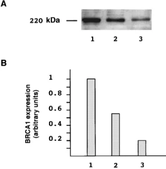
In order to determine the potential relationship between the amount of transcripts and BRCA1 protein, Western blot experiments were performed from whole cell extracts (WCE) of human breast cell lines (HBL-100, MCF-7, BT-20) previously analysed for their BRCA1 mRNA content (Ribieras et al., 1997). The BRCA1 proteins were detected using an anity puri®ed antibody raised against a peptide (aminoacids 1345 to 1362) chosen within the exon 11 of BRCA1. A strong band of 220 kDa is detected in these cell lines (Figure 3), as expected since several authors have described this 220 kDa BRCA1 protein in human cell lines (Chen et al., 1995; Scully et al., 1996). Quanti®cation of the signal obtained for the 220 kDa protein indicated a ratio similar to that obtained in RT ± PCR assays (HBL 100/MCF7/BT-20; 1/0.56/0.24 compared with 1/0.56/0.16 for BRCA1 mRNA content, taking 1 for HBL-100 cell line).
We have, therefore, analysed BRCA1 protein in some tissue samples spanning crucial stages of mammary gland development, i.e. 21st, 25th, 26th and 30th week of gestation. BRCA1 proteins were, also, investigated in an adult mammary gland and in the human breast cell lines. Immunoblots revealed a 220 kDa protein in fetal and adult mammary tissues
Figure 3 BRCA1 expression in human breast cell lines. (a) Immunoblot analysis of the 220 kDa BRCA1 protein; whole cell extracts from human breast cell lines, HBL-100 cells (lane 1), MCF-7 cells (lane 2) and BT-20 cells (lane 3) were separated on a SDS-4 to 12% linear gradient polyacrylamide gel. BRCA1 proteins were probed using the 5HU (1 : 60 diluted) polyclonal antibody directed against the aminoacids 1345 to 1362 of the BRCA1 protein and visualized with the ECL chemiluminescent detection kit (Amersham Life Science, France). (b) The signal corresponding to the BRCA1 band (220 kDa) was normalized, taking 1 for the signal corresponding to the HBL-100 cell lines. Autoradiograms were scanned (UMAX) and the intensity of the band corresponding to the 220 kDa BRCA1 protein was determined using image analyser software (Wayne Rasband, NIH)
Figure 4 Immunoblot analysis of BRCA1 in fetal mammary gland. (a) WCE from four mammary gland samples correspond-ing to the 21st week (lane 1), 25th week (lane 2), 26th week (lane 3), 30th week of gestation (lane 4) and from one sample of adult mammary gland (lane 5) were separated on a SDS-4 to 12% linear gradient polyacrylamide gel. BRCA1 proteins were probed using the 5HU polyclonal antibody and visualized with the ECL chemiluminescent detection kit (Amersham Life Science, France). (b) The signal corresponding to the BRCA1 band (220 kDa) was normalized, taking 1 for the signal corresponding to the adult tissue, as described in the legend of Figure 3
(Figure 4a) indicating that the BRCA1 protein, in human mammary tissues, exhibits an apparent molecular weight similar to that observed in human cell lines (Chen et al., 1995; Scully et al., 1996).
The relative intensity of the bands corresponding to the 220 kDa BRCA1 protein (Figure 4b) indicates that this protein is expressed at a high level between the 21st and the 26th week of fetal life and then, in the 30th week of gestation, this level becomes similar to that observed in the adult mammary gland sample. Control experiments performed with monoclonal antibodies directed against the N-terminal part of the BRCA1 protein (Scully et al., 1996) also indicated, a progressive decrease between the 21st week and 30th week of gestation (data not shown).
Taken together these data indicate that BRCA1 expression is closely associated with the dierentiation of the human mammary gland during fetal life. In addition, quanti®cation of BRCA1 transcripts in kidney, spleen, adrenal gland, lung and pancreas tissue samples from fetuses aged 24 and 30 weeks, indicated that the level of BRCA1 mRNA is lower at the 24th week than in the 30th week (unpublished data). Thus, the temporal pattern of BRCA1 expression in fetal human mammary tissues does not seem to be a consequence of a general activation of this gene. Consistent with this hypothesis, it has been shown in mice embryos, using in situ hybridization methods, that the activation of Brca1 is dependant on the stage of embryo development and on the tissues (Marquis et al., 1995; Blackshear et al., 1998).
In mice, RNase protection assays and Northern blot analysis have shown a 5 ± 10-fold increase in Brca1 transcripts during pregnancy (Lane et al., 1995; Marquis et al., 1995). This up-regulation seems to be
associated with ductular and glandular proliferation (Blackshear et al., 1998) during the ®rst stages of pregnancy. Then, during alveolar epithelial differentia-tion, BRCA1 mRNA becomes nearly undetectable returning, after mammary gland regression, to the level found in virgin mice (Lane et al., 1995; Marquis et al., 1995; Rajan et al., 1997; Blackshear et al., 1998). In situ hybridization has shown that the increase in Brca1 mRNA level occurs in epithelial cells and in adjacent stromal ®broblasts (Blackshear et al., 1998). Since both cell types participate in the morphogenesis of the mammary gland (Sakakura, 1987), all of these data strongly suggest a close association between the postnatal development of mouse mammary gland and BRCA1 expression.
Although the biological function of BRCA1 has not yet been fully determined, evidence for an involvement of BRCA1 in cell cycle checkpoints has been reported by several authors (for review see Bertwistle and Ashworth, 1998). This up-regulation during mammary gland development might suggest that BRCA1 exerts important checkpoint control functions during critical developmental stages.
Acknowledgements
We would like to thank Marc Billaud and Valerie James for discussion and critical reading of the manuscript. F Magdinier is a recipient of the fellowship from the Ligue Nationale contre le Cancer. The present work was supported by the Ligue Nationale contre le Cancer, Comite de SaoÃne et Loire, the Association pour la Recherche contre le Cancer and the CNRS (programme Physique et Chimie du Vivant).
References
Anderson SF, Schegel BP, Nakajima T, Wolpin ES and Parvin JD. (1998). Nat. Genet., 19, 254 ± 256.
Bertwistle D and Ashworth A. (1998). Curr. Opin. Genes Dev., 8, 14 ± 20.
Blackshear PE, Goldsworthy SM, Foley JF, McAllister KA, Bennett M, Collins NK, Bunch DO, Brown P, Wiseman RW and Davis BJ. (1998). Oncogene, 16, 61 ± 68.
Bork P, Hofmann K, Bucher P, Neuwald AF, Altschul SF and Koonin EV. (1997). FASEB J., 11, 68 ± 76.
Callebaut I and Mornon JP. (1997). FEBS Lett., 400, 25 ± 30. Chen Y, Chen CF, Riley DJ, Allred DC, Chen PL, Vonho D, Osborne CK and Lee WH. (1995). Science, 270, 789 ± 791.
Chomczynski P and Sacchi N. (1987). Anal. Biochem., 162, 156 ± 159.
Dawson EK. (1934). Ebind. Med. J., 41, 653 ± 682. Easton D. (1997). Nat. Genet., 16, 210 ± 211.
Ellisen LF and Haber DA. (1998). Annu. Rev. Med., 49, 425 ± 436.
Ford D, Easton DF, Bishop DT, Narod SA, Goldgar DE, Haites N, Milner B, Allan L, Ponder BAJ, Peto J, Smith S, Stratton M, Lenoir GM, Feunten J, Lynch H, Arason A, Barkardottir R, Egilsson V, Black DM, Kelsell D, Spurr N, Devillee P, Cornelisse CJ, Varsen H, Birch JM, Skolnick M, Santibanezkoref MS, Teare D, Steel M, Porter D, Cohen BB, Carothers A, Smyth E, Weber B, Newbold B, Boehnke M, Collins FS, Cannon-Albright LA and Goldgar D. (1994). Lancet, 343, 692 ± 695.
Friedman LS, Ostermeyer EA, Szabo CI, Dowd P, Lynch ED, Rowell SE and King MC. (1994). Nat. Genet., 8, 399 ± 404.
Futreal PA, Liu QY, Shattuck Eidens D, Cochran C, Harshman K, Tavtigian S, Bennett LM, Haugenstrano A, Swensen J, Miki Y, Eddington K, McClure M, Frye C, Weaverfeldhaus J, Ding W, Gholami Z, Soderkvist P, Terry L, Jhanwar S, Berchuck A, Iglehart JD, Marks J, Ballinger DG, Barrett JC, Skolnick MH, Kamb A and Wiseman R. (1994). Science, 266, 120 ± 122.
Girod C and Czyba JC. (1970). Le placenta, la glande mammaire et la lactation. In: Cours sur la biologie de la reproduction. Fascicule II. SIMEP (ed).
Hakem R, de la Pompa JL, Sirard C, Mo R, Woo M, Hakem A, Wakeham A, Potter J, Reitmair A, Billia F, Firpo E, Hui CC, Roberts J, Rossant J and Mak TW. (1996). Cell, 85, 1009 ± 1023.
Lane TF, Deng CX, Elson A, Lyu MS, Kozak CA and Leder P. (1995). Genes Dev., 9, 2712 ± 2722.
Lu M, Conzen SD, Cole CN and Arrick BA. (1996). Cancer Res., 56, 4578 ± 4581.
Magdinier F, Ribieras S, Lenoir GM, Frappart L and Dante R. (1998). Oncogene., 17, 3169 ± 3176.
Marks JR, Huper G, Vaughn JP, Davis PL, Norris J, McDonnell DP, Wiseman RW, Futreal PA and Iglehart JD. (1997). Oncogene, 14, 15 ± 121.
BRCA1 expression in human fetal mammary gland F Magdinier et al 4042
Marquis ST, Rajan JV, Wynshaw-Boris A, Xu J, Yin GY, Abel KJ, Weber BL and Chodosh LA. (1995). Nat. Genet., 11, 17 ± 26.
Merajver SD, Pham TM, Cadu RF, Chen M, Poy EL, Cooney KA, Weber B, Collins FS, Johnston C and Frank TS. (1995). Nat. Genet., 9, 439 ± 443.
Neuhausen SL and Marshall CJ. (1994). Cancer Res., 54, 6069 ± 6072.
Rajan JV, Marquis ST, Gardner HP and Chodosh LA. (1997). Dev. Biol., 184, 385 ± 401.
Ribieras S, Magdinier F, Leclerc D, Lenoir G, Frappart L and Dante R. (1997). Int. J. Cancer, 73, 715 ± 718. Russo J and Russo IH. (1987). Development of the human
mammary gland. In: The mammary gland. Development, Regulation, and Function. Neville MC and Daniel CW (eds). Plenum Press: New York, pp. 67 ± 93.
Saka Y, Esashi F, Matsusaka T, Mochidas S and Yanagida M. (1997). Genes Dev., 11, 3387 ± 3400.
Sakakura T. (1987). Mammary embryogenesis. In: The mammary gland. Development, Regulation and Function. Neville MC and Daniel CW (eds). Plenum Press: New York, pp. 37 ± 66.
Scully R, Chen J, Plug A, Xiao Y, Weaver D, Feunten J, Ashley T and Livingston DM. (1997a). Cell, 88, 265 ± 275. Scully R, Chen J, Ochs RL, Keegan K, Hoekstra M, Feunteun J and Livingston DM. (1997b). Cell, 90, 425 ± 435.
Scully R, Ganesan S, Brown M, DeCaprio JA, Cannistra SA, Feunteun J, Schnitt S and Livingston DM. (1996). Science, 272, 123 ± 125.
Smith SA, Easton DF, Evans DGR and Ponder BAJ. (1992). Nat. Genet., 2, 128 ± 131.
Spillman MA and Bowcok A. (1996). Oncogene, 13, 1639 ± 1645.
Tanner JM. (1962). The development of the reproductive system. In: Growth at Adolescence. Tanner JM (ed.), Blackwell Scienti®c: Oxford, pp. 28 ± 39.
Thakur S, Zhang HB, Peng Y, Le H, Carroll B, Ward T, Yao J, Farid LM, Couch FJ, Wilson RB and Weber BL. (1997). Mol. Cell. Biol, 17, 444 ± 452.
Thompson ME, Jensen RA, Obermiller PS, Page DL and Holt JT. (1995). Nat. Genet., 9, 444 ± 450.
Wilson CA, Payton MN, Elliot GS, Buaas FW, Cajulis EE, Grosshans D, Ramos L, Reese DM, Slamon DJ and Calzone FJ. (1997). Oncogene, 14, 1 ± 16.
Wu LC, Wang ZW, Tsan JT, Spillman MA, Phung A, Xu XL, Yang MC, Hwang LY, Bowcock AM and Baer R. (1996). Nat. Genet., 14, 430 ± 440.
Xu CF, Chambers JA and Solomon E. (1997). J. Biol. Chem., 272, 20994 ± 20997.