Publisher’s version / Version de l'éditeur:
Physical Review A, 58, 1, pp. 426-433, 1998-07-01
READ THESE TERMS AND CONDITIONS CAREFULLY BEFORE USING THIS WEBSITE. https://nrc-publications.canada.ca/eng/copyright
Vous avez des questions? Nous pouvons vous aider. Pour communiquer directement avec un auteur, consultez la première page de la revue dans laquelle son article a été publié afin de trouver ses coordonnées. Si vous n’arrivez pas à les repérer, communiquez avec nous à PublicationsArchive-ArchivesPublications@nrc-cnrc.gc.ca.
Questions? Contact the NRC Publications Archive team at
PublicationsArchive-ArchivesPublications@nrc-cnrc.gc.ca. If you wish to email the authors directly, please see the first page of the publication for their contact information.
NRC Publications Archive
Archives des publications du CNRC
This publication could be one of several versions: author’s original, accepted manuscript or the publisher’s version. / La version de cette publication peut être l’une des suivantes : la version prépublication de l’auteur, la version acceptée du manuscrit ou la version de l’éditeur.
For the publisher’s version, please access the DOI link below./ Pour consulter la version de l’éditeur, utilisez le lien DOI ci-dessous.
https://doi.org/10.1103/PhysRevA.58.426
Access and use of this website and the material on it are subject to the Terms and Conditions set forth at
Time-resolved Coulomb explosion imaging: a method to measure
structure and dynamics of molecular nuclear wave packets
Stapelfeldt, Henrik; Constant, Eric; Sakai, Hirofumi; Corkum, Paul B.
https://publications-cnrc.canada.ca/fra/droits
L’accès à ce site Web et l’utilisation de son contenu sont assujettis aux conditions présentées dans le site LISEZ CES CONDITIONS ATTENTIVEMENT AVANT D’UTILISER CE SITE WEB.
NRC Publications Record / Notice d'Archives des publications de CNRC:
https://nrc-publications.canada.ca/eng/view/object/?id=7efd7a9b-b6cb-42ad-9899-31e9c728b0b5 https://publications-cnrc.canada.ca/fra/voir/objet/?id=7efd7a9b-b6cb-42ad-9899-31e9c728b0b5
Time-resolved Coulomb explosion imaging: A method to measure structure and dynamics
of molecular nuclear wave packets
Henrik Stapelfeldt,1,*Eric Constant,1,2†Hirofumi Sakai,1,3and Paul B. Corkum1
1Steacie Institute for Molecular Sciences, National Research Council of Canada, Ottawa, Ontario, Canada K1A 0R6 2
De´partement de Physique, Faculte´ des Sciences, Universite´ de Sherbrooke, Sherbrooke, Que´bec, Canada J1K 2R1
3Electrotechnical Laboratory, 1-1-4, Umezono, Tsukuba, Ibaraki 305, Japan
~Received 20 August 1997; revised manuscript received 22 January 1998!
Coulomb explosion of molecules induced by an intense femtosecond probe laser pulse provides an approach to measure structure and dynamics of internuclear wave packets on a natural spatial ~Å! and temporal ~fs! scale for molecules. The technique is illustrated by applying it to study photodissociation of I2 initiated by a
femtosecond pump pulse. We report a resolution of ;2–4 Å in the internuclear range ;7–14 Å using an 80-fs probe pulse. We discuss the ultimate spatial and temporal resolution of the technique, as well as the possibili-ties of observing dynamics of dissociating polyatomic molecules. Intense femtosecond laser pulses not only provide a way of probing molecular dynamics but they are also an efficient means to initiate dynamics in, e.g., molecular ionic states. @S1050-2947~98!10106-3#
PACS number~s!: 33.80.Rv, 34.50.Gb, 82.50.Fv
I. INTRODUCTION
Femtosecond time-resolved studies of ultrafast phenom-ena in condensed media or in gases are of great interest in chemistry, physics, and biology @1#. In such studies the main technique applied is pump-probe spectroscopy where a fem-tosecond laser pulse initiates an event of interest and the subsequent evolution is followed by probing the system with another, temporally delayed, femtosecond pulse. In both con-densed phase and gas phase, there is rarely more than one photon used for either the pump step or the probe step since additional photons usually add extra complexity, thereby making the interpretation of the experimental results more difficult.
A different approach to femtosecond ~fs! pump-probe measurements of molecules in the gas phase is now possible @2#. For this approach perturbation theory is abandoned in the probe step and in some cases also in the pump step. It relies on the fact that there are usually two simple limits in the interaction between light and matter. The first one is the widely used perturbation limit in which one or a few photons are involved. The second one is the strong field limit in which a very large number of photons participate. Recent research in atomic and molecular physics shows that ioniza-tion induced by intense laser pulses of short duraioniza-tion can be understood by quasistatic or tunneling models @3,4# that are as simple and intuitive as perturbation theory. Pump-probe experiments in the strong-field limit are virtually unexplored but they have the potential to lead to new insight since stud-ies in this extreme are unrelated to the Fourier transform of traditional frequency-resolved spectroscopic methods.
In this paper we concentrate on the many-photon limit
and demonstrate how an intense probe pulse can be used to measure structure and dynamics of molecules by time-resolved Coulomb explosions. Coulomb explosions of mol-ecules following the collision between a fast molecular beam and a thin foil is used to obtain information about molecular structure @5,6#. Here, we use an intense femtosecond laser pulse rather than a thin foil to induce the Coulomb explosion. Even though the interaction time between the intense laser pulse and the molecule ~equal to the pulse duration! is sig-nificantly longer than the transit time of a molecular ion through the foil ~<1 fs!, it is still short enough to essentially freeze the nuclear motion during ionization. Furthermore, in contrast to the foil method, the laser-induced approach al-lows us to follow the temporal evolution of the molecular structure with a time resolution determined by the duration of the laser pulses.
Recently, we reported the first work on femtosecond time-resolved laser-induced Coulomb explosions in which photo-dissociation of I2 molecules was measured @7#. The basic
idea of the technique is the following. First, an 80-fs @full width at half maximum ~FWHM!#, linearly polarized, 625-nm laser pulse excites a coherent superposition of con-tinuum states ~a dissociative wave packet! by a single-photon transition from the X 1S
g
1 potential to the A 3P
1u potential
~Fig. 1!. Second, the wave packet moves along the A 3P 1u
potential curve with an asymptotic internuclear velocity of ;11.6 Å/ps. Third, the position and the structure of the wave packet is determined by exposing the molecule to an intense, 80-fs, temporally delayed, laser pulse. This causes rapid ejection of several electrons from the molecule correspond-ing to a quasi-instantaneous projection of the wave packet on a repulsive Coulombic state of a multiply charged molecular ion. The Coulomb repulsion between the resulting atomic ions gives rise to the Coulomb explosion. The internuclear distance of the molecule, before the irradiation with the probe pulse, can be determined by measuring the Coulomb energy, released as kinetic energy of the atomic ions since the internuclear distance does not change during the removal *Permanent address: Department of Chemistry, Aarhus
Univer-sity, Langelandsgade 140, DK-8000 Aarhus C, Denmark.
†Permanent address: CPMOH, Universite Bordeaux 1, 351 Cours
de la Liberation, F-33 405 Talence, France.
PRA 58
of the electrons. Furthermore, the distribution of kinetic en-ergies determines the structure of the internuclear wave packet.
The paper is organized in the following way. After a de-scription of the experimental technique in Sec. II we present our experimental results for the photodissociation of I2 con-sisting of kinetic energy spectra measured at different delays between the pump and the probe pulse ~Sec. III!. We explain how these spectra enable us to determine the square of the internuclear wave function at different times after the exci-tation of the wave packet with a resolution of ;2–4 Å in the internuclear range 7–14 Å. A usual concern in femtosecond experiments is nonlinear effects induced by the pump laser. In Sec. IV we discuss how they influence our experimental results. We also discuss how it should be possible to use nonlinear interactions to initiate dynamics in, e.g., molecular ionic states. Section V deals with the present limitations due to space charge effects, and in Sec. VI we discuss the ulti-mate spatial resolution for the Coulomb explosion method. In the last section before the summary we discuss how the technique might be applied to study molecular dynamics in the regime of smaller internuclear separations, and we men-tion the possibilities of extending our approach from di-atomic to polydi-atomic molecules ~Sec. VII!.
II. EXPERIMENTAL TECHNIQUE
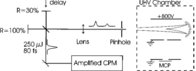
A schematic of the experimental setup is shown in Fig. 2. An amplified colliding pulse modelocked ~CPM! laser pro-vides 80-fs-long ~FWHM of a Gaussian fit! pulses with a pulse energy of ;250mJ. The wavelength is centered around 625 nm, the bandwidth is ;32 meV ~FWHM of a Gaussian fit!, and the repetition rate is 10 Hz. The output pulse of the laser system is split in a Michelson arrangement introducing a controllable delay between the pump and the probe pulse. The relative intensity of the two pulses is controlled by using a partially reflecting mirror in one of the arms ~typical
reflec-tivity ;30%!. The zero delay is determined by cross corre-lation in a 100-mm-thick KDP crystal. The two beams are focused by an f 51.5 m lens through a 500-mm pinhole, which ensures good spatial overlap of the two beams. The pinhole is imaged with a 5-cm-focal-length on-axis parabolic mirror to a focal spot diameter of ;25 mm inside an ultra-high vacuum ~UHV! chamber. Typically, the pump and the probe pulses have peak intensities of ;331013W/cm2and
;931013 W/cm2, respectively.
A pulsed beam of molecular iodine seeded in helium, formed by expanding 0.3 Torr I2 in 750 Torr He through a
;250-mm-diameter stainless steel nozzle, crosses the laser beams 8 cm away from the nozzle. In the present experiment we do not measure the rotational temperature, Trot, or the
vibrational temperature, Tvib, but laser-induced fluorescence studies of supersonic expansion of I2under virtually identical
experimental conditions @8# yielded Trot<10 K and
Tvib<200 K. A vibrational temperature of ;200 K
corre-sponds to a Boltzman distribution with 78% of the I2
mol-ecules in the n50 vibrational level, 17% in the n51 level, and 4% in then52 level. The density of I2molecules in the
laser focus can be estimated by an analytical expression as-suming an ideal free-jet expansion @9#. The result, 631010
molecules per cm3, is likely an overestimate, but indicates
that the actual density is as high as 1010cm23.
The ions produced from the probe laser irradiation are analyzed in a time-of-flight ~TOF! spectrometer @10# that is oriented perpendicular to both the molecular beam and the laser beams. It consists of an accelerating ~electric field ;267 V/cm! and a field-free drift zone equipped with a dual-microchannel-plate detector at the end. Time-of-flight spec-tra are recorded using a digital oscilloscope with a sampling rate of 109per second and an analog bandwidth of 500 MHz.
The flight time of any ion is a measure of its mass-to-charge ratio and its initial velocity along the TOF axis. A 0.5-mm aperture and a 1-mm-wide slit at the exits of the accelerating and the drift region ensure that we only observe fragments with their velocity vector essentially aligned with the TOF axis. Since the measured velocity almost equals the total ve-locity, the kinetic energy Ekinof the ions can be determined.
Also, the aperture and the slit ensure that we can control the polarization of the laser beams with respect to the internu-clear axis of those molecules that are detected. The size of the aperture and the slit and the magnitude of the electric field in the accelerating zone define the acceptance angle ~the maximum angle between the TOF axis and the velocity vec-tor of a detected fragment! and the energy resolution of the spectrometer. The acceptance angle scales as 1/
A
Ekin and is9.6° for an I1 ion with an initial kinetic energy of 1 eV. As
FIG. 1. A schematic of selected potential curves of I2illustrating the formation of the A 3P
1u wave packet and its subsequent
pro-jection by multiphoton ionization onto a repulsive Coulomb poten-tial.
FIG. 2. A schematic of the experimental setup showing the Michelson arrangement and the UHV chamber. The molecular beam propagates perpendicular to the plane of the paper.
a consequence, the collection efficiency of specific fragments is inversely proportional to their kinetic energy in good agreement with our observations. Finally, the energy resolu-tion of the TOF spectrometer causes a broadening of ;30 meV towards lower energies for I1ions independent of their
kinetic energy.
III. EXPERIMENTAL RESULTS
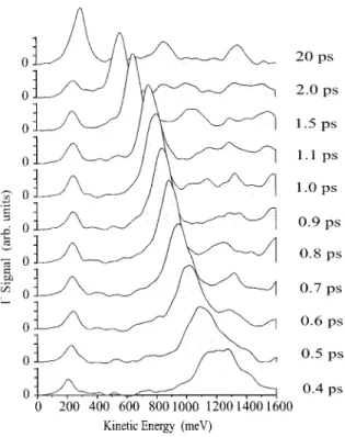
In Fig. 3 we show the kinetic energy spectra of the I1ions
at different delays between the pump and the probe pulses. The pump pulse is polarized perpendicular to the TOF axis. This ensures that molecules aligned parallel to the TOF axis, which are the ones that will contribute to the I1signal ~due
to the directional selectivity of the ion spectrometer!, can be excited to the dissociative A 3P
1u potential since the
X 1Sg1→ A 3P1u transition is perpendicular @11#. The probe
pulse is also polarized perpendicular to the TOF axis. This serves to minimize the ‘‘background’’ signal produced when the probe pulse multiple ionizes those molecules that were not excited by the pump pulse. The multiple ionization of the I2 ground-state molecules leads, among other things, to the
formation of I1 ions with high kinetic energy ~>1 eV!.
These ions are ejected in a narrow cone around the direction of the polarization of the probe laser @10#, and therefore they do not contribute to the detected I1 signal because of the
directional discrimination imposed by the apertures in the spectrometer. Also, metastable I221 ions ~same
mass-to-charge ratio as I1ions! are formed. They give rise to a sharp
peak in the center of the I1part of the TOF spectrum
corre-sponding to the section where very low energy I1fragments
would appear ~<25 meV!. Although the background is mini-mal, we still correct our spectra recorded with both the pump
and the probe pulse by subtracting a spectrum recorded with only the probe pulse.
There is a second reason for keeping the probe pulse po-larized perpendicular to the TOF axis. Since we only observe molecules with the internuclear axis parallel to the TOF axis, the choice of perpendicular polarization of the probe pulse ensures that all observed molecules have their internuclear axis perpendicular to the electric field of the probe pulse. In this case the ionization rate induced by the probe pulse is virtually independent of the internuclear separation R of the dissociating molecule @4,12#.
Two prominent peaks are observed in the spectra ~Fig. 3!. The position of the peak at ;240 meV is independent of the time difference between the pump and the probe pulse and therefore of the wave packet’s internuclear separation. Con-sequently, it must result from the wave packet being pro-jected on an I1-I potential curve since any such curve is
expected to be flat for the internuclear distances considered here ~at 0.4 ps the internuclear distance should be already ;7 Å!. Thus, this peak reflects the distribution of dissociation energies of the wave packet moving on the A 3P
1u potential.
The large peaks at higher kinetic energies are due to pro-jection of the wave packet on an I1-I1 potential curve. As
the time delay is increased the I1-I1 peak moves toward
lower kinetic energies corresponding to Coulomb exploding molecules with larger and larger internuclear separation. At a delay of 20 ps ~uppermost trace! the I1-I1peak almost
co-incides with the I1-I peak, indicating that the internuclear
distance of the dissociating molecule is so large that very little kinetic energy is released when the intense probe pulse ionizes the two widely separated iodine atoms.
The observation of two peaks in the I1 spectrum is
ex-actly what is expected in ionization experiments of dissoci-ating diatomic molecules @13#. If the intensity in a certain part of the laser focus is sufficiently high that the ionization probability of the iodine atom is large, then it is most likely that both iodine atoms are ionized ~projection of the wave packet onto an I1-I1potential!. In lower-intensity regions it
is possible that only one of the two iodine atoms is ionized ~projection onto an I1-I potential!. The observed ratio of the
I1-I1 and I1-I shows that the peak intensity of the probe
pulse is high enough to cause ionization of both iodine atoms in most regions of the focus.
We now discuss how the kinetic energy distribution of the I1-I1peaks in Fig. 3 can be used to determine the
distribu-tion of internuclear distances of the wave packet. The kinetic energy Ekinof an I1ion originating from fragmentation of an
I1-I1state is the sum of the energy from the dissociation E dis
on the initial A 3P
1u potential and the energy released from
the Coulomb explosion ECoulof the I1-I1state:
Ekin5Edis1ECoul. ~1!
Assuming that the laser pulse used for exciting the wave packet is transform limited @14#, the kinetic energy distribu-tion P(Ekin) is given by
P~ Ekin!5
E
P~ Edis!P~ ECoul5Ekin2Edis!dEdis, ~2!FIG. 3. Kinetic energy spectra of I1recorded at different delays
between the pump and the probe laser pulse. The spectra are cor-rected for the dependence of the collection efficiency on the kinetic energy. Each spectrum is an average of 1000 laser shots.
where P(Edis) and P(ECoul) denote the probability distribu-tion of the dissociadistribu-tion energy and the Coulomb energy, re-spectively. Since P(Ekin) and P(Edis) are determined by the
I1-I1 and the I1-I, respectively, P(E
Coul) can be extracted
by deconvolving the two signals. The distribution of internu-clear distances, P(R), is now easily found from P(ECoul)
since ECoul and R are related via
ECoul512 qq
8
e2
4p«0R ~3!
~Coulomb’s law!, where q and q
8
are the charge states of the fragmented ions. In the case of the I1-I1 channel q5q8
51, Eq. ~3! can be written as
R~ Å!57200/ECoul~meV!. ~4!
In Fig. 4 we show the results of the deconvolution—that is, the distribution of internuclear separations of the dissociating molecule at different times after excitation. The figure illus-trates how the wave packet moves from an internuclear sepa-ration of ;7 Å to ;14 Å with a constant internuclear veloc-ity. The width of the wave packet is 2–4 Å for the internuclear separations shown here.
Also shown in Fig. 4 is a classical simulation of the wave packet motion ~broken curves!. The simulation is based on propagating a wave packet excited by an 80-fs ~FWHM! Gaussian pulse on the A 3P
1u potential curve. We use an
existing analytical expression for the A 3P
1u potential
de-rived from spectroscopic studies @15,16#, and the distribution
of dissociation energies is modeled by a functional form of the distribution as determined from the I1-I signal ~see Sec.
IV B!.
There are two important points to note in the comparison between the experimental results and the simulation. First, the observed wave packet appears to move slower than ex-pected from the calculation and, second, the structure of the experimentally determined wave packet is significantly broader than the wave packet obtained from the simulation. Several physical phenomena that may be responsible for the discrepancies between the simulations and the measurements are discussed in the next three sections. These include non-linear effects in the pump step due to the relative high inten-sity of the pump pulse ~Sec. IV!, space charge effects caused by a high charge density in the laser focus after irradiation of the molecules by the probe pulse ~Sec. V!, the finite duration of the probe pulse, which imposes the ultimate spatial reso-lution ~Sec. VI!. In addition, the limited resoreso-lution of the ion spectrometer causes a broadening of the internuclear prob-ability density of the wave packet towards large R values. This is briefly mentioned in Sec. VI.
IV. NONLINEAR EFFECTS IN THE FORMATION OF THE WAVE PACKET
A. Multiphoton excited wave packets
In order to obtain a high transition rate in the single pho-ton process that forms the wave packet, the fluence of the pump pulse must be high. However, this inevitably leads to high intensity of the pump pulse due to its short duration. Nonlinear effects due to the pump pulse should always be a concern in femtosecond experiments. At the intensity of the pump pulse employed in our experiment (;3 31013 W/cm2), the I2molecules are likely excited not just to
the A 3P
1u potential by one-photon absorption but also to
higher-lying potential curves by multiphoton absorption @17#. In general, this will lead to dissociation via different poten-tial curves and therefore to creation of several different wave packets.
Experimentally, multiphoton excitation is revealed by re-cording spectra at very large time delays. Here, the Coulomb energy released is very small and the energy of the peaks in the kinetic energy spectra is almost equal to the dissociation energy of the wave packet. In the 20-ps spectrum of Fig. 3, two peaks, at energies of ;840 and ;1320 meV, are ob-served in addition to the prominent A 3P
1u wave-packet
peak at low energy. The peak at ;840 meV is consistent with two-photon absorption and subsequent dissociation into the atomic fragments I(2P
3/2)-I(2P1/2), which would give
rise to an observed energy of ;758 meV at a delay of 20 ps @18#. The peak at ;1320 meV arises from two-photon exci-tation and subsequent dissociation into the I(2P
3/2)-I(2P3/2)
channel ~expected energy position ;1226 meV! or three-photon excitation and subsequent dissociation into the I(2P
1/2)-I(2P1/2) channel ~expected energy position ;1274
meV!. We note that the energies of the observed peaks tend to be slightly larger than the expected energies of the two- or three-photon excited wave packets. This is consistent with Raman redistribution, discussed in Sec. IV B or with space charge effects ~discussed in Sec. V!.
FIG. 4. The square of the internuclear wave function, uC(R)u2,
of the dissociating I2molecule measured at different times after
excitation by the pump pulse. The full curves are the measurements based on the kinetic energy spectra in Fig. 3. The broken curves represent a simulation of the wave packet motion on the A 3P
1u
potential ~see text!. The amplitudes of the simulated wave packets are scaled to match the observations.
No other two- or three-photon absorption channels can produce wave packets with dissociation energies close to the ones observed. Still, higher-order photon absorption pro-cesses might create wave packets, dissociating into highly excited atomic iodine states, that could account for part of the observed peaks.
In general, the presence of higher-order channels such as two- or three-photon excitation processes caused by the pump pulse adds a background to the pump-probe signal. At the 20-ps delay, where the Coulomb energy is very small, the wave-packet peaks, corresponding to the different dissocia-tion energies, are clearly separated. However, at the smaller delays this is no longer the case since the sum of the Cou-lomb energy and the dissociation energy of the different wave packets might result in almost the same observed ki-netic energy. For instance, at a delay of 0.5 ps, projection of the two-photon excited I(2P
3/2)-I(2P3/2) wave packet onto
an I1-I state gives rise to a kinetic energy of ;1213 meV
whereas the projection of the A 3P
1u wave packet onto an
I1-I1state results in a kinetic energy of ;1170 meV. These
energies are not resolvable in our experiment and cause the relatively broad structure of the I1-I1 peaks observed at a
delay of 0.5 ps ~Fig. 3!. Thus, the presence of multiphoton excited wave packets enhances the width of the experimental internuclear distributions at the 0.4-, 0.5-, and 0.6-ps delays and is one reason for the difference between measurement and simulation in Fig. 4.
Although in our experiment two- and three-photon exci-tation transitions are clearly observable in the 20-ps delay spectrum, such multiphoton processes can be significantly suppressed by lowering the intensity of the pump pulse. The single-photon excitation probability will be reduced, but the experimental signal-to-noise ratio can still be excellent if high repetition rate ~>kHz! lasers are used.
B. Raman redistribution of vibrational population
Another nonlinear interaction that can influence the for-mation of the wave packet is stimulated Raman transitions. Since the bandwidth of the pump pulses ~;32 meV! is larger than the vibrational level spacing ~;26 meV! in the ground electronic state of I2, part of the population in an initial
vi-brational level might be transferred to a higher-lying vibra-tional level by a stimulated Raman transition. Thus, the ini-tial Boltzmann distribution of vibrational levels might be redistributed by one or several Raman processes thereby add-ing vibrational energy to the I2 molecule. This would
mani-fest itself as additional dissociation energy of the wave packet. The I1-I peaks, which reflect the distribution of
dis-sociation energies, are located around ;238 meV and have a FWHM ;98 meV. In comparison, a wave packet excited from the n50 level will be centered around 221 meV and have a FWHM equal to the laser bandwidth ~;32 meV!. Excitation of the small fraction of the Boltzmann distribution that initially resides in higher-lying vibrational levels ~see Sec. II! adds some dissociation energy to the wave packet @19#, but it is not sufficient to explain the observed distribu-tion of dissociadistribu-tion energies.
We would like to stress that even though the observation of enhanced dissociation energies is consistent with stimu-lated Raman transitions redistributing the vibrational
popula-tion a significant contribupopula-tion to the broadening of the I1-I
peaks towards higher energies will come from space charge effects ~Sec. V!. However, Raman transitions should always be considered as a potential interaction mechanism in studies with high intensity and short duration laser pulses @20,21#.
We also observe dissociation energies smaller than what is possible for a laser bandwidth limited excitation of the wave packet from the n50 level. This is due to the finite energy resolution of our ion TOF spectrometer, which al-ways broadens the peaks towards smaller energies. The broadening is estimated to ;30 meV ~Sec. II! in reasonable agreement with the observation of ;197 meV at the low-energy point where the amplitude of the I1-I peaks in Fig. 3
is reduced to 50% of the maximum value.
In the deconvolution procedure, described in Sec. III, we used a functional form of the distribution of dissociation en-ergies. This was obtained by fitting a Gaussian function to the I1-I signal for energies >221 meV, thereby modeling the
effect of the possible population redistribution and space charge effects. For energies <221 meV we simply replaced the distribution function by another Gaussian function with a width equal to the bandwidth of the pump laser pulse. The limited energy resolution of the ion spectrometer and the possible influence of space charge effects on the I1-I peak
prevent this signal from being a perfect image of the actual distribution of dissociation energies. However, once these problems are removed, recording of the I1-I signal will be an
accurate way to determine the distribution of dissociation energies, necessary for the deconvolution procedure. In par-ticular, this eliminates the need for a detailed knowledge of the Franck-Condon overlaps between the vibrational levels of the ground and the final electronic state (A 3P
1u), which
would otherwise be required for a calculation of rather than an experimental determination of the distribution of dissocia-tion energies.
C. Wave-packet dynamics of molecular ionic states induced by intense pump pulses
Rather than decreasing the intensity of the pump laser to suppress nonlinear effects in the excitation process, one can utilize the ability to efficiently drive high-order multiphoton absorption processes with intense femtosecond pulses to populate and initiate dynamics in ionic molecular states. Fur-thermore, it is possible to perform time-resolved spectros-copy of such states by using delayed femtosecond probe pulses to follow their evolution. For instance, we have re-cently employed an intense femtosecond pulse to efficiently excite a wave packet in an I221 state, dissociating into the
fragments I21-I @12#, with the purpose of verifying the
exis-tence of a critical internuclear distance where the ionization rate of molecular ions is strongly enhanced @4#. In order to create the I221 wave packet, the I2 molecules absorbed an
energy from the intense laser field equivalent to >16 photons at 625 nm. This experiment illustrates the approach of utiliz-ing wave-packet technology @22# to study a new problem.
Along the same lines, we recently demonstrated that irra-diation of molecules with intense femtosecond pulses pro-vides a means of producing metastable molecular trications @23#. Thus, it was shown how Cl231, Br231, and I231can be
to an intense (;331014W/cm2), 80-fs-long optical pulse.
The strong field facilitates the triple ionization while the short duration prevents the molecule from dissociating on transient potential curves during the ionization process.
To exert control of the outcome of the nonlinear processes driven by the pump pulse it will be advantageous to use laser beams with a constant spatial intensity profile. Presently, Gaussian shaped beams are employed with the result that, within the focal volume, a variety of different states are ex-cited depending on the maximum intensity reached at a par-ticular position in the focus. Since the signals recorded in our experiment represent an average over the entire focal vol-ume, they consist of contributions from all states excited. Using a laser pulse with an essentially flat spatial profile should ensure a more selective state excitation. This will make the interpretation of the signals easier and increase the population transfer to desired states. A constant transverse intensity profile is achieved, for instance, by selecting the central part of a Gaussian beam with an undersized pinhole and imaging it. In the longitudinal direction the constant in-tensity is obtained by restricting the laser-molecule interac-tion to a dimension much smaller than the Rayleigh length.
We note that a constant spatial intensity profile will also be useful for the probe pulses employed to Coulomb explode the molecule. Here, it should ensure that a wave packet under study is projected primarily onto one type of charge state rather than onto a number of different charge states.
V. SPACE CHARGE EFFECTS
Space charge can influence the velocity distribution of ions when a focused laser beam ionizes a sample of neutral molecules or atoms. Most of the photoelectrons exit the focal region very rapidly, leaving behind a positively charged core of ions. The electrostatic interaction between the positive ions changes their energy spectra and angular distributions. In particular, the kinetic energy of the ions is expected to increase due to the repulsive force between them. Our ex-perimental results are consistent with space charge in the velocity mismatch between the observed and the simulated wave packet ~Fig. 4!.
We can approximate the influence of space charge on the velocity distribution in the analysis of the kinetic energy spectra by assuming that it adds a constant value, Eso, to the
observed kinetic energies: Ekin5ECoul1Edis1Esc.
Repeat-ing the deconvolution procedure with Escas a free parameter,
we obtain a much better agreement between the simulated and the observed internuclear distributions ~Fig. 5! for Esc
equal to 50 meV.
Another indication of space charge comes from the posi-tion of the two high-energy peaks, in the 20-ps spectrum, resulting from the wave packets excited after two- or three-photon absorption. The observed energy positions of these peaks are ;50–100 meV larger than the calculated kinetic energies of the respective channels ~see Sec. IV A!. This is in accordance with the value of Escdetermined in Fig. 5.
Space charge may also influence the I1-I peaks. The
ef-fect should be smaller than in the case of the I1-I1 peaks
since the I1-I signal mainly originates from parts of the laser
focus where the intensity is lower @24# and therefore where the charge density is lower. Nevertheless, the effect is
con-sistent with the broadening of the I1-I peaks towards higher
energies.
We can estimate the space charge energy acquired by the ions in the laser focal volume following the ideas of Am-mosov et al. @25#. The laser focus is approximated by a cyl-inder with a radius of v0 and length l. The electrons are
assumed to exit the focal volume instantly, leaving behind a positively charged volume of approximately uniform charge density, ane, where a is the degree of ionization of each molecule, n is the density of molecules, and e is the elemen-tary charge. As the cloud of ions expands due to Coulomb repulsion the I1ions gain a maximum energy of Ue, where
U is the electrostatic potential felt by the ions at the surface
of the cylinder. Since the length of the cylinder, approxi-mately two times the confocal parameter, 2v02/l, is much larger than v0, U can be calculated by Gauss’s theorem:
Ue5v02ane/2«0, where «0 is the vacuum permittivity.
In-serting the experimental values of v0 ~12.5 mm! and n (6
31010 cm23), and assuminga52 ~the I1-I1channel is the dominant one!, we obtain Ue5170 meV. The agreement with the experimental value of 50–100 meV is satisfactory in light of the simplicity of the space charge model and the uncertainty on the estimate of the density in the molecular beam. Thus, the outcome of the simple calculation corrobo-rates the experimental indication of the non-negligible role of space charge. If high spatial resolution is required future Coulomb explosion experiments should be performed at lower gas density or with a small focal volume.
VI. ULTIMATE SPATIAL RESOLUTION LIMIT
Presently, the spatial resolution of the Coulomb explosion method to measure the size of wave packets is limited by the space charge and the energy resolution of the ion TOF spec-FIG. 5. Same as Fig. 4 except that space charge effects are taken into account in the analysis of the data by assuming that they add 50 meV of kinetic energy to the observed I1ions ~see text for details!.
trometer. Still, Fig. 4 demonstrates a spatial resolution of 2–4 Å for internuclear separations in the range 7–14 Å. Since both effects become more important as the energy of the detected ions decreases, the spatial resolution gets worse for large internuclear distances. However, our spatial resolu-tion is not limited by fundamental physics. The pronounced broadening of the internuclear distributions towards large values for the larger delays ~0.8–1.1 ps! is caused by the energy resolution of the TOF spectrometer. This can be im-proved by a better design of the spectrometer and space charge can be overcome by lowering the target density.
The ultimate resolution limit of our Coulomb explosion technique for measuring wave functions is determined by the effective duration of the probe pulse. The motion of the nu-clei during the probe pulse leads to a blur of the measured internuclear position approximately equal to the product of the internuclear velocity and the effective pulse duration. This effective pulse duration is expected to be only a fraction of the real duration since the multiple ionization only occurs efficiently around the peak intensity of the probe pulse. In our experiment, the wave packet velocity of ;11.6 Å/ps and the 80-fs-long probe pulse should therefore result in an ultimative spatial resolution better than 11.630.080 Å ;0.9 Å. The development of high power pulses of 5–10-fs duration @26# will significantly improve the spatial resolu-tion. Thus, measurements of wave-packet structure with a resolution of a few tenths of an Å seem feasible even for fast internuclear motions ~10–40 Å/ps!.
VII. OUTLOOK
So far we have only considered the measurement of wave packets at relatively large internuclear separations (R >6 Å). This approach ensures that the probe pulse projects the wave packet onto a state that is purely Coulombic. How-ever, at smaller internuclear separations some of the potential curves of molecular ions deviate significantly from a Cou-lombic shape. For instance, many molecular dications and trications exhibit metastable states @23,27,28#. In order to perform wave packet measurements at small internuclear dis-tances, of the precision that we have described, it will be necessary to remove enough electrons from the molecule by the probe pulse that the Coulomb repulsion between the re-sulting atomic ions far exceeds the binding energy of the molecular ion in the absence of Coulomb repulsion. Ideally, the probe pulse should then remove all valence electrons. This can be readily achieved when the molecule consists of atoms with only one or two electrons in the valence shell such as alkali dimers or trimers. For molecules with a larger number of valence electrons, like I2, explosion into highly
charged atomic fragments will be required to ensure that the released kinetic energy approximately corresponds to the Coulomb energy @29#.
One concern about using highly charged atomic ions is that the molecule is likely to pass through highly repulsive states during the removal of the electrons. If the probe pulses are too long, the internuclear motion on the transient repul-sive potential curves will cause a deterioration of the spatial resolution. Thus, observing wave-packet motion at small in-ternuclear separations will require the use of very short, ,20 fs, probe pulses. In addition, as pointed out in Sec. VI, the
nonlinear character of the transition to the highly charged molecular ions implies that the probe process only occurs efficiently during a fraction of the pulse duration correspond-ing to the highest intensities. This should minimize the effect of internuclear motion during the probe pulse and enable studies with a time resolution better than the actual duration of the probe pulse.
One obvious goal in future work is to extend time-resolved Coulomb explosion imaging from studies of di-atomic molecules to studies of polydi-atomic molecules. In the case of polyatomic molecules laser-induced Coulomb explo-sions holds the promise to directly observe three-dimensional evolutions of the nuclear configuration. This is an intriguing subject and one that is not yet well addressed by femtosec-ond spectroscopy. We note, however, that observing three-dimensional structures is a problem that has been confronted, for instance, in beam-foil non-time-dependent Coulomb ex-plosion studies @5,6# and various other fragmentation type experiments @30#. This has led to the development of ad-vanced three-dimensional imaging methods which should be applicable in the laser-induced studies.
Timed Coulomb explosion imaging will not provide as precise values for spectroscopic constants as more traditional spectroscopy methods but wave packet studies are unlikely to require such precision. Optically driven Coulomb explo-sion imaging may have applications in addition to observing molecular dynamics. The technique is very simple and com-patible with molecular beam or ion beam technology. It has a significant advantage over spectroscopy in not requiring any
a priori model for the molecules under study because the
technique gives direct information about the molecular ge-ometry. Thus, Coulomb explosion imaging could be of par-ticular importance for the study of floppy molecules encoun-tered in highly excited states of most molecules or in the case of van der Waals molecules.
VIII. SUMMARY AND CONCLUSION
Femtosecond laser-induced Coulomb explosion imaging is a new method to study dynamics and structure of mol-ecules on a natural temporal ~fs! and spatial ~Å! scale for molecules. It is based on initiating internuclear motion with a femtosecond pump pulse and measuring the time-dependent internuclear position and shape by Coulomb exploding the molecule with an intense, delayed probe pulse. To test the performance of the technique it was applied to study photo-dissociation of iodine molecules. We measured the wave function of I2molecules dissociating in the A 3P1upotential
with a resolution of 2–4 Å in the internuclear range of ;7–14 Å. When space charge effects, present in our experi-ment, are eliminated and the energy resolution of the ion spectrometer is improved it seems feasible to reach the ulti-mate spatial resolution limit given by the product of the in-ternuclear velocity and the effective duration of the probe pulse. This effective pulse duration is smaller than the real duration due to the high nonlinearity of the probe process. Thus, the future use of probe pulses of 5–10 fs duration should make it possible to perform time-resolved studies of internuclear structure with a spatial resolution of a few tenths
of an Å and a time resolution of the order of the optical period of the probe pulse.
Highly nonlinear interactions such as multiphoton ioniza-tion not only provide a method for probing molecular dy-namics but it can also be employed to initiate dydy-namics. Finally, it should be possible to follow photochemical events taking place in polyatomic molecules with the goal of
mea-suring three-dimensional structural changes on an ultrafast time scale.
ACKNOWLEDGMENTS
We would like to acknowledge the technical support from D. Joines and the help and advice from A. Stolow.
@1# Proceedings of the Conference on Ultrafast Phenomena IX, edited by P. F. Barbara, W. H. Knox, G. A. Mourou, and A. H. Zewail ~Springer-Verlag, Berlin, 1994!; Proceedings of the Conference on Ultrafast Phenomena X, edited by P. F. Bar-bara, J. G. Fujimoto, W. H. Knox, and W. Zintu ~Springer-Verlag, Berlin, 1996!.
@2# So far there exists no condensed phase analog.
@3# S. Augst, D. D. Meyerhofer, D. Strickland, and S. L. Chin, J. Opt. Soc. Am. B 8, 858 ~1991!.
@4# T. Seideman, M. Yu. Ivanov, and P. B. Corkum, Phys. Rev. Lett. 75, 2819 ~1995!.
@5# Z. Vager, T. Graber, E. P. Kanter, and D. Zajfman, Phys. Rev. Lett. 70, 3549 ~1993!.
@6# Z. Vager, R. Naaman, and E. P. Kanter, Science 244, 426 ~1989!.
@7# H. Stapelfeldt, E. Constant, and P. B. Corkum, Phys. Rev. Lett.
74, 3780 ~1995!.
@8# G. M. McClelland, K. L. Saenger, J. J. Valentini, and D. R. Herschbach, J. Phys. Chem. 83, 947 ~1979!.
@9# Atomic and Molecular Beam Methods, edited by G. Scoles ~Oxford University Press, Oxford, 1988!, Vol. I, Chap. 2. @10# P. Dietrich, D. T. Strickland, M. Laberge, and P. B. Corkum,
Phys. Rev. A 47, 2305 ~1993!.
@11# R. J. Oldman, R. K. Sander, and K. R. Wilson, J. Chem. Phys.
54, 4127 ~1971!.
@12# E. Constant, H. Stapelfeldt, and P. B. Corkum, Phys. Rev. Lett.
76, 4140 ~1996!.
@13# A small fraction of the dissociating molecules are expected to be projected onto an I1-I21 potential. However, the low
col-lection efficiency of I1ions resulting from this channel makes
the observation difficult.
@14# A chirped pump pulse is not dealt with in this paper. @15# J. Tellinghuisen, J. Chem. Phys. 58, 2821 ~1973!.
@16# At a wavelength of 625 nm there is also a small probability
that the 1P
u
1 potential of I
2is excited @15#. This is not
in-cluded in the simulation since the dissociation dynamics of a wave packet launched on the 1P
u
1potential will be essentially
identical to one launched in the A 3P
1upotential.
@17# In fact, even some ionization of I2is observed when only the pump pulse is applied.
@18# Actually, the Coulomb energy is not completely negligible at a delay of 20 ps. This is taken into account in the calculation of the predicted positions of the peaks, assuming that the I1-I1
explosion channel is the dominant one.
@19# The exact shape of the wave packet will not only depend on the population in the vibrational levels but also on the Franck-Condon overlaps involved in the excitation.
@20# A. M. Weiner, D. E. Leaird, G. P. Wiederbrecht, and K. A. Nelson, Science 247, 1317 ~1990!.
@21# L. D. Noordam, H. Stapelfeldt, D. I. Duncan, and T. F. Gal-lagher, Phys. Rev. Lett. 68, 1496 ~1992!.
@22# I. Sh. Averbukh, M. J. J. Vrakking, D. M. Villeneuve, and A. Stolow, Phys. Rev. Lett. 77, 3518 ~1996!.
@23# H. Sakai et al. ~unpublished!.
@24# P. Dietrich, D. T. Strickland, and P. B. Corkum, J. Phys. B 26, 2323 ~1993!.
@25# M. V. Ammosov, F. A. Ilkov, M. G. Malakhov, and Ch. K. Mukhtarov, J. Opt. Soc. Am. B 6, 1961 ~1989!.
@26# M. Nisoli, S. De Silvestri, and O. Svelto, Appl. Phys. Lett. 68, 2793 ~1996!; M. Nisoli et al., Opt. Lett. 22, 522 ~1997!. @27# P. J. Bruna and J. S. Wright, J. Phys. B 26, 1819 ~1993!. @28# G. Handke, F. Tarantelli, A. Sgamellotti, and L. S. Cederbaum,
J. Chem. Phys. 104, 9531 ~1996!.
@29# C. P. Safvan and D. Mathur, J. Phys. B 27, 4073 ~1994!; D. Mathur et al., ibid. 26, L141 ~1993!.
@30# J. Becker, K. Beckord, U. Werner, and H. O. Lutz, Nucl. In-strum. Methods Phys. Res. A 337, 409 ~1994!.