HAL Id: hal-02292034
https://hal.archives-ouvertes.fr/hal-02292034
Submitted on 20 Sep 2019
HAL is a multi-disciplinary open access
archive for the deposit and dissemination of
sci-entific research documents, whether they are
pub-lished or not. The documents may come from
teaching and research institutions in France or
abroad, or from public or private research centers.
L’archive ouverte pluridisciplinaire HAL, est
destinée au dépôt et à la diffusion de documents
scientifiques de niveau recherche, publiés ou non,
émanant des établissements d’enseignement et de
recherche français ou étrangers, des laboratoires
publics ou privés.
A zwitterionic interpenetrating network for improving
the blood compatibility of polypropylene membranes
applied to leukodepletion
Cheng-Chi Lien, Po-Ju Chen, Antoine Venault, Shuo-Hsi Tang, Ying Fu, Gian
Dizon, Pierre Aimar, Yung Chang
To cite this version:
Cheng-Chi Lien, Po-Ju Chen, Antoine Venault, Shuo-Hsi Tang, Ying Fu, et al..
A
zwitte-rionic interpenetrating network for improving the blood compatibility of polypropylene
mem-branes applied to leukodepletion. Journal of Membrane Science, Elsevier, 2019, 584, pp.148-160.
�10.1016/j.memsci.2019.04.056�. �hal-02292034�
OATAO is an open access repository that collects the work of Toulouse
researchers and makes it freely available over the web where possible
Any correspondence concerning this service should be sent
to the repository administrator: tech-oatao@listes-diff.inp-toulouse.fr
This is an author’s version published in:
http://oatao.univ-toulouse.fr/24234
To cite this version:
Lien, Cheng-Chi and Chen, Po-Ju and Venault, Antoine and Tang, Shuo-Hsi and
Fu, Ying and Dizon, Gian Vincent and Aimar, Pierre
and Chang, Yung A
zwitterionic interpenetrating network for improving the blood compatibility of
polypropylene membranes applied to leukodepletion. (2019) Journal of
Membrane Science, 584. 148-160. ISSN 0376-7388
A zwitterionic interpenetrating network for improving the blood
compatibility of polypropylene membranes applied to leukodepletion
Cheng-Chi Lien
a, Po-Ju Chen
a, Antoine Venault
a,b,∗∗, Shuo-Hsi Tang
a, Ying Fu
a,b,
Gian Vincent Dizon
a,b, Pierre Aimar
c, Yung Chang
a,b,∗aDepartment of Chemical Engineering and R&D Center for Membrane Technology, Chung Yuan Christian University, 200 Chung Pei Rd, Taoyuan 32023, Taiwan bResearch Center for Circular Economy, Chung Yuan Christian University, 200 Chung Pei Rd, Taoyuan 32023, Taiwan
cLaboratoire de Génie Chimique, Université de Toulouse, CNRS, INPT, UPS, 118 route de Narbonne, 31062 Toulouse Cedex 9, France
A R T I C L E I N F O Keywords: PP membranes Interpenetrating network Poly(GMA-co-SBMA) Antifouling Leukodepletion A B S T R A C T
Although widely used in blood-contacting devices, polypropylene (PP) membranes are prone to biofouling by plasma proteins and blood cells. The present study explores the effect of a surface zwitterionization process on the improvement of the biofouling resistance of PP membranes for leukocyte reduction filters. The modification strategy consists in forming an interpenetrating network of poly(glycidyl methacrylate-co-sulfobetaine metha-crylate) (poly(GMA-co-SBMA) around the fibers of coated PP membranes, using a cross-linking agent: ethyle-nediamine (EDA). It is shown that with EDA, a range of poly(GMA-co-SBMA) concentration (1–5 mg/mL) leads to a 0°-water contact angle and high hydration of the networks without affecting the intrinsic porous structure of the material. Besides, the related membranes show excellent resistance to biofouling by Escherichia coli, fi-brinogen, leukocytes, erythrocytes, thrombocytes and cells from whole blood with reductions in adsorption of 97%, 86%, 90%, 95%, 97% and 91%, respectively, compared to unmodified PP. Used in whole blood filtration, it is demonstrated that in the best conditions (5 mg/mL copolymer, with EDA), leukocytes can be efficiently re-moved (> 99.99%) without altering the erythrocytes concentration in the permeate, and that leukodepletion is more efficient than that measured with a commercial hydrophilic PP blood filter (about 50% retention). Physical retention of leukocytes is only efficient if the membrane material is anti-biofouling, and so, does not interact with other blood components able to trigger leukocyte attachment/deformation.
1. Introduction
The development of blood compatible membranes for selective se-paration of blood components has recently gained in momentum as a cost-effective and efficient alternative to centrifugation [1–3]. The mass production of porous matrices suitable to blood separation related ap-plications is not a concern at all, from a structure point of view, because polypropylene (PP) or polysulfone (PSf) are common materials and because highly porous PP and PSf membranes can be prepared using well-known and well-controlled processes, including temperature-duced phase inversion, melt extrusion, stretching or non-solvent in-duced phase separation [4–9].
The difficulty, however, relies on making the materials blood-compatible. Blood inert materials are highly desired for their capability
of preventing any biological response of blood components while ful-filling a particular function for which they have been designed. The adsorption of blood plasma proteins, in particular, can readily occur on hydrophobic solid materials [10,11], then leading to a cascade of bio-logical events which final stage is blood coagulation, that is, death of cells. Therefore, the hydrophobic PP or PSf membranes above-men-tioned have to be chemically modified if one intends to use them in blood contacting devices, to ensure the survival of blood cells but also to minimize process costs associated to plasma proteins/blood cells adhesion (membrane cleaning, membrane replacement, etc.).
The good news is that the design of membranes resisting biofouling by blood components relies on the same principles as the design of membranes resisting general biofouling: the key is to maintain a tight hydration layer at the interfaces wherever the material is likely to be in
∗Corresponding author. Department of Chemical Engineering and R&D Center for Membrane Technology, Chung Yuan Christian University, 200 Chung Pei Rd, Taoyuan 32023, Taiwan.
∗∗Corresponding author. Department of Chemical Engineering and R&D Center for Membrane Technology, Chung Yuan Christian University, 200 Chung Pei Rd, Taoyuan 32023, Taiwan.
E-mail addresses:avenault@cycu.edu.tw(A. Venault),ychang@cycu.edu.tw(Y. Chang).
contact with blood (top surface, and pore walls in particular) [12]. This will prevent water molecules to be expelled from the material surface leaving the space for hydrophobic segments of proteins to interact with the hydrophobic material. Therefore, following the requirements for nonfouling [12], the material has to be hydrophilized in order to strengthen the interactions with water, and it has to contain hydrogen bond acceptors rather than hydrogen bond donors, to promote water trapping and tight hydration. Finally, electrical neutrality must be maintained to minimize electrostatic interactions with charged seg-ments of proteins or cell walls of blood cells. For example, the group of Zhao has published a series of work in which a variety of zwitterionic copolymers were deposited on membranes (polysulfone or poly-ethersulfone) by click chemistry [13], cross-linked polymerization [14] or combination of ATR and click-chemistry [15]. These studies clearly highlighted similar requirements between nonfouling materials and blood-compatible materials.
The body of knowledge available on membrane modifications for general nonfouling applications is quite rich and has kept on attracting a lot of attention over the past recent years [16–27]. Making a hydro-phobic material more hydrophilic requires the use of a hydrophilic or amphiphilic material that has to be anchored on the surface or trapped in the polymeric chains of the matrix material. Anchoring can be simply done by a coating procedure (physical adsorption), which does not involve the use of any costly equipment but leads to low-energy in-teractions between the membrane material and the surface-modifying molecule, or by a grafting process (chemical adsorption), leading to more stable interactions as the surface-modifying molecule is chemi-cally bonded to the membrane, but more complex to implement, harder to run and to scale-up [18,20,23,25,28,29]. Another approach studied in research laboratories and in membrane manufacturing companies is the in-situ modification method that consists in blending the main membrane polymer with the antifouling material [17,21,29–31]. The main advantage of this approach is that it is a one-step membrane preparation/modification process, while coating and grafting require to prepare the membrane first, before applying the surface modification process. The downside of the in-situ modification is the compatibility issues that often arise, in particular when highly hydrophilic units are used, which makes it hard to find a common solvent for the different materials at play. It is tricky when membranes are formed by a phase separation process (commonly used in industry). Compatibility in the solid state is also essential to ensure stability of the interactions once the membrane has been formed. This also adds up to the difficulty of controlling membrane formation mechanisms once a supplementary component is added to original polymer/solvent or polymer/solvent/ non-solvent system as in phase inversion processes. Coating processes
such as dip-coating are viable industrial approaches as very little amount of copolymer is enough for the modification of large surface areas, and, once coating parameters optimized, it is possible to mini-mize the change of structure arising from the deposition of the coating agent.
A variety of nonfouling materials has been presented, either based on poly(ethylene glycol) (PEG) [16–18,21,30,31] or on zwitterions [16,18,20,22,23,25,32–34] such as sulfobetaine methacrylate (SBMA). The advantage of zwitterionic materials over PEG is their higher sta-bility in complex medium. It has also been proven that more water molecules can interact with one zwitterionic molecule than with one PEG molecule [35], which can be essential in view of improving hy-dration of the material to modify. Recently, we presented a novel co-polymer made of glycidyl methacrylate anchoring units and sulfobe-taine methacrylate antifouling segments, used for grafting of versatile interfaces [36]. The obtained poly(GMA-co-SBMA) was shown to be very efficient to mitigate biofouling of surfaces by a number of bio-foulants including proteins, bacteria or blood cells. However, the sur-face had to be activated by a plasma treatment procedure in order to generate hydroxyl or amine functional group that could react with the GMA units. This procedure is likely to lead to partial etching of the surface if it is porous and polymer-made. Besides, this grafting method is hard to control and implement. To ensure the sustainability of the porous structure of PP fibrous membranes and take full advantage of the excellent antifouling properties of poly(GMA-co-SBMA), one has to make use of a coating procedure. On the one hand, coating poly(GMA-co-SBMA) directly on PP membrane would likely lead to the estab-lishment of hydrophobic interactions between GMA segments and PP, but may also be partially unstable. On the other hand, cross-linking the copolymer around the fibers using an appropriate reactants directly in the coating bath would likely improve the surface modification, bene-fiting the coating density and as a result, the ability of the membranes to resist biofouling. Also, coating/cross-linking is a milder and much easier-to-control modification method than grafting.
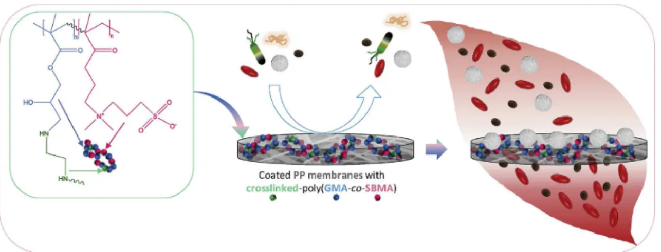
From these considerations, we worked on the design of poly(GMA-co-SBMA)-coated membranes using ethylenediamine (EDA) as a cross-linking agent in the coating bath. The purpose of this work as sketched inFig. 1is to show that the association of poly(GMA-co-SBMA) and EDA can result in the formation of an interpenetrating network enhancing the general antifouling properties and the blood compatibility of PP membranes (Fig. 1). Then, given the significant difference in size be-tween leukocytes and other blood components, it is shown that the as-prepared antifouling and blood compatible membrane can be highly efficient to selectively remove the leukocytes from the blood stream without affecting the erythrocytes concentration.
Fig. 1. Schematic presentation of the objectives of the work: to modify PP membranes with a crosslinked poly(GMA-co-SBMA) polymer, to evaluate the antifouling properties of the resulting membranes, and to apply the membranes in the development of hemocompatible filters for leukocyte removal from whole blood during blood filtration.
2. Materials and methods
2.1. Materials
Sulfobetaine methacrylate monomer was synthesized in our la-boratory. Glycidyl methacrylate monomer, dimethyl sulfoxide (DMSO) and 2,2′-azobis(2-methylpropionitrile) (AIBN) were purchased from Sigma Chemical Co., J.T. Baker and UniRegion Bio-Tech, respectively. Polypropylene non-woven membranes were provided by Mytrex (Taiwan). Ethylenediamine (EDA) was purchased from Sigma-Aldrich. Micro BCA™ protein assay kit was purchased from ThermoFisher Scientific. Whole blood was obtained from a pool of healthy volunteers and provided by the Taipei blood center (123 Lide Road, Beitou District, Taipei City). DI water was produced in the laboratory using a Millipore water purification system.
2.2. Methods
2.2.1. Synthesis and characterization of poly(GMA-r-SBMA)
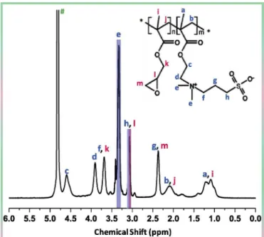
The copolymer was synthesized according to a protocol similar to that earlier reported [36]. A slight modification concerned the nature of the solvent as a DMSO/water mixture was used. Briefly, SBMA, GMA and AIBN were dissolved in a water/DMSO (1/3) solvent mixture in such a way that the total solid content was fixed to 20 wt%, the GMA/ SBMA molar ratio was 20/80, and the total monomer to initiator molar ratio was 100:1. The mixture was homogenized for about 10 min, then degassed using nitrogen, before performing the polymerization reaction at 60 °C for 6 h. The reaction was stopped using an ice bath, and the precipitate repeatedly washed with methanol. The product was freeze-dried, yielding a white powder corresponding to the final poly(GMA-r-SBMA) copolymer. The exact copolymer composition was determined from a 1H NMR analysis (Fig. 2), performed with a 300 MHz Bruker instrument, by integrating the signals at 3.1 ppm (Hh, SBMA and Hl,
GMA) and at 3.3 ppm (He, SBMA). The copolymer was found to be
composed of 74.6 mol% of SBMA and 25.4 mol% of GMA. By comparing the actual composition of the copolymer to that of the reaction mixture, it can be concluded that the two monomers exhibited similar reactivity during the synthesis. Finally, the molecular weight was measured with a G6000 PW XL Viscogel column using an aqueous solution of sodium nitrate 0.1 M as the mobile phase. It was found to be 22. 4 kDa, with a polydispersity index of 2.
2.2.2. Preparation of interpenetrating networks in PP fibrous membranes The modification of PP fibrous membranes with interpenetrating networks of poly(GMA-co-SBMA) is schematized inFig. 3. At first, the copolymer was dissolved in water at ambient temperature. The copo-lymer concentration ranged between 0.1 and 15 mg/mL. After 1 h, the crosslinking agent, ethylene diamine (EDA), was added to the solution and allowed to dissolve with the copolymer at ambient temperature. Then, PP fibrous membranes were added to the solutions for dip-coating. The flask containing the membranes and the dip-coating so-lution was transferred in an oven which temperature was set to 60 °C for crosslinking and interpenetration of the copolymer network. Note that we also performed tests without adding EDA. Therefore, step 2 inFig. 3
was skipped. Eventually, membranes were washed with DI water to remove loosely adhering/uncross-linked copolymer molecules, and dried, and stored at 4 °C until use.
2.2.3. Characterization of PP membranes
The coating density of the modified PP fibrous membranes was determined by weight measurements. Membrane samples (1-cm) were first dried and weighed. After the modification procedure, the mem-brane samples were dried and weighed again. The difference in weight between the uncoated membrane and the coated membrane per unit surface of the membrane sample corresponded to the coating density. 5 independent measurements were performed for each condition of coating. The membrane surfaces were observed by scanning electron microscopy (SEM). The instrument used was a Hitachi S-3000 micro-scope. Prior to observations, virgin and modified PP membrane samples were sputter coated with gold for 100 s. Then, the samples holder was loaded into the SEM chamber, and membranes observed at an accel-erating voltage of 7 kV. The membrane pore size before and after modification was measured with a PMI capillary flow porometer, using Porwick liquid as the intrusion liquid. The porosity was measured by gravimetric analysis, using butanol, as a wetting agent. The membranes were dried and weighed, then immersed 24 h in the wetting liquid, before being weighed again. The porosity can then be evaluated from the knowledge of the different weights (before/after immersion), the density of PP and the density of the wetting agent. The formula used is reported elsewhere [32].
The surface chemistry was analyzed by ATR Fourier-transform in-frared spectroscopy (FTIR) and x-ray photoelectron spectroscopy (XPS). FT-IR analyses were performed on a Jasco FT/IR 6700 instrument. Membranes were fixed to a glass slide positioned on the instrument stage. The number of scans/spectrum and the resolution were set to 32 and 4 cm−1, respectively. Besides, a FT-IR mapping analysis was
con-ducted using the same FT-IR instrument connected to a Jasco IRT-5200 microscope, according to a protocol described elsewhere [32]. Changes compared to our previous study concerned the number of scans and the resolution, set to 64 and 8 cm−1
, respectively, in the present work. Besides, the analysis was performed at 1016 cm−1- 1074 cm−1, range at
which the sulfonate function of SBMA is detected. XPS analyses were run with a PHI Quantera SXM/Auger spectrometer. The method used is similar to that earlier reported [37].
The evaluation of the surface and bulk hydrophilicity of samples was done by measuring their water contact angle (WCA) in air and determining their hydration capacity, respectively. WCA measurements were performed with 4-μL DI-water droplets deposited on the mem-brane surface using an automatic contact angle meter (Kyowa Interface Science Co.). 10 independent tests were run for each membrane. Hydration capacity, expressed in amount of water trapped per unit volume of membrane sample (mg/cm3), was evaluated by gravimetric
measurement. The membrane samples were dried and weighed before being immersed in DI-water for 24 h. Afterwards, superficial water was wiped off and wet membranes weighed again. The difference between the wet and the dry weights per unit volume of membrane corre-sponded to the hydration capacity of the sample at play. 5 independent tests were performed for each type of membrane.
2.2.4. Antifouling and hemocompatible properties of PP membranes Bacterial attachment tests were conducted with Escherichia coli bacteria, genetically modified with a green fluorescent protein. The protocols related to the genetic modification and the culture of bacteria has been described by Hsiao et al. [38]. The attachment tests were then performed as follows. 1-cm-diameter membranes were incubated with 1 mL of bacteria solution in wells of a 24-well plate disposed in an in-cubator which temperature was set to 37 °C. Incubation time was 24 h during which bacterial solution was changed to a fresh solution every 6 h. Afterwards, DI water was used to wash the membranes, and the samples could be observed using an A1R confocal microscope (Nikon). For these observations, the excitation wavelength was set to 488 nm while the emission wavelength was set to 520 nm. 5 independent ob-servations were done, from which the bacterial density could be de-termined using ImageJ®open source software.
The adsorption of fibrinogen on PP and modified PP membranes with a 1 cm2surface area was studied by performing a MicroBCA pro-tein assay (Thermo Scientific Inc.). The samples were positioned in a 24-microtiter plate and incubated with 1 mL of PBS at 37 °C overnight. Then PBS was removed and replaced by 2 mL of a 0.3 mg/mL fibrinogen solution. The incubation of membranes with the proteins was per-formed at 37 °C and last for 2 h. Then, the membranes were rinsed several times with 25 mL of PBS to wash off unabsorbed proteins, before being sonicated for 5 min in 6 mL of a 0.1 wt% sodium n-dodecyl sulfate (SDS) solution. Besides, 150 μL of BCA reagent were mixed with 150 μL of SDS solution in a 96-microwell plate. A calibration curve was de-termined at 562 nm by mixing fibrinogen at varying concentration and BCA reagent. The amount of protein adsorbed to the membranes could then be assessed from the determination of the absorbance at 562 nm of the different solutions after sonication. These measurements were performed using a microplate reader (BioTek Instruments Inc.). The amount of protein adsorbed to each membrane reported in this work corresponds to the average obtained from 6 independent analyses.
Whole blood was either fractionated by centrifugation to obtain red blood cells concentrate, white blood cell concentrates or platelet-rich plasma, or used as is in blood adhesion tests. The method for blood fractionation as performed in this study is published elsewhere [39]. 0.8 mL Of concentrate of interest or of whole blood was incubated at 37 °C for 2 h with membrane samples (diameter: 1 cm) positioned in a well-plate placed in an incubator. Afterwards, blood sample was re-moved and membranes washed with PBS. A solution of glutaraldehyde was used to fix the cells adhering to the membrane samples, operation which was done at 4 °C for 4 h. Finally, the samples were cleaned again with a PBS a solution, and cells adhesion observed by confocal micro-scopy, using the same instrument/settings as for E. coli attachment tests. Furthermore, the quantification of cell densities on the membranes was achieved with ImageJ®software, from the analysis of 5 images obtained
from independent observations.
2.2.5. Blood filtration tests
Virgin, modified PP membrane, and a hydrophilic commercial PP
membrane (Mytrex) were used to filtrate whole blood. Given the large pore size of the membranes, the purpose was to evaluate the leukode-pletion property of the poly(GMA-co-SBMA) interpenetrated mem-branes. Before filtration tests, 13 mm-diameter membrane samples were alternatively washed in ethanol (15 min) and soaked in DI water (15 min). This washing/soaking operation was repeated twice before immersing the membranes in PBS until their use in blood filtration. Membranes were placed in disposable plastic filtration modules. 3 or 5 layers of membrane were stacked in the module. The inlet of the module was connected to a 10 mL-syringe containing whole blood while the permeate was collected in test tubes. Photographs of the fil-tration tests are available at the end of the results and discussion sec-tion. The blood and permeate compositions were determined by com-plete blood count method using a XN 100 instrument (Sysmex Corporation). Leukocytes retention could be determined from the knowledge of the cell counts in blood and in permeate. After filtration tests, the membranes were washed. Cells were then fixed with a glu-taraldehyde solution (2 h at 4 °C), and leukocytes nuclei stained with a 4′,6-diamidino-2-phenylindole dihydrochloride (DAPI) solution (5 mg/ mL) for 15 min. Samples were finally observed by confocal microscopy (CLSM, Nikon A1R, magnification ×200, 405 nm laser).
3. Results and discussion
3.1. Physicochemical characterization of membranes
The effects of poly(GMA-co-SBMA) concentration in the coating bath and of the addition of EDA on the coating density of the mem-branes and on their wettability by water were investigated. It was ob-served in Fig. 4 that increasing the copolymer concentration in the coating bath positively correlated with an increase in the coating den-sity, but larger values were also obtained using EDA: while coating densities larger than 0.25 mg/cm2 were measured with the cross-linking agent, they reached a maximum value of 0.12 mg/cm2without
EDA. This observation reveals the positive role of EDA on the im-mobilization of poly(GMA-co-SBMA) on the surface of PP membranes, by favoring cross-linking between the copolymer chains inter-penetrating the PP fibers. It was further supported by the measurements of surface hydration (WCA) and bulk hydration (hydration capacity). All membranes modified with poly(GMA-co-SBMA) without adding EDA in the coating bath had a WCA of about 125°, almost as high as that of virgin PP membrane (135 ± 2°). At low copolymer concentra-tion and with EDA, the WCA of the membranes remained high but from a 1 mg/mL copolymer concentration in the coating bath and provided the use of EDA (1E sample), the WCA fell to 0, that is, the membrane turned from highly hydrophobic to superhydrophilic. Meanwhile, their capacity to permanently trap water in their bulk, increased drastically from 1E sample as it reached 150 mg/cm3and kept increasing up to
250 mg/cm3. One may also have noticed that the coating density of the membrane prepared by immersion in the 15 mg/mL coating bath con-taining EDA (15E sample) is only slightly larger than the coating
density of the membrane prepared by immersion in the 5 mg/mL coating bath (5E sample). It suggests that the interface is close to be saturated with cross-linked poly(GMA-r-SBMA). As a result, the water wettability (measured by water contact angle or hydration capacity) is similar, hence the plateau observed for hydration. Taken all together, these results suggest that efficient modification is only reached after cross-linking the copolymer chains around the PP fibers, resulting in super-wetting properties. Super-wetting of PP fibers is difficult to ob-tain by a simple wetting procedure. For example, PP membranes coated with a zwitterionic material obtained from the reaction of 3-iodopro-pionic acid with a copolymer of octadecyl acrylate and 2-(dimethyla-mino)ethyl methacrylate units, showed a final WCA of 80°, for a coating density of 0.2 mg/cm2(similar to here) [40]. Similarly, we reported a WCA of 75° for PP membranes coated with a copolymer of zwitter-ionized 4-vinylpyrridine and octadecylacrylate [41]. The composition of the zwitterionic copolymers in these previous studies was indeed different from here, but the results ofFig. 4do highlight that it is not the only parameter affecting hydration of the PP-coated membranes. Instead, formation of a tight network of zwitterionic moieties around the fibers seems to be essential, achievable by a cross-linking procedure following the coating step.
Surface modification did not lead to drastic morphological changes, as the SEM images revealed that the structures of coated membranes were very similar to that of the virgin one (Fig. 5). However, after immersion in 15 mg/mL coating baths containing EDA, the presence of a thin film surrounding PP fibers could be clearly identified, attributed to the cross-linking effect of EDA, since it was not observed for mem-branes modified without the use of EDA. Nonetheless, this change in surface morphology did not impact on the surface pore sizes which
were initially too large (about 10 μm for virgin PP) to be significantly affected by the surface modification process. Similarly, the porosity was only slightly reduced by the coating-crosslinking procedure (from 77% to 72%). This result also implies that the intrinsic membrane separation properties directly arising from the structure are unaffected by the coating procedure, which is highly desired.
In addition to the physical surface properties, we looked into the effect of the coating on the chemical surface properties of the mem-branes. We analyzed the membranes by both FT-IR and XPS, and the related results are presented in Fig. 6. The FT-IR spectra were not modified at low copolymer concentration in the coating bath (1, 5 mg/ mL) without EDA. A same conclusion held with EDA for a concentration of 1 mg/mL. However, at higher concentration without EDA (15 mg/ mL) or with EDA (from 5 mg/mL), several changes were noticed on the FT-IR spectra. The first one is related to the detection of an absorption band at 1730 cm−1, corresponding to the O–C]O functional groups
[32,42,43] brought by the zwitterionic units, while the second change was observed in the range 3100–3600 cm−1, where the hydroxyl
groups of water absorb. This change is an indirect evidence of the presence of the copolymer. Although the membranes were freeze-dried before analysis, remaining water tightly trapped by the zwitterionic units of poly(GMA-co-SBMA) could be detected. It is also worth noting that for membranes immersed in coating baths at a 5 mg/mL and 15 mg/mL copolymer concentration and containing EDA, a clear peak was noticed at 1035 cm−1corresponding to the characteristic vibration
band of the sulfonate head [44]. All in all, this chemical analysis proves the presence of copolymer at the surface of the membranes in some particular conditions of surface modification (copolymer concentration in the coating bath of 15 mg/mL without EDA, and copolymer
concentration in the coating bath of 5 and 15 mg/mL with EDA). During the coating process, part of the copolymer diffused in the bulk of the highly porous membrane and interacted with PP in the layers under-neath the top surface making FT-IR detection more difficult. Clearly, the use of EDA resulted eventually in higher coating density which facilitated the detection of the poly(GMA-r-SBMA).
Note also that for the intended application, there is no need for long-term stability. Indeed, not only blood cells separation is achieved within minutes, but also the filters are disposable, that is, there will be thrown after the operation. Yet, results ofFig. S1show no change in the FT-IR spectra before and after 30 min of immersion in PBS, which proves that the coating is stable enough for the intended application.
The XPS analysis is also useful to support FT-IR results. Here we looked at the N1s, C1s and S2p core-level spectra (Fig. 6b). This ana-lysis is more sensitive, which is clearly proved from the C1s core-level spectra. If one looks at the core-level spectrum of virgin PP, only one peak corresponding to C–C species is observed at 284.3 eV. However, the deconvolution analysis of all other spectra showed the presence of 2 supplementary peaks at BE 286 eV and 288 eV, likely corresponding to
C–O–C and O–C]O species, respectively [32,36]. These two species come from both GMA and SBMA units. The S2p core-level spectra also provide further evidence to the presence of the zwitterionic moieties, as a clear peak was detected on all spectra at BE of about 167.2 eV, cor-responding to the sulfonate functions of SBMA [32,36,45,46]. Besides, the presence of quaternary ammonium functional groups could also be proven from the N1s core-level spectra but clear peaks were only de-tected after using EDA as a cross-linking agent. EDA possesses amine functional groups (secondary amines after cross-linking, and residual primary amine if EDA is not entirely cross-linked) which contributed to the signal of the peak at BE 399.2 eV (C–N species), while the signal at BE 401.8 eV was attributed to the presence of ammonium groups of SBMA [36,45,46].
A mapping analysis of the samples modified using EDA, performed at 1016 cm−1- 1074 cm−1to detect the sulfonate function of SBMA in
the copolymer, confirms the gradually increasing presence of copo-lymer at the surface of the membranes with the copocopo-lymer concentra-tion in the coating bath (Fig. 7). Indeed, while the map of 0.5E sample is deep blue, revealing a low surface copolymer surface density, it turns
Fig. 5. The effect of coating poly(GMA-co-SBMA) with/without cross-linking agent (EDA) on (a) the structure, (b) pore size and (c) porosity of modified PP fibrous membranes. The scale bar is the same for all SEM images.
light blue and then green for 5E and 15E sample, respectively, con-sistent with the increase in copolymer concentration in the modification medium. Besides, one dominating color was seen on samples 0.5 (deep blue) and 5E (light blue), with a few green spots indicating local higher concentration. For 15E, numerous yellow and orange spots reveal re-gions locally highly concentrated in copolymer, which would support the SEM images showing thin deposit on the membrane's surface and so, potential heterogeneous surface modification.
In conclusion of this section on the characterization of surface modified PP membranes, the coating procedure is much more efficient after using cross-linking agent EDA and for concentrations of 5–15 mg/ mL of poly(GMA-co-SBMA) in the coating bath. Coating without EDA resulted in low detectability of the coated layer by both FT-IR and XPS methods.
3.2. Resistance to biofouling: effect of cross-linking agent
After fully characterizing the membranes, we switched our focus on the effect of the copolymer and of the cross-linker on the antifouling properties of membranes. We started with investigating the resistance to bacterial attachment using Escherichia coli modified with a green fluorescent protein as a model bacterium. E. coli is commonly found in wastewaters [47], but is also responsible for medical infections such as urinary tract infections [48]; therefore, it is essential to design mem-brane materials that will efficiently resist to the attachment of these bacteria as contact with E. coli-containing media is frequent. E. coli bacteria have a typical tendency to interact with hydrophobic materials as seen from the results ofFig. 8that highlights that numerous bacteria could be observed on the confocal images of the virgin PP membrane, for which the relative attachment value was set to 100%. As a com-parison, the positive control, TCPS (tissue culture polystyrene) plate, showed a 117% relative adhesion of bacteria. There was, however, a significant decrease in bacterial attachment for all modified PP
membranes. For low (0.1 mg/mL) and high (15 mg/mL) copolymer concentration, there was no positive effect of the addition of EDA as we observed slightly larger bacterial attachment. We hypothesized that at low concentration, the coating density was too low for the cross-linking agent to significantly affect the bonds between the brushed, while at high concentration, the formation of local aggregates as seen from the SEM images, arose in local heterogeneities which promoted bacterial attachment. It is indeed a well-known fact that bacteria have a tendency to adhere better on rough porous surfaces than on smooth dense sur-faces. Therefore, introducing local heterogeneities from a too high content of copolymer/cross-linker in the coating bath has unwanted effect. The results indicate that for samples modified from a 1 or 5 mg/ mL poly(GMA-co-SBMA) concentration in the coating bath, the adhe-sion of bacteria was the lowest and almost comparable to that measured on a polySBMA hydrogel, considered to be an ideal antifouling mate-rial. The addition of EDA decreased E.coli attachment further in such a way that the lowest relative attachment measured was 3 ± 1%. This result is quite remarkable, considering again the highly porous nature of native PP membranes, promoting physical entrapment of the bac-teria. It is also an indirect method to check that the membranes were efficiently modified in these conditions in view of mitigating biofouling by bacteria, since the only biofouling resistant moieties are the PBSMA zwitterionic units brought by the cross-linked copolymer after surface modification.
Other biofoulants that interact with materials at a smaller scale, namely proteins, are also commonly found in most aqueous environ-ments that membranes are likely to be contacted with [49]. In water treatment, food industry or blood filtration, resistance to proteins fouling is one of the keys to the sustainability of membrane processes. Proteins not only easily interact with hydrophobic materials, but also promote the adsorption of other larger biofoulants (bacteria, cells, etc.) depending on the medium at play. Herein, we performed fibrinogen adsorption tests. Fibrinogen is a blood plasma proteins whose
Fig. 7. Mapping analysis of membranes modified using EDA. 4 membrane samples were placed in a square (as depicted by the optical microscopy image) and the region near the interfaces between the membrane samples was scanned to acquire a map containing an analysis of the 4 membranes. The analysis was performed at 1016 cm−1- 1074 cm−1(sulfonate function of SBMA). Maps are color-coded: dark blue color (0 on the scale) corresponds to the absence of functional group while red color (12 on the scale) corresponds to the presence of the functional group at high density. (For interpretation of the references to color in this figure legend, the reader is referred to the Web version of this article.)
interaction with membrane materials will have consequences on the membrane operation (rapid fouling requiring membrane cleaning or replacement) but also on the viability of the surrounding cells (because the fibrinogen is a key component involved in the formation of blood clot, that is, blood cells death [50,51]). A membrane aimed for blood filtration must resist the adsorption of fibrinogen. As established earlier [52], hydration of the membrane is the key to protein resistance. In order for the proteins to interact with a material, water must be ex-pelled first from the surface of the membrane but also from their hy-drophilic segments. Results presented inFig. 9show a drastic decrease
in fibrinogen adsorption after coating the PP fibrous membranes with poly(GMA-co-SBMA) in presence of EDA. From a 1 mg/mL copolymer concentration in the coating bath, the adsorption of fibrinogen was reduced from 6.4 to 2.2 μg/cm2, adsorption value that was further de-creased to 0.9 μg/cm2for the membranes coated using the 5 mg/mL
copolymer concentration in the coating bath. This value compares with polySBMA hydrogel, which supports the efficiency of the copolymer and of the procedure followed to modify the hydrophobic porous fi-brous PP membranes. Besides, we measured again a better efficiency at 5 mg/mL than at 15 mg/mL, which corroborates previous observations with bacteria.
The important reduction in fibrinogen adsorption is a first sign of the efficiency of the modification process to improve the blood com-patibility of the membranes. This test was then followed by an in-depth analysis of blood cells attachment. We performed tests using cells from concentrates as well as we incubated the membranes directly with whole blood. The related quantitative results associated to our confocal analysis are displayed inFig. 10. In all cases, unmodified PP membranes showed very poor blood compatibility and adsorption levels compar-able to the TCPS. The results are again associated to the highly hy-drophobic nature of PP fibrous membranes, arising from both its che-mical composition but also from the porous nature of the materials used. It was therefore not surprising to notice that a slight improvement of the surface/bulk hydrophilicity brought by poly(GMA-co-SBMA) at low concentration in the coating bath led to an obvious decrease in cells affinity with the membranes, and as a result, to a decrease in their adhesion. In general though, all samples modified without the use of EDA showed similar levels of cell resistance, correlating with their si-milar hydrophilicity demonstrated above. However, using EDA and a copolymer concentration of 1 mg/mL and 5 mg/mL, that is, forming the interpenetrating network around PP fibers did lead to a further decrease in cell attachment. For lower concentrations and using EDA, there was no major changes, again correlating with relatively high water contact angle and low hydration. The network is efficient to prevent cell ad-hesion from a certain copolymer concentration only. Notice also that at high concentration (15 mg/mL) and using EDA, cell adhesion increased
Fig. 8. The effect of coating poly(GMA-co-SBMA) with/without cross-linking agent (EDA) on the resistance of membranes to biofouling by Escherichia coli bacteria. Incubation time: 24 h; level of adsorption on TCPS plate: 117 ± 8%; level of adsorption on SBMA hydrogel: 1 ± 0.7%.
Fig. 9. Amount of fibrinogen adsorbed on PP and coated PP membranes, compared with antifouling polySBMA hydrogel, and assessed by a micro BCA™ protein assay.
again, probably due to the lack of surface homogeneity, as also ob-served in the case of bacterial attachment tests and protein adsorption tests.
As a conclusion, although significant improvement in blood com-patibility was obtained with all samples, regardless of the type of modification (with/without EDA) and of the copolymer concentration, the only PP fibrous membranes that could compete with polySBMA hydrogels in terms of blood compatibility was those formed from a copolymer concentration in the coating bath of 5 mg/mL in the pre-sence of EDA. Although earlier studies were published by other groups on the development of PP blood compatible membranes [53–55], the
method used in the present study does not involve γ-irradiation, oxygen plasma treatment or UV irradiation. It is a simpler technique and po-tentially implementable.
3.3. Usage of modified PP membranes as leukodepletion filters
In the previous sections of the manuscript, we showed that mem-brane referred to as “5E” (copolymer concentration: 5 mg/mL, with EDA in the coating bath), presented the best general anti-biofouling properties. Whether blood plasma proteins, bacteria or blood cells (from concentrate or from whole blood) were used, minimum
biofoulants adhesion was measured on this particular sample. The biofouling tests, however, were all conducted in static conditions. The combined results of the antifouling tests and the physical properties of the membranes (large pore size, large porosity) suggest that this coated-PP membranes could be used for a specific application: the physical removal of white blood cells during blood filtration. Leukodepletion can rely on smart interactions between the membrane surface and the material by incorporating charges at the membrane surface in a con-trolled fashion [56], or more simply on molecular sieving if the material surface is also blood compatible. Forming the interpenetrating poly (GMA-co-SBMA) network around the PP fibers greatly enhanced the blood-inert property of PP. Besides, the results displayed inFig. 5 re-vealed that this surface modification had none (or very little) impact on the pore size of the membranes. So, we used the modified-PP mem-branes in blood filtration disposable cells (Fig. 11a). Membranes were stacked as done commercially using either 3 or 5 membrane sheets. Besides, we also performed filtration tests using the unmodified PP and a commercial hydrophilic PP membrane with similar physical proper-ties.
The results show logically show that no blood flow could be ob-tained using the unmodified PP membrane and blood remained in the syringe (Fig. 11a). Nonetheless, surface modification improved the hydrophilicity of the membranes, and permitted blood filtration. The concentration in WBCs in the permeate was determined and it was found to be as low as 0.07 103cells/μL using 5 layers of 5E membrane, corresponding to a 99.99% removal as the initial concentration was 9.39 103cells/μL. Meanwhile, the concentration in RBCs remained
un-affected since the initial RBCs concentration was 4.34 106cells/μL, which suggests that the as-prepared membranes could be used to pre-pare RBCs-rich blood fractions, essential to patient suffering from in-tense bleeding after physical trauma [57]. However the commercial membrane did not permit a good retention of the WBCs.
If one looks carefully at the intrinsic physical properties of the commercial and as-prepared membranes, then results are very sur-prising at a first glance. Firstly, the pore size of the commercial brane is significantly smaller than the pore size of our modified mem-brane (4.3 ± 0.9 μm vs. 10 ± 0.7 μm). Secondly, the porosity of both membranes is in the same range. Thirdly, water wets almost in-stantaneously both membranes. Yet, filtration of whole blood was found to be significantly faster in the case of the commercial membrane than in the case of the modified PP membrane (3 min vs 11 min), while the retention of leukocytes followed an opposite trend (low with com-mercial membrane, high with modified membrane). One could have reasonably expected opposite results. A possible explanation for these conflicting results (with regards to the structure/properties relation-ship) is provided inFig. 12. Starting with commercial membranes, we suspected (and proved afterwards) that they do not resist biofouling. As a result, platelets approaching the membrane and finally making
contact with the material become activated. It is known that platelet can induce leukocytes capture after α-granule P-Selectin has been ex-pressed on the cell membrane [58]. But as white blood cells are de-formable cells (to be able to flow through very small capillaries in vivo), a significant proportion can still penetrate through the small pores under the effect of drag flow. Oppositely, in the case of the modified PP membranes, there is no strong interactions between the membrane material and the leukocytes since we designed low-biofouling mem-branes. It means that there is only physical retention of the cells, re-duced platelet adhesion and little to no leukocyte deformation, but instead, the formation of a cake layer of WBCs which is not strongly adhering to the material due to protective hydration layer forms by the cross-linked copolymer. The WBCs cake layer rapidly blocks the pores, seriously reducing the flux, but can be removed by cleaning the mem-brane. An evidence of the difference in bioinert property is provided in
Fig. 12in the confocal images taken after filtration and washing of the membranes and in the SEM images stressing on different resistance to platelet adhesion. Despite washing, numerous leukocytes cells can be spotted at the surface of the commercial membrane. There are fewer cells on the as-prepared membrane indicating their detachment from the surface of the membranes during the washing procedure, also thanks to reduced platelet adhesion. In light of the results obtained, the mechanism presented inFig. 12 is a possible approach clearly high-lighting that the structure parameters of a membrane should not be considered independently from material-blood component interactions during the filtration of whole blood with the aim to reduce leukocyte concentration in the permeate. Physical retention of leukocytes can only be efficient if there is no chemical interactions occurring in the first place between the membrane material and other blood components able to interact with leukocytes.
4. Conclusions
This work presented a method to efficiently zwitterionize poly-propylene membrane creating an interpenetrating network of a copo-lymer made of glycidyl methacrylate and sulfobetaine methacrylate, poly(GMA-co-SBMA), in presence of ethylenediamine used as a cross-linking agent.
Essential results are listed as follows:
•
Dip-coating of the membranes in a poly(GMA-co-SBMA) bath without the use of ethylenediamine did not permit to efficiently hydrophilize membranes (water contact angle of 125°, hydration capacity below 50 mg/cm3);•
Dip-coating of the membranes in a poly(GMA-co-SBMA) bath con-taining ethylenediamine permitted to generate surfaces with a 0° water contact angle, and a hydration capacity of 250 mg/cm3;•
Except at high copolymer concentration (> 5 mg/mL), little to noFig. 11. Usage of poly(GMA-co-SBMA)-coated membranes in whole blood filtration: assessment of leukodepletion. (a) Photographs of the system, evidencing no permeation using virgin unmodified PP and complete permeation using 3 or 5 layers of modified membranes or commercial hydrophilic membranes; (b) White blood cells and red blood cells concentration in the permeate and white blood cells removal ratio determined from the white blood cells concentration in whole blood (9.39 103cells/μL).
major change in the membrane structure was observed. Besides, the presence of poly(GMA-co-SBMA) was clearly ascertained by XPS measurements;
•
Membranes modified in the best conditions showed excellent re-sistance to biofouling by Escherichia coli, fibrinogen, leukocytes, erythrocytes, thrombocytes and cells from whole blood with re-ductions in adsorption of 97%, 86%, 90%, 95%, 97% and 91%,respectively, compared to unmodified PP;
•
Used in blood filtration, the best membrane (prepared using a co-polymer concentration in the coating bath of 5 mg/mL, in presence of ethylenediamine) demonstrated excellent leukodepletion prop-erties during blood filtration while unaffecting RBCs count. In comparison, unmodified PP did not permit blood flow, while a commercial hydrophilic PP did not act as an efficient leukocyteFig. 12. Possible mechanisms for the retention of leukocytes by the commercial PP and the modified PP membranes. The confocal images were taken after filtration of whole blood and washing of the membrane. The SEM images highlight differences between membranes in resistance behavior to platelet adhesion. The red arrows indicate biofouling, the blue arrows represent antifouling properties. (For interpretation of the references to color in this figure legend, the reader is referred to the Web version of this article.)
barrier.
All in all, the combination of copolymer and surface modification approach (coating and cross-linking) of this study shows great potential for improving the current antifouling performances of polypropylene-based membranes applied to leukodepletion. Besides, the method is likely to be workable with a large variety of hydrophobic porous membranes.
Acknowledgments
The authors would like to acknowledge the Ministry of Science and Technology, Taiwan (MOST 106-2628-E-033-001-MY3, MOST 107-2923-E-033-001, and MOST 108-2923-E-033-001) for their financial support.
Appendix A. Supplementary data
Supplementary data to this article can be found online athttps:// doi.org/10.1016/j.memsci.2019.04.056.
References
[1] M.Y. Jaffrin, C. Legallais, Plasma separation and purification by membrane, in: M. Akay (Ed.), Wiley Encyclopedia of Biomedical Engineering, John Wiley & Sons, Inc., New Your City (USA), 2006ebs 0935.
[2] R. Hirano, K. Namazuda, J. Suemitsu, T. Harashima, N. Hirata, Plasma separation using a membrane, Transfus. Apher. Sci. 56 (2017) 649–653.
[3] A. Ohkubo, T. Okado, Selective plasma exchange, Transfus. Apher. Sci. 56 (2017) 657–660.
[4] K.E. Kinzer, D.R. Lloyd, M.S. Gay, J.P. Wightman, B.C. Johnson, Phase inversion sulfonated polysulfone membranes, J. Membr. Sci. 22 (1985) 1–29.
[5] C. Chandavasu, M. Xanthos, K.K. Sirkar, C.G. Gogos, Fabrication of microporous polymeric membranes by melt processing of immiscible blends, J. Membr. Sci. 211 (2003) 167–175.
[6] W. Yave, R. Quijada, D. Serafini, D.R. Lloyd, Effect of the polypropylene type on polymer–diluent phase diagrams and membrane structure in membranes formed via the TIPS process Part II. Syndiotactic and isotactic polypropylenes produced using metallocene catalysts, J. Membr. Sci. 263 (2005) 154–159.
[7] F. Sadeghi, A. Ajji, P.J. Carreau, Analysis of microporous membranes obtained from polypropylene films by stretching, J. Membr. Sci. 292 (2007) 62–71.
[8] F. Sadeghi, A. Ajji, P.J. Carreau, Microporous membranes obtained from poly-propylene blends with superior permeability properties, J. Polym. Sci., Part B: Polym. Phys. 46 (2008) 148–157.
[9] F. Sadeghi, A. Ajji, P.J. Carreau, Analysis of row nucleated lamellar morphology of polypropylene obtained from the cast film process: effect of melt rheology and process conditions, Polym. Eng. Sci. 47 (2007) 1170–1178.
[10] R.D. Bagnall, Adsorption of plasma proteins on hydrophobic surfaces. I. Albumin and γ-globulin, J. Biomed. Mater. Res. 11 (1977) 947–978.
[11] Y. Ito, M. Sisido, Y. Imanishi, Adsorption of plasma proteins to the derivatives of polyetherurethaneurea carrying tertiary amino groups in the side chains, J. Biomed. Mater. Res. 20 (1986) 1139–1155.
[12] R.G. Chapman, E. Ostuni, S. Takayama, R.E. Holmlin, L. Yan, G.M. Whitesides, Surveying for surfaces that resist the adsorption of proteins, J. Am. Chem. Soc. 122 (2000) 8303–8304.
[13] T. Xiang, R. Wang, W.-F. Zhao, S.-D. Sun, C.-S. Zhao, Covalent deposition of zwit-terionic polymer and citric acid by click chemistry-enabled layer-by-layer assembly for improving the blood compatibility of polysulfone membrane, Langmuir 30 (2014) 5115–5125.
[14] T. Xiang, C.-D. Luo, R. Wang, Z.-Y. Han, S.-D. Sun, C.-S. Zhao, Ionic-strength-sen-sitive polyethersulfone membrane with improved anti-fouling property modified by zwitterionic polymer via in situ cross-linked polymerization, J. Membr. Sci. 476 (2015) 234–242.
[15] T. Xiang, T. Lu, Y. Xie, W.-F. Zhao, S.-D. Sun, C.-S. Zhao, Zwitterionic polymer functionalization of polysulfone membrane with improved antifouling property and blood compatibility by combination of ATRP and click chemistry, Acta Biomater. 40 (2016) 162–171.
[16] S. Rajabzadeh, R. Sano, T. Ishigami, Y. Kakihana, Y. Ohmukai, H. Matsuyama, Preparation of hydrophilic vinyl chloride copolymer hollow fiber membranes with antifouling properties, Appl. Surf. Sci. 324 (2015) 718–724.
[17] Y. Liu, Y. Su, X. Zhao, Y. Li, R. Zhang, Z. Jiang, Improved antifouling properties of polyethersulfone membrane by blending the amphiphilic surface modifier with crosslinked hydrophobic segments, J. Membr. Sci. 486 (2015) 195–206. [18] P.-F. Ren, Y. Fang, L.-S. Wan, X.-Y. Ye, Z.-K. Xu, Surface modification of
poly-propylene microfiltration membrane by grafting poly(sulfobetaine methacrylate) and poly(ethylene glycol); oxidative stability and antifouling capability, J. Membr. Sci. 492 (2015) 249–256.
[19] J.-K. Pi, H.-C. Yang, L.-S. Wan, J. Wu, Z.-K.g Xu, Polypropylene microfiltration membranes modified with TiO2 nanoparticles for surface wettability and
antifouling property, J. Membr. Sci. 500 (2016) 8–15.
[20]T. Xiang, T. Lu, Y. Xie, W.-F. Zhao, S.-D. Sun, C.-S. Zhao, Zwitterionic polymer functionalization of polysulfone membrane with improved antifouling property and blood compatibility by combination of ATRP and click chemistry, J. Membr. Sci. 40 (2016) 162–171.
[21]W. Ma, S. Rajabzadeh, A.R. Shaikh, Y. Kakihana, Y. Sun, H. Matsuyama, Effect of type of poly(ethylene glycol) (PEG) based amphiphilic copolymer on antifouling properties of copolymer/poly(vinylidene fluoride) (PVDF) blend membranes, J. Membr. Sci. 514 (2016) 429–439.
[22]Y.-F. Mi, F.-Y. Zhao, Y.-S. Guo, X.-D. Weng, C.-C. Ye, Q.-F. An, Constructing zwit-terionic surface of nanofiltration membrane for high flux and antifouling perfor-mance, J. Membr. Sci. 541 (2017) 29–38.
[23]D.M. Davenport, J. Lee, M. Elimelech, Efficacy of antifouling modification of ul-trafiltration membranes by grafting zwitterionic polymer brushes, Separ. Purif. Technol. 189 (2017) 389–398.
[24]X. Wang, Z. Wang, Z. Wang, Y. Cao, J. Meng, Tethering of hyperbranched polyols using PEI as a building block to synthesize antifouling PVDF membranes, Appl. Surf. Sci. 419 (2017) 546–556.
[25]D.Y. Zhang, S. Xiong, Y.S. Shi, J. Zhu, Q.L. Hu, J. Liu, Y. Wang, Antifouling en-hancement of polyimide membrane by grafting DEDA-PS zwitterions, Chemosphere 198 (2018) 30–39.
[26]Z. Xu, J. Liao, H. Tang, N. Li, Antifouling polysulfone ultrafiltration membranes with pendent sulfonamide groups, J. Membr. Sci. 548 (2018) 481–489. [27]X. Zhao, N. Jia, L. Chen, L. Liu, C. Gao, Metal-polyphenol coordination networks:
towards engineering of antifouling hybrid membranes via in situ assembly, J. Membr. Sci. 563 (2018) 435–446.
[28]H. Zhang, M. Chiao, Anti-fouling coatings of poly(dimethylsiloxane) devices for biological and biomedical applications, J. Med. Biol. Eng. 35 (2015) 143-14. [29]D. Rana, T. Matsuura, Surface modifications for antifouling membranes, Chem. Rev.
110 (2010) 2448–2471.
[30]F. Gao, G. Zhang, Q. Zhang, X. Zhan, F. Chen, Improved antifouling properties of poly(ether sulfone) membrane by incorporating the amphiphilic comb copolymer with mixed poly(ethylene glycol) and poly(dimethylsiloxane) brushes, Ind. Eng. Chem. Res. 54 (2015) 8789–8800.
[31]Z. Zhou, S. Rajabzadeh, A.R. Shaikh, Y. Kakihana, W. Ma, H. Matsuyama, Effect of surface properties on antifouling performance of poly(vinyl chloride-co-poly(ethy-lene glycol)methyl ether methacrylate)/PVC blend membrane, J. Membr. Sci. 514 (2016) 537–546.
[32]G.V. Dizon, A. Venault, Direct in-situ modification of PVDF membranes with a zwitterionic copolymer to form bi-continuous and fouling resistant membranes, J. Membr. Sci. 550 (2018) 45–58.
[33]A. Venault, C.-H. Hsu, K. Ishihara, Y. Chang, Zwitterionic bi-continuous membranes from a phosphobetaine copolymer/poly(vinylidene fluoride) blend via VIPS for biofouling mitigation, J. Membr. Sci. 550 (2018) 377–388.
[34]T. Xiang, T. Lu, W.-F. Zhao, C.-S. Zhao, Ionic-strength responsive zwitterionic co-polymer hydrogels with tunable swelling and adsorption behaviors, Langmuir 35 (2019) 1146–1155.
[35]J. Wu, W. Lin, Z. Wang, F. Chen, Y. Chang, Investigation of the hydration of non-fouling material poly(sulfobetaine methacrylate) by low-field nuclear magnetic resonance, Langmuir 28 (2012) 7436–7441.
[36]Y.-N. Chou, T.-C. Wen, Y. Chang, Zwitterionic surface grafting of epoxylated sul-fobetaine copolymers for the development of stealth biomaterial interfaces, Acta Biomater. 40 (2016) 78–91.
[37]A. Venault, Y.H. Liu, J.R. Wu, H.S. Yang, Y. Chang, J.Y. Lai, P. Aimar, Low-bio-fouling membranes prepared by liquid induced phase separation of the PVDF/ polystyrene-b-poly (ethylene glycol) methacrylate blend, J. Membr. Sci. 450 (2014) 340–350.
[38]S.W. Hsiao, A. Venault, H.S. Yang, Y. Chang, Bacterial resistance of self-assembled surfaces using PPOm-b-PSBMAn zwitterionic copolymer – concomitant effects of surface topography and surface chemistry on attachment of live bacteria, Colloids Surf., B 118 (2014) 254–260.
[39]J.-F. Jhong, A. Venault, L. Liu, J. Zheng, S.-H. Chen, A. Higuchi, J. Huang, Y. Chang, Introducing mixed-charge copolymers as wound dressing biomaterials, ACS Appl. Mater. Interfaces 6 (2014) 9858–9870.
[40]A. Venault, C.-C. Ye, Y.-C. Lin, C.-W. Tsai, J.-F. Jhong, R.-C. Ruaan, A. Higuchi, A. Chinnathambi, H.-T. Ho, Y. Chang, Zwitterionic fibrous polypropylene as-sembled with amphiphatic carboxybetaine copolymers for hemocompatible blood filtration, Acta Biomater. 40 (2016) 130–141.
[41]A. Venault, K.M. Trinh, Y. Chang, A zwitterionic zP(4VP-r-ODA) copolymer for providing polypropylene membranes with improved hemocompatibility, J. Membr. Sci. 501 (2016) 68–78.
[42]L.P. Zhu, Y.Y. Xu, H.B. Dong, Z. Yi, B.K. Zhu, Amphiphilic PPESK-graft-P(PEGMA) copolymer for surface modification of PPESK membranes, Mater. Chem. Phys. 115 (2009) 223–228.
[43]J. Jin, W. Jiang, Q. Shi, J. Zhao, J. Yin, P. Stagnaro, Fabrication of PP-g-PEGMA-g-heparin and its hemocompatibility: from protein adsorption to anticoagulant ten-dency, Appl. Surf. Sci. 258 (2012) 5841–5849.
[44]M.G. Santonicola, M. Memesa, A. Meszyńska, Y. Mab, G.J. Vancso, Surface-grafted zwitterionic polymers as platforms for functional supported phospholipid mem-branes, Soft Matter 8 (2012) 1556–1562.
[45]W. Zhao, Q. Ye, H. Hu, X. Wang, F. Zhou, Grafting zwitterionic polymer brushes via electrochemical surface-initiated atomic-transfer radical polymerization for anti-fouling applications, J. Mater. Chem. B 2 (2014) 5352–5357.
[46]Y. Chang, W.-Y. Chang, Y.-J. Shih, T.-C. Wei, G.-H. Hsiue, Zwitterionic sulfobetaine-grafted poly(vinylidene fluoride) membrane with highly effective blood compat-ibility via atmospheric plasma-induced surface copolymerization, ACS Appl. Mater.
“A-type” to “B-type” proanthocyanidin interflavan bonds affects extra-intestinal pathogenic Escherichia coli invasion of gut epithelial cells, J. Agric. Food Chem. 62 (2014) 3919–3925.
[49] P.J. Westgate, C. Park, Evaluation of proteins and organic nitrogen in wastewater treatment effluents, Environ. Sci. Technol. 44 (2010) 5352–5357.
[50] K.E. Brummel, S. Butenas, K.G. Mann, An integrated study of fibrinogen during blood coagulation, J. Biol. Chem. 274 (1999) 22862–22870.
[51] S. Kattula, J.R. Byrnes, A.S. Wolberg, Fibrinogen and fibrin hemostasis and thrombosis, Arterioscler. Thromb. Vasc. Biol. 37 (2017) 13–21.
[52] S. Chen, L. Li, C. Zhao, J. Zheng, Surface hydration: principles and applications toward low-fouling/nonfouling biomaterials, Polymer 51 (2010) 5283–5293. [53] X. Hou, X. Wang, Q. Zhu, J. Bao, C. Mao, L. Jiang, J. Shen, Preparation of
polypropylene superhydrophobic surface and its blood compatibility, Colloids Surf., B 80 (2010) 247–250.
[54]R. Li, H. Wang, W. Wang, Y. Ye, Simultaneous radiation induced graft poly-merization of N-Vinyl-2-pyrrolidone onto polypropylene non-woven fabric for im-provement of blood compatibility, Radiat. Phys. Chem. 88 (2013) 65–69. [55]C. Zhang, J. Jin, J. Zhao, W. Jiang, J. Yin, Functionalized polypropylene non-woven
fabric membrane with bovine serum albumin and its hemocompatibility enhance-ment, Colloids Surf., B 102 (2013) 45–52.
[56]Y.W. Chen, A. Venault, J.-F. Jhong, H.-T. Ho, C.-C. Liu, R.-H. Lee, G.-H. Hsiue, Y. Chang, Developing blood leukocytes depletion membranes from the design of bioinert PEGylated hydrogel interfaces with surface charge control, J. Membr. Sci. 537 (2017) 209–219.
[57]G. Liumbruno, F. Bennardello, A. Lattanzio, P. Piccoli, G. Rossetti,
Recommendations for the transfusion of red blood cells, Blood Transfus. 7 (2009) 49–64.
[58]H. Hui, K.A. Fuller, W.N. Erber, M.D. Linden, Imaging flow cytometry in the as-sessment of leukocyte platelet aggregates, Methods 112 (2017) 46–54. Interfaces 3 (2011) 1228–1237.
[47] E.M. Anastasi, B. Matthews, H.M. Stratton, M. Katouli, Pathogenic Escherichia coli found in sewage treatment plants and environmental waters, Appl. Environ. Microbiol. 78 (2012) 5536–5541.