HAL Id: tel-02311628
https://tel.archives-ouvertes.fr/tel-02311628
Submitted on 11 Oct 2019
HAL is a multi-disciplinary open access
archive for the deposit and dissemination of sci-entific research documents, whether they are pub-lished or not. The documents may come from teaching and research institutions in France or abroad, or from public or private research centers.
L’archive ouverte pluridisciplinaire HAL, est destinée au dépôt et à la diffusion de documents scientifiques de niveau recherche, publiés ou non, émanant des établissements d’enseignement et de recherche français ou étrangers, des laboratoires publics ou privés.
Discrimination and Sequencing of Polymers with
Biological Nanopores
Mordjane Boukhet
To cite this version:
Mordjane Boukhet. Discrimination and Sequencing of Polymers with Biological Nanopores. Materials Science [cond-mat.mtrl-sci]. Université de Cergy Pontoise; Albert-Ludwigs-Universität (Freiburg im Breisgau, Allemagne), 2018. English. �NNT : 2018CERG0984�. �tel-02311628�
Discrimination and
Sequencing of Polymers
with Biological Nanopores
Dissertation
to obtain a joint doctoral degree in Physics
of the Albert-Ludwig-Universität Freiburg Germany
and the Université Cergy-Pontoise France
Mordjane Boukhet
September 2018Defended on the 19th of November 2018
Supervisors: Prof. Günter Reiter
Prof. Juan Pelta First Reviewer: Prof. Günter Reiter Second Reviewer: Prof. Juan Pelta
Third Reviewer: Prof. Mathias Winterhalter
Examiners: Prof. Joachim Dzubiella
Dr. Laurent Bacri Prof. Jan C. Behrends Dr. Abdelghani Oukhaled
Abstract
The technique of detection with nanopores at the single molecule level, is one of the most powerful method for the analysis of various molecules, of which biological and synthetic polymers, proteins and peptides, sugar molecules or metal nanoparticles. These pores can also serve as a platform for the study of fundamental physical and biological phenomenons. In the context of molecule analysis, this work, which is experimented using the technique of planar lipid bilayer painting, focuses mainly on the detection of polymers and their utility to portray fundamental processes of the α-hemolysin and aerolysin biological nanopores.
The first results chapter described the probing of flows through α-hemolysin and aerolysin using polyethylene glycols (PEGs) and α-cyclodextrines, and the effects of KCl and LiCl salts on the interaction of PEGs with these pores. One main finding was that there exists a stronger electoosmotic flow in aerolysin, responsible for the transport of the neutral molecules α-cyclodextrines. The second finding was that the dynamics of PEGs with the nanopores are strongly dependent on the salt, showing drastic differences of frequency and dwell times vs. voltage for the two salts, although, the results of detection of mass of PEGs pointed to the fact that the nature of the interaction with the pore is similar in both salts.
The aim of the work presented in the second results chapter, was to detect precision polymers, and find the best conditions, which can lead to their sequencing with nanopores. The homo- an copolymers of poly(phosphodiester)s were probed using α-hemolysin, aerolysin and MspA. The first type of polymers investigated which contained a 3-polythymidine primer and a sequence of comonomers of type (0) showed a strong interaction with the pores that was interpreted as the promotion of ssDNA-primer to the binding with the pore, combined to a high flexibility of the first type of polymers. The polymers which contained alkyne and triazole side chains, were found to have more complex interactions, but interacted for shorter durations with the pore indicating them to be stiffer. The second type of polymers seemed to be clustering in solution due the interaction between side chains, which proved the importance of performing characterization of these molecules in solution using wave scattering in the context of detection and ultimately sequencing.
The study of the third result chapter, focused on the dynamics of small oligonucleotides with the aerolysin pore. The interaction of polyadenines (A3, A4, A5) showed complex dynamics and
kinetics with pore, which was investigated via analysis of the events pattern. The whole process was found to be governed by two binding sites and energy barriers inside the pore that the molecules have to overcome. These results were combined to a developed kinetic model which allowed a complete description of the binding and translocation (or failure of it) of these polyadenines.
The last results chapter described the interaction of bigger polyadenines (A6-A7-A8-A9-A10) with the aerolysin nanopore. The analysis of amplitude of currents of the adenine-induced blocks inside this pore showed an orientation dependent interaction of the molecules with the pore. This orientation dependent interaction started to be apparent for the A7 molecule and became the dominant effect for A9 and A10. Due to the flexibility of ssDNA, this effect is not observed for smaller sized molecules (A6 and below) because of their possibility of reorientation while inside the pore.
Resumé
La technique de détection à l'aide de nanopores au niveau de la molécule unique est l'une des plus puissantes pour l'analyse de diverses molécules, dont les polymères biologiques et synthétiques, les protéines et les peptides, les molécules de sucre ou les nanoparticules métalliques. Ces pores peuvent également servir de plate-forme pour l'étude de phénomènes physiques et biologiques fondamentaux. Dans le cadre de l'analyse de molécules, ce travail, expérimenté en utilisant la technique de la peinture de bicouche lipidique, porte principalement sur la détection des polymères et leur utilité pour sonder les processus fondamentaux des de l'α-hémolysine et de l'aérolysine.
Le premier chapitre de résultats décrit l’analyse des flux à travers l'hémolysine et l'aérolysine à l’aide des polyéthylèneglycols (PEG) et des α-cyclodextrines, ainsi que les effets des sels de KCl et de LiCl sur l'interaction des PEG avec ces pores. L'une des principales conclusions est qu'il existe un flux électoosmotique plus fort dans l'aérolysine, responsable du transport des molécules neutres, les α-cyclodextrines. La seconde constatation concerne la dynamique des PEG avec les nanopores qui semblait être fortement dépendante du sel, montrant des différences drastiques de fréquence et de durée d’interaction en fonction de la tension pour les deux sels, bien que la détection de la masse de PEG dans les deux conditions indique que la nature de l'interaction avec le pore est similaire dans les deux types de sels.
Le but des travaux présentés dans le deuxième chapitre de résultats était de détecter les polymères de précision et à trouver les meilleures conditions pouvant conduire à leur séquençage avec des nanopores. Des homopolymères et copolymères de poly(phosphodiester)s ont été sondés en utilisant l'hémolysine, l'aérolysine et MspA. Le premier type de polymères étudiés contenant une amorce 3-polythymidine et une suite de comonomères de type (0) a montré une forte interaction avec les pores qui a été interprétée comme la promotion de la liaison avec les pores, due à l'amorce d’ADN simple brin, combinée à une grande flexibilité du premier type de polymères. Les polymères qui contenaient des chaînes latérales alcyne et triazole se sont révélés avoir des interactions plus complexes, mais ont interagi pendant des durées plus courtes avec les pores indiquant qu'ils étaient plus rigides. Le second type de polymères semble s’agréger en solution du fait de l’interaction entre les chaînes latérales, ce qui prouve l’importance de la caractérisation de ces molécules en solution par diffusion de rayons, dans le cadre de la détection et finalement de leur séquençage.
L'étude du troisième chapitre de résultats, a porté sur la dynamique de petits oligonucléotides avec le pore d’aerolysine. Les polyadénines (A3, A4, A5) ont montré une dynamique complexe d’interaction avec le pore, qui a été étudiée par l'analyse et la quantification des différentes propriétés des événements. L'ensemble du processus s'est avéré être régi par deux sites de liaison et des barrières énergétiques à l'intérieur du pore que les molécules doivent surmonter. Ces résultats ont été combinés à un modèle cinétique qui a permis une description complète de la liaison et de la translocation (ou son non succès) des polyadénines.
Le dernier chapitre des résultats décrit l’interaction de plus grandes polyadénines (A6-A7-A8-A9-A10) avec l’aérolysine. L’analyse de l'amplitude des courants des blocs induits par l'adénine à l'intérieur de ce pore montre une interaction dépendante de l'orientation des molécules avec le pore. Cette interaction dépendante de l'orientation a commencé à apparaître pour la molécule A7 et est devenue l'effet dominant pour A9 et A10. En raison de la flexibilité de l'ANDsb, cet effet n'est pas observé pour les molécules de plus petite taille (A6 et inférieures) en raison de leur possibilité de réorientation à l'intérieur du pore.
Zusammenfassung
Der Nachweis von Einzelmolekülen mit Nanoporen ist eine der besten Methoden zur Analyse verschiedener Moleküle, darunter befinden sich biologische und synthetische Polymere, Proteine, Peptide, Kohlenhydrate oder Metallnanopartikel. Diese Poren können auch als Plattform für das Studium grundlegender physikalischer und biologischer Phänomene dienen. Auf Basis der Molekülanalyse konzentriert sich diese Arbeit, die von der planaren Lipiddoppelschichten durchgeführt wurde, hauptsächlich auf die Analyse von Polymeren und ihren Nutzen, um grundlegende Funktionsweisen der biologischen Nanoporen α-Hämolysin und Aerolysin zu untersuchen.
Das erste Kapitel beschreibt die Interaktion von PEGs und Cyclodextrinen sowohl mit α-Hämolysin als auch mit Aerolysin, sowie den Einfluß von KCl- und LiCl-Salzen. Ein Ergebnis dabei war, dass es in Aerolysin einen stärkeren elektoosmotischen Fluss gibt, der für den Transport der neutralen α-Cyclodextrin Moleküle verantwortlich gemacht wird.
Als zweiter Befund ergab sich, dass die Dynamik der PEG Moleküle mit den Nanoporen stark vom Typ des verwendeten Salzes abhängig ist. Dies hat drastische Unterschiede in Frequenz und Verweilzeit gegenüber der elektrischen Klemmspannung zur Folge. Jedoch zeigen die Ergebnisse der Massendetektion von PEGs darauf hin, dass die Wechselwirkung mit der Pore bei beiden Salztypen ähnlich ist. Des Weiteren wurde die Identifikation optimaler Sequenzierungsbedingungen von Präzisionspolymeren mit Nanoporen verfolgt. Die Homocopolymere von Poly-(phosphodiestern) wurden mit Hilfe von α-Hämolysin, Aerolysin und MspA analysiert. Der erste Typ der untersuchten Polymere, welcher aus einem 3-Polythymidin-Primer und Comonomeren des Typs 0 besteht, zeigte eine starke Wechselwirkung mit den biologischen Nanoporen. Dies wird als eine Verbesserung der Bindung mit der Pore durch den ssDNS-Primer angesehen.
Die Polymere, die Alkin- oder Triazolseitenketten enthielten, zeigten komplexere Wechselwirkungen. Sie interagierten für kürzere Zeitspannen mit der Pore, was auf eine steifere Polymerstruktur hindeutet. Der zweite Typ von Polymeren schien sich aufgrund der Wechselwirkungen von einzelnen Seitenketten untereinander in Lösung zusammen zu lagern. Dies betont die Bedeutung der, der Sequenzierung vorangehenden, Charakterisierung des Verhaltens individueller Moleküle in Lösung.
Das dritte Ergebniskapitel konzentriert sich auf die Dynamik von kleinen Oligonukleotiden mit der Aerolysin-Pore. Die Wechselwirkung von Polyadeninen (A3, A4, A5) zeigte eine komplexe Wechselwirkung und Dynamik mit der Pore, die durch Analyse des Ereignismusters untersucht wurde. Der gesamte Prozess wurde durch zwei Bindungsstellen und eine Energiebarriere in der Pore bestimmt, die die Moleküle überwinden müssen. Aus diesen Ergebnissen wurde ein kinetisches Modell entwickelt, das eine vollständige Beschreibung der Bindung und Translokation (oder des Versagens davon) von Polyadeninen ermöglichte.
Das letzte Ergebniskapitel beschreibt die Wechselwirkung von größeren Polyadeninen (A6-A7-A8-A9-A10) mit der Aerolysin-Nanopore. Die Stromamplitude der Adenin-induzierten Blockierung innerhalb dieser Pore zeigte eine orientierungsabhängige Wechselwirkung der Moleküle mit der Pore. Diese orientierungsabhängige Wechselwirkung zeigte sich erstmals mit dem A7-Molekül und wurde zum dominierenden Effekt bei den A9 und A10 Molekülen. Aufgrund der Flexibilität der ssDNA wird dieser Effekt für kleinere Moleküle (A6 und darunter) wegen ihrer Fähigkeit zur Reorientierung innerhalb der Pore nicht beobachtet.
To the memory of Loïc Auvray
Who passed away too soon
For all his kindness and help
For everything, thank you
Content
CHAPTER 1: INTRODUCTION, THEORY AND PREVIOUS STUDIES ...1
I. Physics of polymers an confinement effects ... 2
1. Conformation of polymers in solution ... 2
2. Polymers in Confinement and Dynamics ... 5
II. Previous Experiments ... 16
1. Experiences with neutral polymers ... 16
2. Experiments with DNA ... 21
III. Summary ... 26
CHAPTER 2: BIOLOGICAL PORES AND ION CHANNELS ...27
I. Comparison of the different existing nanopores for molecule sensing ... 29
II. Pore forming toxins ... 31
1. Alpha-Hemolysin (α-HL) ... 32
2. Aerolysin ... 36
III. Mycobacterial porins ... 40
1. MspA ... 41
IV. Summary ... 43
CHAPTER 3: EXPERIMENTAL METHODS ...45
I. Lipid types and self-assembly ... 45
II. Planar lipid bilayer painting and methods of current measurement ... 49
1. Classical measurements ... 51
2. Recent method of lipid painting: measurement with MECA-chips ... 57
III. Nanopore insertion into membranes, analyte interaction ... 63
IV. Data analysis ... 64
1. Igor-Pro algorithm ... 65
2. Labview-based software: DetectEvent ... 66
3. Current histograms and characteristic dwell time analysis ... 67
CHAPTER 4: INTERACTION OF PEGS AND CYCLODEXTRINS WITH TWO PROTEIN CHANNELS ...71
I. Analytes : Polyethylene Glycol and Cyclodextrines ... 72
1. Polyethylene Glycol ... 72
2. Cyclodextrins ... 73
II. Results ... 75
2. Measurement of ion selectivity ... 79
3. Interaction of PEGs and aerolysin ... 84
4. Interaction of PEGs and α-hemolysin ... 87
5. Effect of salt on the discrimination of PEGs in aerolysin ... 92
III. Summary ... 95
CHAPTER 5: POLY(PHOSPHODIESTER)S DETECTION AND CHARACTERIZATION ………97
I. Sequence controlled polymers ... 98
II. Scattering Techniques for characterization ... 100
1. Dynamic Light Scattering (DLS) ... 100
2. X-Ray Scattering ... 102
III. Results ... 104
1. Poly(phosphodiester)s with comonomer (0) and the primer sequence of 3-thymidines (P1a and P1b) ... 105
2. Poly(phosphodiester)s with comonomer (0) and the primer sequence 1-thymidine (P1c) ... 120
3. Poly(phosphodiester)s with comonomer (1) and the primer sequence 1-thymidines (P3 and B1) ... 122
4. Poly(phosphodiester)s with comonomer (2) and the primer sequence 1-thymidine (P4) ... 131
5. Interaction with MspA ... 135
IV. Summary ... 138
CHAPTER 6: DYNAMICS OF SSDNA INTERACTION WITH AEROLYSIN: POLYADENINES A3, A4 AND A5...141
I. The ssDNA molecule ... 142
II. Results ... 142
1. Pattern of events in 4M KCl ... 142
2. Analysis of the An-induced events ... 145
3. Model for the description of the dynamics of A3-5 with aerolysin ... 153
4. Extraction of fit values from experimental results ... 160
III. Summary ... 162
CHAPTER 7: ORIENTATION DEPENDENT INTERACTION OF SSDNA WITH AEROLYSIN: A6-10 ...163 I. Results ... 163 1. Dynamics of poly-A6 ... 163 2. Dynamics of poly-A7 ... 166 3. Dynamics of poly-A8 ... 170 4. Dynamics of poly-A9 ... 173 5. Dynamics of poly-A10 ... 176 II. Discussion ... 180 III. Summary ... 181
CONCLUSION...183
REFERENCES ...185
LIST OF PUBLICATIONS ...197
1
Chapter 1: Introduction, theory and previous studies
The study of polymers and their interaction with membrane channels is a relatively recent field that started to be explored in the late 80s and early 90s and allows detection of nanometric particles. This technique is a modernized miniaturized Coulter counter [1], which was used for sensing and sizing of micrometric particle like blood cells. Nanopore based detection has been developed a little after the birth of the Patch Clamp technique of Neher and Sakman [2] in the late 70s and 80s which allowed for the first time to measure currents through single ion channels in biological membrane. Since the first pioneering experiment in DNA detection with the α-hemolysin nanopore [3], various experiments have since been explored in regards to various molecules using biological and solid state nanopores in experiments of which DNA detection and sequencing [4], [5] to synthetic polymer mass spectrometry [6]. More recently, it has been of wide interest to use this method to investigate fundamental physical and biological phenomena, such as the activity of specific enzymes and the unfolding or dynamics of proteins [7]. The detection of molecules with the nanopores relies on various effects, mainly it is possible to detect molecules because of the nature of interaction with the pore namely binding reactions, as opposed to diffusion through the pore (~100 ns) that would not be possible to measure using current-day electronics.The main focus of this thesis is to study the interaction with the pores through electrical measurements, from which is extracted through statistical analysis either the polymer’s or pore’s properties. We focused in this work mainly in the use of biological nanopores, which possess many interesting properties, to study various types of biological and synthetic polymers, for fundamental understandings as well as biotechnological applications in the sensing of polymers.
In this first chapter we introduce concepts of the theory of polymers in relation to our experiments, namely the behavior of polymers in solution and their physical behavior, dynamics in confinement, the fluidic flows through nanopores and the influence of the electrical field in such systems. We also review the state of the art experiments in this field on which a main part of the thesis is based on, with an emphasis on DNA and neutral polymers of PolyEthylene Glycol (PEG).
2 I. Physics of polymers an confinement effects
It is important to understand the physics of polymers in solution in the frame of these experiments. The conformation of polymers in the solvent is a crucial part of the interaction because the experiments are conducted in an aqueous solution, which will directly influence the interaction of the polymer with the pore and some of the main factors which affect the interaction are the conformation and the concentration of polymers in solution. Therefore, it is necessary to consider all the physical aspects in order to carefully and precisely conduct the experiment. We first introduce the main concepts of polymers in solution namely the behavior of polymer in good and bad solvents and explain the influence of the concentration of polymers on solution, from dilute regimes to melts.
1. Conformation of polymers in solution 1.1. Conformation of a neutral chain
The simplest description of a neutral and flexible polymer chain is through the model of a random walk, where the chain is under no external perturbation. The movement of the chain is described by a network dimension d with N step of length a. Here, each monomer moves independently from the other, each step is independent from the previous walking step and the probability of movement is the same in each direction. For example, in d=3 in a cubic network, the probability of a step in a given direction is 1/6.
The mean square distance value between the two extremities of the chain (end-to-end distance) here is derived from the quadratic sum of all steps over the total number N of monomers< >= ∑ < , >, . As each step is independent of j, this equation becomes < >= ∑ < >, which gives the following distance.
3
Figure 1: Model of the random walk of an ideal chain. Extracted from [8]
1.2. Conformation of polymer chains in a solvent and excluded volume effects
In reality, the chain is not totally flexible and there are a number of interactions between the monomers and the solvent that need to be taken into consideration. A polymer chain is constituted of monomers that will interact with each other and with the solvent, the chain is, in this case, semi-flexible. The method used to describe the polymer conformation is the Flory method.
a. Good solvant
Isolated chain – diluted regime
If the concentration of polymers in the solvent is much lower than a critical concentration ∗ ( ∗ ≈ / ), the chains don’t interact with each other (spatially separated). Instead, the interaction between the monomers and the solvent are very strong and the chains are swollen. The radius of one chain is described by the Flory relation:
= (2)
Where the factor = 3/( + 2) and d is the spatial dimension. If d= 3 then = 3/5.
When the polymer chain is diluted in a solvent, the interaction between the monomers will be more or less strong, depending on the strength of interaction with the solvent.
4 Melted Regime
In the case where the concentration of polymers in the solvent is very high ( >> ∗), the main constituent of the solution is the polymer, thus the designation of polymer melt. In this case we use the distance of the ideal chain ( = √ ) because all the excluded volume effects are screened.
Semi diluted Regime
The polymer is in an intermediate regime, ( ~ ∗), excluded volume effects are in effect, but the polymer chains are entangled. Here we introduce a network size , below which the polymers are in a diluted regime and above which they are in a melt configuration.
= ( ) / (3)
a is the monomer size and c is the concentration of the polymer. b. Bad solvent
In the case of a bad solvent, there is no affinity of interaction. The polymer chains minimize the contacts between the monomers and the molecules of the solvent, and the chains are collapsed or in a globular conformation. This is for example the case for proteins that fold into globular conformations, where helixes and sheets interact via hydrogen bonds or S-S bridges.
5 2. Polymers in Confinement and Dynamics
When the polymer is in a confined environment it will take upon specific physical properties; its conformation and behavior will change because of additional imposed constraints. In nanopores many of the polymers studied in this thesis are much bigger than the pore’s diameter thus will be a confined state, it is therefore important to take into consideration this added property in our experiments. In addition to this fact it is possible for the pore to move in this pore under the influence of external forces, for example an electrical or a hydrodynamic force, which also need to be taken into account for the studies that are discussed in the thesis.
In the next paragraphs will be first reviewed the theoretical considerations of a static polymer in confinement and more specifically, in the two examples, where its dimensions are of the same order or much bigger than the pore’s in models developed respectively by Daoud, DeGennes and Brochard and Muthukumar. The dynamics of the polymer under the influence of external forces of electrical or hydrodynamic nature will be discussed in the second part of this section.
2.1. Confinement of a static polymer
a. Polymer and pore with similar dimensions
When a polymer is confined, the external forces acting on it differ from when free in solution. If the pore has dimensions similar to or larger than the polymer dimensions, one can use some theoretical descriptions proposed by Daoud, DeGennes and Brochard.
It is possible to describe a confined static polymer in a pore of diameter D by a ‘‘blob’’ model [9]. If the polymer in a diluted regime with excluded volume effects the Flory Radius is
= / as explained in equation (2). However in the case below when the polymer is confined, in a cylinder of diameter D, its conformation changes because of the decrease of its degrees of freedom and it can be described using the following reasoning:
If ≫ where is the flory radius of the polymer in the bulk (outside the pore) then there are no changes in the chain conformation, but if > , then the chain doesn’t have the same degrees of freedom as in the bulk. The chain can be pictured as a sequence of “blobs”, with a number g of monomers per blob. These blobs have the same diameter than the pore. Because the scale is inferior to the pore’s diameter, inside each blob the monomers cannot sense the constraint
6
due to the confinement and they can move freely and take the same conformation than the polymer in bulk. Following this we can write: ≈ / , which describes the diameter of the blob in relationship to the number of monomers inside the blob
Figure 3: Cartoon of ''Blob'' model for a confined polymer
The total number of blobs is and the total length of the confined chain is = = . A free energy of kBT per blob is needed to confine the chain and we can write:
≈ ≈ / ≈ / (4)
By comparing this free energy with that of an unconfined diluted free chain which is of order kBT leads to calculating the partition coefficient p. The partition coefficient is defined as a ratio of polymer concentration inside and outside the pore at thermodynamic equilibrium and using eq. (4) we write:
≈ ( / ) ≈ ≈ (5)
We clearly see that p decreases exponentially with the degree of polymerization N (polymer sizes) suggesting that larger chains ( ≫ ) do not spontaneously penetrate into the nanopore. As a consequence, in order for the chain to enter the pore one would need to use an external force
7
for injection. This is the case for the many of the polymers (neutral polymers in chapter 3 and polyelectrolytes in chapter 5) presented in this thesis which need to move under the influence of a force in order to penetrate the pore.
b. Polymer through a small hole
A second description of polymers in confinement proposed by Muthukumar [10], [11] is in the case where the polymer is confined in a small hole. This describes the fact that the radius of gyration of the polymer is much bigger that the membrane thickness and the pore’s diameter [11]– [13]. The pore here is embedded within an impermeable membrane, assumed to be an infinite wall. The pore is small enough to accommodate only one monomer at a time and the energy of confinement of the chain is negligible. On one side of the membrane there is m monomers and thus N-m on the other side. The partition function is the product of the two partition functions, as explained by Muthukumar et al. [11]
= ( − ) ( ) (6)
And the partition coefficient for a tail of large enough N segments in the half space is given by:
( ) = (7)
µ is the chemical potential and γ is a critical exponent with different values (depending on the solvent conditions). The critical exponent is γ=1/2 for an ideal solution or γ ≈ 0,69 for a self-avoiding chain (excluded volume effect).
The free energy in this case is
( ) = − ( ) (8)
Replacing the expression of from equation (7) into equation (8), we obtain:
8
The free energy of the whole chain is the sum of energies of two segments (m and N-m monomers), neglecting an unnecessary constant and assuming the critical exponent is the same on both sides, we obtain:
( )
= (1 − ) ln + (1 − ) ln( − ) − Δ (10)
Δ = − , is the difference of the chemical potential of monomers between the two compartments.
This type of description could for example be applied to the translocation of some big polymers in comparatively small pores, in biological processes like through of RNA through the nuclear pore, or in the filtration of voluminous polymers through thin membranes (few nm thick).
Figure 4: Polymer through a small hole, with N-m and m monomers on respective sides, from [11]
2.2. Dynamics of polymers
In general polymers in solution are subjected to two different kinds of forces: a random force that gives rise to the Brownian motion of the chain and a viscous force of the fluid that counter acts the movement of the chain. The viscous force has the form = 6 v, where r is the diameter of the molecule and the viscosity of the polymer solution. Therefore in general in experiments carried in fluids this forces have to be considered in theoretical descriptions. The nanopore experiments is carried in an aqueous environment, where these types of forces need to
9
be taken into account. In addition to these aspects it is again necessary to consider the influence of the confinement on the movement of the polymer under the influence of those forces.
a. Model for ‘‘affine’’ polymer injection at pore’s entrance
The model proposed by Daoudi and Brochard as explained in refs. [14], [15] describes the deformation of a polymer chain under the influence of competitive effects, namely the flux imposed by the fluid on the polymer and the chain relaxation (from entropic origin). At the pore entrance, the hydrodynamic flux is convergent and the flexible molecules are stretched out. The polymer, under certain conditions, is then highly stretched near the pore entrance.
The Zimm relaxation time is ∝ and D is the diffusion coefficient described by = where RF is the gyration radius and is the fluid viscosity. Combining these two expressions gives ∝ (Kirkwood approximation).
The conservation of flux through a sphere from a distance r of the pore can be written as
= 2 v.
We can write the elongational shear related to the fluid velocity as below.
= − v≈ (11)
If the shear rate is low, an isolated object in the fluid is only slightly deformed. If this shear is higher than a critical value ≥ , the deformation is called affine. This means that the solvent imposes the deformation on the macromolecule and the polymer is then deformed analogously to an element of the fluid (high elongational shear). Because the viscous forces are very strong in that region, each monomer follows the fluid movement independently from the others. This critical shear is of the order of the Zimm relaxation frequency ≈ ⇒ ≈ .
This critical shear corresponds to a critical radius from the pore, and the polymer is stretched at a distance ≤ . The deformation of the polymer is affine as long as it is not
10
completely stretched. In that case the stretching out of the chain is proportional to the elongation of a volume of the fluid. The transverse radius of the chain is then equal to:
( ) = (12)
The polymer is sucked into the pore if = ⇒ = ( is the pore diameter). Using the critical value ≈ and injecting it into equation (11) gives a critical flux for polymer injection with ≈ / . In this model no polymer is injected under this critical flux and the entire polymer is sucked into the pore when > . One might notice that is independent of all the polymer’s properties and pore’s dimension.
b. Injection with a hydrodynamic flow
Experiments by Long and Anderson [16] showed that the injection of the polymer happens gradually between the polymer’s withholding and its complete passage inside the pore. Using the reasoning by De Gennes and Brochard [17]:
= + (13)
As showed previously in equation (4) for a chain where the length l is in the confined region, we have:
≈ (14)
The energy is the contribution of the viscous forces on the confined blobs, where each one of the blobs is subject to a force v , where is the fluid viscosity. The total force on all
the confined blobs is = v = v .
We can relate the velocity to the hydrodynamic flux with the relation v = / . The work of the viscous force gives = v /2 , thus
= − v /2 (15)
11
∗ = (16)
this gives
∗ =( ) = (17)
Where = is the critical flux for aspiration of the polymer into the pore.
The same result for the critical flux of injection is found here again, similarly to the previous deformation model.
This critical flow value has been proved experimentally in ref. [18] for DNA injection under a pressure gradient into gold coated polycarbonate membranes.
We have explained the influence of hydrodynamic fluxes in the injection of polymers in the nanopores. However, in the experiments carried in the nanopores, one has to consider the influence of the electrical field on the polymers and their dynamics under these conditions which will be presented in the next section
2.3. Flows dragging polymers and influence of the electrical field
We can distinguish different influences on the movement of polymers in pores under the influence of the electrical field. Two main influences which can drag polymers to and inside the pore are the electroosmotic flow and electrophoresis, we will explicit both of them and their influence on the velocity of the molecules in such a context in the two following paragraphs.
a. Electroosmotic flow in charged channels
The electroosmotic flow is the phenomenon of a fluid movement relative to a charged surface under the influence of an electrical field. Near the charged surface there are more counter ions, when an electric field is applied, the counter ions start moving collectively, dragging molecules from the solvent and creating a flow near the charged surface.
The electroosmotic flow is important to take into consideration for experiments with charged channels or pores. In the case of cylindrical channels with uniformly charged walls, the fluid
12
moves in respect to these walls, creating a plug flow. This flow, if strong enough, is then able to drag analytes along with the solvent.
We can calculate this flow by balancing the forces acting on the fluid, including the electrical driving force which gives the Navier-Stokes equation below:
− ∇ (r) = ( ) (18)
Where is the fluid viscosity, v is the velocity vector, the charge density of the surface, and E the electrical field vector. We omit, in this case, the term of pressure force as we consider that we have a constant pressure in the system (incompressible system).
For a charged planar surface that we assume infinite in the x and z direction, we consider an electrical field in the direction of the plane in the x direction, where is the surface potential. For infinitely long distances along the x and z axis we consider the charge and velocity uniform, so their derivatives are zero in respect to these variables.
Using the Poison equation relating the surface potential and the charge density,
∇. ( ∇ ) + = 0 (19)
Where and are respectively the dielectric permittivity of vacuum and of the fluid. Combining equation (18) and (19) we obtain
− = 0 (20)
Integrating the equation (20) two times, considering that at → ∞, = 0, = 0 and the no slip boundary condition at = 0 thus (0) = 0, we obtain this final equation for the velocity:
( ) = ( ) − (21)
13
In the Debye-Hückel approximation, ( ) falls exponentially with . This yields a plug flow with velocity increasing in the double layer and reaching a maximum outside the double layer, where the potential vanishes. This gives a final velocity of:
( ) = − (22)
This final relation is known as the Helmholtz-Smoluchowski equation.
The value of this flux can be quite important in the case of solid state nanopores with charged walls, and has to be taken into consideration. In the case of biological nanopores, one has to take into account the non-uniformity of charge distribution and calculate this flow as a dependence of the channel ion selectivity. Then, depending on the distribution and density of charge of the channel, this flux might be of more or less importance. It has been shown to play an important role in the transport of molecules in biological nanopores in refs. [19]–[24].
b. Electrophoresis
It is known that a charged particle moving in an electrical field will move towards the electrode with a charge opposite to its own, following a Coulomb force. With that, in the case of a charged particle in a fluid, the situation gets more complex.
In an electrolyte solution a charged particle will move in regards to the electric field, but there would also be additional viscous forces due to the fluid acting on the particle. Because the particle in solution is surrounded by a cloud of counter ions, the velocity of the particle will be derived at the steady state from a balance between an electrical force applied on the cloud of counter ions (the particle's charge and the counter ions cloud charge) and the viscous force due to the fluid.
For simplification we will talk here about the case of a spherical charged particle in solution (it is possible to use this model for simple enough systems), with radius a, we can use the same reasoning for derivation of the velocity and express the amount of charge in a layer of the double layer shell as described in ref. [25]:
= 4 (23)
14
= − 4 (24)
Assuming there is no potential at → ∞
= 4 (25)
The surface charge distribution can is written as:
= − (26)
We can write the surface potential as :
( ) = / (27)
Where A a constant and debye’s length. It is possible to write then using the fact that at the surface the potential is ζ with ζ = / :
= −ζ 1+ 1 (28)
The derivation gives for surface charge distribution
= ζ 1+ 1 (29)
From the definition of distribution of a surface charge at r = a, = , we can derive the zeta potential as below:
=
4 1 +
(30)
or
15
= 4 − 4 ( + ) (31)
This relation means that the potential is the sum of two potentials, one generated from a charge q by the surface charge at r = a and the second generated from a charge -q sphere of radius + which corresponds to the cloud of counter ions distributed in the concentrical layer with Debye length.
Now we can derive the velocity balancing the forces acting on the particle. In the steady state, the electrical force (acting on the net charge of the particle q) and the stokes drag force are balanced which gives
= 6 v (32)
The exact calculation of the velocity is rather complicated and often requires numerical solutions. In the limit of a small or big Debye length (in comparison to the particle size) it is possible to derive a simple form of the velocity, and it can be used in approximation.
Combining equation (32) with equation (30). For a big Debye length limit we obtain:
v =12 (33)
In the case of small Debye length, in other words an infinitely thin double layer, we can neglect curvature effects and we can apply the same reasoning that we applied for the derivation of the electroosmotic velocity (for planar pore walls),
= V + constant (34)
The two velocities only differ in the final integrated form. Here v is the velocity of the fluid parrallel to the surface. The boundary conditions here are = 0 and v = 0 at the edge of the diffuse layer and = and = (the velocity of the particle) at the surface, which gives the surface velocity of
v = (35)
16 II. Previous Experiments
We will discuss in this section, the state of the art experiments which are closely related to the studies carried out in this thesis, especially studies of DNA and PolyEthylene Glycol (PEG) detection, which were one of the firsts to be carried out in this field. We will review the pioneering experiments, which showed the most potential in this field and led to the increasing number of nanopore experiments in the late decades.
1. Experiences with neutral polymers
Krasilnikov and collaborators started to work with synthetic polymers and observe the interactions of these polymers with biological pores in the late 80s. This proved to have great potential for molecular probing and detection, allowing probing of the pore's or polymer's properties. In the 1992 study by Krasilnikov et al. [26] it was shown that the geometry of the α-Hemolysin pore could be probed using polyethylene glycols. This was the first experiment using PEGs with a biological nanopore. Later in 1994, Bezrukov and collaborators [27] used PEGs with another biological nanopore, the alamethicin, to count molecules passing through the pore. These were the first coulter counting experiments done with biological nanopores.
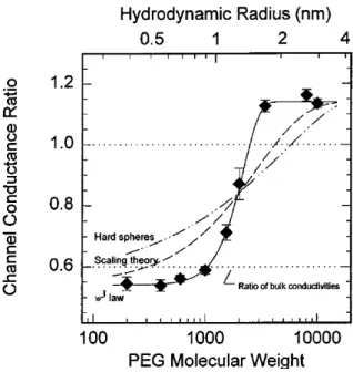
Bezrukov, Kasianowicz et al. [28] later conducted additional PEGs experiments and in 1996 they published a study about the dynamics and free energy of PEGs in the H pore. It was demonstated that the diffusion is not able to describe the interaction of the PEG with the pore and that a polymer had an attractive interaction with the pore, by being reversibly adsorbed to the pore wall. They showed that the predictions using the scaling theory (De Gennes scaling concepts on polymer) and the simple diffusion model of a hard sphere into a cylinder were in disagreement with their results, as shown in Figure 5 (with the two dashed line representing the disagreeing model). The solid line in Figure 5 represents the fitting of the relative average conductances, with the model they proposed in their study, in presence and absence of the polymer in the solution. They were able to extract the partition coefficient ( ) = ( ) representing the average density of monomers inside the pore to those in the bulk, where w is the molecular weight of the PEG and w0 and are extracted from the experimental fitting.
17
Figure 5: Hemolysin conductance ratio & Hydrodynamic Radius vs PEG molecular Weight. Extracted from [28]
They accounted for the polymer pore interaction by using an ''Ad-Hoc'' description of the diffusion coefficient that took into account the polymer’s weight (i.e size) dependence of interaction with the pore (polymer size dependent activation process) with a diffusion coefficient ( ) ∝ / , where RD is the hydrodynamic radius of the polymer. In this paper they suggested that the interaction of the PEG with the pore walls might be of hydrophobic nature, given the usual hydrophobic nature of the PEG-protein interactions. This finding was surprising because of the aqueous nature of this type of channels. This study suggested a complex set of interactions besides diffusion, between the analyte and the pore.
In another study by Merzlyak et al. [29], they showed that using an asymmetrical addition of PEG (one side only of the pore) is able to better probe the pore geometry than in the symmetrical case, where the PEG is present on both sides of the nanopore (the symmetrical case was described in a previous study [26]). Using a filling parameter which describes the ratio of pore length (accessible to the polymer) relative to the whole length of it, ( ) = ( ) ( )
( ) ( )), they plotted it
against the polymers hydrodynamic radii and they performed the experiment by placing the PEG asymmetrically in the cis or the trans side (to probe the differences in the pore’s geometry from both sides). As shown in Figure 6, they observed that the polymers with hydrodynamic radii over 1.2-1.3nm don’t partition into the pore anymore. Based on this result, they concluded an almost similar opening from both sides of the pore. Furthermore, they were able to deduce from this method the channel’s constriction of 0.6-0.7nm and 0.9nm. These results were in close agreement
18
with the light crystallographic results by Song et al. [30], showing the powerful tool that nanopores are for such measurements.
Figure 6: Dependence of Filling coefficient (cis and trans) on the PEG hydrodynamic radius. Ref [29]
Another study by Movileanu et al. [31], showed polymer partitioning into the α-Hemolysin pore for PEG ranging from 940 to 6000 Da. They used the partition coefficient and scaling laws of Daoud and De Gennes and they deduce from their results that molecules with molecular weights higher than 5kDa seem to not partition into the pore.
Salt also showed to play an effect on the interaction of the polymers with the nanopores. Krasilnikov, Kasianowicz and collaborators revealed in different studies that close to the limit of solubility of the salt they observed an enhanced interaction between the PEG and the -hemolysin pore. In 2006 Krasilnikov, Rodrigues and Bezrukov [32] demonstrated the dependency of interaction for different molecular weights of PEG with -hemolysin in a 4M KCl solution. Increasing salt concentration changed polymer partitioning and allowed them to observe time-resolved PEG-induced blockades. They found that dwell times increased with molecular weight until a certain size (~PEG 3400), where the polymer seems to not be completely accommodated in the pore anymore and decreased for higher molecular weights. They explained this phenomenon by the polymer’s tail, which was not accommodated inside the pore and acts like an entropic spring pulling on the trapped part of the polymer.
19
Figure 7: PEG residence time vs PEG molecular weight. PEG is fully accommodated until ~3400, then the entopic
spring pulls on the polymer for higher molecular weights. Ref [32]
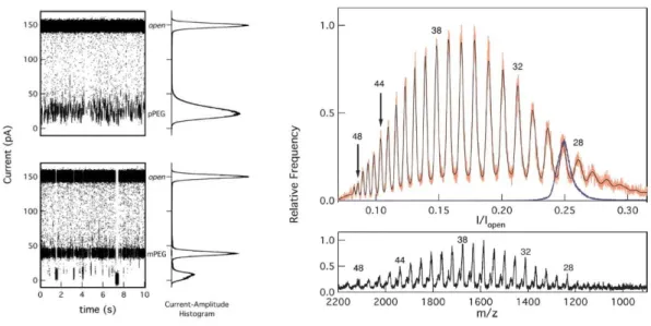
The study of Kasianowicz, Krasilnikov et al. of 2008 [33], showed the mass-dependent interaction of the -Hemolysin and a polydisperse solution of PEG. Using the amplitude of a polymer blockade, they obtained a mass spectrum in agreement with the MALDI-tof spectrum (Figure 8) and they demonstrated that characteristic dwell time values depend on the size of the polymer in solution with differences of one monomer. Indeed, it seemed that the pore was highly sensitive to the size of the polymer and that the interactions between the pore and different molecular weights of the polymer showed clear conductance differences.
20
Figure 8: Conductance and PEG blockades in hemolysin nanopore and corresponding histograms (left) Histogram of
nanopore conductance in the presence of polydisperse PEG1500 and corresponding MALDI TOF. Ref [33]
Later, in a more detailed study about salt concentration effects on the pore-PEGs interaction [34] the authors clearly showed that increased 4M KCl electrolyte concentration increased the on-rates, which became hundreds of times larger than in 1M, while off rates become several hundreds of times smaller. They deduced that the neutral polymers, in this case, can form complexes with ions and that this changed interaction with the -hemolysin pore. In this case, they deduced the ability of PEG to be charged in a high salt condition is because of a non-linear voltage dependency of constant rates. They also observe a high increase in the on-rate constant (and amplitudes) allowing the recognition of single molecules, due to salting out effects, change in polymer conformation, and Coulombic interactions.
These kinds of experiments with increased salt solution were enlarged for different molecules (neutral polymers and polyelectrolytes) and also pore types, as they proved to be powerful tools for the sizing of various analytes [33], [35], [36]. In many of our experiments, we also use a high salt solution to observed enhanced effects of interaction between the pores and our polymers of interest.
Other recent experiments working with neutral polymers include probing the effect of ions on PEG partitioning into the -hemolysin pore [37], showing the importance of alkali cation type in such experiments and another one on the electroosmotic flow in the same channel and PEG
21
[38]. These last ones are closely related to our studies and will be further discussed in the next chapters.
2. Experiments with DNA
The first pioneer experiment in the study of DNA transport through a protein channel was carried out by Kasianowicz et al in 1996. This experiment focused of the study of polynucleotides. In the experiments the authors used DNA and RNA: Poly[A]50 and polyUracil (Poly[U]50), highly negatively charged molecules that were driven by an electrical field towards the pore to achieve translocation [3]. By showing that the lifetime of blockades were proportional to the mean length of the RNA molecules, that the increase of voltage decreases the lifetime of the blockades in general and finally, by using PCR, they demonstrated that ssDNA translocates through the pore in contrast to double stranded DNA, which doesn’t, it was suggested that the molecules completely thread through the pore as extended rods.
Later on, based on their results, they explained the voltage dependence of the capture rate using a Van’t Hoff Arrhenius transition state equation [39].
= (36)
Where is the probability factor ~1, is the frequency factor, ∆U the energy driving the polynucleotide, and the activation energy, i.e the barrier height. Assuming that the electrical energy of the polymer is = | | one can rewrite the rate equation as:
= | | (37)
To get a value for the activation energy they estimated the value of Ro, where Ro is described by . Here, C is the concentration of polymer, D the diffusion coefficient, and A cross section of the channel and l the channel length. The energy barrier was estimated to be ~ 8kBT
Assuming there is only one barrier for polymer entry, it is automatically deduced that an increase in the electrical field reduces the energy required to confine the polymer in the pore. By fitting the results in Figure 9, they obtained a valency of ≈ 2, explaining this result by an average of two nucleotides interacting with the pore entrance and initiating the translocation (as in these
22
conditions one phosphate group bares an average of one negative charge). They explain later that the polynucleotides are captured more frequently from one side than the other due to an asymmetry in the pore. They argue that the confinement barrier is higher on one side in comparison to the other due to a difference in geometry and that there is a higher density of negative charges on one side than on the other, which increases the electrostatic repulsion and thus the barrier for entry.
Figure 9: Rate of DNA blockades from cis and trans vs Voltage (absolute value). Ref [39]
Since the first DNA experiments, enhancing DNA sensing using biological nanopores has been one of the most challenging and important aspects of this field. The most widely used pore for this purpose up until today was the α-Hemolysin pore, which can easily form pores in lipid bilayers. With this pore, very stable recordings could be carried out for long durations of time. Through various experiments it was indeed shown with this particular pore that detection with various molecules could be improved by placing a molecular adapter at the pore entrance [40], [41], by genetically engineering the pore [42], [43]. Moreover, it has later been found by Bayley, Ghadiri, et al. [44] that ssDNA detection can be enhanced by trapping DNA hairpins, achieving measurements in current detection between different ssDNAs, up to one nucleobase.
23
In the study by Clark, Bayley et al. [41] they demonstrated that a covalently attached molecular adapter could be used for continuous detection of indigenous mononucleotides. A -cyclodextrin which narrows the constriction of the beta-barrel and enhances the chemical specificity of the pore allows detection of the single nucleotides of dAMP, dGMP, dCMP, dTMP (2′-deoxyadenosine 5′-monophosphate (dAMP), 2′-deoxycytidine 5′-monophosphate (dCMP), 2′
-deoxyguanosine 5′-monophosphate (dGMP), and 2′-deoxythymidine 5′-monophosphate (dTMP)).
They obtained a resolution up to a single nucleotide (every mononucleotide passing through the pore produced a different current signature) with a confidence interval of 99% in the optimal experiment condition. In a following study of Stoddart, Bayley et al. [43] it was shown that recognition of DNA molecules can be improved using engineered α-Hemolysin pores, and DNA was immobilized using a biotin-steptavidin linkage as (see Figure 10). This study showed that discrimination between types of nucleobases within one DNA molecule could be achieved by three different sites in the α-hemolysin pore, which was not the case for the WT pore.
Figure 10: A. Engineered α-Hemolysin pore ref [43]. a. Current trace with nucleotides blockades. b. Corresponding
histograms ref [41]
Although the α-Hemolysin nanopore was, up until then, the most widely used pore for DNA sequencing, it presented a drawback. Indeed, the β-barrel is about 5 nm long and is able to accommodate up to 10 bases of ssDNA and the current is very much altered by all the nucleotides
24
at once. Even though engineering the pore makes single nucleotide differentiation in a homopolymer sequence containing a single different base possible, as shown in [43], the high fidelity of such measurements for much larger and complex DNA sequences was not proved. This was due to the fact that it was difficult to achieve high signal-noise ratios with such small differences in current detection.
Another pore, which has lately proved to be very powerful for the detection of ssDNA, is the MspA biological pore. This nanopore, which has a different geometrical shape, has a much narrower barrel constriction of 1.2 nm diameter and 0.5 nm long. With this barrel it is possible to accommodate around 4 base pairs, in contrast to 10 bp in the α-Hemolysin β-barrel. The experiments aiming to achieving this technology with MspA were started by Jens Gundlach and collaborators and have since been carried out mainly by the same group. The first experiment carried out was published in 2008 [45], and was the first study where DNA interaction with MspA was shown with a mutant of this pore. Negative charges at the narrowest constriction of the channel were eliminated, which allowed a higher capture of the DNA hairpins in contrast to the WT MspA (very low capture rate because of the repulsion between DNA and negative charges). They showed that the mutant with a positive distribution of charge is the most stable regarding the gating behaviour, and that the duration of interaction between the pore and the ssDNA becomes longer (100µs, in contrast to less than 10µs with the first mutant).
The latter method was further developed by the same group [46], showing that the three bases adjacent to the DNA hairpin are those responsible for the residual current values and that less than 4 bp are participating in the reduction of the pore’s conductance. It was also demonstrated, as a proof of principle, that it is possible to achieve a single nucleotide read out by amplification of the single nucleotide and addition of duplexes of double stranded DNA in between the single stranded DNA nucleotides. This was called duplex interrupted nanopore sequencing: each nucleotide gives rise to a conductance value and then due to the high electrical field in the nanopore, the duplex dissociates, allowing the next nucleotide to move forward in the pore and so on.
25
Figure 11: Detection and translocation of DNA hairpins through the MspA nanopore. Ref [46]
Lately, a new biological pore less explored, the aerolysin nanopore, has been used to size short ssDNA oligonucleotides, detect temperature effects in the transport and detect pepitdes. These ssDNA experiments performed in the group of Yitao Long showed the power of discrimination of the aerolysin pore with short polynucleotides, (3 to 10 adenines) [36] and later they revealed the different sites responsible for the DNA binding through experiments and simulations [47]. This pore, which was not investigated with DNA or peptides before, seemed to have a great potential for such kind of experiments as it is also highly charged. We will discuss this point furthermore into the thesis as we have also used this pore into our studies with DNA as well as neutral polymers and synthetic charged polymers.
Mathé et al and Wanunu et al. [48]–[50] performed other experiments measuring escape times and orientation dependent interactions using DNA hairpins and biological nanopores. These experiments showed the dynamics of DNA hairpins in α-hemolysin with escape times and unzipping of the molecule under the applied voltage. They also observed different dynamics of interaction depending on the 3´ and 5´ orientation interaction of the molecule, with the pore validated by numerical simulations. We will discuss in more detail these last studies in our experimental section.
It is also possible to measure double stranded DNA using solid state nanopores. Different materials and techniques have been developed these past few years to optimize the measurements using these pores. The traditional material used was SiN because of its stable chemical nature and
26
low mechanical stress. The first group that used the SiN nanopore was Golovchenko and collaborators [51], reporting measurements of double stranded DNA in 5nm ion-beam sculpted pores. Other experiments measuring dsDNA have been reported using graphene films [52]–[54]. Using these films was attractive because of the comparable thickness of graphene films and spacing between DNA bases, which make them of great interest for single nucleobase detection. Lately, other techniques have been used coupling biological nanopores with solid state pores [55] or coating SiN surfaces and pores with lipid bilayers [56]. The major challenge with solid state nanopores is the control of the chemistry and charge reproducibility of the pores over the different experiments, which is of a major importance, especially in single molecule detection.
III. Summary
We have reviewed some of the most important experiments, which have led to the big advances in the field of nanopore detection and allowed sizing, sequencing and understanding of some fundamental concepts of the interactions of polymers with ion channels, and more specifically biological nanopores. These studies constitute the basis of most our experiments and will be recurrently referred to in the context of our investigations.
We will explain in the next chapters the different pores that were used in our experiments and their particular properties. We will also present the experimental setup used and technique of data analysis and finally discuss the results obtained during this thesis with the various biological and synthetic polymers studied.
27
Chapter 2: Biological pores and ion channels
Pores are of primary importance for biological studies. Indeed, they represent one of the main pathways of transport for prokaryote and eukaryote cells. The wide classes of pores that exist in nature play essential roles in the regulation and molecular transport through the cell membrane.
These pores can allow the transport of necessary nutrients, regulation of the cell’s processes or signaling between cells and molecule transfer accomplished by various degrees of pore selectivity can critically vary within cell types of the same species and between different species. Internal transport (cytoplasm-nucleus transport) and external cell transport and signaling mechanisms (cell’s external membrane-outer environment) are based on these pore functioning. Because of passive transport (the transport of molecules through diffusion) of small molecules, including ions, most of the pores are ion channels, however some pores only serve as a pathway for the selective transport of ions, and are called ion channels.
One of the main examples of regulatory internal signaling is transport through the nuclear pore [57], [58]. The nucleus which contains all the genetic information needs to have a strong protection from the outer environment. Its pore allows some passive transport (molecules <40kDa, ions, metabolites etc..) but is however highly selective to higher size molecules, through a complex pathway of selectivity governed mainly by nuclear pore proteins called FG-Nups (nucleoporins which contain repeat units of phenylalanine and glycine: FG) organized in a mesh-like structure, and permits transport of ribosomal complexes and external transport of RNA to the cytoplasm.
An instance of cell to cell signaling based on ion transport, is in the case of the nervous system. Neurons ion channels in the case of eukaryotic cells, which transport low amounts of ions, are highly specific to the ion type and are voltage gated (open only when the membrane potential changes). This specificity is crucial for the propagation of nerve impulses. During transmission of these signals one impulse is transferred through a very specific pathway of neurons and these ion channels play a crucial role for the polarization/depolarization of each neuron cell, giving rise to the mechanism of action potentials (threshold mechanism of ion transport necessary for transmission and directionality of signals in the nervous system). The signal which then travels through the neuron (due to membrane depolarization, the voltage gated channels open) to the end of axon is finally transferred through the synapses where a chemical substance is transferred to a
28
receptor opening ligand-gated sodium channels, causing in turn depolarization of the next neuron membrane, which in turn transfers the impulse.
One other main example of ion selective pore is the KcsA pore of the prokaryotic cell
Streptomyces lividans (soil bacteria). It is a voltage-gated potassium channel, and it was the first
K+ channel to be characterized using X-Ray crystallography [59]. Research on this channel, has revealed important mechanisms in ion selectivity and gating function. In addition, part of its structure seemed to be highly conserved within the various K+ channels of prokaryotes and eukaryotes [60], it became a model for the study of potassium channel.
These examples illustrate the complexity and selectivity of such systems that represent important instances necessary to carry processes in biology. However, other pores, which are mostly prokaryotic based are less selective than the previously cited examples, and can serve other purposes than regulation, as it is the case of the mycobacterium porins which serve nutrients transport into the mycobacterium or the pore forming toxins (PFTs), which are toxins that are secreted by the bacteria for host infection purposes. It is important to study these classes of pores to understand their functioning mechanism which is ultimately necessary for biological and medical research application, like drug delivery.
In the frame of our experiments these pores can also be used for different purposes, and they were proved to be powerful tools for sensing and biotechnological applications: because of their high stability in in vitro experiments and their lack of selectivity they can interact with various molecules and various types of polymers ranging from synthetic polymers like Polyethylene Glycol (PEG), or natural polymers like DNA and RNA, to various peptides and proteins, which gives them a high potential for characterization. In addition it is important to understand some processes like translocation of polymers like RNA through the nuclear pore [57], [58], [61] or DNA through viruses capsids [62], [63] for example which use some similar kinetic mechanisms for these processes, and these bacterial pores can be used as model systems to unravel these various biological and physical processes (like confinement, intermolecular forces, etc..).
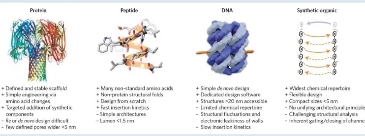
29 I. Comparison of the different existing nanopores for molecule sensing
As briefly mentioned in the first chapter of this work, in the polymer-pore experiments, various types of pores can be used, ranging from biological to solid state nanopore, or other more “exotic” pores constructed from DNA (in the same principle than DNA origami). The various pores and their applications in addition to their advantages and drawbacks are described in refs.[64]–[66]. As explained, biological nanopores, show a high potential for the detection of various molecules: polymers (DNA, synthetic etc..), proteins and are one of the most stable structures for this purpose. Other pores which are solid state nanopores, or pore hybrids, are also used by other groups for detection of polymers for specific advantages and depending on the interest of the user. The following figure [65] and table [66] summarize the advantages and inconvenients of the various existing pores:
Figure 12: Comparison of the four main classes of pores and listing of their advantages and inconvenients. Extracted
30
Table 1: Comparison between the different pores. Extracted from [66]
Based on the characteristics described in Figure 12 and Table 1, we have chosen to use biological pores, because of their high stability in bilayers their sensitivity in detection of polymers and the fact that they are cheap in comparison to other pores. They also have proved over the course of various studies, to be precise tools for the detection of H and D, divalent cations, polyethyelene glycols, sugar molecules, DNA and RNA, various proteins and peptides [6], and demonstrated great potential for the sensing, sizing, sequencing, and study of fundamental processes of the various molecules [6], [64].
We will present next the different pores that were used in the studies namely two PFTS and one mycobarterium porin. We will present their atomistic structures which are commonly determined via X-Ray crystallography (however for one of the presented PFTs -aerolysin- the situation is more complex and will be explicated further on) we will present in detail the various research that has been done to unravel their amino-acidic structure, their geometrical structure as well as their mechanism of insertion into the lipid membranes. It is important to know and understand the structure of these protein pores, in the frame of our molecule-detection experiments, because molecular and geometrical properties directly influence the interaction between the protein-pore and the polymers or molecules that we want to study or sense.
![Figure 1: Model of the random walk of an ideal chain. Extracted from [8]](https://thumb-eu.123doks.com/thumbv2/123doknet/12721473.356676/20.892.232.639.111.388/figure-model-random-walk-ideal-chain-extracted.webp)

![Figure 9: Rate of DNA blockades from cis and trans vs Voltage (absolute value). Ref [39]](https://thumb-eu.123doks.com/thumbv2/123doknet/12721473.356676/39.892.237.578.331.672/figure-rate-dna-blockades-trans-voltage-absolute-value.webp)
![Figure 11: Detection and translocation of DNA hairpins through the MspA nanopore. Ref [46]](https://thumb-eu.123doks.com/thumbv2/123doknet/12721473.356676/42.892.208.660.111.388/figure-detection-translocation-dna-hairpins-mspa-nanopore-ref.webp)
![Figure 19: Extracted from [99] (a) Schematics of symmetrical lipid bilayer with representation of repulsive and attractive attractions](https://thumb-eu.123doks.com/thumbv2/123doknet/12721473.356676/64.892.231.657.111.452/figure-extracted-schematics-symmetrical-representation-repulsive-attractive-attractions.webp)
![Figure 20: extracted from [98] schematics of rearrangement of asymmetrical monolayers](https://thumb-eu.123doks.com/thumbv2/123doknet/12721473.356676/65.892.165.715.104.460/figure-extracted-schematics-rearrangement-asymmetrical-monolayers.webp)
