ORIGINAL ARTICLE
The sensitivity of an interferon-
γ release assay
in microbiologically confirmed pediatric tuberculosis
Kurt Schopfer&Hans L. Rieder&Thomas Bodmer&
Jacqueline F. Steinlin-Schopfer&Yay Chantana&
Peter Studer&Denis Laurent&Beat Richner
Received: 16 March 2013 / Revised: 11 September 2013 / Accepted: 13 September 2013 / Published online: 25 September 2013 # Springer-Verlag Berlin Heidelberg 2013
Abstract This study aimed at determining the sensitivity of a whole blood interferon-γ release assay (IGRA) among children with microbiologically confirmed tuberculosis in a high-burden country. Children with a diagnosis of tuberculosis based on clinical and radiographic assessment were tested with an IGRA in addition to microbiologic examination of appropriate speci-mens for acid-fast bacilli, mycobacterial rRNA, and observa-tion for growth of Mycobacterium tuberculosis on appropriate culture media. Of the 405 children with a clinical diagnosis of tuberculosis, 91 (22.5 %) had microbiologically confirmed tuberculosis, of whom 81 were tested with an IGRA. A positive result was obtained in 43 (sensitivity 53.1 %, 95 % confidence interval 42.3 to 63.6 %), uninfluenced by age, sex, or disease manifestation. Conclusions: The sensitivity of a whole blood
interferon-γ release assay in microbiologically confirmed
pedi-atric tuberculosis was low. An IGRA cannot, thus, be used as rule-in test, but it might be useful to rule in tuberculosis among children in whom tuberculosis is notoriously difficult to con-firm microbiologically.
Keywords Cambodia . Children . Diagnosis . IGRA . Tuberculosis
The diagnosis of pediatric tuberculosis proves difficult in any setting. In high-burden countries with constrained resources, the diagnosis of childhood tuberculosis is often reliant on clinical judgment alone. Even with resources available, the paucibacillary nature of childhood tuberculosis makes its mi-crobiological confirmation a formidable challenge. Demon-stration of bacilli by microscopy, culture, or nucleic acid
amplification is feasible in a minority of cases only [4]. The
yield is particularly poor in very young children [15].
We previously reported on the implementation of labora-tory diagnostic services in a large pediatric referral hospital in
Cambodia [15]. While the focus was on the feasibility of
implementing microbiological services for tuberculosis, an interferon-γ release assay (IGRA) was also included in the evaluation of children with a clinical diagnosis of tuberculosis. The purpose here was to determine the potential contribution of an IGRA, evaluated against the standard of microbiologi-cally confirmed tuberculosis.
Material and methods Setting
The setting has been described [15]. The Jayavarman VII
Hospital in Siem Reap is the largest of the five Kantha Bopha pediatric referral hospitals in Cambodia. Together, these hos-pitals took care of about 120,000 hospitalized and 800,000
ambulatory pediatric patients in 2011 [13]. During the period
from 1 July 2005 through 31 March 2006, all children hospi-talized in this hospital, who are with a clinical diagnosis of tuberculosis were systematically evaluated based on a proto-col ensuring a standardized flow for examinations.
K. Schopfer (*)
:
T. Bodmer:
J. F. Steinlin-Schopfer Institute of Infectious Diseases, University of Bern, Friedbühlstrasse 51, 3010 Bern, Switzerland e-mail: [email protected]K. Schopfer
Scheuermattweg 43, 3043 Uettligen, Switzerland H. L. Rieder
International Union Against Tuberculosis and Lung Disease, 68 blvd Saint-Michel, 75006 Paris, France
H. L. Rieder
Institute of Social and Preventive Medicine, University of Zurich, Hirschenbraben 84, 8001 Zurich, Switzerland
Y. Chantana
:
P. Studer:
D. Laurent:
B. RichnerKantha Bopha Foundation, Phnom Penh, Cambodia, c/o Intercontrol AG, Seefeldstrasse 17, 8008 Zurich, Switzerland
Procedures and ethics considerations
At admission to the hospital, the child's guardian is requested to provide informed and signed (fingerprint) consent for the necessary diagnostic and treatment procedures. This record is kept as a permanent document in the patient's file. Before project implementation, diagnosis of tuberculosis was based on clinical assessment, imaging techniques, and bright field microscopy. A key purpose of this project was upgrading diagnostic mycobacteriological techniques on the same spec-imen with rRNA amplification. The diagnostic procedure was supplemented by an IGRA in children suspected of having tuberculosis to establish the presence of tuberculous infection
to enhance the probability to rule in tuberculosis [2].
Methods
Routine laboratory examinations upon admission included hematology, biochemistry, and urine examinations in accor-dance with locally established hospital standards. Laboratory tests, made after obtaining written consent from the child's guardian, included testing for human immunodeficiency virus
(HIV) with an opt-out approach as recommended [17].
Imag-ing techniques were utilized at the discretion of the clinician subsequent to hospital admission once tuberculosis was clin-ically suspected. Among the children with a clinical diagnosis of tuberculosis supported by radiographic evidence, routine laboratory tests were supplemented by a whole blood IGRA (QuantiFERON®-TB Gold In Tube, Cellestis Ltd., Carnegie, Australia). The IGRAs were done on the same day as blood was collected. A positive IGRA result was defined according to the manufacturer's recommendation.
Tuberculosis-specific laboratory examinations included collections of clinical specimens: a first one on the spot, followed by additional ones to preferably obtain three speci-mens on three consecutive days. All the specispeci-mens were decontaminated and concentrated by centrifugation prior to splitting into aliquots for microscopy (Ziehl–Neelsen method),
rRNA amplification and culture, respectively, as described [15].
A case of microbiologically confirmed (henceforth, shortened
to “confirmed”) tuberculosis was a patient with at least one
positive result by any of the three methods on at least one specimen.
Electronic database and analysis
The data were captured in an EpiData relational database (EpiData Association, version 3.1) and analyzed with EpiData
Analysis version 2.2 (freely available athttp://www.epidata.dk)
and OpenEpi (http://www.openepi.com/OE2.3/Menu/
OpenEpiMenu.htm) as appropriate. The data were abstracted from the paper documents as recorded by health care staff. Up to three disease sites were electronically captured. Electronic
recoding used an algorithm with a predetermined hierarchy of intrathoracic (including pulmonary, intrathoracic lymphatic, and pleural), followed by lymphatic (extrathoracic), soft tissue, osteoarticular, central nervous system (CNS), and finally other sites to obtain a uniform classification for major, second, and
third disease sites. We used the classification “intrathoracic”
rather than“pulmonary” because of the difficulties in childhood
tuberculosis of the respiratory tract to clearly separate tubercu-losis limited to the lung parenchyma (pulmonary) from other intrathoracic manifestations, primarily the intrathoracic lym-phatic system. For categorical variables, we determined propor-tions with 95 % confidence intervals (CI) and odds ratios with 95 % CI for contrasting binomial outcomes. Where deemed appropriate, adjustments for potentially confounding factors were made by stratification using the Mantel–Haenszel proce-dure. For continuous variables, we used standard measures of central tendency such as means and percentiles.
Results
Four hundred and five children had a clinical diagnosis of tuberculosis in the period from 1 July 2005 through 31 March 2006. In 91 (22.5 %), the clinical diagnosis was confirmed
(Table 1). Intrathoracic, lymphatic, soft tissue, and
osteo-articular tuberculosis contributed 316 (89.1 %) of all cases, and 88 (96.7 %) of confirmed cases. The proportion of the confirmed cases among each of these forms was similar (odds ratio of the confirmed intrathoracic tuberculosis versus all other confirmed forms 1.5, 95 % CI 0.9–2.4). Among the 21 cases with tuberculosis of the CNS, only 1 was confirmed and only 2 of the 23 cases with other disease manifestations. However, six of an additional seven patients with CNS tuber-culosis as hierarchically non-primary site had microbiological confirmation.
While 71 cases (17.5 %) were diagnosed among children less than 1 year old, only 4 of the 91 confirmed cases were from this age group. Except for soft tissue tuberculosis, more boys than girls were diagnosed with tuberculosis, reflecting
the admission pattern [15]. Given a diagnosis of tuberculosis,
confirmation was more often obtained among girls than among boys (odds ratio 1.64, 95 % CI 1.03–2.6).
Two or more disease sites were noted in 66 patients (16.3 %). The probability of obtaining confirmation increased with the number of sites affected (significant chi-square for trend). Among the children with intrathoracic tuberculosis, other forms were almost as frequently diagnosed (26 cases) as major second sites as were lymphatic, soft tissue, osteo-articular, and CNS tuberculosis combined (29 cases). Of the 405 children, the HIV test result was recorded in 23 (5.7 %) and was negative in all.
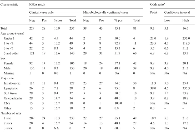
The IGRA results were available for 338 (83.5 %) children: 71 IGRA-positive children, 43 (60.6 %) had confirmed
Ta b le 1 C lini cal an d b act er iologi cal ch ar ac ter isti cs o f tuber culos is pati ents fr om Jayavarman VII H ospit al, S iem Rea p (p er iod o f repr ese ntat ive sa m pl in g is from 1 July 2005 through 31 March 2006) Cha ra cte ri stic H ier ar chi cal ly fir st d is eas e site To ta l In tr athor ac ic L y mpha tic Soft ti ssue O st eoar ti cula r C N S O ther P o s T otal % p os P o s T o tal % pos Pos T otal % pos Pos T otal % pos Pos T otal % pos Pos T otal % pos P o s T otal % pos T o tal 5 6 219 25.6 9 4 1 22.0 1 1 4 6 23.9 1 2 5 5 21.8 1 21 4.8 2 23 8.7 9 1 405 22.5 Age g roup Under 1 4 4 9 8 .2 0 5 0.0 0 7 0 .0 0 4 0.0 0 3 0 .0 0 3 0.0 4 71 5.6 1 to < 3 6 49 12.2 0 7 0 .0 1 3 33.3 3 6 50.0 1 1 100.0 0 4 0 .0 1 1 70 15.7 3 to < 5 6 17 35.3 2 5 40.0 0 9 0 .0 0 1 0.0 0 1 – 00 – 8 3 3 24.2 5 and older 4 0 104 38.5 7 2 4 29.2 1 0 2 7 37.0 9 44 20.5 0 16 0.0 2 16 12.5 6 8 231 29.4 Se x Female 31 101 30.7 5 1 8 27.8 7 25 28.0 5 19 26.3 0 6 0 .0 1 1 0 10.0 4 9 179 27.4 Male 25 1 1 8 21.2 4 2 3 17.4 4 20 20.0 7 36 19.4 1 15 6.7 1 13 7.7 4 2 225 18.7 Miss ing 0 0 – 00 – 01 0 .0 0 0 – 00 – 00 – 01 0 .0 Number of involved sites 1 site 2 7 161 16.8 9 4 0 22.5 5 39 12.8 1 2 5 5 21.8 1 21 4.8 2 0.0 – 56 339 16.5 2 sites 24 53 45.3 0 1 0 .0 6 7 85.7 0 0 – 00 – 00 – 30 61 49.2 3 sites 5 5 100.0 0 0 – 00 – 00 – 00 – 00 – 5 5 100.0 Ma jor second si te Any second site 29 58 50.0 0 1 0 .0 6 7 85.7 0 0 – 00 – 0 0 35 66 53.0 L y mp hatic 5 7 71.4 N A N A N A 0 0 – 00 – 00 – 00 – 5 7 71.4 Soft tissue 3 10 30.0 0 0 – NA N A NA 0 0 – 00 – 00 – 3 1 0 30.0 Osteoarticular 5 6 83.3 0 0 – 6 7 85.7 N A N A N A 0 0 – 00 – 1 1 13 84.6 CNS 5 6 83.3 0 1 0 .0 0 0 – 00 – NA NA NA 0 0 – 5 7 71.4 Other 1 1 2 6 42.3 0 0 – 00 – 00 – 00 – NA NA NA 1 1 26 42.3 IG RA re sult Negative 2 3 138 16.7 2 2 8 7.1 5 34 14.7 6 35 17.1 0 15 0.0 2 17 1 1 .8 38 267 14.2 Positive 27 39 69.2 6 8 75.0 5 8 62.5 4 9 44.4 1 4 25.0 0 3 0 .0 43 71 60.6 Miss ing 6 42 14.3 1 5 20.0 1 4 25.0 2 1 1 18.2 0 2 0 .0 0 3 0.0 1 0 6 7 14.9 CNS central n ervous system, NA not applicable, IG RA inte rf er on-γ re lease assa y, Pos positive, % po s percentag e of positive cases
tuberculosis. Conversely, 38 (14.2 %) of all children with a negative IGRA result had confirmed tuberculosis. To further examine the role of the IGRA, additional analyses were done
among the 338 children with an IGRA result (Table2).
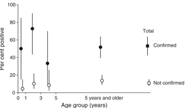
Strat-ifying by diagnostic type of tuberculosis (clinical diagnosis versus confirmed diagnosis), 43 of the 81 confirmed cases had a positive IGRA result (a sensitivity of 53.1 %, 95 % CI 42.3– 63.6 %), as compared to 28 of 257 (10.9 %) children with a clinical diagnosis but without confirmation. The age-specific distribution of IGRA positive showed no predilection for any
age group (Fig.1).
Analyzing the IGRA test results by site of disease, we found overlapping confidence intervals for the proportion of a positive IGRA result among patients with intrathoracic tuberculosis only, intrathoracic plus extrathoracic, and extra-thoracic tuberculosis only.
The odds ratio of finding a positive IGRA result among the microbiologically confirmed versus the clinically diagnosed
only cases was 9.2 (Table3). While the ratio varied somewhat
by age group, sex, and number of disease sites (one versus two, three or more), these variations were small, and adjust-ment for all these variables did not make any significant difference (odds ratio 7.6, 95 % CI 3.7 to 15.7).
Discussion
The diagnosis of tuberculosis in children is fraught with problems in the operating characteristics, notably test sensi-tivity, of virtually any assay being used. While systems based
on the identification of molecular methods [12,15] hold great
promise, the yield remains very low even under optimized conditions, and complex sample collection procedures are required (sputum induction and gastric lavage with subse-quent concentration). The problem is compounded the
youn-ger the children are [15].
Table 2 Univariate analysis of interferon-γ release assay result by case definition (clinical only versus microbiologically confirmed), Jayavarman VII Hospital, Siem Reap (period of representative sampling is from 1 July 2005 through 31 March 2006)
Characteristic IGRA result Odds ratioa
Clinical cases only Microbiologically confirmed cases Point Confidence interval Neg Pos % pos Total Neg Pos % pos Total Low High Total 229 28 10.9 257 38 43 53.1 81 9.3 5.1 16.6 Age group (years)
Under 1 42 2 4.5 44 2 2 50.0 4 21.0 1.9 236.0 1 to <3 44 5 10.2 49 3 8 72.7 11 23.5 4.7 118.3 3 to <5 22 2 8.3 24 4 2 33.3 6 5.5 0.6 51.2 5 and older 121 19 13.6 140 29 31 51.7 60 6.8 6.8 13.7 Sex Female 92 14 13.2 106 18 24 57.1 42 8.8 3.8 20.1 Male 136 14 9.3 150 20 19 48.7 39 9.2 4.0 21.3 Missing 1 0 0.0 1 0 0 NA 0 NA NA NA Major site Intrathoracic 115 12 9.4 127 23 27 54.0 50 11.3 5.0 25.4 Lymphatic 26 2 7.1 28 2 6 75.0 8 39.0 4.5 335.3 Soft tissue 29 3 9.4 32 5 5 50.0 10 9.7 1.7 53.8 Osteoarticular 29 5 14.7 34 6 4 40.0 10 3.9 0.8 18.8 CNS 15 3 16.7 18 0 1 100.0 1 NA NA NA Other 15 3 16.7 18 2 0 0.0 2 0.0 – – Number of sites 1 site 209 24 10.3 233 22 27 55.1 49 10.7 5.3 21.6 2 sites 20 4 16.7 24 14 13 48.1 27 4.6 1.3 17.3 3 sites 0 0 NA 0 2 3 60.0 5 NA NA NA
Adjusted odds ratio of a positive IGRA result by case definition, adjusted for age group, sex (one case with missing information excluded), and number of sites: 7.6 (95 % confidence interval 3.7 to 15.7), suggesting no confounding by the variables considered for adjustment
Neg negative, Pos positive, % pos percentage of positive cases
a
The difficulty in obtaining microbiological confirmation of tuberculosis also affects the choice of the gold standard to evaluate any new diagnostic technique in childhood
tuberculo-sis. As we have shown [15] even when using different
tech-niques on multiple specimens, tuberculosis could not be con-firmed in more than three quarters of children with a clinical diagnosis. This diagnostic shortcoming potentially introduces a selection bias in test evaluation. Conversely, a large number of children with microbiological confirmation were available in this study, providing a fairly sturdy estimate for the test sensitivity.
Thus, a high index of suspicion coupled with clinical acumen remains the most important prerequisite for the diag-nosis of tuberculosis in children. The frequency with which extrathoracic tuberculosis as the sole presentation was
identified in our setting indicates that tuberculosis was high on the list of the differential diagnosis. It is not surprising that the frequency of confirmation increased if more than one disease site was involved because the likelihood of
tuberculo-sis increases with multi-system disease [16]. We were
inter-ested in learning to what extent an IGRA could be of assis-tance in the diagnosis of tuberculosis in children as demon-strated in this comparatively large series of 81 confirmed tuberculosis cases. This issue has been addressed by others
[1,3,5,6,8,9,11]. A limitation to any study of tuberculosis in
children is the gold standard of microbiological confirmation. It is not surprising that sample sizes in most studies have commonly been relatively small. Of the above seven studies,
the one from South Africa [9] was the largest (involving 57
cases), the other six included fewer than 30 children. Small sample sizes result in relatively large reported variations in IGRA sensitivity, in these studies ranging from 56 to 93 %.
The issue has also been a subject of a meta-analysis [10].
Unfortunately, that analysis mixed“confirmed and/or
proba-ble active” tuberculosis and thus essentially invalidated it. Among our 81 children with confirmed tuberculosis, only 43 had a positive IGRA, a disappointingly low sensitivity of 53 %. Therefore, an IGRA test result cannot be used as a rule-out test for tuberculosis and does not substitute microbi-ological investigation. Conversely, because of the great diffi-culty in obtaining microbiological confirmation among chil-dren, a positive result can be useful to rule in tuberculosis. While only 6 % of our patients had their HIV status recorded, we have no evidence that there was a selection bias for the transcription. All 23 of these patients had a negative HIV test result. This suggests that HIV infection cannot explain the limited sensitivity of the IGRA in our patient population. The low frequency of positivity was independent of age, sex, and multi-system disease. Our study was not designed to deter-mine IGRA specificity, but specificity is of lesser concern as it is precisely this characteristic where the test offers a large
advantage over tuberculin skin testing [10]. While research
points to the future possibility to distinguish latent tuberculosis
infection and active disease [7], none of the currently available
immunological tests has that discriminatory ability [14].
In this setting, the sensitivity of the whole blood IGRA for diagnosing active tuberculosis was (remarkably) low and barely exceeded 50 % in microbiologically confirmed pediatric tuber-culosis. This poor sensitivity was not related to age, sex, and disease manifestation or to HIV infection. Nevertheless, given the very low yield of microbiological investigations in the very young child with suspected tuberculosis, the addition of even a low-sensitivity test may substantially improve the accuracy of the diagnosis in children under the age of 3 years.
Acknowledgments We thank the personnel at the Collaborating Insti-tute of Infectious Diseases in Switzerland: Jasmin Portmann introduced the diagnostic technologies, assisted in building up the operation of the Table 3 Odds ratio of a positive IGRA result among microbiologically
confirmed versus non-confirmed cases (crude and adjusted results) Variable(s) adjusted for Odds ratio
Point estimate 95 % confidence interval Low High Crude, non-adjusted 9.2 5.1 16.6 Adjusted for age 8.3 4.6 15.0 Adjusted for sex 9.0 5.0 16.2 Adjusted for multiple sites 8.5 4.5 16.0 Adjusted for age and sex 8.2 4.5 14.9 Adjusted for age and multiple sites 7.4 3.9 14.3 Adjusted for sex and multiple sites 8.5 4.5 16.2 Adjusted for age, sex, and multiple sites 7.6 3.7 15.7 Jayavarman VII Hospital, Siem Reap; period of representative sampling is from 1 July 2005 through 31 March 2006
Age group (years) 0 1 3 5
Per cent positive
0 20 40 60 80 100
5 years and older
Total
Confirmed
Not confirmed
Fig. 1 Age-specific proportion of positive IGRA results, by case defini-tion (clinical only versus microbiologically confirmed). Filled symbols denote cases with microbiological confirmation; hollow symbols denote clinically diagnosed cases without microbiological confirmation. Jayavarman VII Hospital, Siem Reap; period of representative sampling is from 1 July 2005 through 31 March 2006
tuberculosis laboratory, and trained the technical staff. Annemarie Hilty was responsible for the laboratory cultures. Doris Schopfer and Simon Lüthi were in charge of the reagent production, planning and logistics of shipments, and uninterrupted supply of material and equipment. The project was funded by a grant from the University of Bern and the Kantha Bopha Foundation.
Conflict of interest The authors declare that they have no conflict of interest.
References
1. Bianchi L, Galli L, Moriondo M, Veneruso G, Becciolini L, Azzari C, Chiappini E, de Martino M (2009) Interferon-γ release assay im-proves the diagnosis of tuberculosis in children. Pediatr Infect Dis J 28:510–514
2. Canadian Thoracic Society (2013) Canadian tuberculosis standards, 7th edition. Can Respir J 20(Suppl A):1A–174A
3. Cruz AT, Geltemeyer AB, Starke JR, Flores JA, Graviss EA, Smith KC (2011) Comparing the tuberculin skin test and T-SPOT.TB blood test in children. Pediatrics 127:e31–e38
4. Cruz AT, Starke JR (2010) Pediatric tuberculosis. Pediatrics Rev 31: 13–26
5. Detjen AK, Keil T, Roll S, Hauer B, Mauch H, Wahn U, Magdorf K (2007) Interferon-γ release assays improve the diagnosis of tuberculo-sis and nontuberculous mycobacterial disease in children in a country with a low incidence of tuberculosis. Clin Infect Dis 45:322–328 6. Dogra S, Narang P, Mendiratta DK, Chaturvedi P, Reingold AL,
Colford JM, Riley LW, Pai M (2007) Comparison of a whole blood interferon-γ assay with tuberculin skin testing for the detection of tuberculosis infection in hospitalized children in rural India. J Infection 54:267–276
7. Harari A, Rozot V, Bellutti Enders F, Perreau M, Mazza Stalder J, Nicod LP, Cavassini M, Calandra T, Blanchet CL, Jaton K, Faouzi M, Day CL, Hanekom WA, Bart PA, Pantaleo G (2011) Dominant TNF-α+ Mycobacterium tuberculosis-specific CD4+T cell responses discrimi-nate between latent infection and active disease. Nat Med 17:372–376
8. Kampmann B, Whittaker E, Williams A, Walters S, Gordon A, Martinez-Alier N, Williams B, Crook AM, Hutton AM, Anderson ST (2009) Interferon-γ release assays do not identify more children with active tuberculosis than the tuberculin skin test. Eur Respir J 33: 1374–1382
9. Liebeschuetz S, Bamber S, Ewer K, Deeks J, Pathan AA, Lalvani A (2004) Diagnosis of tuberculosis in South African children with a T-cell-based assay: a prospective cohort study. Lancet 364:2196–2203 10. Mandalakas AM, Detjen AK, Hesseling AC, Benedetti A, Menzies D (2011) Interferon-gamma release assays and childhood tuberculosis: systematic review and meta-analysis. Int J Tuberc Lung Dis 15: 1018–1032
11. Markova R, Drenska R, Minchev P, Todorova Y, Ciccozzi M, Amicosante M (2011) Association of age with the level of response in the QuantiFERON-TB Gold In-Tube assay for children with active tuberculosis. New Microbiol 34:81–85
12. Nicol MP, Workman L, Isaacs W, Munro J, Black F, Eley B, Boehme CC, Zemanay W, Zar HJ (2011) Accuracy of the Xpert MTB/RIF test for the diagnosis of pulmonary tuberculosis in children admitted to hospital in Cape Town, South Africa: a descriptive study. Lancet Infect Dis 11:819–824
13. Richner B (2012) Annual report of the Foundation of Children's Hospitals Kantha Bopha, Dr. med. Beat Richner. Annual Report 2011. http://www.beatrichner.ch/pdf/Jahresberichte. Accessed August 13, 2012
14. Ruhwald M, Aabye MG, Ravn P (2012) IP-10 release assays in the diagnosis of tuberculosis infection: current status and future direc-tions. Expert Rev Mol Diagn 12:175–187
15. Schopfer K, Rieder HL, Bodmer T, Steinlin-Schopfer JF, Chantana Y, Somathea T, Studer P, Laurent D, Richner B (2012) Laboratory diagnosis of tuberculosis in a large pediatric hospital in Cambodia. Int J Tuberc Lung Dis 16:503–509
16. Scully RE, Mark EJ, McNeely WF, McNeely BU (1988) Case records of the Massachusetts general hospital. Weekly clinicopatho-logical exercises. Case 35–1988. 63-year-old asplenic man with myelofibrosis, fever, and bloody diarrhea. N Engl J Med 319:564– 574
17. World Health Organization, UNAIDS (2007) Guidance on provider-initiated HIV testing and counselling in health facilities. World Health Organization Document 1–56