HAL Id: hal-03161407
https://hal.archives-ouvertes.fr/hal-03161407
Submitted on 9 Mar 2021
HAL is a multi-disciplinary open access
archive for the deposit and dissemination of sci-entific research documents, whether they are pub-lished or not. The documents may come from teaching and research institutions in France or abroad, or from public or private research centers.
L’archive ouverte pluridisciplinaire HAL, est destinée au dépôt et à la diffusion de documents scientifiques de niveau recherche, publiés ou non, émanant des établissements d’enseignement et de recherche français ou étrangers, des laboratoires publics ou privés.
human 5-HT4 receptor mRNA
Marjorie Maillet, Monique Gastineau, Pascal Bochet, Marie-Liesse
Asselin-Labat, Eric Morel, Jean-Noël Laverrière, Anne-Marie Lompré,
Rodolphe Fischmeister, Frank Lezoualc’H
To cite this version:
Marjorie Maillet, Monique Gastineau, Pascal Bochet, Marie-Liesse Asselin-Labat, Eric Morel, et al.. Functional studies of the 5’-untranslated region of human 5-HT4 receptor mRNA. Biochemical Journal, Portland Press, 2005, 387 (2), pp.463-471. �10.1042/BJ20040744�. �hal-03161407�
Functional studies of the 5’-untranslated region of the human
5-HT
4receptor mRNA
Marjorie Maillet, Monique Gastineau, Pascal Bochet, Marie-Liesse Asselin-Labat§, Eric
Morel, Jean-Noël Laverrière#, Anne-Marie Lompré, Rodolphe Fischmeister & Frank Lezoualc’h*
Cardiologie Cellulaire et Moléculaire, INSERM U-446, INSERM U-461§, IRF-75, Université Paris-Sud, Faculté de Pharmacie, F-92296 Châtenay-Malabry;
#CNRS UMR 7079, Université Pierre et Marie Curie, Paris; France
*Corresponding author. INSERM U-446, Faculté de Pharmacie, 5, Rue J.-B. Clément, F-92296 Châtenay-Malabry Cedex France, Fax 33-1-46 83 54 75
SYNOPSIS
The serotonin 5-HT4 receptor is a member of the seven transmembrane-spanning G
protein-coupled family of receptors and mediates many cellular functions both in the central nervous system and at the periphery. In this study, we isolated and characterized the 5’-flanking region of the human 5-HT4 receptor (h5-HT4). We demonstrate the existence of a novel exon which
corresponds to the 5’ untranslated region (UTR) of the h5-HT4 receptor gene. RNase
protection analysis and RT-PCR experiments performed on human atrial RNA demonstrated that the major transcription start site of the h5-HT4 receptor gene is located at –3185 bp
relative to the first ATG codon. In addition, a 1.2 kb promoter fragment which drives the transcription of the 5-HT4 receptor was characterised. The promoter region lacks TATA and
CAAT canonical motivefs in the appropriate location but contains putative binding sites for several transcription factors. Transient transfection assays revealed that the [–3299/–3050] gene fragment possesses the ability to promote the expression of the luciferase reporter gene in human cell lines. In contrast, the promoter was silent in monkey COS-7 cells, indicating the requirement of specific factors to initiate transcription in human cells. In addition to the promoter element, enhancer activity was found in a region [-220/-61] located in the long 5’-UTR. Mutational analysis, gel shift and transfection assays identified a Nkx2.5 like binding site as a regulatory sequence of this enhancer. Our results suggest a complex regulation of the h5-HT4 receptor gene expression involving distinct promoters and non-coding exons.
Key Words: Serotonin – Promoter - Transcriptional regulation – Arrythmia - Nkx2.5 -
cyclic AMP
Abbreviations used: 5-HT, 5-hydroxytryptamine; HEK, human embryonic kidney; GPCR, G
protein-coupled receptors, h5-HT4 receptor, human 5-HT4 receptor; MEF-2, myocyte
enhancer factor-2; PCR, polymerase chain reaction; RPA, RNase protection assay; RT, reverse transcriptase, UTR, untranslated region.
INTRODUCTION
Serotonin (5-HT) is a major neurotransmitter both in the central nervous system and peripheral tissues. Of the 14 mammalian 5-HT receptor subtypes identified so far, all are G-protein-coupled receptors (GPCRs) to the exception of the 5-HT3 receptor that belongs to the
superfamily of ligand-gated ion channels [1]. The 5-HT4 receptor was first described as a
potent 5-HT receptor able to induce cAMP production in primary neuronal cultures from mouse colliculi [2]. Subsequent pharmacological studies have demonstrated the existence of 5-HT4 receptor responses in the heart, adrenal gland, bladder and digestive tract of various
species including human [3,4]. In mammalian brain, activation of the 5-HT4 receptor
increases memory and learning [5]. Accordingly, the 5-HT4 receptor stimulates acetylcholine
release in rat frontal cortex [6,7] and regulates the processing of the amyloid precursor protein [8,9]. In peripheral tissues, the 5-HT4 receptor has been shown to regulate gastrointestinal
tract motility, intestinal electrolyte secretion and bladder contraction [4].
In the heart, 5-HT4 receptors exert strong positive chronotropic, inotropic and lusitropic
effects in human and pig atria. However, unlike β-adrenergic receptors, the 5-HT4 receptor
exerts its effects exclusively on the atrial tissue and has no functional effects on ventricles [10,11]. In addition, the cardiac effects of the 5-HT4 receptor are restricted to human and pig
atria and are absent froma large number of laboratory animals, such as rat, guinea pig, rabbit and frog [12,13]. Regarding 5-HT4 receptor associated cardiac disorders, there are in vivo, in vitro, and clinical evidence that these receptors can trigger atrial arrhythmia [14]. Therefore,
the regulation of 5-HT4 receptor expression is of particular importance to understand their
involvement in this cardiac disorder.
Several groups have isolated and sequenced C-terminal splice variants of the human 5-HT4 (h5-HT4) receptor. Eight C-terminal isoforms and one internal splice variant have been
currently identified and named h5-HT4(a), h5-HT4(b), h5-HT4(c), h5-HT4(d), h5-HT4(e), h5-HT4(f),
h5-HT4(g), h5-HT4(n) and h5-HT4(hb) [15-19]. These receptors are heptahelical transmembrane
receptors that are positively coupled to adenylyl cyclase. The h5-HT4 receptor gene is located
on chromosome 5, bands 5q31-q33 [20] and contains 5 exons encoding the common transmembrane domains of the h5-HT4 receptor as well as eight alternatively spliced cassettes
which code for the internal and C-terminal splice variants [17]. The 5’-untranslated region (5’-UTR) of the h5-HT4 receptor gene has recently been characterised from human placental
cDNA. The 5’-UTR is extremely long in this tissue since it reached more than 5100 bp upstream from the translation start site and consists of 25 exons and a part of exon 26 which
includes the first methionine (Figure 1A) [21]. Thus, according to this study the entire h5-HT4
receptor gene spans more than 700 kb and is divided in at least 38 exons.
To date, there is no detailed information on the identification of the cis-acting elements which may be involved in the regulation of h5-HT4 receptor expression. Therefore, to further
understand the regulation of the h5-HT4 receptorgeneand its potential implication in cardiac
disorders such as atrial arrhythmia, we characterised its 5’-UTR in human atrial RNA. RT-PCR experiments and RNA protection assay showed a major transcription start site located within intron 25 at –3185 bp relative to the first ATG codon. In addition to the promoter element, enhancer activity was found within intron 25 but in a region (-220/-61) located in the 5’-UTR. Mutational analysis, gel shift and transfection assays identified a Nkx2.5 like binding site as a regulatory sequence of this enhancer.
MATERIALS AND METHODS
Surgery
All protocols for obtaining human tissue were approved by the ethics committee of GREBB institute (Hôpital de Bicêtre, Université de Paris-Sud). Specimens of right atrial appendages were obtained from patients undergoing heart surgery for coronary artery diseases or valve replacement at the Institut Hospitalier Jacques Cartier (Massy, France).
Cell culture and products
The human neuronal neuroblastoma cell line IMR32, human embryonic kidney (HEK) and
human cervix epitheloid carcinoma (HeLa) cells were purchased from ATCC (Rockville, USA) and were grown at 37°C in Dulbecco’s modified Eagle’s medium (DMEM) supplemented with 10% foetal calf serum (FCS), 1% non essential amino acids and antibiotics in humidified 5% CO2. COS-7 cells were cultured as previously described [15]. Primary
cultures of neonatal rat ventricular myocytes were prepared from heartsof 2- to 3-day-old Sprague-Dawley rat pups. Briefly, hearts were digested with collagenase and pancreatin. Myocytes were then purified over a Percollgradient and were plated at a density of 1 x 10⁶ cells per 35-mm dish and cultured overnight in DMEM containing 5% FCS and 10% horse serum. All media, media supplements and sera were obtained from Life Technologies Inc. PCR Reactions were performed with High Fidelity Taq DNA polymerase (Roche Diagnostics, Meylan, France).
Oligonucleotides
Numbering is relative to the first ATG codon. Primers used for the different experiments were designed based on the sequences of the 5-HT4 receptor cDNA (GenBank accession no
Y12507) and the BAC clone CTB-160O22 (GenBank accession no AC008627.7): Sense primers: –2106 (position –2106/–2084); –2340 (position –2340/–2318); –2711 (position – 2711/–2689); –2762 (position –2762/–2740); –2837 (position –2837/–2815); –2994 (position –2994/–2972); –3093 (position –3093/–3071); –3134 (position –3134/–3112); –3192 (position –3192/–3170); –3299 (position –3299/–3277); antisense primers: +40 (position +40/+20); +131 (position +131/+113); –2166 (position –2166/–2188); –2318 (position – 2318/–2341); –2392 (position –2392/–2415); –2970 (position –2970/–2947). The sequence of S124, S125, S137, S138 and β-actin primers have been previously described [15, 21].
Reverse Transcription – Polymerase Chain Reaction (RT-PCR)
Total RNA was prepared from human atrial tissues and cell lines using the Trizol RNA purification system (Life Technologies Inc.). After purification, RNA was treated with DNase I (Life Technologies Inc.) and five µg of total RNA were then hybridized with oligo(dT) primer and reverse transcribed using Superscript reverse transcriptase II (Life Technologies Inc.). The resulting single strand cDNAs were used as templates in two successive PCR reactions using the different primers indicated in the text. Both PCR reactions were performed using the following cycle conditions: denaturation for 1 min at 94°C, annealing for 30 s at 58°C, and extension for 1 min 30 sec at 72°C with a final extension step for 8 min at 72°C. The PCR products were electrophoresed on 1-2% agarose gel containing 0.01% ethidium bromide and photographed under U.V. irradiation.
RNase protection assay (RPA)
Total RNA from human atrium was analysed by RPA using the RPA III kit (Ambion, Austin, TX, USA) and an antisense RNA probe directed against the 5'-flanking sequence (–3362/– 2970). Template DNA required for the synthesis of the RNA probe was obtained by PCR amplification of the BAC clone CTB-160O22 (Research Genetics, Huntsville, USA; GenBank accession no AC008627.7). The RNA probe was transcribed (Maxiscript T7 kit; Ambion) in presence of [α-32P]UTP 800 Ci/mmol (Perkin Elmer Life Science products, France) and purified on a 5% polyacrylamide gel. The radiolabeled RNA probe was then hybridised with 50 µg of total RNA from human atrium or HEK cells at 42°C for 18 h. After digestion with RNase A and RNase T1, protected RNA fragments were separated on an 8 M urea/7% acrylamide gel. The gel was exposed to X-Omat AR film for 3 days (Eastman Kodak) at -80°C.
Reporter construct and luciferase assay
DNA fragments were constructed by PCR using the BAC clone CTB-160O22 (GenBank accession no AC008627.7) as a template and were cloned into the luciferase reporter vector, pGL3 basic in either KpnI/BglII or KpnI/XhoI restriction sites (Promega). The authenticity of the nucleotide sequences was confirmed by DNA sequencing. For cell lines, transient transfections were carried out in 24 well plates using polyethylenimine as previously described [22]. Primary culture of rat neonatal cardiomyocytes maintained 2 days in vitro
instructions. After transfection,myocytes were cultured overnight in DMEM with 5% horse serum,then in serum-free medium. After 24 h, cells were harvested andassayed for luciferase activity. Luciferase measurements were carried out using the Promega luciferase assay system according to manufacturer instructions in a Lumat luminometer. Results are expressed relative to the luminescence obtained with the promoter-less pGL3 basic vector. Each transfection experiment was done in quadruplicate, repeated at least 3 times and was normalized per mg of cell protein.
Site directed mutagenesis
Mutations in the Oct-1, MEF-2 and Nkx2.5 consensus binding sites within region [-220/-61] were introduced using QuickChangeTM site-directed mutagenesis kit (Statagene) using the pGL3[-220/-61] construct as a template. The sequences of the mutated oligonucleotides used were as follows (sense strand): Mut Oct-1 5’-GGCTGTACCATACGT
CAACGCGCCAAAAGCTTTTTGCTT-3’, Mut MEF-2 5’-GTA
TTGTTTATTCCCTATTTC CACAAA-3’, Mut Nkx2.5 5’-CAAAACAAG
TCACGGTAGGAAGCAATA-3’.
Electrophoretic mobility shift assay and identification of DNA-binding Proteins
Nuclear extracts were prepared from IMR32 cell line as previously described [23]. Nkx2.5 protein was prepared by coupled in vitro transcription/translation of a T7-driven Nkx2.5 plasmid in reticulocyte lysate by using a TNT kit (Promega). The Nkx2.5 gel retardation
probe was constructed by annealing complementary synthetic oligonucleotides
(5’-AAACAAGTCATAATAGGAAGCA-3’). The oligonucleotide probe was end-labelled with
γ-[32P]ATP (3000 Ci/mmol) (Perkin Elmer Life Science products, France) and T4
polynucleotide kinase (Promega) and purified on a G-25 column. The following double-stranded oligonucleotides containing the mutated consensus sequences were used for
competition assays: Nkx2.5 mut (5’-AAACAAGTCACGGTAGGAAGCA-3’). Ets-1 probe
has already been described [42]. Nuclear extracts (20 µg) or 3µl of programmed lysatewere incubated with 0.5 ng of [32P] double-stranded probe in 20 µl of 20 mM HEPES pH 7.9, 100 mM NaCl, 20 mM KCl, 0.2 mM EDTA, 0.2 mM EGTA, 1 mM dithiothreitol, 0.5 mM phenylmethylsulfonyl fluoride, 10 % glycerol and 1 µg of poly(dI-dC) (Amersham Biosciences). For competition studies,before the addition of Nkx2.5 probe, nuclear extracts werepreincubated for 10 min at room temperature with a 50-fold excessof unlabeled Nkx2.5 mut and Ets-1 oligonucleotides. Reactions mixtures were incubated for 30 min at room
temperature and DNA-protein complexes were resolved on a 6% nondenaturing polyacrylamide gel at 20 mA for 3 hr in 0.5× TBE (45 mM Tris-borate and 1 mM EDTA).
Gels were vacuum-dried and were visualized using the Molecular Dynamics Storm
PhosphorImager (Amersham Biosciences).
DNA affinity purification of nuclear proteins were performed using 300 µg of nuclear extracts The 5’-biotinylated oligonucleotides MEF2-Control
(AAGCTCGCTCTAAAAATAA-CCCTGTCCCTGGT), Mut-MEF2-Control
(AAGCTCGCTCTAAGGCTAACCCTGTCCC-TGGT) and MEF2-5-HT4-R (TTAGTATTGTTTATTAAATATTTCCACAAAAC), were
first coupled to streptavidine agarose beads (Sigma) for 1 hr at 4°C. Nuclear extracts were then incubated for 2 hrs with the precoated beads. Beads were washed three times and boiled in reducing sample buffer to elute the bound proteins. Proteins were separated on a 10 % polyacrylamide gel and electroblotted onto Amersham Pharmacia Biotech PVDF membrane. Proteins were detectedusing anti-MEF-2 polyclonal antibody (Santa Cruz, H-300, 1:500) and visualized by the chemiluminescencesystem (AmershamBiosciences).
Statistical analysis
An unpaired Student´s t-test was used to evaluate differences between means; differences were considered significant when p < 0.05.
RESULTS
Isolation of the 5’ end of the h5-HT4 receptor cDNA
To further identify the 5’ end of the h5-HT4 receptor cDNA in human atria, we carried out
successive RT-PCR experiments on total RNA extracted from human atria using various oligonucleotide primers based on the h5-HT4 receptor genomic DNA sequence (Genbank
accession no AC008627.7). Because h5-HT4 receptor mRNA are weakly expressed in their
different target tissues, especially in the atrium [15,24], we performed nested PCR to amplify the 5’ upstream sequence. To prevent any amplification of genomic DNA, sense and antisense primers were respectively selected on intron 25 and exon 27 (Figure 1A). Using the sense/antisense primers [–2340/+131] and [–2106/ +40] in consecutive PCR reactions, a 2 kb DNA fragment was amplified (data not shown). It displayed full homology with the h5-HT4
receptor genomic DNA sequence (Genbank accession no AC008627.7) indicating that the 5’ UTR of the h5-HT4 receptor in human atria extended at least upstream of position –2340 bp
from the ATG codon (data not shown). The 5’ UTR was further explored by carrying out nested PCR on cDNA using sense primers located upstream position –2340 (Figure 2A). Using as templates the products of a first PCR performed with the couple of primers [–2837/– 2166], a second nested PCR performed with the sense/antisense primers [–2762/–2318] led to the amplification of products of expected sizes (Figure 2B). Similarly, two PCR reactions performed consecutively with couple [–2762/–2166] and couple [–2711/–2318] amplified the predicted products (Figure 2B). In contrast, no PCR product was synthesized from reverse transcribed RNA with couple of primers [–3299/–2166] followed by [–2837/–2318] although an obvious fragment of the expected size was amplified from genomic DNA (Figure 2B). The specificity of the PCR products was confirmed by Southern blot using the 32P-5'-end-labeled internal –2392 primer (Figure 2B, right panel). Altogether these results indicated that the transcription start site of the h5-HT4 receptor gene in human atria was located within a region
between –3299 bp and –2837 bp from the ATG codon.
Identification of the transcription start site of the h5-HT4 receptor gene
To delineate the region containing the transcription start site, PCRs were performed using 3’ antisense primers adjacent to primer –2340 and progressively further 5’ sense primers (Figure 3). cDNA from human atria and human genomic DNA were compared. The nested PCR products obtained from this analysis are shown in Figure 3A. The primer pair [–3093/–2392] allowed the amplification of a DNA fragment of 701 bp from the primary PCR product
amplified with the primer pair [–3134/–2318]. In contrast, no PCR product was observed with primers [–3192/–2392] and [–3134/–2392]. This was not due to inefficient amplification since all primer pairs generated products of appropriate size using human genomic DNA as a template (Figure 3A, right panel). To map the transcription start site with another approach, we performed RPA analysis with total RNA isolated from human atrial tissues or HEK cells using a 32P-labelled RNA probe encompassing the sequence –3362/–2970 of the h5-HT4
receptor gene. As shown in Figure 3B (lane 1), the 392 bp probe hybridised with human atrial RNA was partially protected against degradation and gave a 200 bp fragment. This result demonstrated the presence of one major start site within the 5-HT4 receptor gene in human
atria around position –3185. The absence of any specific signal in the lane corresponding to the RNA extracted from HEK cells is in accordance with the observation that these cells do not express the h5-HT4 receptor as assessed by RT-PCR (Figure 3B). Altogether these results
indicate that the main transcriptional start site of the h5-HT4 receptor gene resides between –
3192 and –3134 bp in human atria.
Next, to check whether this transcriptional start site was unique to the heart, we carried out nested RT-PCR experiments on RNA extracted from human placenta (Figure 3C). In this tissue the major transcriptional start site of the h5-HT4 receptor has been previously located in
exon 1 which is at more than 450 kb from the ATG codon [21]. As shown in Figure 3C, no PCR product was obtained in human placenta with primers [–2837/–2166] followed by [– 2762/–2318] whereas a fragment of 444 bp was amplified from human atrial cDNA. Similarly, the primer pair [–3093/–2392] allowed the amplification of a DNA fragment of 700 bp from the human heart cDNA product amplified with the primer pair [–3134/–2318] (Figure 3A). No PCR product was observed when the same experiment was performed on cDNA of human placenta (data not shown). We also demonstrated the presence in heart and placenta tissues of cDNA corresponding to the constitutively expressed β-actin gene (Figure 3C), as well as the absence of actin PCR product in a control without reverse transcriptase (Figure 3C). Therefore, signals obtained in our study were not due to any contaminating genomic DNA since no bands were observed when RNA was directly amplified. Altogether, these data indicate that the long 5’ UTR and the –3185 bp start site are unique to the heart. With respect to the finding of Hiroi et al. [21], we named this new long exon which contains the 5’ UTR located on intron 25 and exon 26, exon 1 bis (Figure 3D, upper panel).
Interestingly, we found that the start site present in human placenta appeared also to be used in human atria since we were able to detect by PCR a 220 bp product which corresponds to a DNA fragment lying between exon 1 and exon 4 (Figure 3D, lower panel). This nested
PCR was performed using primers [S137/S124] and [S138/S125] which have been previously shown to specifically amplify these exons [21]. These results suggest that the h5-HT4 receptor
gene may use several transcription initiation sites (Figure 3D, upper panel).
Transcriptional activity of the h5-HT4 receptor promoter
The genomic 5’ flanking sequence lying between position –4298 and –3100 did not contain any canonical TATA sequence nor a CAAT box (Figure 1B). Nevertheless, to test whether the region located upstream of exon 1 bis behaved as a functional promoter, three 5’ deletion fragments were generated by PCR and subcloned into the promoter-less vector pGL3-Basic (Figure 4). The largest DNA fragment corresponded to sequence –4298/–3050 while the others corresponded to shorter proximal sequences extending from –3639 to –3050 and –3299 to –3050 (Figure 4). To date, there is no in vitro cardiac cellular system expressing endogenous 5-HT4 receptors which could allow such a functional study. Therefore, the
promoter activities of the luciferase constructs were evaluated by transient transfection in three human cell lines, IMR32, HeLa and HEK cells as well as in monkey COS-7 cells. Among these cell lines, only human neuroblastoma IMR32 cells express 5-HT4 receptor
mRNA ([8], and data not shown). After transfection in COS-7 cells, the different reporter constructs did not produce any significant increase in luciferase activity compared to the control pGL3-basic vector (Figure 4) although a strong signal was detected upon transfection of the cytomegalovirus (CMV) promoter linked to the luciferase gene (data not shown). In contrast, all three constructs activate luciferase expression to approximately 1.5- to 7-fold above control value in transiently transfected human HeLa, HEK and IMR32 cells (Figure 4). The highest relative luciferase expression (up to 8 fold the control value with –3299/–3050 construct) was observed in HeLa cells (Figure 4). The sequential 5’ deletions of the region from –4298 to –3299 significantly increased promoter activity in the three human cell lines suggesting the presence of repressor elements in this promoter domain (Figure 4).
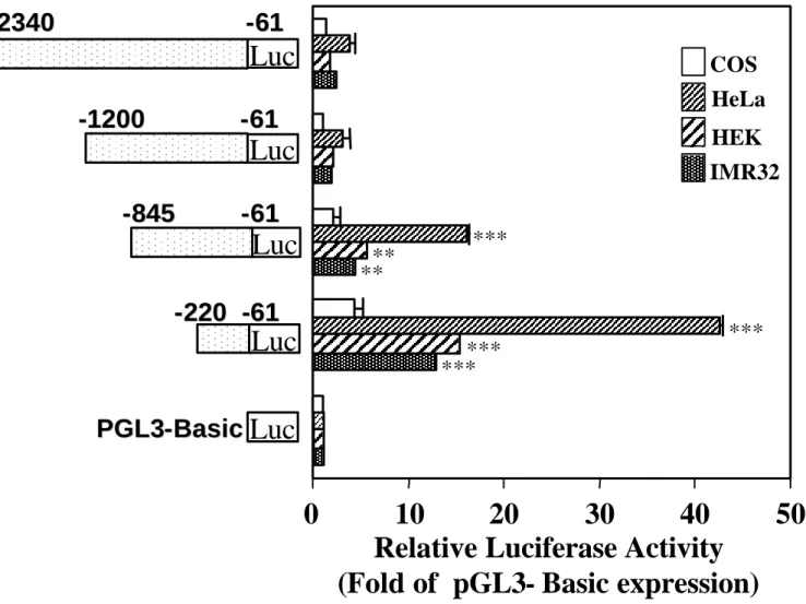
Transcriptional activity of exon 1 bis of the h5-HT4 receptor
The h5-HT4 receptor comprised more than 3.1 kb in its 5’ UTR located in exon 1 bis. Since
transcriptional regulation by exons has been reported for several genes [25, 26], we examined the promoter activity of the long 5’ UTR of the h5-HT4 receptor. To this end, we generated by
PCR a series of constructs containing serial 3’ deletions of exon 1 bis which were sub-cloned into the pGL3-Basic vector (Figure 5). The constructs were then transiently transfected into
HeLa, HEK, IMR32 and COS-7 cells and assayed for luciferase activity (Figure 5). As observed with previous constructs, no luciferase activity was detected in COS-7 cells with any of these new constructs (Figure 5). A modest, not significant increase in luciferase activity was obtained with the two constructs, –2340/–61 and –1200/–61 when transfected in human cell lines (Figure 5). Deletion of the region –1200 to –845 led to a significant enhancement in transcriptional activity in human cell lines as compared with the pGL3-basic vector (Figure 5). Most importantly, deletion from –845 to –220 produced a greater than 10-fold increase in luciferase activiy in IMR32 and HEK cells (Figure 5). Again, the highest relative luciferase activity was observed in HeLa cells (more than 40 fold the control value) (Figure 5).
An Nkx2.5 like element is required for optimal transcriptional activity of the [-220/-61] region
The prediction of potential regulatory elements within the [-220/-61] region was determined by using MatInspector [27] and the TFSearch [28]. We found that it contains putative binding sites for the transcription factors Oct-1, MEF-2 and Nkx2.5. Therefore, we examined whether mutations in the binding sites of these transcription factors affected the enhancer activity of the [-220/-61] region. Mutations of the Oct-1 consensus binding site did not significantly influence the transcriptional activity of the [-220/-61] region in IMR32 cells (Fig 6A) although these mutations have been previously shown to be effective in the loss of Oct-1 binding [43]. A mutation in the MEF-2 like binding site induced a slight decrease of the transcription activity (30%) (Fig 6A). However, we were unable to show any binding of MEF-2 proteins to this DNA sequence when we performed DNA affinity purification of nuclear proteins extracted from IMR32 cells (data not shown). Mutations in the Nkx2.5 like binding site reduced the enhancer activity of the [-220/-61] region by 70% (Fig 6A). These results indicate that the Nkx2.5 like consensus sequence plays a major role in the transcriptional activity of the [-220/-61] region.
Gel mobility shift assay was conducted to determine whether the Nkx2.5 consensus sequence was able to bind nuclear proteins. Two major complexes were formed with the radio-labelled Nkx2.5 probe and proteins extracted from IMR32 cells (Figure 6B, lane 1). All bound proteins can be displaced by 50-fold excess unlabeled Nkx2.5 consensus sequence (Figure 6B, lane 3). Competition with a mutant Nkx2.5 binding site failed to completely abolish complex formation (Figure 6B, lane 2) as did an excess of unlabelled consensus Ets-1 oligonucleotide (Figure 6B, lane 4). To confirm that Nkx2.5 protein binds to the Nkx2.5 consensus sequence located in the [-220/-61] region, an electrophoretic mobility shift assay
was performed with in vitro translated Nkx2.5 protein and Nkx2.5 wild type (WT) and mutated (Mut) probes. As shown in Figure 6C, Nkx2.5 protein was bound to the WT probe and to a much lesser extent to the Mut probe . These results demonstrated that Nkx2.5 protein specifically binds to this Nkx2.5 like sequence.
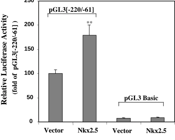
Finally, to test the potential involvement of the transcription factor Nkx2.5 in the regulation of the transcriptional activity of the [-220/-61] region in cardiac cells, we carried out co-transfection experiments in primary neonatal rat ventricular myocytes. Figure 6D shows that transfection of an expression vector encoding Nkx2.5 enhanced the transcriptional activity of the [-220/-61] region in primary neonatal cardiomyocytes. These data indicate that Nkx2.5 may participate in the regulation of 5-HT4 receptor gene expression.
DISCUSSION
In the present study we report the functional characterization of the 5'-flanking region of the h5-HT4 receptor. RT-PCR experiments combined with the sequencing of the 5'-flanking
region of the h5-HT4 receptor showed that the long 5’-UTR (3.1 kb) lays between the
transcription initiation site and the first ATG. Such long 5’-UTR may reduce RNA translation and lead to low levels of expressed transcripts [29] as it is the case for the h5-HT4 receptor in
the heart [24,30]. Indeed, self-complementary sequences can form stable stem-loop structures that interfere with the assembly of the preinitiation complex. These stem loops can be further stabilized by the interaction with RNA-binding proteins. Furthermore, the presence of ATG codons located upstream of the start site can inhibit translation by causing premature initiation and thereby preventing the ribosome from reaching the ATG start codon [31].
Sequence analysis of the 5'-untranslated flanking region of the h5-HT4 receptor gene
indicated that the region upstream of the putative transcriptional start site does not contain any of the typical characteristics of promoter regions such as a TATA box, CAAT box, or the GC-rich region. The TATA-less promoter is characteristic of GPCRs and examples include promoters of the serotonin 5-HT2A and 5-HT2C receptors [32,45]. Interestingly, inspection of
the sequence around the transcription initiation site revealed a consensus sequence for an initiator element (INR) which consists of the sequence TTCACTTT (Figure 1B) [33]. The INR is known to be functionally analogous to the TATA box since it determines the site of transcription and its direction [33].
In our study, we found that, in human atrium, the long 5’-UTR of the h5-HT4 receptor
mRNA is located –3185 bp upstream of the translation initiation site and is encoded by a single exon (exon 1 bis). This start site is unique to the heart and not present in placenta. However, the start site previously described in placenta was also detected in the heart (Figure 3B) suggesting that the h5-HT4 receptor gene may use multiple transcription initiation sites.
This is in accordance with previous studies showing that 5-HT1A, 5-HT2A and 5-HT2C receptor
genes have several transcription start sites [32,44,45]. Therefore, the 5'-UTR of the h5-HT4
receptor is a result of the use of the two alternate promoters. It does not change the protein-coding region, but may affect the transcription efficiency as well as the spatial and temporal expression of the h5-HT4 receptor. The reason for the complex arrangement of numerous start
sites spread over an extended domain remains obscure. Perhaps these mRNAs are differentially regulated at the level of translation in different cell types. Alternatively, the transcription initiation sites may be regulated independently, providing a potential mechanism
to differentially control transcription of the gene in different cell types and tissues regions as it has been shown for several genes [46].
Transient transfection assays with a series of the h5-HT4 receptor promoter-luciferase
constructs identified a ~1.2 kb fragment of 5'-nontranscribed sequence that was active as a promoter in the human cell lines, IMR32, HEK and HeLa and silent in COS-7 cells. These data suggest that this DNA fragment is necessary to drive cell specific expression of the 5-HT4 promoter. Progressive deletion of the h5-HT4 promoter resulted in an increase in
luciferase activity in human cells but not in monkey COS-7 cells. This indicates that the sequences immediately upstream of the transcription start site probably contain positive elements that are required to enhance transcription in the human cell lines tested. Surprisingly, we found that the DNA region –845/–61 of exon 1 bis increased luciferase activity upon transfection in human cells. Deletion of the region located between –845 and – 220 further enhanced luciferase expression indicating the presence of strong positive elements between position –220 and –61. Analysis of the [–220, –61] region by MatInspector and TransFac programs revealed that it contains putative binding sites for several transcription factors such as Oct-1 and Nkx2.5. Mutational analysis of either consensus sequences showed that the Nkx2.5 like binding site strongly regulates transcriptional activity of the [-220/–61] region. EMSA demonstrates specific binding of nuclear proteins to a portion of this region containing the Nkx2.5 binding site.
Nkx2.5 (also called Csx) is a member of the NK class of homeodomain proteins required for cardiac commitment, differentiation and morphogenesis [36]. This transcription factor is continually expressed from embryonic stage to adulthood in human heart [37]. Nkx2.5 has been shown to regulate transcription of markers of cardiac hypertrophy such as the skeletal α -actin [38] and the atrial natriuretic factor [39]. However, Nkx2.5 by itself is a weak transactivator and requires additional interactions with other transcriptions factors such as serum response factor or GATA-4 [40,41]. The potential MEF-2 binding site located in the proximity of the Nkx2.5 binding site would have been a good partner since MEF-2 response elements have been reported in several cardiac promoters and their mutations have been shown to decrease promoter activity [34,35]. However, when we performed DNA affinity purification using the MEF-2 like binding site as a probe, we were not able to detect any binding of MEF-2 proteins to this DNA sequence (data not shown). Thus the partners of NKx2.5 have to be defined.
In summary, we have identified a second transcription start site of the h5-HT4 receptor
gene which is under the control of a promoter region located at more than 3 kb from the first ATG codon. We also provide evidence that the long 5’UTR isolated in our study is only coded by one exon in human atrium. The [-220/-61] region contains a Nkx2.5 like element which regulates the transcriptional activity of the h5-HT4 receptor gene.
ACKNOWLEDGMENTS
We wish to thank Dr. Patrick Donzeau-Gouge, Institut Hospitalier Jacques Cartier, Service de Chirurgie Cardiaque, F-91349 Massy Cedex, France, for providing human atrial tissues. We are grateful to Dr. Izumo, Department of Medicine, Harvard Medical School, Boston, USA for providing expression vector encoding Nkx2.5
REFERENCES
1 Hoyer, D., Hannon, J. P. and Martin, G. R. (2002) Molecular, pharmacological and functional diversity of 5-HT receptors. Pharmacol. Biochem. Behav. 71, 533-554
2 Dumuis, A., Bouhelal, R., Sebben, M., Cory, R. and Bockaert, J. (1988) A nonclassical 5-hydroxytryptamine receptor positively coupled with adenylate cyclase in the central nervous system. Mol. Pharmacol. 34, 880-887
3 Langlois, M. and Fischmeister, R. (2003) 5-HT4 receptor ligands: applications and new
prospects. J. Med. Chem. 46, 319-344.
4 Hegde, S. S. and Eglen, R. M. (1996) Peripheral 5-HT4 receptors. FASEB J. 10, 1398-1407
5 Marchetti, E., Dumuis, A., Bockaert, J., Soumireu-Mourat, B. and Roman, F. S. (2000) Differential modulation of the 5-HT(4) receptor agonists and antagonist on rat learning and memory. Neuropharmacology 39, 2017-2027
6 Maillet, M., Robert, S.J. and Lezoualc’h, F. (2004) New insights into 5-HT4 receptor: a
novel therapeutic target for Alzheimer’s disease? Current Alz. Res. 1, 82-86.
7 Lezoualc'h, F. and Robert, S. J. (2003) The serotonin 5-HT4 receptor and the amyloid
precursor protein processing. Exp. Gerontol. 38, 159-166
8 Robert, S. J., Zugaza, J. L., Fischmeister, R., Gardier, A. M. and Lezoualc'h, F. (2001) The Human Serotonin 5-HT4 Receptor Regulates Secretion of Non- amyloidogenic Precursor Protein. J. Biol. Chem. 276, 44881-44888
9 Maillet, M., Robert, S. J., Cacquevel, M., Gastineau, M., Vivien, D., Bertoglio, J., Zugaza, J. L., Fischmeister, R. and Lezoualc'h, F. (2003) Crosstalk between Rap1 and Rac regulates secretion of sAPPα. Nat. Cell. Biol. 5, 633-639
10 Jahnel, U., Rupp, J., Ertl, R. and Nawrath, H. (1992) Positive inotropic response to 5-HT in human atrial but not in ventricular heart muscle. Naunyn Schmiedebergs Arch. Pharmacol. 346, 482-5
5-Hydroxytryptamine stimulates human isolated atrium but not ventricle. Eur. J. Pharmacol. 230, 103-105
12 Kaumann, A. J. (1991) 5-HT4-like receptors in mammalian atria. J. Neural. Transm. 34 , 195-201
13 Ouadid, H., Seguin, J., Dumuis, A., Bockaert, J. and Nargeot, J. (1992) Serotonin increases calcium current in human atrial myocytes via the newly described 5-hydroxytryptamine4 receptors. Mol. Pharmacol. 41, 346-351
14 Yusuf, S., Al-Saady, N. and Camm A. J. (2003) 5-hydroxytryptamine and atrial fibrillation: how significant is this piece in the puzzle? J Cardiovasc. Electrophysiol. 14, 209-214.
15 Blondel, O., Gastineau, M., Dahmoune, Y., Langlois, M. and Fischmeister, R. (1998) Cloning, expression, and pharmacology of four human 5-hydroxytryptamine 4 receptor isoforms produced by alternative splicing in the carboxyl terminus. J. Neurochem. 70, 2252-2261
16 Claeysen, S., Sebben, M., Becamel, C., Bockaert, J. and Dumuis, A. (1999) Novel brain-specific 5-HT4 receptor splice variants show marked constitutive activity: role of the C-terminal intracellular domain. Mol. Pharmacol. 55, 910-920
17 Bender, E., Pindon, A., van Oers, I., Zhang, Y. B., Gommeren, W., Verhasselt, P., Jurzak, M., Leysen, J. and Luyten, W. (2000) Structure of the human serotonin 5-HT4 receptor gene and cloning of a novel 5-HT4 splice variant. J. Neurochem. 74, 478-489 18 Mialet, J., Berque-Bestel, I., Eftekhari, P., Gastineau, M., Giner, M., Dahmoune, Y.,
Donzeau-Gouge, P., Hoebeke, J., Langlois, M., Sicsic, S., Fischmeister, R. and Lezoualc'h, F. (2000) Isolation of the serotoninergic 5-HT4(e) receptor from human heart and comparative analysis of its pharmacological profile in C6-glial and CHO cell lines. Br. J. Pharmacol. 129, 771-781
19 Vilaro, M. T., Domenech, T., Palacios, J. M. and Mengod, G. (2002) Cloning and characterization of a novel human 5-HT(4) receptor variant that lacks the alternatively spliced carboxy terminal exon. RT-PCR distribution in human brain and periphery of multiple 5-HT(4) receptor variants. Neuropharmacology 42, 60-73
20 Claeysen, S., Faye, P., Sebben, M., Lemaire, S., Bockaert, J., Dumuis, A. and Taviaux, S. (1997) Assignment of 5-hydroxytryptamine receptor (HTR4) to human chromosome 5 bands q31-q33 by in situ hybridization. Cytogenet. Cell. Genet. 78, 133-140
21 Hiroi, T., Hayashi-Kobayashi, N., Nagumo, S., Ino, M., Okawa, Y., Aoba, A. and Matsui, H. (2001) Identification and characterization of the human serotonin-4 receptor gene promoter. Biochem. Biophys. Res. Commun. 289, 337-344
22 Boussif, O., Lezoualc'h, F., Zanta, M. A., Mergny, M. D., Scherman, D., Demeneix, B. and Behr, J. P. (1995) A versatile vector for gene and oligonucleotide transfer into cells in culture and in vivo: polyethylenimine. Proc. Natl Acad. Sci. U S A 92, 7297-7301 23 Schreiber, E., Matthias, P., Muller, M. M. and Schaffner, W. (1989) Rapid detection of
octamer binding proteins with 'mini-extracts', prepared from a small number of cells. Nucleic Acids Res. 17, 6419-6423
24 Medhurst, A. D., Lezoualc'h, F., Fischmeister, R., Middlemiss, D. N. and Sanger, G. J. (2001) Quantitative mRNA analysis of five C-terminal splice variants of the human 5-HT(4) receptor in the central nervous system by TaqMan real time RT-PCR. Mol. Brain Res. 90, 125-134
25 Boukouvala, S., Price, N., Plant, K.E. and Sim, E. (2003) Structure and transcriptional regulation of the Nat2 gene encoding for the drug-metabolizing enzyme arylamine N-acetyltransferase type 2 in mice. Biochem. J. 375, 593–602
26 Stickeler, E., Fraser, S. D., Honig, A., Chen, A. L., Berget, S. M. and Cooper, T. A. (2001) The RNA binding protein YB-1 binds A/C-rich exon enhancers and stimulates splicing of the CD44 alternative exon v4. EMBO J. 20, 3821-3830
27 Quandt, K., Frech, K., Karas, H., Wingender, E. and Werner, T. (1995) MatInd and MatInspector: new fast and versatile tools for detection of consensus matches in nucleotide sequence data. Nucleic Acids Res. 23, 4878-4884
28 Heinemeyer, T., Wingender, E., Reuter, I., Hermjakob, H., Kel, A. E., Kel, O. V., Ignatieva, E. V., Ananko, E. A., Podkolodnaya, O. A., Kolpakov, F. A., Podkolodny, N. L. and Kolchanov, N. A. (1998) Databases on transcriptional regulation: TRANSFAC, TRRD and COMPEL. Nucleic Acids Res. 26, 362-370
29 Meijer, H. A. and Thomas, A. A. (2002) Control of eukaryotic protein synthesis by upstream open reading frames in the 5'-untranslated region of an mRNA. Biochem. J.
367, 1-11.
30 Kaumann, A. J., Lynham, J. A. and Brown, A. M. (1996) Comparison of the densities of 5-HT4 receptors, beta 1- and beta 2- adrenoceptors in human atrium: functional implications. Naunyn Schmiedebergs Arch. Pharmacol. 353, 592-595
31 Cazzola, M. and Skoda, R. C. (2000) Translational pathophysiology: a novel molecular mechanism of human disease. Blood 95, 3280-3288
32 Zhu, Q. S., Chen, K. and Shih, J. C. (1995) Characterization of the human 5-HT2A receptor gene promoter. J. Neurosci. 15, 4885-4895
33 Smale, S. T. (1997) Transcription initiation from TATA-less promoters within eukaryotic protein-coding genes. Biochim. Biophys. Acta. 1351, 73-88
34 Akazawa, H. and Komuro, I. (2003) Roles of cardiac transcription factors in cardiac hypertrophy. Circ. Res. 92,1079-1088. Genes Dev. 6, 1783-1798
35 Di Lisi, R., Millino, C., Calabria, E., Altruda, F., Schiaffino, S. and Ausoni, S. (1998) Combinatorial cis-acting elements control tissue-specific activation of the cardiac troponin I gene in vitro and in vivo. J. Biol. Chem. 273, 25371-25380
36 Schwartz, R. J. and Olson, E. N. (1999) Building the heart piece by piece: modularity of cis-elements regulating Nkx2-5 transcription. Development 126, 4187-4192
37 Shiojima, I., Komuro, I., Mizuno, T., Aikawa, R., Akazawa, H., Oka, T., Yamazaki, T. and Yazaki, Y. (1996) Molecular cloning and characterization of human cardiac homeobox gene CSX1. Circ. Res. 79, 920-929
38 Chen, C. Y. and Schwartz, R. J. (1995) Identification of novel DNA binding targets and regulatory domains of a murine tinman homeodomain factor, Nkx-2.5. J. Biol. Chem.
270, 15628-15633
39 Durocher, D., Chen, C. Y., Ardati, A. , Schwartz, R. J. and Nemer, M. (1996) The atrial natriuretic factor promoter is a downstream target for Nkx-2.5 in the myocardium. Mol. Cell. Biol. 16, 4648-4655
40 Sepulveda, J. L., Belaguli, N., Nigam, V. , Chen, C. Y., Nemer, M. and Schwartz, R. J. (1998) GATA-4 and Nkx-2.5 coactivate Nkx-2 DNA binding targets: role for regulating early cardiac gene expression. Mol. Cell. Biol. 18, 3405-34015
41 Sepulveda, J.L., Vlahopoulos, S., Iyer, D., Belaguli, N. and Schwartz, R. J. (2002) Combinatorial expression of GATA4, Nkx2-5, and serum response factor directs early cardiac gene activity. J. Biol. Chem. 277, 25775-257782
42 Hadri, L., Ozog, A., Soncin, F. and Lompre, A. M. (2002) Basal transcription of the mouse sarco(endo)plasmic reticulum Ca2+-ATPase type 3 gene in endothelial cells is controlled by Ets-1 and Sp1. J. Biol. Chem. 277, 36471-36478
43 Sebastian, S., White, J.A., Wilson, J.E. (1999) Characterization of the rat type III hexokinase gene promoter. A functional octamer 1 motif is critical for basal promoter activity. J. Biol. Chem. 274, 31700-31706
44 Parks, C. L. and Shenk, T. (1996) The serotonin 1A receptor gene contains a TATA-less promoter that responds to MAZ and Sp1. J. Biol. Chem. 271, 4417–4430
45 Xie, E., Zhu, L., Zhao, L. and Chang. L. S. (1996) The human serotonin 5-HT2C 35,
551–561
46 Chiu, I. M., K. Touhalisky, C. and Baran. C. (2001) Multiple controlling mechanisms of FGF1 gene expression through multiple tissue-specific promoters. Prog. Nucleic Acid Res. Mol. Biol. 70, 155-174
FIGURE LEGENDS
Figure 1. A) 5’ gene organization of the h5-HT4 receptor in human atria. An arrow indicates
the ATG translation initiation codon. B) Nucleotide sequence (GenBank accession no AC008627.7) of the 5’ upstream flanking region of the h5-HT4 receptor gene showing the
position of various putative transcription factor binding sites (underlined). Bases relative to the ATG translation initiation codon (in boldface, designated +1) are numbered on the left and on the right. The intron sequence is shown in small letters and the coding sequence in bold letters.
Figure 2. Isolation of a long 5’ UTR in the 5-HT4 receptor gene in human atria. A) Upper
Panel. A schematic diagram representing the positions of sense (dark arrows) and antisense (grey arrows) primers on intron 25 used for RT-PCR experiments. The open arrow indicates the position of the probe used for the Southern blot. B) Results of agarose gel electrophoresis of nested PCR products are shown on the left panel. Molecular size markers are indicated in bp. Primer pairs used for the nested PCR are represented into brackets. The specificity of the PCR products were analysed by Southern blotting using the 32P-5'-end-labeled internal –2392 primer. A 6 h exposure of the autoradiogram is shown on the right panel. gDNA; human genomic DNA.
Figure 3. Identification of the region of the transcription start site of the 5-HT4 receptor
gene. A) Results of agarose gel electrophoresis of cDNA and genomic DNA (gDNA) of
nested PCR products are shown on the left and right lower panels, respectively. Molecular size markers indicated in bp were run with the PCR products. The positions of primers are shown in the upper panel. Sense and antisense primers are in dark and grey arrows respectively. CT, PCR performed with water instead of cDNA is a negative control. B) RNase protection analysis of the h5-HT4 receptor gene. The labelled RNA probe [–3362, –2970] was
hybridised to 60 µg of human atrium RNA (lanes 1), 60 µg of HEK cells RNA (lanes 2), 60 µg of tRNA (lanes 3), 60 µg of tRNA without RNase A (lanes 4). The RNA molecular weight in bp is shown in lane 5. The position of a major protected band and its size is indicated by an arrows on the left. Asterisks show non-specific background. C) Agarose gel electrophoresis of PCR products showing that the –3185 bp start site is unique to the heart. The PCR experiment was performed on cDNA of human placenta and atrium using primers [–2837/–2166] followed by [–2762/–2318]. These primers are specific of Exon 1A. Positive control was
performed using human actin primers on RNA samples treated with (+RT) or without (-RT) reverse transcriptase. D) The upper panel shows the schematic representation of the 5-HT4
receptor gene corresponding to exons 1-26 and exon 1 bis (not to scale). Each grey box corresponds to one exon. Exon1 bis contains the 5’ UTR located on intron 25 (white box) and exon 26, and the coding region of exon 26. The two arrows indicate the possible transcription start sites. Lower panel, results of agarose gel electrophoresis of PCR products showing the presence of exons 1-4 in human atrium. Molecular size markers are indicated in bp.
Figure 4. Transcriptional activity of the h5-HT4 receptor promoter. The upper panel
shows a diagram of the 5’ flanking region of the h5-HT4 receptor gene containing the large
exon 1 bis encoding the long 5’ UTR (–3185, +26). The schematic diagram on the lower left represents each deletion construct of the h5-HT4 receptor promoter linked to the luciferase
gene which were transfected into different cell lines (right panel). Results are expressed as relative luciferase activity expressed as fold expression over the promoter-less pGL3 Basic vector. Values reported in the figure represent the mean ± S.E.M of four independent transfections performed in triplicates. Bars bearing “*” (p<0.05), “**” (p<0.01), “***”(p<0.001) are statistically different from the control pGL3 Basic vector.
Figure 5. Functional analysis of the 5’ UTR of the h5-HT4 receptor gene in different cell
lines. 5’ deletion construct of exon 1 bis were fused to the pGL3 Basic vector and transfected
into the indicated cell lines. Schematic representation of the constructs tested and the relative luciferase activity are indicated on the left and right panel, respectively. Results are expressed as relative luciferase activity expressed as fold expression over the promoter-less pGL3 Basic vector. Values reported in the figure represent the mean ± S.E.M of four independent transfections. Bars bearing “**” (p<0.01), “***”(p<0.001) are statistically different from the control pGL3 Basic vector.
Figure 6. Identification of a Nkx2.5 like binding site that regulated the transcriptional activity of the [-220/-61] region. A) Effects of mutations in the potential MEF-2, Nkx2.5 and
Oct-1 binding sites of the [-220/-61] region in exon 1 bis. Wild type (WT) and mutant constructs of the [-220/-61] region were transfected into IMR32 cells and luciferase activity was then assayed. Results are expressed as a percentage of the wild type pGL3[-220/-61] luciferase activity. Values represent the mean ± S.E.M of three independent transfections.
Bars bearing “*” (p<0.05), “***”(p<0.001) are statistically different from the WT pGL3[-220/-61]. B) Electrophoretic mobility shift assay of nuclear extracts from IMR32 cells. 0.5 ng of [32P] labelled Nkx2.5 probe were incubated with 20 µg of nuclear extracts in the absence of competitor (lane 1) or in the presence of a 50 fold excess of unlabeled oligonucleotides (lanes 2, 3 and 4). Lane 2, mutated (Mut) Nkx2.5 cold probe; lane 3, wild type Nkx2.5 probe; lane 4, Ets-1 consensus probe (lane 4). Opened arrows indicate the two specific complexes formed.
C) Wild type (WT) and mutated (Mut) Nkx2.5 probes were incubated with in vitro translated
Nkx2.5 protein. WT probe had a high binding affinity of Nkx2.5 protein, and mutations into the probe strongly reduced the binding affinity of Nkx2.5 protein. D) Effects of Nkx2.5 on the transcriptional activity of the [-220/-61] region in primary neonatal rat ventricular myocytes. Cardiomyocytes were co-transfected with 1 µg of control expression vector or 1 µg of Nkx2.5 expression vector and 1 µg of pGL3[-220/-61] or pGL3[-220/-61] basic at two days in vitro. Values reported in the figure represent the mean ± S.E.M of three independent transfections. The bar bearing “**” (p<0.01) is statistically different from the control expression vector.
B
-4299 CATACACACC AGGTCTTAAT GGATTTGAAT TGGCAGAATT TTTAGAAGTG ACACAACTTG TACCTGAGCA TAGGAGATGC TCTGAGTTGA TTGTGTTTTT -4200 -4199 AATTAGCTAG GCAGTGTTTT GTGAAGAAGC CAGAAATATA AGTATTCAAG ATAAAAGCAC TGGTTCCTTC CACATCCCCC TCACCCTATG CAATCTTCCC -4100
Oct-1
-4099 AGCAAGAGAT GTTCCTATAA TCATGGCTCT GATTAGTTCT CCCTGGTGAG TCCTGCAGAA AATAGTCAGG CATAATCAAT TAATCAAATT AGGATAGGTA -4000 -3999 CTAATCATCA ACTCCTCCTT CCTCTGTTCC TTTCTCTCTG ATAAAATGCA TGCTATCTCA CTGGTTTGTT GAAAGGCAAT CATTTTAAAT TTGCTCATTG -3900
GATA-1 C/EBP
-3899 GGTTTCAGTG CAAAAAATGT AACAATCTAT ATGCTATGAA TATACAAGAT TTTCCTTGTG TTGTAACATT TTCACATGCA GTTTTAATGA TACAAACTTT -3800
Oct-1
-3799 AGGTTTTAAT GGCTTTTAAA GCTGAAACTG CCCAGCTTTT CTTCCTTTTA ATCTGCAAAA CAATACAATA TTGATTTTAT TGTATATTAC TCAGCATTCA -3700 -3699 TTTGCTTGCT GACTGGAACA CAACTCAAAC TGGCTTAAGG AGGCAAAAAG AATTTGTTGG CTCTAGTAAT TAGGACATTC AAGTGTAAAA TTATCTTAAG -3600
MEF2
-3599 ACACTGCTGG ATCTTAAGCC TCAAATTATG TAGTCAGGAC ACTGGCATTC CCTGTGTTGT GGCTCTGCCT TCCTTTGGGT TAGTTTTCTT CTCAGACAAG -3500
NF-κB
-3499 CTCCTCCCAT GGGGTGGCAA TGATGGCCAC TAGCAGCTTC AAACTGTCTG ACAACACCAG AGGAAATACA GAGTCTTTCT AGCAAAAATT CTAGGACTGG -3400 NFAT
-3399 CTTTCCCTGG TCCAGCCAGT AAGCTAAGGG ATTCTTGTTG CCAGGAATGG ACTATTCTGA TCGACCAGCC CTGCTTATGT GTTCATCACT GGAGCTAGGG -3300 -3299 ATTGGAGAAG GGAAGAGGTG GTTCCAGTAA GGAAAATCCA GACATTATTC TCAGAGAAAG ATGAAATAGA TGGATGGACA GCAGAAAATT ATAGTGGATG -3200
NFAT GATA-3
-3199 TTCACTTTCA TCAGGATACT CTGTTGCTCC AGTGGGAGGA CGTATAATAA GATGACAAGA ATGATGTAAG CTAAAGCTCG AGTTTTGGAA TTCATAGCCG -3100 INR
-3099 TGAATTCTAT TCCCAGATTG GTCAAGTGCT GCTGTGTATG ACTTTGGCAA TTCAATTACC CTCTCTGACC CTCAGTCTCT TCATCTATAG GATGGAGGTT -3000 -2999 GGACTCGGAG GTTGGACTCA AAGGTTGGAG GAGGAGAAAA AGGGCAATGT CTGGTACTTA TTAACACGTA GTAAAGGGCA ACTAATTAAG ATATAATACT -2900 -2899 TTTGCTATTC TTTAAATTAC AATATTCTAT TCTGAGATGG TCCCCTTCCT CCCAGTGTGA AAGTCAAAAA CTCCTAAGCC TCAGTTTTCT CATTTGCAAA -2800 -2799 ACTAATGAAC TACTTAGTTG ACTCAATAAA TATCTATGAT GAGGACACAA TATACCAGGC ATTTCTCGAG GGCACTGAAG ATGTCACAGT GATCAAGACA -2700 -2699 GTGTCCTACG CCCTCTTGAA ATTTATATTC TAGTGAGCAA GACAGATCAC AAATAAAGAA GCAATTGACT ATATATATAT ATATATATAT ATATATACTG -2600 -2599 GGAAGAAAAA AATAGAACAG AGAAAGAGGA TAGACATTTT TAGAAAATGG GGTGTGAAAT TGTAGTTAGG TGTTGAGAGA AGACCTGTCT GAGGAAGTGA -2500 -2499 TATTTGAGCG GCACCCCTAA GGAAGTGAGA GAGTAATCTA CATGGTCACA TTCCAGGGAA AAGAGACAAA GTGAAAAGGC CCTGAGATGG GAACCTACGG -2400 -2399 TTACGGCCCA GGAAAGCAGC AAAACCAGTG AGCTGGTGGG TGGGAAAGGG GAGGGGCATG GCTACAGTTG AGTTTGGAGA AGAGTGGGGG CCAGATCATG -2300 -2299 TAAGGTCTGG TTAGCTATGA TAAGGAATTT GGATTGTATG CTAAGTGTGA TAGGAAGCCA CTAGAATTTT ATGCAAGCAA GAAATGACCT GACTTACATT -2200 -2199 TAAAAAATAT CATTCTGGCA GGAGACAGAT TGGGGGTGTG GGTGAGTAAT TCAGAGAAAG AAATTGGTGG CTTGGACCCA GATGCTGGGT CCAGAGGTGA -2100 -2099 TGAGAAGGGT CAGGAGTAGG AATAATTTTC TTCCTTTCTT TTACTTTTTT TCTTTCTTTC TTTTTCTTGA GACAGGGTCT CACTTTGTCA CCCAGGCTGG -2000 -1999 AGTACAGTAG CGTGATCTTG GCTCACAGCA GCCTCAACCT CCTGGGCTCA GGTGATCCTC CCACCTCAGC CCCCCAGGTA GCTGGAACTA CAGGCATATA -1900 -1899 TCACCACGCC CATCTAATTT TTTGTGGAGA TGGGGTTTTG CCACGTTGCC CAGGCTGGTC TCAAACTCCT GGGCTCAAGC GATCCACTCA CCTAGGCCCC -1800 -1799 CCAAAGTGTT GGGATTATAG GCATGAGCTA CTGCCCCCGG CCAGGAATCA TTTTCAAAGT AGAGAGAATG AGTTTGCTGA TGGTTTGGTC TTAGGGTATG -1700 -1699 AGAAGAAGTT TTTCTGGTCT GAACCACTGG GTAAATGGTG GGCCATTTAC TGAGATAGAG AACACTGGGG AAGAGCAGGT TTTTGTGTCG GAGATGAGAA -1600 -1599 AGGAAGAGTT TAGTTTGAAA TGCTTTTTAA TCATCCATCT TGAGATGCTG GATGGGCACT GTCTACACCT GGGAAAGTCT CAACGTAGGG ATAAAATTTT -1500 -1499 GGGAGTCTAC AGTGAATAGT ATTGTAATTG TACTGTATTT TCAGCCACCA GATGGATGTG ATCCTCTCAG GAAAAATTTT AGATAGACAA GGAAAGAGGA -1400 -1399 TTGAGACCTG TCACTTCGAT AGTTAGGGCT TTGTACTTGT ATATTTCCAG CAAAATATTA TTCTACATTC AAAATATAAA ACAGACAGAT GTGGACAGAG -1300 -1299 CTACTTTCCT TCAGATTAGG GGAGGGGGCT CAGAGTTCTG CAGGTGGAGC CTTTCCCATG CCTGCCCCAG GCCCAGAAAT GCCCATGGGA GCTCCTGTTA -1200 -1199 AGTTTAGAGG ATCTAAGAAA CAAACATCGA AACACACAGA GAGTCGAGGT CCTCTCCATA TCTAGTGATC TGCCATTCTT TTCGTGGCTG CCAGCCACAT -1100 -1099 CTGTTTAGCT CACCCTGGCC AAGGTGTCCA AGGGCCCAGG CTGAGTTTCA TGGTTTTTTT ATGTAGATAA CAGATTTCCA GGTTCTTCTT GGTAAGATTC -1000 -999 AACTAGAAAA CTAGCAAACA GAACTATAGA ATATCAGAGC TAGGAGTGAA CTCAGAGATT ATTTTTTTCC AATCTCTTCT CTTTACTGAG AAGTGAATGA -900 -899 TGATGTTCAG AAGGAAAAGG GACTTGCTCA TGGTGACCCT AGAAGAGACA GCCGGACCCT GCCCCTACAC TTACTGATTC ATCATGCTGT GGAACAGATG -800 -799 TGATGAAGAT ATAAAAATGC AAAAATGGAA CTGAACAATT AGTAAAGAGG GTTGATTTTG TGGAGATCAG TGAATGCCCA TCCATTAAAG AGTGTGAGGT -700 -699 CTTCTGAGGG TCTGTGATAG AGGAACTTTG GTAATTTGAA GTTAACTCTC CTTCGATTGG GAATTCTTAG CCTGGGGTTC CTGGATGGGC CTGGGATTCC -600
C/EBP
-599 TGGATTCCTG AGTTCAAGGA ATCATGGACT CTCTGGAAAG TGTGCATGTG TGTGCATGCG TATCTATTTT TTTCTGGGCA GACAGTTCTC AAGAGTTTAC -500 GRE
-499 TAGATTCTCA AAAGAATCTG TGATTTCCTA CCCTTAAAAT GATTTAAACA TTTTTTTAAA ATCCTCATAG GAACAAAATG GCCATTTTGG GCAATATACT -400 -399 ATTTTAAGCA TAAGAATGTA GCTACCTATC TTCACTAATT ACTGTCCTAA TGCAGATCAT AAAAATAAGC CATGTTTCTG GAAAGAACAT AAATAACAAT -300 -299 AAATGGAGCA AAAAGAAGCA CCCATATGTT TTAATCACAA AGCGATCAGT AGGGTGTAAA TAAACCTTTT GTGACACTCG GCTGTACCAT ACGTCAATAT -200
Oct-1
-199 GCCAAAAGCT TTTTGCTTTA GTATTGTTTA TTAAATATTT CCACAAAACA AGTCATAATA GGAAGCAATA GTTATTTGAG TTTTTTAAAA CAAGGACTTG -100
MEF2 Nkx2.5
-99 TATTTTTCAA TGCTGCTTTC TTTTATAACA TCTTTTTACT TCCCCCATTT TAGGACCCTG TGGGCATTGA AATCCAACTC ACTCATGCTT ATTTCCTGTA 0 +1 ATGGACAAAC TTGATGCTAA TGTGAGgtat gttacctttc tgtacatatt gtgttgtaaa ctgagaacat ttctgctatg aagactctaa aaatctgttg 100 +101 ttgtagagtt ataaagttaa tggatgtgga ttttctttga tgtttcatat ataatcatgt tcacagtact gtttgattat tagtgttttg tcatttctgt 200
Figure 1
+27 +1 +26 INTRON 26 ~85 kbp ATG +152 -47 EXON 26 EXON 27 INTRON 25 ~25 kbp +27 +1 +26 INTRON 26 ~85 kbp ATG +152 -47 EXON 26 EXON 27 INTRON 25 ~25 kbpFigure 2
500Marker
gDNA
cDNA
[ -2837; -2318] [ -2837; -2318] [ -2762; -2318] [ -2711; -2318] 750 250 2000 1000 1500 3000gDNA
cDNA
[ -2837; -2318] [ -2837; -2318] [ -2762; -2318] [ -2711; -2318]B
Agarose gel
Southern blot
+1 +26

![Figure 2500MarkergDNAcDNA[ -2837; -2318][ -2837; -2318][ -2762; -2318][ -2711; -2318]7502502000100015003000 gDNA cDNA[ -2837; -2318][ -2837; -2318][ -2762; -2318] [ -2711; -2318]B](https://thumb-eu.123doks.com/thumbv2/123doknet/14541006.535425/29.892.85.819.213.1101/figure-markergdnacdna-gdna-cdna-b.webp)