HAL Id: hal-02145586
https://hal.archives-ouvertes.fr/hal-02145586v2
Submitted on 12 Sep 2019
HAL is a multi-disciplinary open access
archive for the deposit and dissemination of sci-entific research documents, whether they are pub-lished or not. The documents may come from teaching and research institutions in France or abroad, or from public or private research centers.
L’archive ouverte pluridisciplinaire HAL, est destinée au dépôt et à la diffusion de documents scientifiques de niveau recherche, publiés ou non, émanant des établissements d’enseignement et de recherche français ou étrangers, des laboratoires publics ou privés.
Killing from the inside: Intracellular role of T3SS in the
fate of Pseudomonas aeruginosa within macrophages
revealed by mgtC and oprF mutants
Preeti Garai, Laurence Berry, Malika Moussouni, Sophie Bleves,
Anne-Béatrice Blanc-Potard
To cite this version:
Preeti Garai, Laurence Berry, Malika Moussouni, Sophie Bleves, Anne-Béatrice Blanc-Potard. Killing from the inside: Intracellular role of T3SS in the fate of Pseudomonas aeruginosa within macrophages revealed by mgtC and oprF mutants. PLoS Pathogens, Public Library of Science, 2019, 15 (6), pp.e1007812. �10.1371/journal.ppat.1007812�. �hal-02145586v2�
☯These authors contributed equally to this work. *anne.blanc-potard@umontpellier.fr
Abstract
While considered solely an extracellular pathogen, increasing evidence indicates that Pseu-domonas aeruginosa encounters intracellular environment in diverse mammalian cell types, including macrophages. In the present study, we have deciphered the intramacrophage fate of wild-type P. aeruginosa PAO1 strain by live and electron microscopy. P. aeruginosa first resided in phagosomal vacuoles and subsequently could be detected in the cytoplasm, indi-cating phagosomal escape of the pathogen, a finding also supported by vacuolar rupture assay. The intracellular bacteria could eventually induce cell lysis, both in a macrophage cell line and primary human macrophages. Two bacterial factors, MgtC and OprF, recently iden-tified to be important for survival of P. aeruginosa in macrophages, were found to be
involved in bacterial escape from the phagosome as well as in cell lysis caused by intracellu-lar bacteria. Strikingly, type III secretion system (T3SS) genes of P. aeruginosa were down-regulated within macrophages in both mgtC and oprF mutants. Concordantly, cyclic di-GMP (c-di-GMP) level was increased in both mutants, providing a clue for negative regulation of T3SS inside macrophages. Consistent with the phenotypes and gene expression pattern of mgtC and oprF mutants, a T3SS mutant (ΔpscN) exhibited defect in phagosomal escape and macrophage lysis driven by internalized bacteria. Importantly, these effects appeared to be largely dependent on the ExoS effector, in contrast with the known T3SS-dependent, but ExoS independent, cytotoxicity caused by extracellular P. aeruginosa towards macro-phages. Moreover, this macrophage damage caused by intracellular P. aeruginosa was found to be dependent on GTPase Activating Protein (GAP) domain of ExoS. Hence, our work highlights T3SS and ExoS, whose expression is modulated by MgtC and OprF, as key players in the intramacrophage life of P. aeruginosa which allow internalized bacteria to lyse macrophages.
a1111111111 a1111111111 a1111111111
OPEN ACCESS
Citation: Garai P, Berry L, Moussouni M, Bleves S,
Blanc-Potard A-B (2019) Killing from the inside: Intracellular role of T3SS in the fate of Pseudomonas aeruginosa within macrophages revealed by mgtC and oprF mutants. PLoS Pathog 15(6): e1007812.https://doi.org/10.1371/journal. ppat.1007812
Editor: Joan Mecsas, Tufts University, UNITED
STATES
Received: January 18, 2019 Accepted: May 2, 2019 Published: June 20, 2019
Copyright:© 2019 Garai et al. This is an open access article distributed under the terms of the
Creative Commons Attribution License, which permits unrestricted use, distribution, and reproduction in any medium, provided the original author and source are credited.
Data Availability Statement: All relevant data are
within the manuscript and its Supporting Information files.
Funding: This work is supported by Vaincre La
Mucoviscidose (http://www.vaincrelamuco.org/) (RF20150501356/1/1/47 and RIF20170502042) and Association Gregory Lemarchal (https:// association-gregorylemarchal.org/). PG is supported by the Association Me´diterrane´e Infection (http://www.mediterranee-infection.com/
Author summary
The ability of professional phagocytes to ingest and kill microorganisms is central to host defense andPseudomonas aeruginosa has developed mechanisms to avoid being killed by
phagocytes. While considered an extracellular pathogen,P. aeruginosa has been reported
to be engulfed by macrophages in animal models. Here, we visualized the fate ofP. aerugi-nosa within cultured macrophages, revealing macrophage lysis driven by intracellular P. aeruginosa. Two bacterial factors, MgtC and OprF, recently discovered to be involved in
the intramacrophage survival ofP. aeruginosa, appeared to play a role in this cytotoxicity
caused by intracellular bacteria. We provided evidence that type III secretion system (T3SS) gene expression is lowered intracellularly inmgtC and oprF mutants. We further
showed that intramacrophageP. aeruginosa uses its T3SS, specifically the ExoS effector, to
promote phagosomal escape and cell lysis. We thus describe a transient intramacrophage stage ofP. aeruginosa that could contribute to bacterial dissemination.
Introduction
Pathogenic bacteria are commonly classified as intracellular or extracellular pathogens [1]. Intracellular bacterial pathogens, such asMycobacterium tuberculosis or Salmonella enterica,
can replicate within host cells, including macrophages. In contrast, extracellular pathogens, such asYersinia spp., Staphylococcus aureus, Pseudomonas aeruginosa or streptococci, avoid
phagocytosis or exhibit cytotoxicity towards phagocytic cells, to promote their extracellular multiplication. However, recent data have emphasized that several extracellular pathogens can enter host cellsin vivo, resulting in a phase of intracellular residence, which can be of
impor-tance in addition to the typical extracellular infection. For example, althoughYersinia spp.
sub-vert the functions of phagocytes from the outside, these bacteria also subsub-vert macrophage functions within the cell [2]. Once considered an extracellular pathogen, it is now established thatS. aureus can survive within many mammalian cell types including macrophages [3,4] and the intramacrophage fate ofS. aureus has been deciphered [5,6]. Moreover, an intracellular phase within splenic macrophages has been recently shown to play a crucial role in initiating dissemination ofStreptococcus pneumoniae, providing a divergence from the dogma that
con-sidered this bacterial pathogen a classical example of extracellular pathogens [7].
The environmental bacterium and opportunistic human pathogenP. aeruginosa is
respon-sible for a variety of acute infections and is a major cause of mortality in chronically infected cystic fibrosis (CF) patients. The chronic infection ofP. aeruginosa and its resistance to
treat-ment is largely due to its ability to form biofilms, which relies on the production of exopolysac-charides (EPS), whereas the type III secretion system (T3SS) is reported to play a key role in the pathogenesis of acuteP. aeruginosa infections [8]. Four T3SS effectors (ExoU, ExoS, ExoT, ExoY) have been identified so far, ExoS being nearly always mutually exclusive with the potent cytotoxin ExoU and more prevalent than ExoU [9–11]. ExoS has a dual function as it contains a GTPase activating protein (GAP) domain as well as an ADP ribosyltransferase (ADPRT) domain [10]. An intracellular step in airway epithelial cells has been proposed to occur before the formation of biofilm during the acute phase of infection [12,13] and the intracellular stage ofP. aeruginosa within cultured epithelial cells has been fairly studied [14–16]. The principle thatP. aeruginosa is not exclusively an extracellular pathogen has been convincingly
estab-lished by the recent use of advanced imaging methods to track bacteria within epithelial cells [17]. The T3SS, more specifically its effector ExoS, has been implicated in the formation of membrane blebs-niche and avoidance of acidified compartments to allow bacterial
Intramacrophage fate of Pseudomonas aeruginosa
). MM is supported by Vaincre La Mucoviscidose (RIF20170502042). The funders had no role in study design, data collection and analysis, decision to publish, or preparation of the manuscript.
Competing interests: The authors have declared
generally mediated by regulation of the bacterial genes involved in physiological adaptation to the microenvironment. The identification of mutants lacking the ability to survive within mac-rophages and the study ofP. aeruginosa gene expression inside macrophages is critical to
determine bacterial players in this step. We have previously uncovered MgtC as a bacterial fac-tor involved in the intramacrophage survival ofP. aeruginosa [25,27]. In agreement with this intramacrophage role, expression ofPseudomonas mgtC (PA4635) gene is induced when the
bacteria reside inside macrophages [25]. MgtC is known to promote intramacrophage growth in several classical intracellular bacteria, includingSalmonella typhimurium where it inhibits
bacterial ATP synthase and represses cellulose production [28–30], and is considered as a clue to reveal bacterial pathogens adapted to an intramacrophage stage [30,31]. In addition, a recent study has implicated the outer membrane protein OprF in the ability of otopathogenicP. aeru-ginosa strains to survive inside macrophages [26]. OprF is an outer membrane porin that can modulate the production of various virulence factors ofP. aeruginosa [32,33].
In the present study, we investigated the fate ofP. aeruginosa within macrophages using
wild-type PAO1 strain, which lacks ExoU, along with isogenicmgtC and oprF mutants. We
also explored, for the first time, the regulation of T3SS genes whenP. aeruginosa resides inside
macrophages. The T3SS and ExoS effector, whose expression was found to be modulated by MgtC and OprF intracellularly, were seemingly involved in this intracellular stage, playing a role in vacuolar escape and cell lysis caused by intracellular bacteria.
Results
Intracellular
P. aeruginosa can promote macrophage lysis
We previously visualized fluorescentP. aeruginosa residing within fixed macrophages [25]. To investigate the fate ofP. aeruginosa after phagocytosis in a dynamic way, we monitored
macro-phages infected with fluorescent bacteria using time-lapse live microscopy. J774 macromacro-phages were infected with wild-type PAO1 strain expressing GFP grown exponentially in LB medium (Multiplicity of infection or MOI = 10). After 25 minutes of phagocytosis, several washes were performed to remove non-internalized bacteria and gentamicin was added to kill the remain-ing extracellular bacteria. Microscopic observation of infected macrophages up to 3 hours showed cell lysis with increasing time (Fig 1), starting between 1.5 and 2 hours post-phagocy-tosis, which can be attributed to intracellular bacteria as gentamicin was retained throughout the experiment. No lysis of uninfected cells present in the same field was observed (Fig 1and
S1 Fig), as expected from an event due to intracellular bacteria rather than extracellular bacte-ria. The cell lysis appeared to take place within a rapid time frame of few seconds, as shown by the movie (S1 Movie), after which the bacteria seemed to be released from the host cell. We also infected human monocyte-derived macrophages (HMDMs) with GFP producing PAO1 strain under similar conditions and found lysed infected cells after 2 hours of phagocytosis (S2
Fig). Hence, the phenomenon of lysis of macrophages by intracellular PAO1 is not restricted to J774 murine cell line, but is extendable to primary human macrophages.
We further examined intracellularP. aeruginosa within macrophages in more detail using
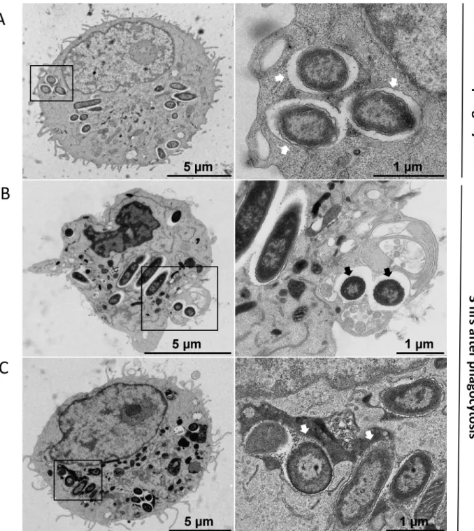
transmission electron microscopy (TEM). J774 macrophages infected with wild-type PAO1 strain, were subjected to fixation at early time post-infection (30 min after phagocytosis) or at later time of infection (3 hrs after phagocytosis). After phagocytosis,P. aeruginosa was present
in membrane bound vacuoles inside macrophages (Fig 2A). At a later time point, some bacte-ria could be found in the cytoplasm with no surrounding membrane, suggesting disruption of the vacuole membrane (Fig 2B). The infected macrophage was damaged, displaying highly condensed chromatin and membrane blebbing, and lacking pseudopodia. Healthy infected cells were also observed, where bacteria were mostly found in vacuoles partially or totally filled with heterogeneous electron dense material, suggesting that the vacuole has fused with lyso-somes (Fig 2C). To confirm this, we examined the association between fluorescent PAO1 bac-teria and the LysoTracker probe during infection using fixed macrophages. Bacbac-teria
colocalizing with LysoTracker could be visualized (S3 Fig), thereby corroborating the TEM observation of fusion of vacuoles with lysosomes and the localization of bacteria in acidified compartment.
TEM analyses allowed us to conclude that wild-type PAO1 strain has the ability to reside within vacuoles and possibly escape from the phagosome into the cytoplasm, and promote cell damage. A rapid cell lysis event caused by intracellularP. aeruginosa visualized by live
micros-copy revealed that phagocytosed bacteria can escape from macrophage through cell lysis.
Intracellular
mgtC and oprF mutants are compromised in cell lysis
Our previous results based on gentamicin protection assay on J774 infected macrophages and counting of colony forming units (CFU) indicated thatmgtC mutant (generated in the PAO1
background) survived to a lesser extent than the wild-type strain [25]. More recently, anoprF
mutant of an otopathogenic strain ofP. aeruginosa was also found to be defective in
intracellu-lar survival in mouse bone marrow macrophages based on gentamicin protection assay [26]. To analyze the phenotypes ofoprF mutant in the PAO1 background towards J774
macro-phages and compare that withmgtC mutant, we used here a previously described oprF mutant
generated in PAO1 strain [25,34].
Based on the finding that intracellularP. aeruginosa can cause macrophage lysis, we
devel-oped a suitable assay using fluorescent microscopic analysis to quantify cell damage caused by intracellular bacteria. Macrophages were infected with fluorescent bacteria, treated with
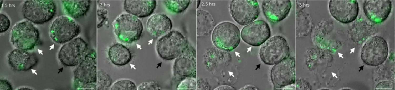
Fig 1. Live imaging of macrophages infected withP. aeruginosa. J774 macrophages were infected with PAO1 wild-type (WT) strain expressing GFP. Time
lapse imaging was started at 1.5 hrs post-phagocytosis. Cells were maintained in DMEM supplemented with gentamicin at 37˚C and 5% CO2throughout
imaging. White arrows point at the cells that harbor intracellular bacteria and undergo lysis between 1.5 hrs and 3 hrs post-phagocytosis. Black arrow shows an uninfected and unlysed cell. Scale bar is equivalent to 10μm.
https://doi.org/10.1371/journal.ppat.1007812.g001
gentamicin for 2 hours after phagocytosis, fixed and stained with phalloidin-TRITC to label F-actin and visualize macrophage morphology. A clear cortical red labeling was seen in most
Fig 2. Transmission electron micrographs (TEM) ofP. aeruginosa within macrophages. J774 macrophages were infected with P. aeruginosa for
30 min (A) or 3 hrs (B and C) and subjected to TEM (left panels). Black rectangles show intracellular bacteria that are shown at higher
magnification in the right panels. A. At early time after phagocytosis, most of bacteria were found inside membrane bound vacuoles (white arrows). B. At later time, some bacteria can be observed in the cytoplasm with no surrounding membrane suggesting disruption of the vacuole membrane (black arrows). The infected macrophage in panel B shows an abnormal morphology, with highly condensed chromatin and membrane blebbing, but no pseudopodia. C. At later time, bacteria can also be found in vacuole partially or totally filled with heterogeneous electron dense material (white arrows), suggesting that the vacuole has fused with lysosomes. The infected macrophage shown in panel C appears normal.
cells, which is indicative of the plasma membrane-associated cortical actin. Few infected cells appeared lysed due to the loss of cortical actin staining (Fig 3A), which agrees with the obser-vation of lysed macrophages in time lapse fluorescence microscopy (Fig 1). The loss of cortical actin staining was found to be due to internalized bacteria because upon arresting phagocyto-sis, by treating macrophages with cytochalasin D, bacteria were not internalized and loss of cortical actin staining was not observed (S4 Fig). Hence, any delayed effect of extracellular bac-teria occurring before gentamicin treatment or a contribution of extracellular bacbac-teria that would resist gentamicin treatment can be ruled out. Quantification of the number of cells without phalloidin labelling showed highest value for cells infected with wild-type strain, inter-mediate withmgtC mutant and lowest with oprF mutant (Fig 3B). Thus, both intracellular
mgtC and oprF mutants appeared compromised for cell lysis. This feature is not linked to a
Fig 3. Visualization (A) and quantification (B) of lysed infected cells upon phalloidin labeling. GFP expressing PAO1 WT,
ΔmgtC and ΔoprF strains were used for infecting J774 macrophages. Gentamicin was added after phagocytosis and cells were fixed at 2 hrs post-phagocytosis, stained with phalloidin and imaged with fluorescent microscope. DAPI was used to stain the nucleus. Cells that have intracellular bacteria, but lack the phalloidin cortical label were considered as lysed by intracellular bacteria (shown by arrows). Scale bar is equivalent to 10μm. After imaging, infected cells were counted and percentage of lysed cells with intracellular bacteria out of total number of cells was plotted for each strain. Error bars correspond to standard errors from three independent experiments. At least 200 cells were counted per strain. The asterisks indicateP values (One way ANOVA, where all strains were compared to WT using Dunnett’s multiple comparison post-test,�P <0.05 and���P <0.001), showing statistical significance with respect to WT.
https://doi.org/10.1371/journal.ppat.1007812.g003
lower internalization rate of the mutant strains, becausemgtC mutant is known to be more
phagocytosed than wild-type strain [25] and a similar trend was found foroprF mutant (not
shown). Since T3SS has been involved in the intracellular life ofP. aeruginosa in other cell
types [17], we next monitored the expression of T3SS genes during the residence of bacteria within macrophages.
Down-regulation of T3SS gene expression in
mgtC and oprF mutants inside
macrophages
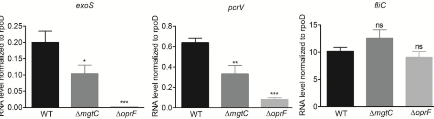
TheP. aeruginosa oprF mutant was reported to be defective in the secretion of T3SS effectors
in liquid culture and in the production of PcrV, the T3SS needle tip protein [32,34], but the effect of OprF on transcription of T3SS genes has not been tested so far. We thus investigated the expression of the effector geneexoS along with the gene pcrV in oprF mutant strain in
com-parison to wild-type strain upon macrophage infection. As a control, we tested flagellin coding genefliC, which is not part of the T3SS regulon, but was proposed to be secreted by the T3SS
[35]. Expression of bothpcrV and exoS genes was found to be remarkably reduced in the oprF
mutant (Fig 4). Strikingly, themgtC mutant also exhibited significantly reduced expression of
these two T3SS genes (Fig 4), although to a lesser extent than theoprF mutant, indicating an
unexpected interplay between MgtC and T3SS. On the other hand,fliC expression was not
altered in both mutants.
Since we observed a decreased transcriptional level of T3SS genes in theoprF mutant, we
further examined the link between intramacrophage expression of T3SS and cell lysis in this mutant. A recombinant plasmid allowing IPTG-inducible overproduction of ExsA, a master transcriptional activator of T3SS genes [36], was introduced in theoprF mutant. Upon
induc-tion ofexsA expression with 0.01 mM IPTG, oprF mutant promoted macrophage lysis like the
wild-type strain (S5 Fig), supporting a model whereby the effect of OprF is the result of its pos-itive regulatory effect on T3SS expression.
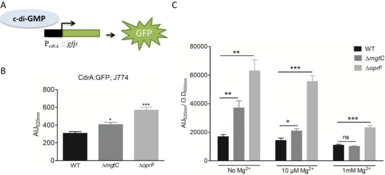
To address the mechanism behind the downregulation of transcription of T3SS inoprF and mgtC mutant strains, we evaluated the level of the second messenger c-di-GMP, as it is known
to participate in T3SS repression inP. aeruginosa [37]. TheoprF mutant was already reported
to have high production of c-di-GMP in liquid culture [34]. We used a fluorescence-based
Fig 4. Expression of T3SS genes inP. aeruginosa strains residing in J774 macrophages. J774 macrophages were infected with PAO1 WT, ΔmgtC and ΔoprF
strains. After phagocytosis, cells were maintained in DMEM supplemented with gentamicin. RNA was extracted from bacteria isolated from infected macrophages 1 hr after phagocytosis. The level ofexoS, pcrV and fliC transcripts relative to those of the house-keeping gene rpoD was measured by qRT-PCR and plotted on the Y-axis. Error bars correspond to standard errors from at least three independent experiments. The asterisks indicateP values (One way ANOVA, where all strains were compared to WT using Dunnett’s multiple comparison post-test,�P <0.05,��P <0.01,���P <0.001 and ns = P >0.05 or non-significant), showing statistical significance with respect to WT.
reporter (Fig 5A) that has been validated to gauge c-di-GMP level insideP. aeruginosa [38]. The pCdrA::gfp plasmid was introduced into wild-type and mutant strains, and fluorescence
was measured in infected macrophages (Fig 5B). BothoprF and mgtC mutants exhibited
signif-icantly increased activity of thecdrA promoter comparatively to wild-type strain, indicative of
higher levels of c-di-GMP than the wild-type strain. To better appreciate the differences moni-tored within macrophages, we also measured fluorescence of strains grown in culture medium with various magnesium concentrations (Fig 5C). TheoprF mutant exhibited a two to
three-fold increase in the activity of thecdrA promoter comparatively to wild-type strain, which is in
the same range as the increase observed within macrophages (Fig 5B), and agrees with pub-lished data obtained with this indirect strategy and direct c-di-GMP measurement [34]. Under magnesium limitation, a condition known to inducemgtC expression, the mgtC mutant also
showed increased activity of thecdrA promoter comparatively to wild-type strain, with a
two-fold increase in the absence of Mg2+, and a minor, but significant, increase in the presence of 10μM Mg2+(that is similar to what is observed within macrophages). On the other hand, the level of fluorescence of wild-type strain andmgtC mutant was identical in medium
supple-mented with 1 mM Mg2+, a condition that preventsmgtC expression [25]. Taken together, these results indicate that the production of c-di-GMP is increased relatively to wild-type strain in bothoprF and mgtC mutants, with a more pronounced effect for oprF, when bacteria
reside within macrophages, thus providing a mechanistic clue for the negative effect on T3SS gene expression.
Fig 5. Measurement of c-di-GMP level inΔmgtC and ΔoprF mutants. PAO1 WT, ΔmgtC and ΔoprF harboring reporter plasmid pCdrA::gfp, which expresses
GFP under the control of the promoter of c-di-GMP responsive genecdrA (A), were used to infect J774 cells and fluorescence was measured (B). After phagocytosis, DMEM containing 300μg/ml of amikacin was added to eliminate extracellular bacteria. Fluorescence (excitation, 485 nm and emission, 520 nm) was measured 1 hour after phagocytosis and plotted as arbitrary units (AU). Error bars correspond to standard errors from four independent experiments. The asterisks indicateP values (One way ANOVA, where all strains were compared to WT using Dunnett’s multiple comparison post-test,�P <0.05 and���P <0.001), showing statistical significance with respect to WT. (C) PAO1 WT,ΔmgtC and ΔoprF harboring reporter plasmid pCdrA::gfp, were grown in NCE medium with varying concentration of Mg2+. After 1 hour of growth, fluorescence (excitation, 485 nm and emission, 520 nm) of the culture was measured
along with its OD600nm. The fluorescence was plotted as arbitrary units (AU520nm) after normalizing with OD600nm. Error bars correspond to standard errors
from three independent experiments. The asterisks indicateP values showing statistical significance with respect to WT in the respective condition, using Student’s t test (ns =P >0.05,�P <0.05,��P <0.01 and���P <0.001).
https://doi.org/10.1371/journal.ppat.1007812.g005
A T3SS mutant is defective for cell death driven intracellularly in an
ExoS-dependent manner
Given the effect of bothoprF and mgtC deletions on the expression of T3SS genes within
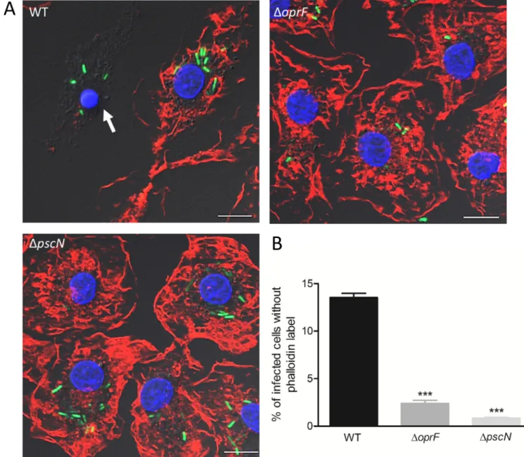
mac-rophages, we investigated the fate of a T3SS mutant upon phagocytosis. We first used apscN
mutant that is defective for the ATPase function of the T3SS machinery [36]. Intracellular T3SS mutant did not induce loss of cortical phalloidin staining, as shown by fluorescent imag-ing and subsequent quantification, indicatimag-ing lack of cell lysis (Fig 6A and 6B). Thus, the phe-notype of T3SS mutant is consistent with that ofmgtC and oprF mutants and is in correlation
with their level of T3SS gene expression. We further examined the relevance of our findings to primary human macrophages, by assessing the lysis of HMDMs driven intracellularly by wild-typeP. aeruginosa or oprF and pscN mutants. The count of intracellularly lysed cells was
signif-icantly different between wild-type and each mutant strain (Fig 7), with a similar trend to that of J774 macrophages (Fig 3andFig 6), thus confirming the importance of T3SS in intracellu-larly driven lysis of primary human macrophages as well.
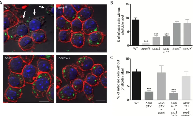
To address the implication of T3SS effector proteins in this process, we used mutant strains for individual effector genesexoS, exoT and exoY, and exoSTY triple mutant. The intracellular
lysis of macrophages was found to be reduced for theexoSTY triple mutant and to a similar
extent for theexoS mutant (Fig 6A and 6B), suggesting a major contribution of ExoS.
Fig 6. Assessment of role of T3SS and its effectors in cell lysis induced by intracellular PAO1. J774 macrophages were infected with GFP expressing strains
as indicated. After phagocytosis, cells were maintained in DMEM supplemented with gentamicin. Cells were imaged 2 hrs post-phagocytosis after staining with phalloidin (A). Scale bar is equivalent to 10μm. Lysis was quantified (B & C) by counting infected cells lacking cortical labeling (indicated by arrows). Percentage of lysed cells with intracellular bacteria out of total number of infected cells was plotted. Error bars correspond to standard errors from at least three independent experiments. At least 200 cells were counted per strain. The asterisks indicateP values (One way ANOVA, where all strains were compared to WT using Dunnett’s multiple comparison test,���P <0.001), showing statistical significance with respect to WT.
Accordingly,exoT and exoY mutants behave similarly to wild-type strain (Fig 6B). Moreover, complementation of theexoSTY mutant with exoS restored the wild-type phenotype (Fig 6C). Although the lysis byexoS mutant strain was substantially higher than that of pscN mutant,
these results suggest that the T3SS-mediated intracellular lysis of macrophages byP. aeruginosa
relies mainly on the T3SS effector ExoS. Thus, this ExoS-dependent cytotoxic effect mediated by intracellular bacteria differs from the classical T3SS-dependent cytotoxicity driven by extra-cellular bacteria towards macrophages that has been reported to be mostly independent of ExoS [39–41]. To confirm this difference between intracellular and extracellular bacteria
Fig 7. Visualization and quantification of lysis of infected primary human macrophages driven intracellularly byP. aeruginosa strains. HMDMs were
infected with GFP expressing PAO1 WT,ΔoprF, and ΔpscN strains. After phagocytosis, cells were maintained in RPMI supplemented with gentamicin. Cells were imaged 2 hrs post-phagocytosis after staining with phalloidin (A) and lysis was quantified (B) by counting infected cells lacking cortical labeling (indicated by arrows). Scale bar is equivalent to 10μm. Percentage of lysed cells with intracellular bacteria out of total number of infected cells was plotted. Error bars correspond to standard errors from two independent experiments. At least 200 cells were counted per strain. The asterisks indicateP values (One way ANOVA, where all strains were compared to WT using Bonferroni’s multiple comparison test,���P <0.001), showing statistical significance with respect to WT.
https://doi.org/10.1371/journal.ppat.1007812.g007
exoSTY mutant producing either ADPRT (GAP-) or GAP (ADPRT-)
domain of ExoS. Our results clearly showed that the strain retaining GAP activity of ExoS but lacking ADPRT activity (ΔexoSTY + exoS ADPRT-) could complement the phenotype to an extent similar to the strain complemented withexoS (Fig 6C), which is also equivalent to the wild-type. To strengthen our findings, we performed trypan blue exclusion assay after 2hrs of gentamicin treatment post-phagocytosis. The results are consistent with phalloidin assay, sup-porting the implication of ExoS, and more specifically its GAP domain, for macrophage death induced by intracellular bacteria (S7 Fig, panel B). This contrasts with the results of trypan blue exclusion assay performed immediately after phagocytosis, where a low level of T3SS-dependent cell death is observed, but not linked to ExoS function (S7 Fig, panel A).
Hence, our results indicate that, in contrast to extracellularP. aeruginosa, intracellular P. aeruginosa uses an ExoS-dependent T3SS mediated mechanism to promote macrophage lysis.
Moreover, only the GAP activity of ExoS is required for this process.
P. aeruginosa T3SS, oprF and mgtC mutants displayed lower vacuolar
escape than wild-type PAO1
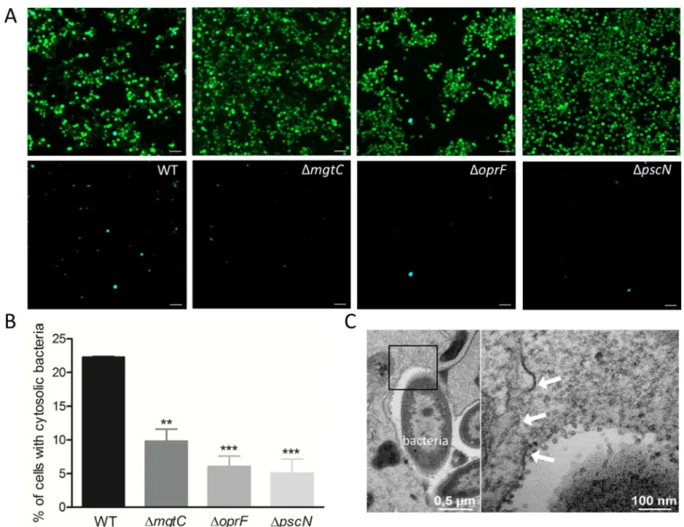
The phagosomal environment of the macrophage is hostile for bacterial pathogens and TEM analysis indicated that PAO1 can be found in the cytoplasm, suggesting escape from the pha-gosomal vacuole (Fig 2B). We decided to address the intracellular role of T3SS, OprF and MgtC in the phagosomal escape ofP. aeruginosa to the cytoplasm of macrophages. To monitor P. aeruginosa escape from phagosome, we used the CCF4-AM/β-lactamase assay that has been
developed for tracking vacuolar rupture by intracellular pathogens [43]. This assay takes advantage of the natural production ofβ-lactamase by P. aeruginosa [44], which can cleave a fluorescentβ-lactamase substrate, CCF4-AM, that is trapped within the host cytoplasm. CCF4-AM emits a green fluorescence signal, whereas in the presence ofβ-lactamase activity, a blue fluorescence signal is produced. The detection of blue fluorescent cells at 2 hrs post-phagocytosis indicated bacterial escape from the phagosome to the cytoplasm (Fig 8A). The escape of wild-type strain was compared to that ofmgtC, oprF and pscN mutants by
quantify-ing the percentage of blue fluorescent cells out of total green fluorescent cells. Wild-type strain showed significantly higher percentage of phagosomal escape than all mutants tested (Fig 8B). No significant difference in terms of cleavage of CCF4-AM was measured for the mutants with respect to wild-type in liquid cultures (S8 Fig), indicating that the lower amount of blue fluo-rescent cells with the mutants is not due to reduced production of endogenousβ-lactamase. In agreement with the finding of phagosomal escape by CCF4-AM/β-lactamase assay, ruptured phagosomal membrane could be visualized by TEM in macrophages infected with PAO1 wild-type strain (Fig 8C). BothexoS and exoSTY effector mutants also displayed a low percentage of
phagosomal escape, which appeared similar to that ofpscN mutant (S9 Fig). These results indi-cate that the T3SS, through the ExoS effector, plays role in the escape ofP. aeruginosa from the
phagosome to the cytoplasm. The effect of MgtC and OprF in this process may as well be mediated by their effect on T3SS gene expression (Fig 4).
Cumulatively, our results support a T3SS-dependent vacuolar escape forP. aeruginosa,
leading to the localization of bacteria in the cytoplasm and cell lysis as depicted in the proposed model (Fig 9).
Discussion
The ability of professional phagocytes to ingest and kill microorganisms is central to innate immunity and host defense.P. aeruginosa is known to avoid being killed by phagocytes
through the destruction of immune cells extracellularly as well as avoidance of phagocytosis
Fig 8. Assessment of access ofP. aeruginosa to host cytosol using phagosome escape assay. J774 macrophages were infected with PAO1 WT, ΔmgtC, ΔoprF
andΔpscN strains. After phagocytosis, cells were stained with CCF4-AM in presence of gentamicin. 2 hrs post-phagocytosis, the cells were imaged with 10X objective using FITC and DAPI channels. Upon escape of bacteria from phagosome to the cytosol, the CCF4-AM FRET is lost, producing blue color. (A) Representative pictures are shown with the following channels: Total cell population is shown in merged green and blue cells, whereas cells with cleaved CCF4 probe are shown in blue in the lower panel. Scale bar is equivalent to 50μm. (B) Images were analyzed and quantified by Cell Profiler software to calculate the percentage of blue cells out of total green cells. At least 200 cells were counted per strain. Error bars correspond to standard errors from three independent experiments. The asterisks indicateP values (One way ANOVA, where all strains were compared to WT using Dunnett’s multiple comparison post-test,��P <0.01 and���P <0.001), showing statistical significance with respect to WT. (C) Electron micrograph showing a disrupted vacuole membrane (white arrows) in a macrophage infected with PAO1 strain. The right panel shows higher magnification of the black square in the left panel.
https://doi.org/10.1371/journal.ppat.1007812.g008
[20]. However,P. aeruginosa has been reported to be engulfed by macrophages in animal
infection models [21–23]. In addition,P. aeruginosa has been visualized in phagocytes in cell
culture models in several studies, where MgtC and OprF have been shown to be involved in the ability ofP. aeruginosa to survive in cultured macrophages [25,26]. The virulence ofP. aer-uginosa mgtC mutant can be restored in zebrafish embryos upon macrophages depletion,
sug-gesting that MgtC acts to evade phagocytes [25]. Interestingly, a similar behavior has been reported for a T3SS mutant in the same infection model [21]. We show here that MgtC and OprF regulate T3SS whenP. aeruginosa resides in macrophages and we describe a novel
strat-egy used byP. aeruginosa to escape from macrophages that relies on a T3SS-dependent cell
lysis induced by intracellular bacteria (Fig 9).
Using electron microscopy, we demonstrate that upon phagocytosis,P. aeruginosa PAO1
strain resides in membrane bound vacuoles, whereas a cytosolic location can be observed at later time of infection, which corroborates the observation of an otopathogenicP. aeruginosa
clinical strain in HMDMs [26]. Microscopic analysis of live and fixed cells revealed macro-phage lysis driven by intracellular bacteria, with J774 macromacro-phage cell line as well as HMDMs. This
Fig 9. Model for intramacrophage fate ofP. aeruginosa. Phagocytosed P. aeruginosa PAO1 first resides in a vacuole, before escaping the phagosome
and promoting macrophage lysis. This cell lysis driven by intracellularP. aeruginosa involves the T3SS and more specifically ExoS. MgtC and OprF act positively on the expression of T3SS, possibly by reducing c-di-GMP level, a negative regulator of T3SS expression. Thereby T3SS and its effector ExoS play a role in phagosomal escape and cell lysis. Further work will be required to address secretion of ExoS from intracellularly expressed T3SS as well as identify host targets.
cell lysis is a rapid process associated with the loss of cortical actin from plasma membrane. We pro-pose that the cell lysis induced by intracellularP. aeruginosa is linked to the phagosomal escape of
bacteria as indicated by the observation of cytosolic bacteria and ruptured phagosomal membrane by TEM as well as CCF4-AM/β-lactamase based phagosomal rupture assay.
To better characterize theP. aeruginosa factors involved in its intramacrophage fate, we
investigated intracellular expression of T3SS genes inmgtC and oprF mutants. Expression of
T3SS genes upon macrophage infection was significantly decreased inmgtC mutant and
abro-gated inoprF mutant. The production of T3SS effectors or needle component was previously
known to be altered in theoprF mutant [32,34], but a direct effect at the transcriptional level was not investigated before. This regulation could be mediated by c-di-GMP, a known nega-tive regulator of T3SS expression [37], as the reporter assay revealed an increased level of c-di-GMP in bothoprF and mgtC mutants upon monitoring infected macrophages, with a more
pronounced effect foroprF mutant. An increased level of c-di-GMP in oprF mutant is
consis-tent with previous results obtainedin vitro [34]. The moderate, but significant, increase in the level of c-di-GMP inmgtC mutant in macrophages with respect to wild-type strain is equivalent
to the fold increase obtained in low-Mg2+cultures. It is of interest to note that aSalmonella mgtC mutant also exhibited increased c-di-GMP level intracellularly and under low-Mg2+ con-dition [29]. TheP. aeruginosa mgtC and oprF mutants showed a moderate and a more
pro-nounced decrease, respectively, in cytotoxicity driven by intracellular bacteria and phagosomal escape. These phenotypes could be linked to the pattern of T3SS expression inmgtC and oprF
mutants because a T3SS mutant appeared to lack cytotoxicity driven by intracellular bacteria and showed reduced phagosomal escape. Accordingly, inducing expression ofexsA gene, a
mas-ter activator of T3SS, inoprF mutant promoted macrophage lysis driven by intracellular bacteria
to a similar level to that of wild-type strain, supporting the model whereby the phenotype of
oprF mutant is the result of a negative regulation of T3SS expression.
Our data indicate that the T3SS-mediated cytotoxicity driven by intracellularP. aeruginosa
is largely dependent on the ExoS effector. ExoS, which has a dual function [10], was known to play a role in the intracellular life ofP. aeruginosa in cell types other than macrophages. The
T3SS and ExoS are indeed key factors for intracellular replication ofP. aeruginosa in epithelial
cells, with a main role of the ADPRT domain in the formation of replicative niche in mem-brane blebs [18,19,45]. ExoS and ExoT ADPRT domains also promote bacterial survival in neutrophils [46], by having a protective role against NADPH-oxidase activity [47]. However, the effect of T3SS towards macrophages was so far restricted to the well-known cytotoxicity caused by extracellularP. aeruginosa [39,48–50]. InP. aeruginosa strains lacking ExoU toxin,
such as PAO1, this cytotoxicity is due to inflammasome activation, which is dependent on T3SS translocation apparatus, but is independent of the ExoS effector [39–41,51]. Accordingly, this T3SS-dependent ExoS-independent cytotoxity was observed in our assays when extracel-lular bacteria were not removed. In contrast, upon removal of extracelextracel-lular bacteria, a T3SS-mediated cytotoxicity implicating ExoS was uncovered. Hence, ExoS appears to be associated with cell damage only when it is secreted from internalized bacteria. Moreover, phagosomal rupture assay indicated that ExoS plays role in the escape ofP. aeruginosa from the phagosome
to the cytoplasm and we propose that upon phagosomal rupture, the cytoplasmic location ofP. aeruginosa induces inflammasome, possibly through the release of bacterial
lipopolysaccha-rides (LPS) in the cytoplasm, which would promote cell death. Therefore, in addition to the inflammasome activation caused by extracellularP. aeruginosa and, as very recently described,
by intracellular T3SS-negativeP. aeruginosa in the context of long-term infection [52], our study suggests that inflammasome and subsequent macrophage death can also be caused by intracellular T3SS-positiveP. aeruginosa. Further studies will be required to characterize
inflammasome activation and pathways that lead to cell death.
domain have similar biochemical activities, these effectors may differ in their localization inside host cells or they may interact with different host factors to bring about their effect. This is supported by the fact that apoptosis is induced in epithelial cells by ExoT GAP domain, a feature that was not reported for ExoS GAP domain [55] and by our finding that, unlike ExoS, ExoT is not involved in the intramacrophage phenotypes. Importantly, we show that the extent of phagosomal escape ofpscN and exoSTY mutants was similar to that of exoS mutant alone,
suggesting that ExoS is the main effector protein involved in the exit ofP. aeruginosa from the
phagosome. In epithelial cells, ExoS has been involved in avoidance of acidified compartment [18,19]. Considering our findings, ExoS may allow avoidance of acidification in macrophages and may also contribute to the vacuolar escape in epithelial cells. Further studies will be required to decipher in more detail, the function of ExoS inside macrophages, including a bet-ter understanding of its role in phagosomal escape and identification of its host targets within macrophages. ExoS-positive strains represent a large number of clinical isolates but the num-ber of ExoS-negative strains is nevertheless substantial (about one third of isolates), especially among strains associated with bacteremia [10,11,56]. However, strains lacking ExoS usually encode the potent cytotoxin ExoU, thus exhibiting high toxicity towards eukaryotic cells from outside and less susceptibility to encounter an intracellular stage.
In conclusion, our results indicate thatP. aeruginosa shares common feature with other
so-called extracellular pathogens, such asS. aureus, which can reside transiently within
macro-phages [4] and require bacterial factors to survive this stage [57]. The present study uncovered bacterial factors allowing internalizedP. aeruginosa to lyse macrophages. This should let
bacte-ria to evade macrophages and an important issue now is to better evaluate the contribution of intramacrophage stage to disease outcome duringP. aeruginosa infection. Survival of P. aerugi-nosa within macrophages and subsequent bacterial release may play a role in the establishment
and dissemination of infection. There is also evidence that intracellular survival may contrib-ute to persistence of the infection by creating a niche refractory to antibiotic action [24], highlighting the potential importance of this overlooked phase ofP. aeruginosa infection.
Materials and methods
Bacterial strains and growth conditions
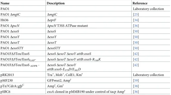
Bacterial strains and plasmids are described inTable 1.P. aeruginosa mutant strains (all in the
PAO1 background) have been described and phenotypically characterized previously (Table 1). TheoprF mutant (strain H636) is derived from PAO1 wild-type strain H103 [34], which exhibits the same level of ExoS secretion under T3SS-inducing conditions as PAO1, the isogenic strain formgtC and T3SS mutants (S10 Fig).P. aeruginosa was grown at 37˚C in Luria
broth (LB). Growth in magnesium-defined medium was done in NCE-minimal medium [58] supplemented with 0.1% casamino acids, 38 mM glycerol, without MgSO4or containing
10μM or 1mM MgSO4. Plasmid pMF230 expressing GFP constitutively [59] (obtained from
Addgene), was introduced inP. aeruginosa by conjugation, using an E. coli strain containing
helper plasmid pRK2013. Recombinant bacteria were selected onPseudomonas isolation agar
(PIA) containing carbenicillin at the concentration of 300μg/ml.
Infection of J774 macrophages
J774 cells (murine macrophage cell line J774A.1, gifted by Gisèle Bourg, Inserm U 1047, Nıˆmes, France) were maintained at 37˚C in 5% CO2in Dulbecco’s modified Eagle medium
(DMEM) (Gibco) supplemented with 10% fetal bovine serum (FBS) (Gibco). The infection of J774 macrophages byP. aeruginosa was carried out essentially as described previously [25]. Mid-log phaseP. aeruginosa grown in LB broth was centrifuged and resuspended in PBS to
infect J774 macrophages (5×10⁵cells/well) at an MOI of 10. After centrifugation of the 24-well culture plate for 5 min for synchronization of infection, bacterial phagocytosis was allowed for 25 min. Cells were washed three times with sterile PBS and fresh DMEM medium supple-mented with 400μg/ml gentamicin was added and retained throughout the infection.
Infection of human primary macrophages
Purified monocytes isolated as described from blood of healthy donors were frozen in liquid nitrogen [60,61]. Cells were thawed for experiment and seeded onto 24-well plates at a density of 7x10⁵per well in complete culture medium (RPMI containing 10% FCS) and differentiated into macrophages with rh-M-CSF (10 ng/ml) (purchased from Al-Immuno tools) for 7 days. HMDMs were infected with exponentially growingP. aeruginosa cultures (OD600= 0.8) at an
MOI of 10, as described above.
Live microscopy
J774 macrophages were seeded in ibidiμ-slide (8 wells) in DMEM medium supplemented with 10% FBS and infected withP. aeruginosa PAO1 expressing GFP as described in the
Table 1. Bacterial strains and plasmids used in the study.
Name Description Reference
PAO1 Laboratory collection
PAO1ΔmgtC ΔmgtC [25]
H636 ΔoprF [34]
PAO1ΔpscN ΔpscN T3SS ATPase mutant [36]
PAO1ΔexoS ΔexoS [50]
PAO1ΔexoY ΔexoY [50]
PAO1ΔexoT ΔexoT [50]
PAO1ΔexoSTY ΔexoSTY [50]
PAO1FΔ3Tox/ExoS ΔexoS ΔexoT ΔexoY attB::exoS [42] PAO1FΔ3Tox/ExoSGAP- ΔexoS ΔexoT ΔexoY attB::exoS-R146K [42]
PAO1FΔ3Tox/ExoSADPR- ΔexoS ΔexoT ΔexoY
attB::exoS-E379D/E381D
[42]
pRK2013 Tra+, Mob+, ColE1, Kmr Laboratory collection
pMF230 GFPmut2, Ampr [59]
pTn7CdrA::gfpC Ampr, Gmr [38]
pSBC6 exsA cloned in pMMB190 under control of tacp Ampr [36]
https://doi.org/10.1371/journal.ppat.1007812.t001
were fixed for 4 hrs at room temperature with 2.5% gluteraldehyde in cacodylate buffer 0.1 M pH 7.4 with 5mM CaCl2, washed with cacodylate buffer, post-fixed for 1 hr in 1% osmium
tetroxide and 1.5% potassium ferricyanide in cacodylate buffer, washed with distilled water, followed by overnight incubation in 2% uranyl acetate, prepared in water. Dehydration was performed through acetonitrile series and samples were impregnated in epon 118: acetonitrile 50:50, followed by two times for 1 hr in 100% epon. After overnight polymerization at 60˚C, coverslips were detached by thermal shock with liquid nitrogen. Polymerization was then pro-longed for 48 hrs at 60˚C. Ultrathin sections of 70 nm were cut with a Leica UC7 ultramicro-tome (Leica microsystems), counterstained with lead citrate and observed in a Jeol 1200 EXII transmission electron microscope. All chemicals were from Electron Microscopy Sciences (USA) and solvents were from Sigma. Images were processed using Fiji software.
Colocalization of
P. aeruginosa with acidic compartments
Macrophages were infected with GFP labelledP. aeruginosa as described above. After 2.5 hrs
of gentamicin treatment, infected J774 cells were washed twice with PBS and incubated with 50 nM Lysotracker red DND-99 (Molecular Probes) in DMEM for 10 min to stain lysosomes. Cells were then washed with PBS and fixed with 4% paraformaldehyde in PBS and mounted on glass slides in Vectashield (Vector Laboratories, Inc) with 40,6-diamidino-2-phenylindole
(DAPI) to stain the nucleus. The slides were examined using an upright fluorescence micro-scope (Axioimager Z2, Zeiss) equipped with an Apotome 1 for optical sectioning. A 63X Apochromat Objective (NA 1.4) was used, transmitted light was acquired using differential interference contrast (DIC), Fluorescein isothiocyanate (FITC) filter was used to visualize GFP expressing bacteria and Lysotracker red fluorescence was acquired using a texas red filter set.
Phalloidin labeling
J774 macrophages were seeded on glass coverslips and infected with GFP expressing bacteria as described in the previous sections. For cytochalasin treatment, DMEM containing 2μM cytochalasin D (Sigma) was added to the macrophages 1 hour before infection and maintained during phagocytosis. After phagocytosis, the cells were maintained in 1μM cytochalasin D till the end of the experiment. For untreated control, 0.2% DMSO (solvent control) in DMEM was added to the cells before and during phagocytosis. After phagocytosis, cells were main-tained in 0.1% DMSO. After fixation with 4% paraformaldehyde (EMS, USA) in PBS for 5 min, cells were washed once with PBS and permeabilized by adding 0.1% triton X-100 for 1 min 30 sec. Cells were then washed once with PBS and incubated with 1μg/ml Tetramethylr-hodamine B isothiocyanate (TRITC)-labeled phalloidin (Sigma-Aldrich) in PBS for 30 min in dark. Cells were washed twice with PBS and coverslips were mounted on glass slides in Vecta-shield with DAPI (Vector Laboratories, Inc). The slides were examined using an upright
fluorescence microscope (Axioimager Z1, Zeiss) equipped with an Apotome 1 for optical sec-tioning. A 63X Apochromat Objective (NA 1.4) was used and transmitted light was acquired using DIC. FITC and texas red filters were used to visualize GFP expressing bacteria and phal-loidin respectively. Cell nuclei were visulalized using DAPI filter. Images were processed using ZEN software (Zeiss). Cells were counted manually, where infected cells lacking the phalloidin stain were considered as lysed. Percentage of such lysed cells with intracellular bacteria out of total number of infected cells was calculated and plotted for each strain.
Strains containing pSBC6 plasmid (expressingexsA under the control of tac promoter)
were grown before infection in LB or in LB supplemented with 0.01 mM IPTG. Because these strains did not harbor GFP producing plasmid, the number of total cells without phalloidin label was counted and percentage out of total number of cells was plotted. The percent of lysed cells for wild-type strain was similar to that found with GFP positive PAO1 strain.
LDH cytotoxicity assay
The cytotoxicity was assessed by release of LDH from infected J774 macrophages infected, using the Pierce LDH cytotoxicity assay kit (Thermo Scientific). Macrophages were infected for 2 hrs at an MOI of 10 as described above, except that cells were seeded in a 96 well plate and extracellular bacteria were not removed. The assay was performed on 50μl of the culture supernatant according to manufacturer’s instructions. LDH release was obtained by subtract-ing the 680 nm absorbance value from 490 nm absorbance. The percentage of LDH release was first normalized to that of the uninfected control and then calculated relatively to that of uninfected cells lysed with Triton X-100, which was set at 100% LDH release.
Trypan Blue exclusion test of cell viability
The membrane impermeable dye Trypan Blue was used to quantify cell viability after phagocy-tosis or after 2 hrs of gentamicin treatment following phagocyphagocy-tosis. Trypan Blue stain (0.4%) was added in 1:1 ratio with PBS for 3 min at room temperature, replaced by PBS and the cells were imaged using an optical microscope in bright field mode. Dead cells appear blue as they take up the stain, in contrast to healthy cells that appear transparent because of the exclusion of the dye. Cells were counted and the percentage of dead cells out of total cells (blue + color-less) was calculated.
RNA extraction and quantitative RT-PCR (qRT-PCR)
For bacterial RNA extraction from infected J774, 6.5x10⁶macrophages were seeded into a 100 cm2tissue culture dish and infected at an MOI of 10 as described above. 1 hour after phagocy-tosis, cells were washed three times with PBS, lysed with 0.1% Triton X100 and pelleted by cen-trifugation at 13000 rpm for 10 min at 15˚C. Bacteria were resuspended in 500μl PBS and the non resuspended cellular debris was discarded. 900μl of RNA protect reagent (Qiagen) was added and incubated for 5 min. The sample was centrifuged at 13000 rpm for 10 min. Bacteria in the pellet were lysed with lysozyme and RNA was prepared with RNeasy kit (Qiagen). Superscript III reverse transcriptase (Invitrogen) was used for reverse transcription. Controls without reverse transcriptase were done on each RNA sample to rule out possible DNA con-tamination. Quantitative real-time PCR (q-RT-PCR) was performed using a Light Cycler 480 SYBR Green I Master mix in a 480 Light Cycler instrument (Roche). PCR conditions were as follows: 3 min denaturation at 98˚C, 45 cycles of 98˚C for 5 sec, 60˚C for 10 sec and 72˚C for 10 sec. The sequences of primers used for RT-PCR are listed inS1 Table.
could be obtained in comparison to the blank. Fluorescence was plotted for each strain in terms of arbitrary units (AU). CdrA activity of all strains was also measured in liquid cultures under changing concentrations of magnesium. All strains, grown overnight in LB, were diluted in LB and grown until OD600of 0.6, washed in NCE medium without magnesium and resuspended in
NCE medium containing 1 mM, 10μM or no magnesium for 1 hour in 96 well plate (Greiner, Flat-Bottom). Their fluorescence (excitation, 485 nm and emission, 520 nm) and OD600nmwere
measured. Fluorescence (AU520nm) was normalized to OD600nmand plotted for each strain.
CCF4 fluorometric assay to monitor the escape of
P. aeruginosa in host cytosol
The vacuole escape assay was adapted from the CCF4 FRET assay [43] using the CCF4-AM Live-Blazer Loading Kit (Invitrogen) and an image-based quantification [62]. Briefly, J774 macrophages were seeded in 96 well plate (Greiner, Flat-Bottom), containing 5x10⁴cells per well. Overnight bac-terial cultures were subcultured in LB with 50μg/ml of ampicillin to enhance the expression of beta-lactamase, present naturally inP. aeruginosa. Infection was carried out as mentioned in the
previous sections at the MOI of 10. After phagocytosis, the cells were washed thrice with PBS to remove extracellular bacteria. 100μl of HBSS buffer containing 3 mM probenecid and gentamicin (400μg/ml), was added in each well. The substrate solution was prepared by mixing 6 μl of CCF4-AM (solution A), 60μl of solution B and 934 μl of solution C. 20 μl of the substrate solution was added to each well and the plate was incubated in dark at 37˚C with 5% CO2. After 2 hrs, the
cells were imaged as described in the Live Imaging section, using a 10X objective. FITC and DAPI channels were used to visualize CCF4-FRET (Green) and loss of FRET (Blue) respectively. Each sample was taken in triplicate and image acquisition was performed by automated random acqui-sition. Images were analyzed by Cell Profiler software to calculate the number of blue and green cells. The threshold for detection of blue signal by the software was normalized to uninfected con-trol i.e. no blue cells could be detected in the uninfected concon-trol. The percentage of blue cells, rep-resenting the cells with cytosolic bacteria, out of total green cells was plotted. All strains were tested for their ability to cleave CCF4in vitro, before carrying out the vacuole rupture assay.
Over-night cultures grown in LB were subcultured at the ratio of 1:20 in LB with 100μg/ml of ampicil-lin. After 2 hours of growth, the cultures were centrifuged and resuspended in PBS. 100μl of this was aliquoted in 96 well plate (Greiner, Flat-Bottom) and 20μl of CCF4 substrate solution (A+B +C) was added. Tecan fluorimeter (Spark 20M) was used to measure fluorescence (excitation, 405 nm and emission, 450 nm) using PBS as blank. Blue fluorescence (AU450nm) was observed for all
strains and none of the mutants exhibited significantly lower value than the wild-type strain.
Ethics statement
Monocytes were issued from blood of anonymous donors obtained from the French blood bank (Etablissement Franc¸ais du Sang, approval EFS-OCPM n˚ 21PLER2018-0057).
Supporting information
S1 Fig. Live imaging of macrophages infected withP. aeruginosa. J774 macrophages were
infected with PAO1 wild-type strain expressing GFP. Time lapse imaging was started at 1.5 hrs post-phagocytosis. Cells were maintained in DMEM supplemented with gentamicin, at 37˚C and 5% CO2throughout imaging. White arrows point at infected cells that undergo lysis,
whereas black arrows indicate uninfected cells that do not lyse. Images were taken between 1.5 hrs and 3 hrs post-phagocytosis as shown on the panels. Scale bar is equivalent to 10μm. (PDF)
S2 Fig. Live imaging of primary human macrophages infected withP. aeruginosa. HMDMs
were infected with PAO1 wild-type (WT) strain expressing GFP. Time lapse imaging was started at 1.5 hrs post-phagocytosis. Cells were maintained in RPMI supplemented with genta-micin at 37˚C and 5% CO2throughout imaging. White arrows point at the cells that harbor
intracellular bacteria and undergo lysis between 1.5 hrs and 3 hrs post-phagocytosis. Black arrow shows an uninfected and unlysed cell. Scale bar is equivalent to 20μm.
(PDF)
S3 Fig. Colocalization ofP. aeruginosa with a probe that labels acidic compartments. J774
macrophages were infected with PAO1 expressing GFP. After 2.5 hrs of gentamicin treatment, infected J774 cells were incubated with Lysotracker for 10 min, a red fluorescent weak base that accumulates in acidic compartments. Cells were then fixed and imaged with fluorescence microscope. (A) The image shows individual panels for Differential Interference Contrast (DIC), lysosomal compartment (red), bacteria expressing GFP (green), the nucleus (blue) and merged image of all channels. The solid arrow shows colocalization of bacteria with lysotracker and dashed arrow shows non-colocalization. Scale bar is equivalent to 5μm. (B) 3D-recon-structed image of the same area of (A).
(PDF)
S4 Fig. Visualization (A) and quantification (B) of lysed infected cells upon cytochalasin D treatment. GFP expressing PAO1 was used for infecting J774 macrophages. DMEM
con-taining 2μM cytochalasin D was added to the macrophages 1 hour before infection and main-tained during phagocytosis. After phagocytosis, cells were mainmain-tained in 1μM cytochalasin D in DMEM supplemented with gentamicin till the end of the experiment. 0.2% DMSO in DMEM was added to the cells as solvent control before and during phagocytosis. After phago-cytosis, cells were maintained in 0.1% DMSO in DMEM with gentamicin, fixed 2 hrs post-phagocytosis, stained with phalloidin and imaged with fluorescent microscope. DAPI was used to stain the nucleus. Cells that have intracellular bacteria, but lack the phalloidin cortical label, were considered as lysed by intracellular bacteria (shown by arrows). Scale bar is equiva-lent to 10μm. Percentage of lysed cells with intracellular bacteria out of total number of cells was plotted. Error bars correspond to standard errors from two independent experiments. At least 200 cells were counted per strain. The asterisks indicateP values (Student’s t-test, ��P <0.01), showing statistical significance with respect to DMSO control.
(PDF)
S5 Fig. Quantification of dying cells infected withP. aeruginosa strains overexpressing exsA. PAO1 WT, ΔoprF, PAO1 WT + pexsA and ΔoprF + pexsA strains were used for infecting
J774 macrophages. Expression ofexsA was induced by adding 0.01 mM IPTG for the strains
PAO1 WT + pexsA and ΔoprF + pexsA. Gentamicin was added after phagocytosis and cells
were fixed at 2 hrs post-phagocytosis, stained with phalloidin and imaged with fluorescent microscope. DAPI was used to stain the nucleus. Cells that lacked the phalloidin cortical label
of total uninfected cells lysed with Triton X-100, which was set at 100% LDH release. Error bars correspond to standard errors (SE) from at least four independent experiments. The asterisks indicateP values (One way ANOVA, where all strains were compared to WT using Dunnett’s
multiple comparison post-test,��P <0.01), showing statistical significance with respect to WT.
(PDF)
S7 Fig. Quantification of lysed cells by staining with Trypan Blue. J774 macrophages were
infected with the strains as indicated. After phagocytosis, cells were maintained in DMEM sup-plemented with gentamicin. Cells were stained with trypan blue at (A) 30 min or (B) 2 hrs post-phagocytosis and imaged. Lysed cells were quantified by counting cells stained with try-pan blue and the percentage of lysed cells out of total number of cells was plotted. Error bars correspond to standard errors from at least three independent experiments. At least 400 cells were counted per strain. The asterisks indicateP values (One way ANOVA, where all strains
were compared to WT using Dunnett’s multiple comparison test,�P <0.05,��P <0.01 and ���P <0.001), showing statistical significance with respect to WT.
(PDF)
S8 Fig. Assessment ofβ-lactamase activity in liquid culture. PAO1 WT, ΔmgtC, ΔoprF,
ΔpscN, ΔexoS and ΔexoSTY strains grown in presence of ampicillin, were incubated with CCF4-AM for 1 hour. The blue fluorescence generated as a result of loss of FRET of CCF4 was measured (excitation, 420 nm and emission, 450 nm) and plotted as arbitrary units (AU). Error bars correspond to standard errors from four independent experiments. All strains were compared to WT using One way ANOVA, Dunnett’s multiple comparison post-test. No sig-nificant difference was found between the mutant strains and WT.
(PDF)
S9 Fig. Phagosome escape assay of T3SS mutants. J774 macrophages were infected with
PAO1 WT,ΔpscN, ΔexoS and ΔexoSTY strains. After phagocytosis, cells were stained with CCF4-AM in presence of gentamicin. 2 hrs post-phagocytosis, the cells were imaged with 10X objective using FITC and DAPI channels. Upon escape of bacteria from phagosome to the cytosol, the CCF4-AM FRET is lost, producing blue color. Images were analyzed and quanti-fied by Cell Profiler software to calculate the percentage of blue cells out of total green cells. At least 200 cells were counted per strain. Error bars correspond to standard errors from four independent experiments. The asterisks indicateP values (One way ANOVA, where all strains
were compared to WT using Dunnett’s multiple comparison post-test,�P <0.05), showing
sta-tistical significance with respect to WT. (PDF)
S10 Fig. Comparison of secreted protein profiles and ExoS production under T3SS-induc-ing conditions in two PAO1 strains. Immunodetection of the ExoS effector in culture
supernatant of PAO1 and PAO1 H103 (isogenic WT strain foroprF mutant) grown in
expo-nential phase at 37˚C under T3SS-inducing (− Ca2+) or non-inducing (+ Ca2+) conditions. The bacterial culture supernatant (equivalent of 1 OD600unit) was loaded on an SDS gel
con-taining 10% polyacrylamide. As control, cellular fraction (equivalent of 0.1 OD600unit) was
loaded. Upper panels show Western blot using anti-ExoS antibodies (Soscia et al., 2007; doi:
10.1128/JB.01677-06) and lower panels show Coomassie stained gels. (PDF)
S1 Movie. Live microscopy movie imaging cell lysis in real time. J774 macrophages were
infected with PAO1 WT strain expressing GFP. Cells were maintained in DMEM supple-mented with gentamicin, at 37˚C and 5% CO2throughout imaging. Imaging was started at 3
hrs post-phagocytosis and continued for 10 min with interval of 30 sec between frames. The time frame is displayed in the movie in the format of minutes:seconds:milli seconds. The movie shows quick lysis of macrophages harboring intracellular bacteria occurring within a minute.
(MOV)
S1 Table. List of primers used for RT-PCR.
(PDF)
Acknowledgments
We thank Sylvie Chevalier (Rouen), Ina Attre´e and Sylvie Elsen (Grenoble) for providing strains and plasmids, the Montpellier RIO Imaging microscopy platform for photonic Micros-copy and the Electron MicrosMicros-copy facility of the University of Montpellier (MEA) for sample preparation and transmission electron microscopy. We thank Be´rengère Ize and Laila Gan-noun-Zaki for critical reading of the manuscript, Matteo Bonazzi and Fernande Siadous for their expertise with the CCF4 assay, Chantal Soscia for testing ExoS secretion.
Author Contributions
Conceptualization: Preeti Garai, Laurence Berry, Anne-Be´atrice Blanc-Potard. Data curation: Preeti Garai.
Formal analysis: Preeti Garai, Malika Moussouni. Funding acquisition: Anne-Be´atrice Blanc-Potard.
Investigation: Preeti Garai, Laurence Berry, Malika Moussouni, Anne-Be´atrice Blanc-Potard. Methodology: Preeti Garai, Laurence Berry, Anne-Be´atrice Blanc-Potard.
Project administration: Anne-Be´atrice Blanc-Potard. Resources: Sophie Bleves, Anne-Be´atrice Blanc-Potard. Supervision: Anne-Be´atrice Blanc-Potard.
Validation: Preeti Garai, Anne-Be´atrice Blanc-Potard. Visualization: Preeti Garai, Malika Moussouni.
Writing – original draft: Preeti Garai, Anne-Be´atrice Blanc-Potard.
Writing – review & editing: Preeti Garai, Laurence Berry, Malika Moussouni, Sophie Bleves,
Anne-Be´atrice Blanc-Potard.
5. Flannagan RS, Heit B, Heinrichs DE (2016) Intracellular replication of Staphylococcus aureus in mature phagolysosomes in macrophages precedes host cell death, and bacterial escape and dissemination. Cell Microbiol 18: 514–535.https://doi.org/10.1111/cmi.12527PMID:26408990
6. Jubrail J, Morris P, Bewley MA, Stoneham S, Johnston SA, et al. (2016) Inability to sustain intraphagoly-sosomal killing of Staphylococcus aureus predisposes to bacterial persistence in macrophages. Cell Microbiol 18: 80–96.https://doi.org/10.1111/cmi.12485PMID:26248337
7. Ercoli G, Fernandes VE, Chung WY, Wanford JJ, Thomson S, et al. (2018) Intracellular replication of
Streptococcus pneumoniae inside splenic macrophages serves as a reservoir for septicaemia. Nat
Microbiol.
8. Klockgether J, Tummler B (2017) Recent advances in understanding Pseudomonas aeruginosa as a pathogen. F1000Res 6: 1261.https://doi.org/10.12688/f1000research.10506.1PMID:28794863 9. Engel J, Balachandran P (2009) Role of Pseudomonas aeruginosa type III effectors in disease. Curr
Opin Microbiol 12: 61–66.https://doi.org/10.1016/j.mib.2008.12.007PMID:19168385
10. Hauser AR (2009) The type III secretion system of Pseudomonas aeruginosa: infection by injection. Nat Rev Microbiol 7: 654–665.https://doi.org/10.1038/nrmicro2199PMID:19680249
11. Feltman H, Schulert G, Khan S, Jain M, Peterson L, et al. (2001) Prevalence of type III secretion genes in clinical and environmental isolates of Pseudomonas aeruginosa. Microbiology 147: 2659–2669.
https://doi.org/10.1099/00221287-147-10-2659PMID:11577145
12. Garcia-Medina R, Dunne WM, Singh PK, Brody SL (2005) Pseudomonas aeruginosa acquires biofilm-like properties within airway epithelial cells. Infect Immun 73: 8298–8305.https://doi.org/10.1128/IAI. 73.12.8298-8305.2005PMID:16299327
13. Sana TG, Hachani A, Bucior I, Soscia C, Garvis S, et al. (2012) The second type VI secretion system of
Pseudomonas aeruginosa strain PAO1 is regulated by quorum sensing and Fur and modulates
internal-ization in epithelial cells. J Biol Chem 287: 27095–27105.https://doi.org/10.1074/jbc.M112.376368
PMID:22665491
14. Mittal R, Grati M, Gerring R, Blackwelder P, Yan D, et al. (2014) In vitro interaction of Pseudomonas
aeruginosa with human middle ear epithelial cells. PLoS One 9: e91885.https://doi.org/10.1371/ journal.pone.0091885PMID:24632826
15. Kazmierczak BI, Jou TS, Mostov K, Engel JN (2001) Rho GTPase activity modulates Pseudomonas
aeruginosa internalization by epithelial cells. Cell Microbiol 3: 85–98. PMID:11207623
16. Angus AA, Lee AA, Augustin DK, Lee EJ, Evans DJ, et al. (2008) Pseudomonas aeruginosa induces membrane blebs in epithelial cells, which are utilized as a niche for intracellular replication and motility. Infect Immun 76: 1992–2001.https://doi.org/10.1128/IAI.01221-07PMID:18316391
17. Kroken AR, Chen CK, Evans DJ, Yahr TL, Fleiszig SMJ (2018) The Impact of ExoS on Pseudomonas
aeruginosa Internalization by Epithelial Cells Is Independent of fleQ and Correlates with Bistability of
Type Three Secretion System Gene Expression. MBio 9.
18. Angus AA, Evans DJ, Barbieri JT, Fleiszig SM (2010) The ADP-ribosylation domain of Pseudomonas
aeruginosa ExoS is required for membrane bleb niche formation and bacterial survival within epithelial
cells. Infect Immun 78: 4500–4510.https://doi.org/10.1128/IAI.00417-10PMID:20732998 19. Heimer SR, Evans DJ, Stern ME, Barbieri JT, Yahr T, et al. (2013) Pseudomonas aeruginosa utilizes
the type III secreted toxin ExoS to avoid acidified compartments within epithelial cells. PLoS One 8: e73111.https://doi.org/10.1371/journal.pone.0073111PMID:24058462
20. Lovewell RR, Patankar YR, Berwin B (2014) Mechanisms of phagocytosis and host clearance of
Pseu-domonas aeruginosa. Am J Physiol Lung Cell Mol Physiol 306: L591–603.https://doi.org/10.1152/ ajplung.00335.2013PMID:24464809