A d-Amino Acid at the N-Terminus of a Protein
Abrogates Its Degradation by the N-End Rule Pathway
The MIT Faculty has made this article openly available.
Please share
how this access benefits you. Your story matters.
Citation
Rabideau, Amy E., and Bradley L. Pentelute. “A d-Amino Acid at the
N-Terminus of a Protein Abrogates Its Degradation by the N-End
Rule Pathway.” ACS Central Science 1.8 (2015): 423–430. © 2015
American Chemical Society
As Published
http://dx.doi.org/10.1021/acscentsci.5b00308
Publisher
American Chemical Society (ACS)
Version
Final published version
Citable link
http://hdl.handle.net/1721.1/110605
Terms of Use
Article is made available in accordance with the publisher's
policy and may be subject to US copyright law. Please refer to the
publisher's site for terms of use.
A
D
‑Amino Acid at the N‑Terminus of a Protein Abrogates Its
Degradation by the N
‑End Rule Pathway
Amy E. Rabideau and Bradley L. Pentelute
*
Department of Chemistry, Massachusetts Institute of Technology, 18-596, 77 Massachusetts Avenue, Cambridge, Massachusetts 02139, United States
*
S Supporting InformationABSTRACT: Eukaryotes have evolved the ubiquitin (Ub)/ proteasome system to degrade polypeptides. The Ub/ proteasome system is one way that cells regulate cytosolic protein and amino acids levels through the recognition and ubiquitination of a protein’s N-terminus via E1, E2, and E3 enzymes. The process by which the N-terminus stimulates intracellular protein degradation is referred to as the N-end rule. Characterization of the N-end rule has been limited to only the natural L-amino acids. Using a cytosolic delivery
platform derived from anthrax lethal toxin, we probed the stability of mixed chirality proteins, containing one D-amino
acid on the N-terminus of otherwise allL-proteins. In all cases, we observed that one N-terminalD-amino acid stabilized the cargo
protein to proteasomal degradation with respect to the N-end rule. We found that since the mixed chirality proteins were not polyubiquitinated, they evaded N-end-mediated proteasomal degradation. Evidently, a subtle change on the N-terminus of a natural protein can enhance its intracellular lifetime.
■
INTRODUCTIONThe chirality of biomolecules in nature is critical for substrate recognition, protein binding, and product formation. While E3 ubiquitin (Ub) ligases of the Ub/proteasome system have promiscuous substrate binding sites, the chirality of protein substrates has never been investigated.1−3 Since the homeo-stasis of a cell’s protein and amino acid concentrations is regulated by the proteasome, perturbation of the proteasome’s activity through substrate modification can affect the intra-cellular equilibrium. Here, we investigated the effect of one mirror imageD-amino acid on the N-terminus of otherwise all L-proteins on proteasomal degradation after delivery of the
mixed chirality proteins into the cytosol of cells.
Varshavsky and co-workers have characterized the N-end rule as it relates to the intracellular stability of proteins.4−6 According to the N-end rule, the identity of the N-terminal amino acid mediates the selective degradation of specific proteins through the Ub/proteasome system. The N-terminal degradation signals are termed N-degrons, which can range from stabilizing to destabilizing residues. Key destabilizing residues include type 1 (R, K, and H) and type 2 (L, F, Y, W, and I) residues, while D, E, N, Q, and C can be destabilizing after modifications such as acetylation or arginylation. N-degrons are recognized by N-recognins, or E3 Ub ligases, which interact with E2 Ub conjugating enzymes to polyubiquitinate proteins for proteasomal degradation.6To date, the N-end rule has been defined only forL-amino acids.7−10In a recent study
by Sriram et al. to identify inhibitors of the N-end rule, the authors demonstrated that two mixed chirality dipeptides
containing an N-terminalDArg did not affect protein stability to
the same extent as dipeptides containing an N-terminalLArg.3 While this study suggested that the N-end rule is stereospecific, it does not provide direct evidence that thesefindings will hold for an intact protein. The main challenge with probing the stability of proteins containing non-natural functionalities at the N-terminus is the delivery of such proteins into the cytosol. To overcome this challenge, we utilized a platform derived from nature that enables the delivery of different types of proteins into the cytosol of cells.
Anthrax lethal toxin from Bacillus anthracis utilizes the protective antigen (PA) pore to deliver lethal factor (LF) into the cytosol.11Protein translocation by anthrax lethal toxin has been extensively characterized. In short, to obtain entry into the cell, PA binds to an anthrax receptor on the cell surface and forms the PA prepore.12−17LF binds to the PA prepore, then the entire complex is endocytosed, and endosomal acidification triggers a conformational rearrangement of the PA prepore to form a pore.18,19LF translocates through the pore via a charge state dependent Brownian ratchet into the cytosol.13,20The N-terminal domain of LF (LFN) is sufficient to bind to the PA
prepore, but does not cause any intracellular toxicity.21 The PA/LFNdelivery system has been engineered to deliver various
peptide, protein, and small molecule cargoes into the cell cytosol. Previous work has shown that numerous peptides and
Received: September 10, 2015
Published: November 11, 2015
Research Article
http://pubs.acs.org/journal/acscii
proteins, including those composed of D-amino acids, can be
efficiently delivered through the PA pore.22−25
The majority of eukaryotic proteins from ribosomal trans-lation are composed of L-amino acids and achiral glycine. In
order to study the intracellular stability of proteins containing
D-amino acids, we used sortase A (SrtA) from Staphylococcus
aureus or native chemical ligation (NCL) to ligate oneD-amino
acid onto the N-terminus of L-proteins that can then be
delivered in a PA dependent manner.26 Furthermore, we incorporated a cleavable linker that releases the cargo protein from LFNafter translocation into the cytosol further allowing us to characterize N-terminal D-amino acids on proteins other
than LFN, including A-chain of diphtheria toxin (DTA)27,28and a designed ankyrin repeats protein (DARPin).29,30We opted to use a hindered disulfide cleavable linker that was small in structure such that it could translocate through the PA pore efficiently and be readily cleaved in the reducing environment of the cytosol.31
■
RESULTSSortase A Attaches One D-Amino Acid onto the
N-Terminus of LFN-DTA. The X-LFN-DTAmut constructs were
produced through enzyme-mediated ligation of XALPSTGG onto the N-terminus of the LFN-DTAmut. The N-terminal
amino acid (X) represents a natural L-amino acid (LX) or its
mirror imageD-amino acid (DX), while the remaining residues
wereL-stereochemistry (Figure 1a). Each XALPSTGG peptide
was ligated to G5-LFN-DTAmut using Staphylococcus aureus
sortase A to yield XALPSTG5-LFN-DTAmutconstructs (X-LFN -DTAmut; Figure 1b). A one-pot ligation scheme was used for
each reaction.22,32
One N-Terminal D-Amino Acid Stabilizes LFN-DTA to
Proteasomal Degradation. We used the A chain of diphtheria toxin (DTA) as a first measure of proteasomal
degradation inside cells with less protein synthesis inhibition, inferring that the cargo was degraded more rapidly. Similar assays have been used to understand how the N-end rule affects toxin stability in the cytosol of cells.7For our experiments, we used a mutant form of DTA (E148S; DTAmut) that is 300-fold
less active than wild-type DTA,33 allowing us to detect differences in cytosolic lifetime of each X-LFN-DTAmut over a
wider dynamic range. Since wild-type DTA can neutralize its substrate in minutes, DTAmut enabled analysis of various
substrates after a 6-h time period. DTA inhibits protein synthesis by ADP ribosylating elongation factor-2.27,28 To corroborate our observations with DTA as the read-out, we used an orthogonal assay based on Western blot analysis of the cytosolic fraction (Figure 1c).34
For the protein synthesis inhibition assay, Chinese hamster ovary (CHO-K1) cells were treated with 10-fold serial dilutions of each construct for 6 h to allow for sufficient buildup of the translocated material in the cytosol and to observe DTAmut
activity. After translocation, the cells were washed and treated with3H-Leu in leucine-free medium for 1 h to detect DTA
mut
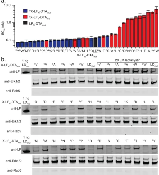
activity. The fraction of protein synthesis with respect to DTAmut was measured with a scintillation counter (Figure S1 and Table S1). According to Figure 2a, regardless of chirality, stabilizing amino acids such as A and V on the N-terminus of LFN-DTAmut had similar EC50 values as the positive control (LFN-DTAmut), which contains LA at the N-terminus.
Furthermore, destabilizing residues likeLW on the N-terminus
of LFN-DTAmut had significantly higher EC50 values than the
control (i.e., less DTA activity) while the DW-LF
N-DTAmut
construct displayed activity comparable to the control, which suggests thatD-amino acids act as stabilizing residues.
To further confirm that our observations were a result of proteasomal degradation, we used Western blot analysis. The X-LFN-DTAmut constructs were delivered into CHO-K1 cells, lysed using digitonin lysis buffer, and analyzed by Western blot. Digitonin is a nonionic detergent used to permeabilize the plasma membrane, while the membrane-bound organelles remain intact. CHO-K1 cells were treated with X-LFN-DTAmut
constructs in the presence of PA for 6 h and were lysed using a digitonin lysis buffer and then analyzed by Western blot. The Western blot was immunostained with an LF antibody for stability analysis, and then stained for the cytosolic proteins, Erk1/2, and the early endosomal protein, Rab5. A low level of Rab5 and a high level of Erk1/2 demonstrate efficient cytosolic extraction. Based on thefindings inFigure 2b, the Western blot results corroborated the protein synthesis inhibition data and showed significant differences in cytosolic protein levels, where
D-amino acids proved to be stabilizing regardless of the side
chain identity. These data support the end rule for N-terminal L-amino acids, while all N-terminal D-amino acids
stabilized X-LFN-DTAmut to degradation.
As a control, CHO-K1 cells were treated with select conjugates (LV-, DV-, LA-, DA-, LW, and DW-LF
N-DTAmut) in
the presence of lactacystin, a proteasome inhibitor. The samples treated with lactacystin all showed strong anti-LF bands, indicating that the proteasome played a key role in degrading theLX-LF
N-DTAmutconstructs but had no observable effect on
the DX-LF
N-DTAmut constructs. Furthermore, these data
indicated that each construct translocated efficiently into the cells, regardless of the N-terminal amino acid.
To verify the mechanism of translocation and endosome escape, we incubated the X-LFN-DTAmut constructs with a mutant PA (PA[F427H]),35 a vacuolar H+-ATPase inhibitor
Figure 1. Intracellular stability was monitored for X-LFN-DTA
constructs delivered through protective antigen pore. (a) LX- (left) andDX- (right) amino acids ligated to the N-terminus of LFN-DTA
(LFN, green, pdb is 1J7N; DTA, orange, pdb is 1DTP). (b)
XALPSTGG peptides, where X represents either an L- or D-amino acid, are ligated onto G5-LFN-DTA using sortase A (SrtA) to form
X-LFN-DTA constructs. (c) Translocation of X-LFN-DTA constructs is
achieved using protective antigen (PA) of anthrax toxin.
ACS Central Science Research Article
DOI: 10.1021/acscentsci.5b00308 ACS Cent. Sci. 2015, 1, 423−430 424
(bafilomycin A1), or at 4 °C with CHO-K1 for 6 h. In all cases, no material was found to translocate into the cytosol by Western blot (Figure S2). These controls indicated that delivery of the X-LFN-DTAmut constructs into the cytosol is
dependent on functional endocytic machinery and PA. Moreover, we found that our observations were not cell-specific. After translocation, we observed protein stabilization in human embryonic kidney cells (HEK-293T) and human cervical cancer cells (HeLa), similar to the stabilization observed in CHO-K1 cells (Figure S3).
Proteasomal Stabilization Is Not an Artifact of the Sortag. SrtA ligation adds a short linkage (i.e., LPSTG5)
between the N-terminal amino acid (X) and the start of the protein. We used native chemical ligation (NCL)36to prepare constructs with native N-terminal sequences for comparison with sortagged proteins. For our analysis, LFN-DTAmut was synthesized containingL-alanine at the N-terminus (wild-type)
and compared to NCL synthesized constructs containing DA,
LW, or DW.37
Each NCL construct was translocated in CHO-K1 cells, and their protein stability was compared to that of the sortagged conjugates using Western blot. For the NCL reaction
we installed a Cys residue at position 17 that was later alkylated with bromoacetamide, which does not affect translocation. Based on the Western blot of the translocated material in
Figure S4, both native and sortagged constructs containingLA,
DA, andDW had similar protein stability, whileLW in both cases
was degraded. These observations indicated that stabilization of LFN-DTAmut through the incorporation of one N-terminal D
-amino acid is not an artifact of the sortag.
LFN-DTA with One N-terminal D-Amino Acid Is Stable
In Vitro. To support our cytosolic studies, we analyzed the in vitro rates of degradation of the X-LFN-DTAmut constructs in rabbit reticulocyte lysate (RRL). Pure X-LFN-DTAmutproteins
were incubated in the presence of 70% RRL at 37°C. Samples at various time points were pulled and analyzed by Western blot using LF andβ-actin antibodies (Figure 3a). As indicated in Figure 3b, only LW-LFN-DTAmut experienced significant
protein degradation after 120 min. These data further support the in vivo protein synthesis inhibition and Western blot analyses, which collectively suggest that N-terminal D-amino
acids stabilize LFN-DTAmut to protein degradation.
Figure 2.One N-terminalD-amino acid on LFN-DTA enhances protein stability. (a) Translocation X-LFN-DTA constructs was analyzed by protein
synthesis inhibition assay in CHO-K1 cells after 6 h (n = 3). EC50values from the protein synthesis inhibition assay were graphed for allLX-LFN
-DTA orDX-LFN-DTA constructs. EC50values (and error bars) were determined using a Boltzmann distributionfit. (b)LV-,DV-,LA-,DA-,LW-, and DW-LF
N-DTAmutwere translocated into CHO-K1 cells in the presence of 20 nM PA for 6 h, then extracted using digitonin lysis buffer, and analyzed
by Western blot. As a proteasomal inhibitor, 20μM lactacystin was used. Translocation of allLX-LF
N-DTA orDX-LFN-DTA constructs was analyzed
by Western blot. ACS Central Science
LFN-DTA with One N-Terminal D-Amino Acid Is Not
Ubiquitinated. Polyubiquitination of proteins by the E1, E2, and E3 enzymes is a critical step before proteasomal degradation.38In order to identify the mode in which proteins with N-terminalD-amino acids are stabilized, we used a
pull-down assay. Protein constructs containing biotin (X-K(bio)-LFN-DTAmut, where K(bio) represents biotinylated lysine and
X representsLV,LW,DW,LR, orDR) were synthesized (Figure
S5 and Figure S6). Each construct was incubated with 70% RRL for 10 min to allow for polyubiquitination followed by pull-down using streptavidin beads and Western blot analysis (Figure 3c). Streptavidin and anti-ubiquitin staining indicated that only the constructs containing N-terminalLW andLR were ubiquitinated, while the negative control LV- as well as both
DW- and DR-K(bio)-LF
N-DTAmut constructs contained no
detectable ubiquitination. These results indicate that destabiliz-ing amino acids like LW and LR are recognized by the Ub/ proteasome system and are readily degraded, whileDW andDR are not ubiquitinated (Figure 3d).
N-Terminal Stabilization Is Not Protein-Specific. In order to study the stabilization of proteins other than LFN, we incorporated a cleavable linker to separate the attached cargo from LFN once the entire construct translocated into the cytosol. The cleavable linker allowed for the intracellular stabilization of different types of cargo to be explored using the PA/LFN delivery platform. We used a hindered disulfide
cleavable linker to increase its stability toward reduction outside of the cell.31While hindered disulfide bonds have a wide range of reduction rates, the penicillamine−cysteine bond was chosen
since it is more stable than unhindered disulfide bonds, but can be readily reduced in the cytosol over the time scale of our experiments (Figure S7).
Enabled by the hindered disulfide cleavable linker, we analyzed the stability of X-DTAmut and X-DARPin protein cargo after translocation into the cell cytosol. For our analyses, 1-X (Figure 4a) and 2-X (Figure 4b) conjugates were synthesized containing X-DTAmut and X-DARPin linked to LFN through a hindered disulfide, respectively, where X
representsLV,DV, LA,DA, LW, and DW. The protein stability
of X-DTAmut was analyzed using the protein synthesis inhibition assay after translocation in CHO-K1 cells for 6 h. According to the results in Figure S8, the protein synthesis inhibition of X-DTAmut was stabilized through the addition of
one N-terminal D-amino acid. These data were confirmed
through the use of Western blot analysis. CHO-K1 cells were treated and lysed using the same conditions previously described. According to the anti-LF immunostaining inFigure 4c, there was no detectable full-length material (1-X) after the 6 h incubation. These results indicated sufficient cleavage of the hindered disulfide material in the reducing environment of the cytosol after translocation. The bands corresponding to cleaved LFN further demonstrated the reduction. The presence of
bands corresponding to DTA in Figure 4c corroborated the protein synthesis inhibition assay data; the cleaved X-DTAmut
proteins in which X represents aD-amino acid were stabilized to
degradation upon cleavage from LFN. A similar analysis was
made for the X-DARPin protein cargo (2-X), in which we demonstrated the stabilization of biotinylated DARPin using
Figure 3.One N-terminalD-amino acid prevents ubiquitination of LFN-DTA. (a) The stability ofLV-,DV-,LA-,DA-,LW-, andDW-LFN-DTAmut(4
ng) was monitored in 70% RRL over time at 37°C and then analyzed by Western blot. (b) The concentration of X-LFN-DTAmut(%) was plotted
against time, based on the Western blot in panel a. (c) X-K(bio)-LFN-DTAmutconstructs (1μM; X represents LV,LW,DW, LR, andDR) were
incubated in 70% RRL for 10 min at 37°C and then pulled down using streptavidin beads for 1 h. Elution samples were analyzed by Western blot (streptavidin and anti-ubiquitin staining).
ACS Central Science Research Article
DOI: 10.1021/acscentsci.5b00308 ACS Cent. Sci. 2015, 1, 423−430 426
one N-terminal D-amino acid (Figure 4d). Both DTAmut and
DARPin proteins were stabilized using one N-terminalD-amino
acid suggesting that this phenomenon is not protein-specific.
■
DISCUSSIONWe have demonstrated that one D-amino acid at the
N-terminus of a protein abrogates its proteasomal degradation by the N-end rule pathway. This phenomenon was evident for LFN-DTAmut, DTAmut, and DARPin proteins delivered into the cytosol using the PA/LFNdelivery system. Ourfindings suggest
that stabilization using D-amino acids at the N-terminus of
proteins that follow the N-end rule is not protein-specific. Thus, we update the N-end rule to include D-amino acids as
stabilizing residues. Since proteins with an N-terminalD-amino
acid were not polyubiquitinated, our observations hold for proteins that follow the N-end rule and rely on ubiquitination for degradation. Further investigation of the promiscuity of E3 Ub ligases using noncanonical L-amino acids such as
selenocysteine or hydroxyproline would provide insight into E3 substrate specificity. We believe that the inclusion of N-terminalD-amino acids can be expanded to stabilize biologics
prone to degradation via the N-end rule as well as to extend the intracellular half-lives of therapeutic proteins.
In addition to D-amino acids, we observed that LPro
effectively abrogated the degradation of LFN-DTAmut. The
original N-end rule studies were performed in vitro and relied on protein fusions such as beta-galactosidase to the C-terminus of ubiquitin.39 After translation, deubiquitinating enzymes (DUBs) cleaved the protein fusions, allowing each derivative to be analyzed. Although proline is a naturally occurring amino acid, the ubiquitin−proline bond is inefficiently cleaved by naturally occurring DUBs.40 Using the PA/LFN delivery
platform, we delivered both LP-LF
N-DTA and DP-LFN-DTA.
Our results demonstrated that bothL- andD-stereoisomers had
an equivalent stabilizing effect. As a result, proteins can be stabilized to the same extent as those containing N-terminalD
-amino acids through the naturalLPro residue.
In the Ub/proteasome system, an E3 Ub ligase forms a complex with an E2 Ub conjugating enzyme, in order to conjugate ubiquitin onto the fated protein.41,42Specifically, the UBR box domain within E3 ubiquitin ligases recognizes protein substrates containing type 1 destabilizing residues, while the N-domain recognizes protein substrates containing type 2 destabilizing residues. The Kd for the interaction between an
E3 Ub ligase from Saccharomyces cerevisiae (Ubr1) and peptide substrates containing type 1 N-terminal destabilizing residues has been determined to be ∼1 μM.2 This low affinity makes binding experiments challenging and our attempts to investigate binding unsuccessful. Nevertheless, analysis of crystal structures of the UBR box domains from the Ubr1 and Ubr2 E3 ubiquitin ligases revealed critical hydrogen bonds between the first two residues of the substrates.43,44 We hypothesized that inverted stereochemistry at theα carbon of the N-terminal residue would interrupt the hydrogen bonding network, prevent substrate binding in the UBR box, and inhibit ubiquitination. Consistent with this hypothesis, through a pull-down assay, we demonstrated that LFN-DTAmut constructs
containing one N-terminal D-amino acid were not
ubiquiti-nated, while the constructs containing an N-terminalL-amino
acid were polyubiquitinated. These experiments provide direct evidence for the N-end rule in nature. In this study, each construct contained the same residue in the second position (i.e.,LAla). Based on analyses of the Ubr crystal structures, it is clear that the second amino acid plays a supportive role in substrate recognition. Exploration into the effect of the second position amino acid’s identity (e.g., stereochemistry) is underway.
The biological properties of mixed chirality proteins have previously been unexplored due to the plasma membrane, which acts as a barrier between the extracellular and intracellular environments. The development of the PA/LFN
delivery system has provided access to a new chemical space within the cytosol of cells. The PA/LFN delivery system has been used to deliver a variety of cargoes into the cytosol. Many examples of delivery incorporate the protein, peptide, or small molecule cargo on the C-terminus of LFN, leaving cargo
attachment to the N-terminus of LFN relatively
unex-plored.22,23,25,45 Through these studies, we have found that the PA pore can tolerate short peptide modifications at the N-terminus of LFN. Furthermore, we analyzed the intracellular stability of two different proteins, DTAmut and DARPin, by
Figure 4.N-terminalD-amino acid stabilization is not limited to LFN.
(a) Molecular composition of X-DTAmutconjugated to LFNthrough a
hindered disulfide (1-X), where X represents G5,LV,DV,LA,DA,LW,
orDW. (b) Molecular composition of X-DARPin conjugated to LF N
through a hindered disulfide (2-X), where X representsLV,DV,LA,DA, LW, or DW. (c) CHO-K1 cells were treated with 100 nM 1-X
conjugates in the presence of 20 nM PA for 6 h, then extracted using digitonin lysis buffer, and analyzed by Western blot. The absence of full-length material suggests that each construct was appropriately reduced in the cytosol. Furthermore, LFN (LA as the native
N-terminus) and X-DTAmutbands indicated cleavage and stabilization of
the X-DTAmut cargo with one N-terminal D-amino acid. The
postincubation medium was analyzed by Western blot to indicate the stability of the hindered disulfide over the time of the experiment. (d) CHO-K1 cells were treated with 100 nM 2-X conjugates in the presence of 20 nM PA for 6 h, then extracted using digitonin lysis buffer, and analyzed by Western blot using anti-LF and streptavidin staining. LFNand X-DARPin bands indicated cleavage and stabilization
of the X-DARPin cargo with one N-terminalD-amino acid. ACS Central Science
installing a hindered disulfide cleavable linker between LFNand the protein cargo. The hindered disulfide was shown to be readily reduced in the cell cytosol, freeing the cargo for interactions with intracellular substrates. This type of cleavable linker permits any cargo to be tethered onto LFNfor delivery followed by traceless release inside the cell. Previous explorations of translocation have involved the delivery of proteins from N- to C-termini. For thefirst time, by use of a cleavable linker, we demonstrated that the PA pore is capable of translocating protein cargo from C- to N-termini, providing further support to PA’s payload promiscuity. Utilizing the PA/ LFNdelivery system, studies are ongoing to explore the effect of
mirror image amino acids on ubiquitin-independent protein degradation including unstructured or destabilizing regions of proteins.23
D-Amino acid incorporation into polypeptides occurs in
nature, albeit infrequently when compared to L-amino acid
incorporation. Select organisms including bacteria and some eukaryotes utilize racemases to convertL- toD-amino acids or
nonribosomal protein synthetases to site specifically insert aD
-amino acid within a growing peptide chain.46,47 The precise roles of naturally occurring D-amino acid containing
poly-peptides remain an area of investigation; however, early studies have shown that polypeptides containing D-amino acids are
often more active compared to their L-counterparts.46
Furthermore, naturally occurring cyclic peptides which would also evade the N-end rule have been demonstrated to have enhanced stability in cells.48 Perhaps nature has evolved unexpected ways to circumvent the N-end rule.
■
METHODSMaterials. Peptides were synthesized using Fmoc-protected
L- andD-amino acids, N,N,N
′,N′-tetramethyl-O-(1H-benzotria-zol-1-yl)uronium hexafluorophosphate (HBTU), and 1-[bis- (dimethylamino)methylene]-1H-1,2,3-triazolo[4,5-b]-pyridinium 3-oxid hexafluorophosphate (HATU) purchased from Creosalus and ChemImpex. Dimethylformamide, piper-idine, diisopropylethylamine, trifluoroacetic acid, and triisopro-pylsilane were purchased from VWR or Sigma-Aldrich. All cloning was accomplished using the QuikChange Lightning kit (Agilent) or HiFi DNA Taq Polymerase (LifeTechnologies) and pET SUMO Champion kit (LifeTechnologies). All proteins were expressed in BL21(DE3) from LifeTechnologies. All medium for tissue culture was from LifeTechnologies, and fetal bovine serum was from Sigma-Aldrich. For Western blots, nitrocellulose membranes (GE), filters (BioRad), and PBS blocking buffer (LI-COR) were used. We used the following primary and secondary antibodies: LF (Santa Cruz), DTA (abcam), Erk1/2 (Cell Signaling), Rab5 (Cell Signaling), β-actin (Sigma-Aldrich), ubiquitin (Santa Cruz), donkey anti-goat IRdye800 (LI-COR), donkey anti-goat IRdye680 (LI-COR), goat anti-mouse IRdye800 (LI-COR), goat anti-mouse IRdye680 (LI-COR), goat rabbit IRdye680, goat anti-rabbit IRdye800 COR), and streptavidin IRdye680 (LI-COR). Unless specified otherwise, all other reagents were purchased from VWR, Sigma-Aldrich, or LifeTechnologies.
Sortase A Mediated Ligation of X-LFN-DTAmut
Con-structs. The X-LFN-DTAmutconstructs were synthesized using
the one-pot SrtA-mediated ligation strategy with an evolved SrtA (SrtA*), as reported in Liao et al.22,26G5-LFN-DTAmutwas ligated onto each XALPSTGG peptide using the following conditions: 50 μM G5-LFN-DTAmut, 1 mM XALPSTGG, and 5μM SrtA* in SrtA buffer (50 mM Tris pH 7.5, 150 mM NaCl
and 10 mM CaCl2). These conditions were optimized to maximize the amount of product formed with respect to G5
-LFN-DTAmut. The sortagging reactions were incubated at room
temperature for 25 min, then NiNTA was added to each reaction mixture, and the mixtures were rotated for an additional 5 min to remove the SrtA* from the reaction mixtures. At the completion of the reaction, the samples were spinfiltered at 4 °C and then buffer exchanged three times into 20 mM Tris pH 7.5 and 150 mM NaCl to remove the excess peptide. LC−MS was used to analyze the purity of each X-LFN
-DTAmutconstruct.
Protein Synthesis Inhibition Assay with X-LFN-DTAmut
Constructs. Chinese hamster ovary (CHO-K1) cells (ATCC) were grown in F-12K medium containing 10% (v/v) fetal bovine serum and 1× penicillin−streptomycin at 37 °C and 5% CO2. For the protein synthesis inhibition assay, 20,000
CHO-K1 cells were plated per well in 96-well plates 16 h before the assay. Each X-LFN-DTAmutconstruct was diluted 10-fold and in
triplicate, and then PA was added to each well for a final concentration of 20 nM. The plates were incubated for 6 h at 37°C and 5% CO2. After incubation, the medium was removed
and the cells were washed three times with PBS. Leucine-free F-12K containing 3H-leucine (1 μCi mL−1, PerkinElmer) was added to each well and incubated for 1 h at 37°C and 5% CO2.
The radioactivity was removed, and the cells were washed three times with PBS and suspended in scintillationfluid. Scintillation counting was used to measure the amount of3H-Leu present,
which is indicative of DTA activity (i.e., fraction of protein synthesis). For each sample, the scintillation counts were normalized to a PA only control. The data were fitted with a sigmoidal Boltzmannfit using OriginLab software.
Translocation and Western Blot Analysis with X-LFN
-DTAmutConstructs. For Western blot analysis, 200,000
CHO-K1 cells were plated per well in 12-well plates 16 h prior to treatment. Cells were treated with 100 nM X-LFN-DTAmut
construct in the presence of 20 nM PA in serum-containing F-12K for 6 h at 37 °C and 5% CO2. In select experiments,
lactacystin was used to inhibit the proteasome. For this treatment, cells were preincubated with 20μM lactacystin for 1 h at 37°C and 5% CO2and then subsequently treated with the
X-LFN-DTAmut constructs in the presence of PA. After translocation, the medium was removed and 0.25% trypsin− EDTA was added to each well for 5 min at 37°C and 5% CO2 to remove any nonspecifically bound material from the cell surface as well as lift the cells from the plate. The cells were washed twice with PBS at 500g for 2 min at room temperature. In order to obtain the cytosolic fraction, cells were lysed according to the conditions previously reported. In brief, the cells were lysed using 50μg mL−1digitonin in buffer containing 75 mM NaCl, 1 mM NaH2PO4, 8 mM Na2HPO4, 250 mM sucrose, and protease inhibitor cocktail (Roche) for 10 min on ice and then spun down at 4°C for 10 min.
The extracted lysates were analyzed by Western blot. Nitrocellulose membrane and filters were soaked in buffer containing 48 mM Tris-HCl, 39 mM glycine, 0.0375% SDS (v/ v), and 20% methanol (v/v). Proteins from the gel were transferred to the membrane at 17 V for 1 h using a TE 70 semi-dry transfer unit (GE Healthcare). After transfer, the membrane was blocked for 2 h at room temperature with blocking buffer (LI-COR) and then incubated with the appropriate primary antibody (LF, Erk1/2, or Rab5) in TBST (50 mM Tris-HCl, 150 mM NaCl, 0.1% Tween 20 (v/v)) overnight at 4°C. The membranes were washed three
ACS Central Science Research Article
DOI: 10.1021/acscentsci.5b00308 ACS Cent. Sci. 2015, 1, 423−430 428
times with TBST, then stained with a secondary antibody, and imaged using an Odyssey infrared imaging system (LI-COR). The efficiency of lysis was analyzed by anti-Erk1/2 (cytosolic protein) and anti-Rab5 (early endosome) immunostaining.
In Vitro Stability of X-LFN-DTAmut Constructs. The in
vitro stability of X-LFN-DTAmutconstructs (where X represents LV,DV,LA,DA,LW, orDW) was analyzed in rabbit reticulocyte
lysate (RRL). Each X-LFN-DTAmut construct (4 ng) was
incubated in a 70% RRL solution for up to 120 min. Time points were pulled at 0, 10, 60, and 120 min. At each time point, 2μL of each sample was added to 20 μL of 1× loading dye andflash frozen. Time points were analyzed by Western blot, which was immunostained with LF andβ-actin antibodies. The bands were quantified using LI-COR Image Studio software. The rate of degradation graph was plotted according to normalized values.
Streptavidin Pulldown of Ubiquitinated Constructs. In order to analyze the ubiquitination of the biotinylated constructs, 1 μM X-K(bio)-LFN-DTAmut was incubated in 70% RRL (20 μL total volume) at 37 °C for 10 min. The samples were then incubated with 20μL of Dynabeads MyOne Streptavidin C1 beads (LifeTechnologies; washed twice with 200μL of 50 mM HEPES pH 7.1, 200 mM KCl, 10% glycerol, 0.02% NP-40) for 1 h at room temperature. After incubation, the beads were washed twice with the same HEPES buffer and then eluted in 20μL of 2× loading dye for 10 min at 95 °C. Samples were analyzed by Western blot, which was stained with streptavidin and ubiquitin antibody.
Stabilization of X-DTAmut or X-DARPin after
Trans-location. The hindered disulfide conjugates (1-X and 2-X) were synthesized through C-terminal penicillamine (C*) on LFN(LFN-C*) and a C-terminal cysteine (C) on X-DTAmutor
X-DARPin. A three-step ligation strategy was optimized for the synthesis of the hindered disulfide conjugates: 1, sortagging to form LFN-C*; 2, sortagging to form X-DTAmut-C-Ellman’s
X-DARPin-C-Ellman’s; and 3, oxidation to form 1-X or 2-X conjugates, where X is any amino acid on the N-terminus of DTAmutor DARPin. Complete ligation details can be found in theSupporting Information.
As a first measure of X-DTAmut’s protein stability, we used the protein synthesis inhibition assay with 1-X conjugates. CHO-K1 cells were treated with 10-fold dilutions of 1-X conjugates for 6 h in the presence of 20 nM PA for 6 h at 37°C and 5% CO2. After 6 h, the cells were washed three times with PBS and then treated with leucine-free F-12K medium containing 3H-leucine (1 μCi mL−1, PerkinElmer) for 1 h at
37 °C and 5% CO2. The cells were washed three times and
then resuspended in scintillationfluid, and3H radioactivity was
counted. For each sample, the scintillation counts were normalized to a PA only control.
The protein stability of both X-DTAmutand X-DARPin was
determined using Western blot analysis. CHO-K1 cells were treated with 100 nM 1-X or 2-X (where X representsLV,DV, LA,DA,LW, orDW) in the presence of 20 nM PA for 6 h at 37
°C and 5% CO2. After 6 h, the medium was removed and
0.25% trypsin−EDTA was added to each well for 5 min at 37 °C and 5% CO2. The cells were washed twice with PBS at 500g
for 2 min at room temperature. The cytosolic fraction was extracted using the digitonin lysis conditions and analyzed by Western blot as previously described. The membrane was stained with LF, DTA or streptavidin, Erk1/2, and Rab5 antibodies and then stained with the appropriate secondary antibodies prior to imaging.
■
ASSOCIATED CONTENT*
S Supporting InformationThe Supporting Information is available free of charge on the
ACS Publications websiteat DOI:10.1021/acscentsci.5b00308. Western blot analyses, rate of reduction of 1-X, and LC− MS characterization (PDF)
■
AUTHOR INFORMATIONCorresponding Author
*E-mail:blp@mit.edu.
Notes
The authors declare no competingfinancial interest.
■
ACKNOWLEDGMENTSThis research was generously supported by MIT start-up funds, the MIT Reed Fund, NSF CAREER Award (CHE-1351807), and a Damon Runyon Cancer Research Foundation award for B.L.P. and a National Science Foundation Graduate Research Fellowship for A.E.R. We would like to thank Prof. R. John Collier (Harvard) for his continued support. We thank the NERCE facility (Grant: U54 AI057159) for expressing some toxin proteins and Prof. Douglas Lauffenburger (MIT) for the use of the Odyssey Infrared Imaging System. We also thank Prof. Alexander Varshavsky (CalTech), Dr. Jang-Hyun Oh (CalTech), Dr. Xiaoli Liao (MIT), Dr. Faycal Touti (MIT), and Mr. Ethan Evans (MIT) for helpful conversations.
■
REFERENCES(1) Tasaki, T.; Mulder, L. C. F.; Iwamatsu, A.; Lee, M. J.; Davydov, I. V.; Varshavsky, A.; Muesing, M.; Kwon, Y. T. A family of mammalian E3 ubiquitin ligases that contain the UBR box motif and recognize N-degrons. Mol. Cell. Biol. 2005, 25, 7120−7136.
(2) Xia, Z.; Webster, A.; Du, F.; Piatkov, K.; Ghislain, M.; Varshavsky, A. Substrate-binding sites of UBR1, the ubiquitin ligase of the N-end rule pathway. J. Biol. Chem. 2008, 283, 24011−24028.
(3) Sriram, S.; Lee, J. H.; Mai, B. K.; Jiang, Y.; Kim, Y.; Yoo, Y. D.; Banerjee, R.; Lee, S.-H.; Lee, M. J. Development and Characterization of Monomeric N-End Rule Inhibitors through In Vitro Model Substrates. J. Med. Chem. 2013, 56, 2540−2546.
(4) Bachmair, A.; Finley, D.; Varshavsky, A. In vivo Half-Life of a Protein Is a Function of Its Amino-Terminal Residue. Science 1986, 234, 179−186.
(5) Gonda, D. K.; Bachmair, A.; Wunning, I.; Tobias, J. W.; Lane, W. S.; Varshavsky, A. Universality and structure of the N-end rule. J. Biol. Chem. 1989, 264, 16700−16712.
(6) Varshavsky, A. The N-end rule pathway and regulation by proteolysis. Protein Sci. 2011, 20, 1298−1345.
(7) Falnes, P. O.; Olsnes, S. Modulation of the intracellular stability and toxicity of diphtheria toxin through degradation by the N-end rule pathway. EMBO J. 1998, 17, 615−625.
(8) Gupta, P. K.; Moayeri, M.; Crown, D.; Fattah, R. J.; Leppla, S. H. Role of N-Terminal Amino Acids in the Potency of Anthrax Lethal Factor. PLoS One 2008, 3, e3130.
(9) Bachran, C.; Gupta, P. K.; Bachran, S.; Leysath, C. E.; Hoover, B.; Fattah, R. J.; Leppla, S. H. Reductive Methylation and Mutation of an Anthrax Toxin Fusion Protein Modulates its Stability and Cytotoxicity. Sci. Rep. 2014,DOI: 10.1038/srep04754.
(10) Baker, R. T.; Varshavsky, A. Inhibition of the N-end rule pathway in living cells. Proc. Natl. Acad. Sci. U. S. A. 1991, 88, 1090− 1094.
(11) Young, J. A. T.; Collier, R. J. Anthrax toxin: Receptor binding, internalization, pore formation, and translocation. Annu. Rev. Biochem. 2007, 76, 243−265.
(12) Feld, G. K.; Thoren, K. L.; Kintzer, A. F.; Sterling, H. J.; Tang, II; Greenberg, S. G.; Williams, E. R.; Krantz, B. A. Structural basis for ACS Central Science
the unfolding of anthrax lethal factor by protective antigen oligomers. Nat. Struct. Mol. Biol. 2010, 17, 1383−U1245.
(13) Krantz, B. A.; Finkelstein, A.; Collier, R. J. Protein translocation through the anthrax toxin transmembrane pore is driven by a proton gradient. J. Mol. Biol. 2006, 355, 968−979.
(14) Scobie, H. M.; Rainey, G. J. A.; Bradley, K. A.; Young, J. A. T. Human capillary morphogenesis protein 2 functions as an anthrax toxin receptor. Proc. Natl. Acad. Sci. U. S. A. 2003, 100, 5170−5174.
(15) Nassi, S.; Collier, R. J.; Finkelstein, A. PA(63) channel of anthrax toxin: An extended beta-barrel. Biochemistry 2002, 41, 1445− 1450.
(16) Milne, J. C.; Furlong, D.; Hanna, P. C.; Wall, J. S.; Collier, R. J. Anthrax protective antigen forms oligomers during intoxication of mammalian-cells. J. Biol. Chem. 1994, 269, 20607−20612.
(17) Klimpel, K. R.; Molloy, S. S.; Thomas, G.; Leppla, S. H. Anthrax toxin protective antigen is activated by a cell-surface protease with the sequence specificity and catalytic properties of furin. Proc. Natl. Acad. Sci. U. S. A. 1992, 89, 10277−10281.
(18) Krantz, B. A.; Trivedi, A. D.; Cunningham, K.; Christensen, K. A.; Collier, R. J. Acid-induced unfolding of the amino-terminal domains of the lethal and edema factors of anthrax toxin. J. Mol. Biol. 2004, 344, 739−756.
(19) Miller, C. J.; Elliott, J. L.; Collier, R. J. Anthrax protective antigen: Prepore-to-pore conversion. Biochemistry 1999, 38, 10432− 10441.
(20) Jiang, J.; Pentelute, B. L.; Collier, R. J.; Zhou, Z. H. Atomic structure of anthrax protective antigen pore elucidates toxin trans-location. Nature 2015, 521, 545−549.
(21) Arora, N.; Leppla, S. H. Residues 1−254 of anthrax toxin lethal factor are sufficient to cause cellular uptake of fused polypeptides. J. Biol. Chem. 1993, 268, 3334−3341.
(22) Liao, X.; Rabideau, A. E.; Pentelute, B. L. Delivery of Antibody Mimics into Mammalian Cells via Anthrax Toxin Protective Antigen. ChemBioChem 2014, 15, 2458−2466.
(23) Rabideau, A. E.; Liao, X.; Pentelute, B. L. Delivery of mirror image polypeptides into cells. Chemical Science 2015, 6, 648−653.
(24) Milne, J. C.; Blanket, S. R.; Hanna, P. C.; Collier, R. J. Protective antigen-binding domain of anthrax lethal factor mediates translocation of a heterologous protein fused to its amino- or carboxy-terminus. Mol. Microbiol. 1995, 15, 661−666.
(25) Rabideau, A. E.; Liao, X.; Akçay, G.; Pentelute, B. L. Translocation of Non-Canonical Polypeptides into Cells Using Protective Antigen. Sci. Rep. 2015,DOI: 10.1038/srep11944.
(26) Chen, I.; Dorr, B. M.; Liu, D. R. A general strategy for the evolution of bond-forming enzymes using yeast display. Proc. Natl. Acad. Sci. U. S. A. 2011, 108, 11399−11404.
(27) Collier, R. J.; Kandel, J. Structure and activity of diphtheria toxin. 1. Thiol-dependent dissociation of fraction of toxin into enzymically active and inactive fragments. J. Biol. Chem. 1971, 246, 1496.
(28) Wilson, B. A.; Collier, R. J. Diphtheria-toxin and Pseudomonas aeruginosa exotoxin A - active-site structure and enzymatic mechanism. Curr. Top. Microbiol. Immunol. 1992, 175, 27−41.
(29) Parizek, P.; Kummer, L.; Rube, P.; Prinz, A.; Herberg, F. W.; Pluckthun, A. Designed Ankyrin Repeat Proteins (DARPins) as Novel Isoform-Specific Intracellular Inhibitors of c-Jun N-Terminal Kinases. ACS Chem. Biol. 2012, 7, 1356−1366.
(30) Tamaskovic, R.; Simon, M.; Stefan, N.; Schwill, M.; Pluckthun, A.; Wittrup, K. D.; Verdine, G. L. Designed ankyrin repeat proteins (DARPins) from research to therapy. Methods Enzymol. 2012, 503, 101−134.
(31) Kellogg, B. A.; Garrett, L.; Kovtun, Y.; Lai, K. C.; Leece, B.; Miller, M.; Payne, G.; Steeves, R.; Whiteman, K. R.; Widdison, W.; Xie, H.; Singh, R.; Chari, R. V. J.; Lambert, J. M.; Lutz, R. J. Disulfide-Linked Antibody-Maytansinoid Conjugates: Optimization of In Vivo Activity by Varying the Steric Hindrance at Carbon Atoms Adjacent to the Disulfide Linkage. Bioconjugate Chem. 2011, 22, 717−727.
(32) Popp, M. W.; Antos, J. M.; Grotenbreg, G. M.; Spooner, E.; Ploegh, H. L. Sortagging: a versatile method for protein labeling. Nat. Chem. Biol. 2007, 3, 707−708.
(33) Wilson, B. A.; Reich, K. A.; Weinstein, B. R.; Collier, R. J. Active-site mutations of diphtheria toxin: effects of replacing glutamic acid-148 with aspartic acid, glutamine, or serine. Biochemistry 1990, 29, 8643−8651.
(34) Adam, S. A.; Marr, R. S.; Gerace, L. Nuclear-protein import in permeabilized mammalian-cells requires soluble cytoplasmic factors. J. Cell Biol. 1990, 111, 807−816.
(35) Sun, J.; Lang, A. E.; Aktories, K.; Collier, R. J. Phenylalanine-427 of anthrax protective antigen functions in both pore formation and protein translocation. Proc. Natl. Acad. Sci. U. S. A. 2008, 105, 4346− 4351.
(36) Dawson, P. E.; Muir, T. W.; Clark-Lewis, I.; Kent, S. B. Synthesis of proteins by native chemical ligation. Science 1994, 266, 776−779.
(37) Pentelute, B. L.; Barker, A. P.; Janowiak, B. E.; Kent, S. B. H.; Collier, R. J. A Semisynthesis Platform for Investigating Structure-Function Relationships in the N-Terminal Domain of the Anthrax Lethal Factor. ACS Chem. Biol. 2010, 5, 359−364.
(38) Bachmair, A.; Varshavsky, A. The degradation signal in a short-lived protein. Cell 1989, 56, 1019−1032.
(39) Varshavsky, A. Ubiquitin fusion technique and related methods. Methods Enzymol. 2005, 399, 777−799.
(40) Gilchrist, C. A.; Gray, D. A.; Baker, R. T. A ubiquitin-specific protease that efficiently cleaves the ubiquitin-proline bond. J. Biol. Chem. 1997, 272, 32280−32285.
(41) Hershko, A.; Ciechanover, A. The ubiquitin system for protein degradation. Annu. Rev. Biochem. 1992, 61, 761−807.
(42) Bartel, B.; Wunning, I.; Varshavsky, A. The recognition component of the N-end rule pathway. EMBO J. 1990, 9, 3179−3189. (43) Choi, W. S.; Jeong, B.-C.; Joo, Y. J.; Lee, M.-R.; Kim, J.; Eck, M. J.; Song, H. K. Structural basis for the recognition of N-end rule substrates by the UBR box of ubiquitin ligases. Nat. Struct. Mol. Biol. 2010, 17, 1175−1181.
(44) Matta-Camacho, E.; Kozlov, G.; Li, F. F.; Gehring, K. Structural basis of substrate recognition and specificity in the N-end rule pathway. Nat. Struct. Mol. Biol. 2010, 17, 1182−1187.
(45) Pentelute, B. L.; Sharma, O.; Collier, R. J. Chemical dissection of protein translocation through the anthrax toxin pore. Angew. Chem., Int. Ed. 2011, 50, 2294−2296.
(46) Bai, L.; Sheeley, S.; Sweedler, J. V. Analysis of Endogenous D-Amino Acid-Containing Peptides in Metazoa. Bioanal. Rev. 2009, 1, 7−24.
(47) Ollivaux, C.; Soyez, D.; Toullec, J.-Y. Biogenesis of D-amino acid containing peptides/proteins: where, when and how? J. Pept. Sci. 2014, 20, 595−612.
(48) Cascales, L.; Craik, D. J. Naturally occurring circular proteins: distribution, biosynthesis and evolution. Org. Biomol. Chem. 2010, 8, 5035−5047.
ACS Central Science Research Article
DOI: 10.1021/acscentsci.5b00308 ACS Cent. Sci. 2015, 1, 423−430 430