HAL Id: hal-03209592
https://hal.archives-ouvertes.fr/hal-03209592
Submitted on 27 Apr 2021
HAL is a multi-disciplinary open access
archive for the deposit and dissemination of
sci-entific research documents, whether they are
pub-lished or not. The documents may come from
teaching and research institutions in France or
abroad, or from public or private research centers.
L’archive ouverte pluridisciplinaire HAL, est
destinée au dépôt et à la diffusion de documents
scientifiques de niveau recherche, publiés ou non,
émanant des établissements d’enseignement et de
recherche français ou étrangers, des laboratoires
publics ou privés.
detector domain of the GacS histidine kinase controlling
biofilm formation in Pseudomonas aeruginosa
Ahmad Ali-Ahmad, Firas Fadel, Corinne Sebban-Kreuzer, Moly Ba, Gauthier
Pélissier, Olivier Bornet, Françoise Guerlesquin, Yves Bourne, Christophe
Bordi, Florence Vincent
To cite this version:
Ahmad Ali-Ahmad, Firas Fadel, Corinne Sebban-Kreuzer, Moly Ba, Gauthier Pélissier, et al..
Struc-tural and functional insights into the periplasmic detector domain of the GacS histidine kinase
con-trolling biofilm formation in Pseudomonas aeruginosa. Scientific Reports, Nature Publishing Group,
2017, 7, pp.11262. �10.1038/s41598-017-11361-3�. �hal-03209592�
www.nature.com/scientificreports
Structural and functional insights
into the periplasmic detector
domain of the GacS histidine kinase
controlling biofilm formation in
Pseudomonas aeruginosa
Ahmad Ali-Ahmad
1, Firas Fadel
1,2, Corinne Sebban-Kreuzer
2, Moly Ba
2, Gauthier Dangla
Pélissier
2, Olivier Bornet
2, Françoise Guerlesquin
2, Yves Bourne
1, Christophe Bordi
2&
Florence Vincent
1Pseudomonas aeruginosa is an opportunistic pathogenic bacterium responsible for both acute and
chronic infections and has developed resistance mechanisms due to its ability to promote biofilm formation and evade host adaptive immune responses. Here, we investigate the functional role of the periplasmic detector domain (GacSPD) from the membrane-bound GacS histidine kinase, which is one of
the key players for biofilm formation and coordination of bacterial lifestyles. A gacS mutant devoid of the periplasmic detector domain is severely defective in biofilm formation. Functional assays indicate that this effect is accompanied by concomitant changes in the expression of the two RsmY/Z small RNAs that control activation of GacA-regulated genes. The solution NMR structure of GacSPD reveals a distinct
PDC/PAS α/β fold characterized by a three-stranded β-sheet flanked by α-helices and an atypical major loop. Point mutations in a putative ligand binding pocket lined by positively-charged residues originating primarily from the major loop impaired biofilm formation. These results demonstrate the functional role of GacSPD, evidence critical residues involved in GacS/GacA signal transduction system that regulates biofilm
formation, and document the evolutionary diversity of the PDC/PAS domain fold in bacteria.
To cope with environmental changes and develop colonization strategies, bacteria have evolved several sens-ing systems, includsens-ing cell-surface signalsens-ing systems, quorum senssens-ing, cyclic di-GMP, and the predominant two-component signal-transduction systems (TCS). By modulating cellular functions in response to environ-mental changes, TCSs play essential roles for the adaptation and survival of organisms1, 2. Typically, a TCS com-prises a membrane-embedded histidine kinase sensor (HK), which acts mainly as a dimeric assembly, and a cognate response regulator (RR). Detection of an environmental stimuli by the HK detector domain triggers autophosphorylation of the HK cytoplasmic domain3, 4, leading to activation of a phosphorelay mechanism end-ing onto the cognate RR to mediate expression of various target genes5.
P. aeruginosa is a major opportunistic pathogen, responsible for nosocomial infections causing severe
infec-tions in vulnerable patients such as those with cystic fibrosis or hospitalized with cancer, severe burns and in intensive care units. P. aeruginosa is able to switch from a planktonic (free swimming) to a sessile (biofilm) life-style and several TCSs play a critical role in controlling this switch6, 7. In the free-swimming state responsible for acute infection, bacteria can cross host barriers and proliferate inside the host using motility and virulence factors that are secreted in the extracellular space or directly injected into the host cells using the type III secre-tion systems8. Chronic infection is characterized by formation of an antibiotic-resistant biofilm in which intricate bacterial communities are embedded within a matrix of exopolysaccharides and DNA9. In this particular state, bacteria concomitantly secrete toxins delivered by the type VI secretion system (H1-T6SS), which are used to kill and compete with other species in a crowded and enclosed community10–13.
1CNRS, Aix Marseille Univ, AFMB, Marseille, France. 2LISM, IMM, Aix-Marseille Univ and CNRS, Marseille, 13402,
France. Ahmad Ali-Ahmad and Firas Fadel contribute equally to this work. Correspondence and requests for materials should be addressed to C.B. (email: bordi@imm.cnrs.fr) or F.V. (email: florence.vincent@afmb.univ-mrs.fr) Received: 21 June 2017
Accepted: 17 August 2017 Published: xx xx xxxx
Many reports have described a balance between expression of molecular determinants involved in chronic infection (biofilm) and those involved in acute infection (cytotoxicity). In P. aeruginosa, the HK/RR pair made by the GacS/GacA TCS, which plays a central role for controlling the transition state between the two infection types, is antagonistically modulated by three other histidine kinase sensors LadS, RetS, and PA161114, 15. The calcium-responsive LadS HK activates GacA by using GacS as a direct phosphorelay mechanism to promote chronic infection16, 17. Conversely, the RetS HK blocks GacA activation by impeding GacS autophosphorylation to promote acute infection18. In this regulatory scheme, the PA1611 HK permits GacS activation by preventing the interfering effects of the RetS HK on the GacS signalling pathway19, 20.
Within the HK family, activation of this phosphorylation cascade requires recognition of an external signal molecule by a highly variable detector domain, which can be embedded in the cytoplasmic, the inner membrane or the periplasmic space3, 4. The nature of the signal sensed by the HKs are broad and can include nutriments, ions, temperature or redox state21. During the last decade, several detector domains have been characterized and various structural families have been proposed despite sequence discrepancy. In turn, three large families of detector domains have been defined according to sequence similarity and fold: 1- an α-helical fold like the E. coli NarX detector domain, 2- a β-sheet fold like the P. aeruginosa RetS periplasmic detector domain and 3- a mixed α/β fold named PAS-like/PhoQ, DcuS and CitA (PDC) domain3, 22–24.
The GacS HK harbors a N-terminal transmembrane α-helix, followed by a periplasmic detector domain (GacSPD) tailed by a second transmembrane α-helix connected to a large cytoplasmic region25. Unlike classical HKs made of a single cytoplasmic transmitter domain (H1), the unorthodox GacS HK consists of a transmitter domain (H1) linked to the two phosphotransfer receiver (D1) and transmitter (H2) domains. Once activated, the GacA RR positively and exclusively controls expression of two unique target genes encoding the two small non-coding RsmY and RsmZ RNAs26. These two RsmY and RsmZ RNAs sequesters the small RNA-binding protein RsmA, a translational repressor of genes regulating biofilm, such as the polysaccharide pel and psl27, 28, the type VI secretion system (H1-T6SS) and associated virulence factors, or cytotoxicity such as the type III secretion system (T3SS)29, 30.
The GacS HK possesses a periplasmic 126-residue detector domain which is proposed to recognize a yet unknown signal and transmit structural rearrangements onto the transmembrane helices, leading to activation of the phosphorelay cascade25. While the series of events occurring in this regulatory mechanism have been well documented at the molecular level, as exemplified by the calcium-responsive LadS HK17, the architecture of GacSPD and the molecular determinants underlying signal response remain to be investigated. Here, we report the functional and structural characterization of P. aeruginosa GacSPD. We show that a P. aeruginosa mutant strain lacking GacSPD exhibits altered biofilm formation and rsm gene expression. The solution nuclear magnetic res-onance (NMR) structure of GacSPD reveals an atypical PDC/PAS-like domain fold that consists of a 3-stranded β-sheet flanked by 3 α-helices on one face and a major loop region on the opposite face. Mapping of conserved surface-exposed residues identifies a putative functional pocket that could act as a ligand-binding site created primarily by residues from the major loop. NMR relaxation experiments indicate that this major loop is confor-mationally dynamic in solution, suggesting that ligand-induced conformational changes may occur. Mutation of three residues lining this putative binding pocket causes severe defects in biofilm formation and rsm genes expres-sion, suggesting a functional role of these residues in the downstream GacS/GacA signal transduction system. Overall, these results unveil the functional role of GacSPD and document the evolutionary diversity of PDC/PAS domain fold in bacteria. They provide new insights into the central role of the P. aeruginosa GacS/GacA TCS to control bacterial lifestyle through the GacS-mediated signaling transduction mechanism.
Results and Discussion
The GacS periplasmic detector domain is required for GacS function.
To evaluate the functional role of the GacSPD domain in activation of the GacS/GacA signaling pathway in P. aeruginosa, we performed phe-notypic analysis related to biofilm formation using the PAKgacSΔPD strain harboring a GacS HK variant lacking102 residues, of the periplasmic detector domain (Table S1). The PAKgacSΔPD strain exhibits severe defects in
biofilm formation as determined by crystal violet staining, a phenotype similar to the PAKΔgacS mutant strain (Fig. 1a). To better quantify the amount of biofilm produced by the PAKgacSΔPD strain, we in-depth analyzed the
biofilm morphology of the three WT, PAKΔgacS and PAKgacSΔPD strains by confocal laser scanning microscopy
using DAPI-labelled cells (Fig. 1b). Consistent with the crystal violet-based assay, biofilm image analysis of the PAKgacSΔPD strain evidences a lack of biofilm structure reminiscent of the PAKΔgacS strain, corresponding to
around 4-fold reduction in biofilm thickness compared to a compact multilayer structure of the WT PAK strain (Fig. 1b). Next, we examined whether deletion of GacSPD impairs the GacS/GacA TCS signaling pathway. We thus monitored the expression profiles of the two rsmZ and rsmY genes by introducing the rsmY–lacZ and rsmZ–lacZ transcriptional fusions in the PAKgacSΔPD strain. Analysis of the level of β-galactosidase activity, measured at
various growth stages, revealed a reduced rsmY and rsmZ promoter activity in the PAKgacSΔPD strain compared
to the parental WT PAK strain (Fig. 2a). Hence, expression level of the two sRNAs was decreased by 61-fold and 11-fold for rsmY and rsmZ, respectively, in the gacSΔPD mutant PAK strain compared to the WT strain (Fig. 2a). Since up-regulation of rsmY and rsmZ genes leads to T3SS repression and T6SS up-regulation14, 31, we also exam-ined whether a PAKgacSΔPD mutant strain could affect T3SS or T6SS expression. By monitoring expression level
of vgrG1b or exoS, which are two specific components of T6SS and T3SS, respectively, we found a 6.1-fold induc-tion of T3SS associated to a 7.1-fold repression of T6SS in the GacSΔPD HK variant (Fig. 2b), consistent with a similar expression level of these two components in the PAK ΔgacS strain.
To evidence the functionality of the GacS variant lacking the periplasmic detector domain produced by the PAKgacSΔPD strain, we checked the capacity of the LadS HK to activate expression of rsm genes. We had pre-viously reported that the cytoplasmic region of GacS HK is required for the LadS HK to activate biofilm for-mation and expression of rsmY and rsmZ genes16. Thus, we overexpressed the full-length LadS protein using
www.nature.com/scientificreports/
the pBBRladS plasmid in the WT, PAKΔgacS and PAKgacSΔPD strains containing the rsmY-lacZ or rsmZ-lacZ
fusion. Overexpression of LadS results in a significant increase of biofilm formation and activity of both rsm fusions in the WT and PAKgacSΔPD strains (Fig. S1), suggesting that a GacS variant lacking the periplasmic detector domain is responsive to LadS activation. Taken together, these data demonstrate the functional role of the GacS periplasmic dectector domain to activate the GacS/GacA signaling pathway and modulate expression of the two rsm genes for production of the T3SS or T6SS effectors. The fact that the GacS periplasmic domain from the root-colonizing P. fluorescens CHA0 strain was shown to not have essential activity for the GacS/GacA sign-aling pathway32 argues for possible different functions within a group of evolutionary-related proteins. We thus generated a phylogenic tree using 227 sequence homologues of the GacS periplasmic detector domain (Fig. S2). As evidenced by the phylogenic tree, the periplasmic domains of P. fluorescens and P. aeruginosa GacS appear to have early diverged within a Pseudomonas group reinforcing the hypothesis that these proteins can evolve a new or modified function. This is consistent with the recent observation that even closely related LadS orthologues are unable to sense calcium pointing to the acquisition of new functions by the periplasmic detector domains during evolution of this HK family10.
The NMR solution structure of GacS
PDreveals a distinct PDC α/β fold domain.
To decipher the architecture and functional determinants of GacSPD (Met38-Gly164), a construct harboring an N-terminal TEV-cleavable His6-tag was expressed in E. coli and purified as previously reported33. Size-exclusion chromatog-raphy (SEC)-MALS analysis reveals a dominant monomeric population with an estimated molecular weight of 15 kDa (Fig. S3). 15N- and 13C-labelled GacSPD produced in 15N and 13C-labelled minimal medium was used to achieve a complete resonance assignment of GacSPD obtained using standard multidimensional triple resonance NMR experiments33.The GacSPD solution structure is based on 1299 non-redundant and unambiguously nuclear Overhauser effect (NOE)-derived distance restraints, 94 dihedral angle restraints and 27 hydrogen bond restraints (Table S2). The final ensemble of 20 best low-energy NMR structures has a rmsd value of 0.95 Å for the backbone (N, Cα and Co) atoms, and exhibits no obvious NOE violations and dihedral violations >0.5 Å. The rmsd value of backbone residue atoms in regular secondary structure elements is 0.35 Å for 524 atoms.
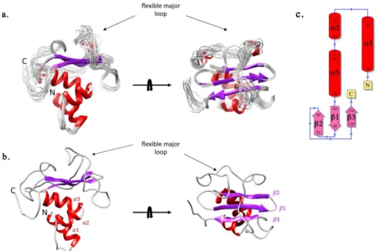
The GacSPD solution structure consists of a central three-stranded antiparallel β-sheet flanked by three N-terminal α-helices on one side and a major loop on the opposite side. The α3-β1/2-loop-β3 topology found in GacSPD is reminiscent of the α3-β2-α1/2-β3-α topology found in other extracytoplasmic PDC/PAS domains (Fig. 3). In fact, the GacSPD three-stranded β-sheet differs from the extended five-stranded β sheet typically found in canonical PDC/PAS fold structures34 (Fig. S4). The major 49-residue length loop (Gly99-Leu148), which
Figure 1. Deletion of the GacS periplasmic detection domain affects biofilm formation. (a) Biofilm production
in glass tubes (upper panel) is illustrated and quantified after crystal violet-staining (lower panel). Biofilm levels represent mean values (with error bars) obtained from three independent experiments. *, **, *** and ns refer to p < 0.05, p < 0.01, p < 0.001 and non-significant difference, respectively, according to the Wilcoxon-Mann-Whitney tests. (b) Biofilm formation is monitored by confocal laser scanning microscopy after 12 h. The extracted z images and their respective xy and xz planes are shown.
connects β2 to β3, wraps one face of the β-sheet and part of the outer face of helix α3 that is tightly packed against the opposite face of the β-sheet34, 35 (Fig. 3). Despite the sparse NOE contacts for the major loop region that mainly comprise intra-residue and sequential contacts (Fig. S5), nine long-range NOE distance restraints could be identified between residues from the loop and the β-sheet (Thr123/Gly93, Thr123/Arg94, His124/Gly93, His124/Arg94, Leu125/Gly93, Leu125/Arg94, Gly131/His97, Gly131/Thr86, Ala139/Trp150).
To evaluate the dynamics behavior of GacSPD and its major loop in solution, we determined the NMR relax-ations properties on the pico to nanosecond timescale of the backbone amides. Most residues showed {1H-15N} NOE ratio values above 0.8 indicating highly defined structures with low flexibility (Fig. 4). In contrast, residues in the major loop (as well as in the N- and C-termini) displayed NOE ratio values below 0.4 together with shorter transversal relaxation time (T1) and longer longitudinal relaxation time (T2), which reflect higher flexibility. These results are in excellent agreement with NOE-based secondary structures and confirm the inherent flexibil-ity of the GacSPD major loop.
Structural comparison evidences a conserved residue pattern despite fold variation.
A hidden Markov model (HMM)-based profile search identified several periplasmic domains adopting a PDC fold such as the CitA detector kinase (CitAp domain), the methyl-accepting chemotaxis protein from Geobacter sulfurreducens and the DcuS sensor kinase (DcuSp domain), as the closest structural homologs (HHpred true-positive Prob >97.4% Figs 5and S4). Next, pairwise structural comparison between GacSPD and CitAp, DcuSp, the Salmonella typhimurium metal binding domain (PhoQ), the Halorhodospira halophile photoactive yellow protein (PYP) and the Geobacter
sulfurredu-cens methyl-accepting binding domains (GSU0582 and GSU0935) showed an average rmsd value of 2.78 ± 0.4 Å for
42 CAs atoms of the central β-sheet, compared to a value of 2.1 ± 0.5 Å between various PAS and PDC domains34–42.
Figure 2. Effect of the deletion of the GacS detection domain on rsm, T3SS and T6SS gene expression. (a)
Activities of the rsmZ–lacZ (left) and rsmY–lacZ (right) transcriptional chromosomal fusions were monitored at different growth stages in the PAK WT (open triangle), PAKΔgacS (open square) or PAKgacSΔPD (open circle) strain. The corresponding β-galactosidase activities are expressed in Miller units as the mean values (with error bars) of three independent experiments. (b) Transcript levels of VgrG1b (T6SS; dark blue bar) and ExoS (T3SS; light blue bar) were monitored in the PAK, PAK∆gacS and PAKgacSΔPD strains using RT-qPCR; fold change is displayed for the two mutant strains compared to PAK strain. *, ** and *** refer to p < 0.05, p < 0.01 and p < 0.001, respectively, according to the moderated t-tests.
www.nature.com/scientificreports/
Besides this overall fold conservation, the striking structural differences between GacSPD and other PDC/PAS detector domains reside in the number of β-strands from the central β-sheet and the presence of a major loop (Gly99-Leu148) covering the outer face of the β-sheet at the apical side of GacSPD (Fig. S4). In other PDC-type detector domains, such as CitAp, DcuSp and PhoQ, the corresponding loop shows high variability in length and is often limited to two small α-helices containing up to twelve residues that contribute to ligand specificity43 (Fig. S4). In fact, an overlay of PDC-containing domain structures reveals that GacSPD has the longest, 49-residues length, loop region (Fig. 5a), which exhibits a pronounced flexibility in solution as demonstrated by NMR
Figure 3. NMR solution structure of GacSPD. (a) A superimposition of 20 representative structures of GacSPD corresponding to the minimal rmsd of all protein backbone N, Cα and CO atoms. (b) GacSPD lowest-energy structure. The structure contains a central β-sheet containing three β-strands (β1 84–89, β2 93–98 and β3 150– 154) and three N-terminal α-helices (α1 38–50, α2 57–63 and α3 65–76). A major loop, indicated by a black arrow, links β2 to β3 and wraps the apical side of the β-sheet. The N- and C-termini are labelled. (c) Topology scheme of GacSPD.
Figure 4. 15N NMR backbone relaxation data of GacSPD. (a) Per residue 15N T1 longitudinal relaxation times. (b) Per residue 15N T2 transverse relaxation times. (c) Per residue heteronuclear NOE ratios along with the location of GacSPD secondary structural elements shown as red cylinder for α-helix and violet arrow for β-strand.
relaxation data. Such loop flexibility has been reported for other PDC homologues, such as the citrate-free form of CitAp, where a major disordered loop of more than 30 residues, not detected in the crystal structure, showed strong line broadening and low 1H/15N signals from backbone amide groups by NMR spectroscopy. Interestingly, stabilization of this loop occurs in the presence of citrate, that also promotes folding of the nearby C-terminal helix44. Thus, one could propose that conformational changes of the major loop could also occur in GacSPD upon ligand binding, as evidenced for CitAp44.
A sequence alignment of GacSPD with the closest structural homologues CitAp and DcuSp evidences conser-vation of the GacSPD Arg94 and His97 pair of interacting residues in these closest structural homologs. In fact, the corresponding residue pairs in DcuSp (Arg107, His110) and CitAp (Arg66, His69) are key residues for binding the negatively-charged C4 and C6-dicarboxyalte ligands, respectively (Fig. 5b and c)35, 39. Moreover, a multiple sequence alignment using 227 GacS periplasmic detector domains from the Pseudomonas genus reveals conservation of three residues (His97, His133 and Trp150) within the Pseudomonas genus, while Arg94 is not conserved. With the excep-tion of the Trp150 indole ring stably anchored in the hydrophobic core, Arg94, His97 and His124 form a triad of interacting residues on top of the β-sheet facing the major loop and define a pocket that could represent a putative ligand-binding site (Figs 5 and 6). His124 is a conserved basic residue within the Pseudomonas aeruginosa group and can be substituted with an arginine (Fig. 6b). The solvent-exposed His133 is located within the major loop at one edge of this putative binding site. Given the striking conservation pattern of the Arg-His residue pair in GacSPD, we performed 1H-15N HSQC titration experiments of GacSPD with a series of related negatively charged molecules, e.g. citrate, fumarate as well as other Krebs cycle intermediates reported to affect the GacS/GacA signaling pathway45, 46, and other molecules or ions (Table S5), but no binding with GacSPD was observed.
GacS
PDharbors a functional surface region within the major loop.
To assess the functional role of these five GacSPD residues, we first checked the impact of mutations into alanine on the GacSPD fold using NMR 1H-15N HSQC experiments. With the exception of the GacSPD W150A variant that was found to be misfolded due to its major role in GacSPD folding and stability, the three fingerprints recorded for a GacSPD double mutant (Arg94Ala and His97Ala) and each of the H124A and H133A single mutant did not reveal an alteration of the overall GacSPD fold. compared to WT GacSPD (Fig. S6).We next generated the four gacSR94A, gacSH97A, gacSH124A and gacSH133A mutant PAK strains to assess the
poten-tial role of these residues for biofilm formation. Combined analyzes using crystal violet staining and confocal microscopy revealed that the two GacS His97Ala and His133Ala mutants abolish biofilm formation compared to the WT PAK strain (Fig. 7). The biofilm morphology of these two gacSH97A and gacSH133A PAK strains exhibits a
strong reduction compared to the WT PAK strain (Figs 1b and 7b), corresponding to single isolated colonies as observed for the ΔgacS PAK strain (Fig. 1b). The gacSH124A mutant PAK strain showed an altered biofilm
mor-phology compared to the WT strain, indicating a functional role of the pocket-lining His124 residue in biofilm formation. By contrast, the gacSR94A mutant PAK strain showed biofilm morphology similar to WT PAK strain
(Figs 1 and 7b), indicating that mutating Arg94 does not impair biofilm formation.
Figure 5. Structural comparison. (a) Superimposition of GacSPD (colored secondary structure) and two structural homologues, E. coli DcuSp (dark grey), and K. pneumonia CitAp (light grey). (b) Close-up views of the conservation pattern made by the two positively-charged residues between GacSPD, CitAp and DcuSp. (c) Structural sequence alignment of GacSPD, CitAp and DcuSp. The conserved Arg and His residues are highlighted with orange stars.
www.nature.com/scientificreports/
To further confirm the functional role of these residues, we examined the effect of these four GacSPD mutants on the expression of the GacS/GacA TCS target genes, as already performed with the GacSΔPD PAK strain for monitoring rsmZ/rsmY expression and T6SS and T3SS activation or repression. In turn, the rsmY–lacZ and rsmZ–
lacZ transcriptional fusions were introduced into the four gacSR94A, gacSH97A, gacSH124A and gacSH133A mutant PAK
strains. Expression level of the two RsmY and RsmZ sRNAs, as measured by the β-galactosidase activity assay at various growth stages, was decreased in the three gacSH97A, gacSH124A and gacSH133A mutant PAK strains by
around 4.6-fold, 1.7-fold and 2.2-fold, respectively for RsmZ and around 3-fold, 5.7-fold and 6-fold, respectively for RsmY, compared to the WT PAK strain (Fig. 8a). In parallel we also examined whether the gacSR94A, gacSH97A,
gacSH124A and gacSH133A mutant strains could affect expression of T3SS and T6SS. By monitoring the expression
level of T6SS (vgrG1b) or T3SS (exoS), we found a 6.2, 2.3 and 6.1-fold induction of T3SS associated to a 6.3, 3.8 and 7.1-fold repression of T6SS in the gacSH97A, gacSH124A and gacSH133A mutants, respectively (Fig. 8b).
In summary, we unveiled a functional surface region within GacSPD involved in the activation of the GacS/ GacA signaling pathway in P aeruginosa, arguing for a functional role of the periplasmic detector domain of the central GacS HK. The GacSPD NMR solution structure reveals an atypical PDC/PAS-like domain fold that consists of a 3-stranded β-sheet flanked by 3 α-helices and a major loop. This functional region consists of a
Figure 6. Mutation locations in GacSPD solution structure. (a) A GacSPD cartoon representation with the selected mutated residues shown as sticks. (b) Multiple sequence alignment of 227 GacS periplasmic detector domains from the Pseudomonas Genus. The logo representation was generated with the Skylign web-server62. Location of the mutated residues (Arg94, His97, His124, His133 and Trp150) is indicated by a red dot.
positively-charged pocket at the apical side and is defined by at least the three residues His97 H124, and H133 residues, which clearly involves the major loop and one face of the central β-sheet. Interestingly, while mutation of the invariant Arg94 residue in CitA/DcuS/GacS did not affect the P. aeruginosa adherence, mutation of the
Figure 7. Effect of mutations in the GacS detection domain on biofilm formation. (a) Biofilm production in glass
tubes (upper panel) is illustrated and quantified after crystal violet staining (lower panel). The corresponding levels of biofilm production represent means values (with error bars) obtained from three independent experiments. *, **, *** and ns referred to p < 0.05, p < 0.01 and p < 0.001 and non-significant difference, respectively, according to the Wilcoxon-Mann-Whitney tests. (b) Biofilm formation monitored by confocal laser scanning microscopy after 12 h. Extracted z images and their respective xy and xz planes are shown.
www.nature.com/scientificreports/
three His97, H124 and His133 GacS residues, the latter two being not conserved in CitA/DcuS, severely altered biofilm formation. We could thus propose that the ligand binding site in GacSPD does not occur at the same site as the closest structural homologues CitAp and DcuSp. This is consistent with a functional diversity of the GacS periplasmic detector domain acquired during evolution of the PDC/PAS domain-containing HK.
Materials Methods
Bacterial strains and culture conditions.
Strains and plasmids used for this study are listed in Tables S1and S3, respectively. Strains were grown at 37 °C in LB medium or in M63 medium supplemented with 1 mM MgCl2, 0.5% casa-aminoacids, 0.2% glucose. Recombinant plasmids were introduced into P. aeruginosa genome through conjugative transfer using pRK2013. Transconjugants were selected on Pseudomonas isolation agar (PIA;Difco Laboratories) supplemented with appropriate antibiotics. Kanamycin (Km) at 50 µg/ml and tetra-cycline (Tc) at 150 µg/ml supplemented with streptomycin47 at 2 mg/ml were used for E. coli and P. aeruginosa strains, respectively.
Construction of chromosomal variants and mutants.
To generate a gacSΔPD PAK strain harbouring a chromosomal copy of a gacS variant devoid of a periplasmic domain, DNA fragments corresponding to the upstream and downstream sequences (approximately 500 pb) of the deleted region were amplified from the PAKFigure 8. Effect of mutations in the GacS detection domain on rsm genes, T3SS and T6SS expression. (a)
Activities of the rsmZ–lacZ (left panel) and rsmY–lacZ (right panel) transcriptional chromosomal fusions were monitored at different growth stages in the PAKWT, PAKΔgacS, PAKgacSR94A, PAKgacSH97A, PAKgacSH124A
PAKgacSH133A strains. The corresponding β-galactosidase activities are expressed in Miller units and correspond
to mean values (with error bars) obtained from three independent experiments. (b) Transcript levels of VgrG1b (T6SS; black bar) and ExoS (T3SS; white bar) were monitored in the PAK, PAKΔgacS, PAKgacSR94A,
PAKgacSH97A, PAKgacSH124A and PAKgacSH133A strains using RT-qPCR and fold change is displayed for the four
mutant strains compared to the PAK strain. *, ** and *** refer to p < 0.05, p < 0.01 and p < 0.001, respectively, according to the moderated t-tests.
genomic DNA using appropriate oligonucleotide pairs (Table S4). The PCR products were cloned into the pCR2.1 vector (Invitrogen) using the SLIC method. After DNA sequencing, the cloned region was digested by BamH1 and ApaI and inserted into the linearized pKNG101 vector, resulting in the PKNG101 gacSΔPD plasmid.
To engineer the four gacSR94A, gacSH97A, gacSH124A, gacSH133A, mutant PAK strains, each harbouring a point
alanine mutation into a chromosomal copy of the gacS gene, the upstream and downstream sequences (approx-imately 500 pb) were amplified from the PAK genomic DNA using the appropriate pairs of primers (Table S4). The forward and reverse PCR products were linked and cloned into the suicide pKNG101 vector using the SLIC strategy, yielding the four gacSR94A, gacSH97A, gacSH124A and gacSH133A PKNG101 plasmids. The resulting suicide
plasmid was introduced into the P. aeruginosa genomic DNA through conjugative transfer by a three-partner pro-cedure using pRK2013. The deletion mutants were obtained by a double selection: first on LB agar supplemented with Irgasan (25 μg/mL) and streptomycin (1000 μg/mL) at 37 °C followed by a NaCl-free LB agar containing 6% sucrose at 30 °C. The genome of each mutant was extracted and the targeted DNA fragment was amplified and verified by sequencing.
RT-qPCR.
The WT, ΔgacS, gacSΔPD, gacSR94A, gacSH97A, gacSH124A, gacSH133A PAK strains were grown at 37 °Cunder agitation until OD600 reached 4. Total cellular RNA from a 10 mL culture medium was isolated using the pureYield RNA midiprep system (Promega), cleaned up and concentrated using the RNeasy kit (Qiagen). RNA yield, purity and integrity were further evaluated on Nanodrop and Experion devices. Reverse transcription was performed on 2 μg of RNA using the SuperScript III first-strand synthesis system (Invitrogen). Real-time PCR runs were carried out on a CFX96 Real-Time System (Bio-Rad). Cycling parameters of the real-time PCR were 98 °C for 2 min, followed by 45 cycles of 98 °C for 5 s and 60 °C for 10 s, ending with a melting curve from 65 °C to 95 °C to evaluate amplification specificity. To determine the amplification kinetics of each product, fluorescence derived from incorporation of EvaGreen into the double-stranded PCR products was measured at the end of each cycle using a SsoFast EvaGreen Supermix 2X Kit (Bio-Rad). The data were analyzed using Bio-Rad CFX Manager Software 3.0 (Bio-Rad). The uvrD gene was used as a reference for normalization, in particular because transcrip-tion of uvrD is fairly stable in bacteria exposed to antibiotics even at relatively high concentratranscrip-tions48.
Biofilm assay.
The P. aeruginosa adherence assay was performed in individual glass tubes containing 1 mL of medium as described previously31. Bacteria were grown in M63 medium supplemented with 1 mM MgCl2, 0.5% casa-amino acids, 0.2% glucose under static conditions at 30 °C. After 12 hours, the cultures were incubated with 1% Crystal Violet for 10 min to stain the attached bacteria and washed twice. Staining was extracted by treatment with 400 μL 95% ethanol. Subsequently, 600 μL of water were added and OD570 was measured. All quantification assays were performed at least in triplicate.
Measurements of β-galactosidase activity.
Strains carrying the lacZ transcriptional fusions were grown at 37 °C in agitated LB medium. Bacterial cells were collected by centrifugation at different growth times. The β-galactosidase activity was measured using the method of Miller49.Confocal laser scanning microscopy analysis of biofilm.
P. aeruginosa strains were grown in an 8-wellchambered coverglass (Lab-teK II). 200 μL of M63 derivate medium containing a bacterial suspension at an OD600 of 0.1 was incubated at 30 °C. After 12 h, the medium was aspired from the corner of each chamber and rinsed twice by adding 200 μL of sterile PBS to remove unattached cells. Prior to observation, bacteria were fixed with 4% paraformaldehyde and stained using 4′,6′-diamidino-2-phenylindole (DAPI) for 15 min. Images were taken at locations of biofilm formation using a confocal laser scanning microscope Confocal Olympus FV1000. Positions were chosen for sagittal sections (xz position) to minimize experimental variability. The number of images for the 3D biofilm observation within each stack depended on biofilm thickness. All confocal images were analysed using the imageJ software50.
Cloning, expression and purification of GacS
PDfor structural determination.
The DNA sequence coding for the periplasmic detector domain of the GacS HK (Met38-Gly164) was amplified from the extracted PAK genome by using the appropriate primers (Table S4). The PCR products were cloned into the pLic03 vector linearized by BamH1 using the LIC method51 yielding the pLic03_GacSsp coding for GacSPD fused to a 6xHis tag and a tobacco etch virus (TEV) at the N terminal region. Expression and purification of GacSPD were performed as described33.Structure calculation.
The NMR sample contained 0.8 mM protein concentration (90% H2O, 10% D2O) in 150 mM NaCl, 50 mM phosphate buffer, pH7. Recorded spectra were analyzed with CARA on the basis of the previously published backbone amide and side chain resonances assignment33. The approximate inter-proton dis-tances were obtained from the 2D NOESY, 13C NOESY-HSQC and 3D 15N NOESY-HSQC spectra recorded using a mixing time of 150 ms. A total number of 1299 restraints were used for structure calculations. Hydrogen bonds were identified by recording long-range JNC’ HNCO-COSY52 and a series of 15N-1H HSQC spectra using a sample freshly dissolved in D2O; 27 nonexchangeable amides were located in regions of defined secondary structures. Hydrogen bond constraints were introduced using distance restraints in the range of 2.7–3.0 Å and 1.8–2.0 Å for O-N and O-HN, respectively. Moreover, 94 additional dihedral restraints were calculated using TALOS+ based on 1Hα, 13Cα, 13Cβ, 13C’ and 15N chemical shifts53. Input data and structure calculation statistics are summarized in Table S2. The accuracy of the NMR models has been assessed based on the traditional criteria for successful structure calculation using the program CYANA54. 100 structures were generated and the 20 lowest-energy struc-tures were selected and each subjected to restrained molecular dynamics using the Amber 4.1 force field within the SANDER module of Amber 10. Water molecules were stripped off and energy terms were calculated for thewww.nature.com/scientificreports/
protein using AMBER55. Non-bonded interaction cutoff was 15 Å for the restrained MD runs. The final structural ensemble was analysed using PROCHECK-NMR56. Structure coordinates have been deposited to the Protein Data Bank under accession number 5O7J.
Relaxation study.
15N T1, T2, and NOE NMR relaxation measurements were performed at 25 °C on a Bruker Avance 600 MHz using a 2 mM GacSPD sample (90% H2O, 10% D2O) in 150 mM NaCl, 50 mM phos-phate buffer, pH 7.0. T1 data and T2 data were both acquired with ten relaxation delays (10, 20, 50, 100, 200, 300, 400, 600, 800 and 1000 ms and 17.6, 35.2, 52.8, 70.4, 88.0, 105.6, 123.2, 140.8, 158.4 and 176.0 ms, respectively). Experimental 15N heteronucleaur NOE values were determined from the intensity ratios of amide signals of inter-leaved 2D 1H-15N HSQC spectra with and without a 5 sec saturation period. Relaxation times were calculated as reported by Farrow and coworkers57 using an exponential fit of single exponential decays to peak intensity values:
I = I0exp(−t/T) where T = T1 or T2, and I = resonance intensity at time t. Steady-state 1H−15N NOE ratios were calculated using the r = I/I0 expression.
Ligand binding using
1H-
15N HSQC titration.
Putative ligand molecules (see Table S5) were tested at 298 K using 150 μM of 15N-labelled GacSPD native protein and mutants (50 mM Na2HPO4/NaH2PO4 buffer pH 7, 50 mM NaCl) with a ligand concentration range of 0.1–20 mM.
Bioinformatics analysis.
227 GacS protein sequence homologues belonging to the Pseudomonas genus were retrieved from Uniprot (ftp://ftp.uniprot.org, Feb 2017) and aligned with Muscle58 and downloaded from the Uniprot FTP server (ftp://ftp.uniprot.org) in February, 2017. From the multiple alignments, a selection of GacS periplasmic domains was made with Gblock Server59. Maximum likelihood trees were generated with PhyML-SMS60 using the LG empirical amino acid substitution model of evolution61 and 1000 bootstrap replicates. In parallel, the resulting trees were compared with that obtained from the downloaded 16S rRNA sequences. The tree parameters including topology were optimized. The sequences of the periplasmic domains that belong to the same branch as P. aeruginosa GacS were retained to generate the sequence logo using the Skylign web-server62.References
1. Stock, A. M., Robinson, V. L. & Goudreau, P. N. Two-component signal transduction. Annu Rev Biochem 69, 183–215 (2000). 2. Jung, K., Fried, L., Behr, S. & Heermann, R. Histidine kinases and response regulators in networks. Curr Opin Microbiol 15, 118–124
(2012).
3. Cheung, J. & Hendrickson, W. A. Sensor domains of two-component regulatory systems. Current opinion in microbiology 13, 116–123 (2010).
4. Zhang, Z., Liu, Q. & Hendrickson, W. A. Crystal structures of apparent saccharide sensors from histidine kinase receptors prevalent in a human gut symbiont. FEBS Journal 281, 4263–4279 (2014).
5. Mitrophanov, A. Y. & Groisman, E. A. Signal integration in bacterial two-component regulatory systems. Genes Dev 22, 2601–2611 (2008).
6. Murray, T. S., Egan, M. & Kazmierczak, B. I. Pseudomonas aeruginosa chronic colonization in cystic fibrosis patients. Curr Opin
Pediatr 19, 83–88 (2007).
7. Gellatly, S. L. & Hancock, R. E. Pseudomonas aeruginosa: new insights into pathogenesis and host defenses. Pathog Dis 67, 159–173 (2013).
8. Furukawa, S., Kuchma, S. L. & O’Toole, G. A. Keeping their options open: acute versus persistent infections. J Bacteriol 188, 1211–1217 (2006).
9. Costerton, J. W., Stewart, P. S. & Greenberg, E. P. Bacterial biofilms: a common cause of persistent infections. Science 284, 1318–1322 (1999).
10. Mougous, J. D. et al. A virulence locus of Pseudomonas aeruginosa encodes a protein secretion apparatus. Science 312, 1526–1530 (2006).
11. Hood, R. D. et al. A type VI secretion system of Pseudomonas aeruginosa targets a toxin to bacteria. Cell Host Microbe 7, 25–37 (2010).
12. Moscoso, J. A., Mikkelsen, H., Heeb, S., Williams, P. & Filloux, A. The Pseudomonas aeruginosa sensor RetS switches type III and type VI secretion via c-di-GMP signalling. Environ Microbiol 13, 3128–3138 (2011).
13. Russell, A. B. et al. Type VI secretion delivers bacteriolytic effectors to target cells. Nature 475, 343–347 (2011).
14. Goodman, A. L. et al. A signaling network reciprocally regulates genes associated with acute infection and chronic persistence in
Pseudomonas aeruginosa. Dev Cell 7, 745–754 (2004).
15. Ventre, I. et al. Multiple sensors control reciprocal expression of Pseudomonas aeruginosa regulatory RNA and virulence genes. Proc
Natl Acad Sci USA 103, 171–176 (2006).
16. Chambonnier, G. et al. The Hybrid Histidine Kinase LadS Forms a Multicomponent Signal Transduction System With the GacS/ GacA two component System in Pseudomonas aeruginosa. Plos genetics (2016).
17. Broder, U. N., Jaeger, T. & Jenal, U. LadS is a calcium-responsive kinase that induces acute-to-chronic virulence switch in
Pseudomonas aeruginosa. Nat Microbiol 2, 16184 (2016).
18. Goodman, A. L. et al. Direct interaction between sensor kinase proteins mediates acute and chronic disease phenotypes in a bacterial pathogen. Genes Dev 23, 249–259 (2009).
19. Kong, W. et al. Hybrid sensor kinase PA1611 in Pseudomonas aeruginosa regulates transitions between acute and chronic infection through direct interaction with RetS. Mol Microbiol 88, 784–797 (2013).
20. Bhagirath, A. Y. et al. Characterization of the Direct Interaction between Hybrid Sensor Kinases PA1611 and RetS That Controls Biofilm Formation and the Type III Secretion System in Pseudomonas aeruginosa. ACS Infect Dis 3, 162–175 (2017).
21. Zschiedrich, C. P., Keidel, V. & Szurmant, H. Molecular Mechanisms of Two-Component Signal Transduction. J Mol Biol (2016). 22. Cheung, J. & Hendrickson, W. A. Structural analysis of ligand stimulation of the histidine kinase NarX. Structure 17, 190–201
(2009).
23. Zhang, Z. & Hendrickson, W. A. Structural characterization of the predominant family of histidine kinase sensor domains. J Mol Biol
400, 335–353 (2010).
24. Vincent, F. et al. Distinct oligomeric forms of the Pseudomonas aeruginosa RetS sensor domain modulate accessibility to the ligand binding site. Environ Microbiol 12, 1775–1786 (2010).
25. Heeb, S. & Haas, D. Regulatory roles of the GacS/GacA two-component system in plant-associated and other gram-negative bacteria. Molecular plant-microbe interactions 14, 1351–1363 (2001).
26. Brencic, A. & Lory, S. Determination of the regulon and identification of novel mRNA targets of Pseudomonas aeruginosa RsmA.
Mol Microbiol 72, 612–632 (2009).
27. Friedman, L. & Kolter, R. Two genetic loci produce distinct carbohydrate-rich structural components of the Pseudomonas aeruginosa biofilm matrix. J Bacteriol 186, 4457–4465 (2004).
28. Vasseur, P., Vallet-Gely, I., Soscia, C., Genin, S. & Filloux, A. The pel genes of the Pseudomonas aeruginosa PAK strain are involved at early and late stages of biofilm formation. Microbiology 151, 985–997 (2005).
29. Bleves, S., Soscia, C., Nogueira-Orlandi, P., Lazdunski, A. & Filloux, A. Quorum sensing negatively controls type III secretion regulon expression in Pseudomonas aeruginosa PAO1. J Bacteriol 187, 3898–3902 (2005).
30. Engel, J. & Balachandran, P. Role of Pseudomonas aeruginosa type III effectors in disease. Curr Opin Microbiol 12, 61–66 (2009). 31. Bordi, C. et al. Regulatory RNAs and the HptB/RetS signalling pathways fine-tune Pseudomonas aeruginosa pathogenesis. Mol
Microbiol 76, 1427–1443 (2010).
32. Zuber, S. et al. GacS sensor domains pertinent to the regulation of exoproduct formation and to the biocontrol potential of
Pseudomonas fluorescens CHA0. Mol Plant Microbe Interact 16, 634–644 (2003).
33. Ali-Ahmad, A. et al. NMR assignments of the GacS histidine-kinase periplasmic detection domain from Pseudomonas aeruginosa PAO1. Biomol NMR Assign (2016).
34. Möglich, A., Ayers, R. A. & Moffat, K. Structure and signaling mechanism of Per-ARNT-Sim domains. Structure 17, 1282–1294 (2009).
35. Cheung, J. & Hendrickson, W. A. Crystal structures of C4-dicarboxylate ligand complexes with sensor domains of histidine kinases DcuS and DctB. J Biol Chem 283, 30256–30265 (2008).
36. Cho, U. S. et al. Metal bridges between the PhoQ sensor domain and the membrane regulate transmembrane signaling. J Mol Biol
356, 1193–1206 (2006).
37. Pokkuluri, P. R. et al. Structures and solution properties of two novel periplasmic sensor domains with c-type heme from chemotaxis proteins of Geobacter sulfurreducens: implications for signal transduction. J Mol Biol 377, 1498–1517 (2008).
38. Borgstahl, G. E., Williams, D. R. & Getzoff, E. D. 1.4 A structure of photoactive yellow protein, a cytosolic photoreceptor: unusual fold, active site, and chromophore. Biochemistry 34, 6278–6287 (1995).
39. Reinelt, S., Hofmann, E., Gerharz, T., Bott, M. & Madden, D. R. The structure of the periplasmic ligand-binding domain of the sensor kinase CitA reveals the first extracellular PAS domain. J Biol Chem 278, 39189–39196 (2003).
40. Cheung, J., Bingman, C. A., Reyngold, M., Hendrickson, W. A. & Waldburger, C. D. Crystal structure of a functional dimer of the PhoQ sensor domain. J Biol Chem 283, 13762–13770 (2008).
41. Zhou, Y. F. et al. C4-dicarboxylates sensing mechanism revealed by the crystal structures of DctB sensor domain. J Mol Biol 383, 49–61 (2008).
42. Affandi, T., Issaian, A. V. & McEvoy, M. M. The Structure of the Periplasmic Sensor Domain of the Histidine Kinase CusS Shows Unusual Metal Ion Coordination at the Dimeric Interface. Biochemistry 55, 5296–5306 (2016).
43. Shah, N. et al. Reductive evolution and the loss of PDC/PAS domains from the genus Staphylococcus. BMC Genomics 14, 524 (2013). 44. Sevvana, M. et al. A ligand-induced switch in the periplasmic domain of sensor histidine kinase CitA. J Mol Biol 377, 512–523
(2008).
45. Takeuchi, K. et al. Small RNA-dependent expression of secondary metabolism is controlled by Krebs cycle function in Pseudomonas
fluorescens. J Biol Chem 284, 34976–34985 (2009).
46. Dacheux, D. et al. Activation of the Pseudomonas aeruginosa type III secretion system requires an intact pyruvate dehydrogenase aceAB operon. Infect Immun 70, 3973–3977 (2002).
47. Smith, H. Questions about the behaviour of bacterial pathogens in vivo. Philos Trans R Soc Lond B Biol Sci 355, 551–564 (2000). 48. Jo, J. T., Brinkman, F. S. & Hancock, R. E. Aminoglycoside efflux in Pseudomonas aeruginosa: involvement of novel outer membrane
proteins. Antimicrob Agents Chemother 47, 1101–1111 (2003).
49. Sambrook, J., Fritsch, E. F. & Maniatis, T. Molecular cloning: a laboratory manual (Cold spring harbor laboratory press, 1989). 50. Schneider, C. A., Rasband, W. S. & Eliceiri, K. W. NIH Image to ImageJ: 25 years of image analysis. Nature methods 9, 671–675
(2012).
51. Aslanidis, C. & de Jong, P. J. Ligation-independent cloning of PCR products (LIC-PCR). Nucleic Acids Res 18, 6069–6074 (1990). 52. Cordier, F. & Grzesiek, S. Direct observation of hydrogen bonds in proteins by interresidue 3h J NC scalar couplings. Journal of the
American Chemical Society 121, 1601–1602 (1999).
53. Shen, Y., Delaglio, F., Cornilescu, G. & Bax, A. TALOS+: a hybrid method for predicting protein backbone torsion angles from NMR chemical shifts. J Biomol NMR 44, 213–223 (2009).
54. Güntert, P., Mumenthaler, C. & Wüthrich, K. Torsion angle dynamics for NMR structure calculation with the new program DYANA.
J Mol Biol 273, 283–298 (1997).
55. Go tz, A. W. et al. Routine microsecond molecular dynamics simulations with AMBER on GPUs. 1. Generalized born. Journal of
chemical theory and computation 8, 1542–1555 (2012).
56. Laskowski, R. A., Rullmannn, J. A., MacArthur, M. W., Kaptein, R. & Thornton, J. M. AQUA and PROCHECK-NMR: programs for checking the quality of protein structures solved by NMR. J Biomol NMR 8, 477–486 (1996).
57. Farrow, N. A. et al. Backbone dynamics of a free and a phosphopeptide-complexed Src homology 2 domain studied by 15N NMR relaxation. Biochemistry 33, 5984–6003 (1994).
58. McWilliam, H. et al. Analysis Tool Web Services from the EMBL-EBI. Nucleic Acids Res 41, W597–W600 (2013).
59. Castresana, J. Selection of conserved blocks from multiple alignments for their use in phylogenetic analysis. Mol Biol Evol 17, 540–552 (2000).
60. Guindon, S., Delsuc, F., Dufayard, J. F. & Gascuel, O. Estimating maximum likelihood phylogenies with PhyML. Methods Mol Biol
537, 113–137 (2009).
61. Le, S. Q. & Gascuel, O. An improved general amino acid replacement matrix. Mol Biol Evol 25, 1307–1320 (2008).
62. Wheeler, T. J., Clements, J. & Finn, R. D. Skylign: a tool for creating informative, interactive logos representing sequence alignments and profile hidden Markov models. BMC Bioinformatics 15, 7 (2014).
Acknowledgements
The authors thank Dr. A. Favier (IBS, Grenoble) and the TGIR-RMN-THC FR3050 CNRS (Grenoble, France) for data collection, and Dr. E. Goemaere for cloning advice in the LIC vectors. We also thank Yann Denis for technical assistance at the transcriptome platform of the Institut de Microbiologie de la Méditerranée. This work was supported by the CNRS, the French National Agency for Research (ANR-14-CE09-0005), a joint grant from the VLM and the Gregory Lemarchal association (RF20150501359/1/1/72) and the French Infrastructure for Integrated Structural Biology (FRISBI) ANR-10-INSB-05-01. A. A. Ahmad was a recipient of a PhD grant from the French Ministry of Research/AMU and VLM/Lemarchal association.
www.nature.com/scientificreports/
Author Contributions
A.A.A., F.F. and C.K. experimentation (NMR, mutagenesis), interpretation, manuscript writing. F.V., C.B., Y.B. and F.G. conceptualization, manuscript writing. M.B., O.B. and G.D.P experimentation (NMR, bioinformatics, microscopy).
Additional Information
Supplementary information accompanies this paper at doi:10.1038/s41598-017-11361-3
Competing Interests: The authors declare that they have no competing interests.
Publisher's note: Springer Nature remains neutral with regard to jurisdictional claims in published maps and
institutional affiliations.
Open Access This article is licensed under a Creative Commons Attribution 4.0 International
License, which permits use, sharing, adaptation, distribution and reproduction in any medium or format, as long as you give appropriate credit to the original author(s) and the source, provide a link to the Cre-ative Commons license, and indicate if changes were made. The images or other third party material in this article are included in the article’s Creative Commons license, unless indicated otherwise in a credit line to the material. If material is not included in the article’s Creative Commons license and your intended use is not per-mitted by statutory regulation or exceeds the perper-mitted use, you will need to obtain permission directly from the copyright holder. To view a copy of this license, visit http://creativecommons.org/licenses/by/4.0/.