HAL Id: tel-01743809
https://tel.archives-ouvertes.fr/tel-01743809
Submitted on 26 Mar 2018
HAL is a multi-disciplinary open access archive for the deposit and dissemination of sci-entific research documents, whether they are pub-lished or not. The documents may come from teaching and research institutions in France or abroad, or from public or private research centers.
L’archive ouverte pluridisciplinaire HAL, est destinée au dépôt et à la diffusion de documents scientifiques de niveau recherche, publiés ou non, émanant des établissements d’enseignement et de recherche français ou étrangers, des laboratoires publics ou privés.
Function and plasticity of NKp46 expressing innate
lymphoid cells
Thomas Verrier
To cite this version:
Thomas Verrier. Function and plasticity of NKp46 expressing innate lymphoid cells. Human health and pathology. Université Sorbonne Paris Cité, 2016. English. �NNT : 2016USPCC173�. �tel-01743809�
1
Thèse de doctorat
de l’Université Sorbonne Paris Cité
Préparée à l’Université Paris Diderot
Ecole doctorale Bio-‐SPC (ED N°157) Institut Pasteur Paris -‐ Unité d’Immunité Innée
Function and Plasticity of NKp46 expressing
Innate Lymphoid Cells
Thomas Verrier
Thèse de doctorat d’immunologie
Dirigée par James P. Di Santo
Présentée et soutenue publiquement à l’Institut Pasteur (Paris) le 30 Septembre 2016
Président du jury : Lantz, Olivier PhD – Institut Curie (Paris) Rapporteurs : Sirard, Jean-‐Claude PhD – Institut Pasteur (Lille)
Braud, Veronique PhD – Institut de Pharmacologie Moléculaire et Cellulaire (Nice) Examinateurs : Eberl, Gerard Pr. – Institut Pasteur (Paris)
Anderson, Graham Pr. – Institute of Immunology and Immunotherapy (Birmingham)
Directeur de thèse : Di Santo, James PhD – Institut Pasteur Paris
This work is licensed under a Creative Commons Attribution-‐NonCommercial-‐ NoDerivatives 4.0 International License.
2
-Note to the reader :
This manuscript presents my personal interpretations of complex biological system. My assumptions rely on selected scientific publications and extended bibliographic research, which is by no mean exclusive, and might become outdated in view of new discoveries.
AhR AHR AMP BM BMDC CBFB CCR CD CEC CHILP CLP CYP CX3CR1 CXCL CXCR DC DN DP DR DSS E4BP4 EILP ER FL Flt3 Fut2 GFP GM-CSF HA HEK-293T HFD HMG HSC IDO1 IFNar IFNGR IL ILC kD KLRG1 KO Lck Lin LN LSK
Aryl hydrocarbon Receptor
Airway Hyper Responsiveness Antimicrobial peptide Bone Marrow
Bone Marrow derived Dendritic Cell
Core Binding Factor β
« CC » Chemokine Receptor Cluster of Differenciation
Colonic Epithelial Cell Common Helper-like ILC progenitor Common Lymphoid Progenitor
Cytochrome p450 « CX3C » Chemokine Receptor 1 « CXC » Chemokine Ligand « CXC » Chemokine Receptor Dendritic Cells Double Negative Double Positive Death Receptor Dextran Sodium Sulfate
E4 promoter Binding Protein 4
Early ILC Progenitor
Endoplasmic Reticulum Fetal Liver Fms-like Tyrosine Kinase 3 Fucosyltransferase Green Fluorescent Protein Granulocytes Macrophages Colony Stimulating Factor
Hemaglutininn
Human Embryonic Kidney 293 T cells
High Fat Diet
High Mobility Group
Hematopoitic Stem Cells
Indoleamine 2,3- dioxygenase IFN α Receptor Interferon γ Receptor
Interleukin Innate Lymphoid cells
kiloDalton Killer-cell Lectin-like Receptor subfamily G Knock-out Lymphocyte Specific protein tyrosine kinase Lineage Lymph Nodes
LT LTi MCMV mLN MHC Ncr1 Nfil3 NK NR PLZF PMA PP RA RAG ROR S1pr5 SCID SG SLO SP TCF-1 TCR TGF TL1A TLR TRAIL TSLP SCF Sca-1 WT XCL1 Lymphotoxin
Lymphoid Tissue inducer
Mouse Cytomegalovirus mesenteric Lymph Nodes Major Histocompatibility Complex Natural Cytotoxicity Receptor 1
Nuclear Factor Interleukin 3
Natural Killer
Nuclear Receptors Promyelocytic Leukemia Zinc Finger
Phorbol 12-Myristate 13-Acetate Peyer’s Patches Retinoic Acid Recombination Activating Gene
Retinoic Acid-related orphan receptor
Sphingosine 1-phosphate receptor 5 Severe Combined Immunodeficiency Syndrome Salivary Glands Secondary Lymphoid Organs
Single Positive
T-cell Factor 1
T Cell Receptor
Transforming Growth Factor Tumor Necrosis Factor-Like Ligand 1A Toll Like Receptor Tumor Necrosis Factor Related Apoptosis-inducing Ligand
Thymic Stromal-derived Lymphopoietin Stem Cell Factor
Stem Cell Antigen 1 Wild Type « Xc » Chemokine Ligand Cell population Miscellaneous Surface molecule Transcription factor Secreted molecule
Index
Introduction : A problem of nomenclature 11
Dissecting hematopoisis 16
Chapter I : Paving the way for an ILC fate 19
Tcf-1 : EILPing ILC 23
Nfil3 around the clock 25
Tox Tox Tox 26
Id2 : CHILP vous plait 27
PLZF : Same Same but different 29
Common Gamma Chain : One to rule them all 30 I GATA3 my mind 32
T-bet or not T-bet 33
Eomessing around 34
RORγt : Let him roar again 36 Tissue Residency – Home Sweet Home 38 Chapter II : Activation & Function, Godspeed you! 39
IL-12 & IL-1 Family : Twelve plus one, not eleven plus two 42 IFNγ : An army of one 43
Perforin/Granzyme : Cytotoxicity is my city 45
GM-CSF: GMetting CeriouSF 47
IL-17: Sweet Seventeen 48
IL-22: Border control 50
Chapter III : ILC1 to 3 53
NKp46: The marker of the beast 56
ILC1 day 57
ILC1b Natural (born) Killer 58
Immature ILC1a 59
ILC1ab : Real inbetweener or just a faker ? 60
ILC3: Prying open my third eye 61
Lymphoid Tissue Inducer: Initiate 62
NKp46+ ILC3: the last but not the least 64
Published Articles 67 Article n°1 : The Chemokine Receptor CXCR6 Controls the Functional Topography 69
of Interleukin-22 Producing Intestinal Innate Lymphoid Cells
Article n°2 : Phenotypic and Functional Plasticity of Murine Intestinal NKp46+ Group 3 93 Innate Lymphoid Cells
Article n°3 : Notch Signaling in Group 3 Innate Lymphoid Cells Modulates their Plasticity 107
Introduction : A problem of nomenclature
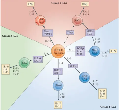
Innate lymphoid cells (ILC) are critical immune effectors the early phases of immune responses (Fig.1). These RAG independent immune effectors respond to external stimuli solely through germ-line encoded surface receptors, which trigger their rapid cytokine production. ILCs originate from an Id2-dependent precursor and are separated in three groups reminiscent of helper T (TH) cells. TH1 cells and
Group 1 ILC (ILC1) participate in the defense against intracellular pathogens, TH2
cells and group 2 ILC (ILC2) contribute to the establishment of allergic responses
and promote the expulsion of intestinal parasite while TH3 cells and Group 3 ILC
(ILC3) promote mucosal homeostasis, anti-fungal and anti-bacterial responses.
Figure 1 : Overview of the different ILC group
“Innate lymphoid cells — a proposal for uniform nomenclature”
Hergen Spits, David Artis, Marco Colonna, Andreas Diefenbach, James P. D Nature Reviews Immunology 13, 145-149 (February 2013) | doi:10.1038/nri3365 i Santo, Gerard Eberl, Shigeo Koyasu, Richard M. Locksley, Andrew N.J.
McKenzie, Reina E. Mebius, Fiona Powrie & Eric Vivier.
With respect to the timeline of ILC discovery, the first ILC to be described were a subset of ILC1. In 1975, Kiessling and colleagues described a lymphocyte population poised to “naturally kill” tumors in vitro without any requirement for prior antigenic
stimulation1. Some features of these cells, also known as Natural Killer (NK), were used
as a basis upon the discovery of other ILC subsets. In 1992, the identification of unknown lymphocytes that resided in neo-natal lymph nodes further led to their characterization
as Lymphoid Tissue inducer cells (LTi)2, which are the prototypical member of ILC3. ILC2
were described 20 years later, and were first named “natural helper”3 as opposed to Natural
Killer, but also “nuocytes”4. As each group named overlapping population differently, an
uniform nomenclature was proposed in 20135 (Fig.1). Since then, the number of reports
on “innate lymphoid cells” kept increasing bringing more and more insight as to their function and diversity (Graph.1). Due to this dynamic, group investigating ILC subsets had to constantly adjust by adding markers to distinguish subsets that were thought to be homogenous population. As such, I propose to use an informal nomenclature that aims to reduce confusion while being as accurate as possible.
The presented work focuses on ILC1 and ILC3
biology in C57BL/6 mice.
Marker
Ligand
ILC1
ILC2
ILC3
CD45
NA ++ ++ +CD90
NA +/++* ++ ++Sca-1
NA +* + +*CD3 (T Cells)
NACD19 (B Cells)
NAROR
γt
NA +*** - +GATA-3
NA + ++ +T-bet
NA + - +*Eomes
NA +* --IL-17RB
IL-25 - +-T1-ST2
IL-33 - +-CD212
IL-12; IL-23R + +** +IL-18R
IL-18 + - NAIL-23R
IL-23 - - +IL-1R
IL1α; IL-1β NA NA +CD122
IL-2; IL-15 + NA NACD127
IL-7; TSLP +* + +CD117
SCF +* +* +CCR6
CCL20 - - +*CXCR6
CXCL16 +* - +CXCR3
NA +* NA +*CD49a
NA +* - +*CD49b
NA +* --TRAIL
DR5 +* --CD4
NA - - +*NKp46
Unknown + - +*NK1.1
NA + --KLRG1
NA +* +*-MHC-II
TCR +** +* +* * Subset related ** Following Activation *** Plasticity NA Not Adressed Lineage Markers -Transcription Factor Cytokine Responsiveness Steering MoleculesILC1 regroups three main subsets of NK1.1+ NKp46+ cells (Fig.2), but no consensus for the nomenclature of these cells has been formally established yet. Out of commodity, I therefore propose a designation based on the expression of the transcription factor Eomes and the integrins CD49a and CD49b that will be used onwards. The features that justify this classification will be addressed in the ILC1 section. I will refer to Eomes- CD49a+ ILC1 that resides in the liver and the intestinal mucosa as ILC1a, to Eomes+ CD49b+ ILC1, which
are commonly known as circulating cytotoxic NK cells as ILC1b, and to Eomes+ CD49a+
CD49b+ ILC1 that populates the Salivary Gland and the Uterine decidua as ILC1ab. In the
case in which the ILC1 subset is not clearly determined (ND), I will refer to as ILC1ND.
CD49b NKp46 CD49a NK1.1
ILC1
a Eomes NKp46 CD49a NK1.1ILC1
ab Eomes NKp46 NK1.1 CD49bILC1
b Salivary Glands Uterus Bone Marrow Spleen Liver Intes=neILC3 contains two lineages with a distinguishable ontogeny and different dependency on transcription factors (Fig.3) , which will be further explained in the ILC3 section. Fetal CCR6+ T-bet- ILC3 LTi-like cells can be dissected according to CD4 expression, and will be referred to as CD4- or CD4+ ILC3. CCR6- T-bet+ ILC3 that arise post-natally
contain NKp46- and NKp46+ ILC3, and will be referred to as NKp46- or NKp46+ ILC3. As
CCR6 and T-bet expression by ILC3 were reported after the discovery of ILC3, some studies
solely relied on CD4 and NKp46 expression. In this case, CD4- NKp46- ILC3, which include
CCR6+ and T-bet+ will be referred to as DN ILC3. If the ILC3 subset is not defined, it will be refered to as ILC3ND. T-bet CCR6 CD4 CCR6
CD4
+CD4
-‐NKp46
-‐ NKp46 T-betNKp46
+DN ILC3
E13.5 (Embryo) Birth Weanling Age AdultFigure 3 : Group 3 ILC include fetal CCR6+ Lti-like cells and T-bet dependent ILC3
16
Hematopoitic stem cells mature into effector cells through the sequential integration of external signals. Several methods permit to dissect the molecular actors (i.e. cytokines or transcription factors) involved in this differenciation process, and to understand the hierarchy between immune populations.
In vitro culture and adoptive transfer both require isolation of cells, which are either
cultured in media with selected cytokines, or injected into recipient mice, respectively. While the signals that drive immune cells development are controlled to some extent in
vitro, adoptive transfer provides a more complex environment that resembles physiological
conditions. If primary cells from genetically modified mice are used as a cellular source, these methods are now crucial to demonstrate both the responsiveness of immune cells to extrinsic signals and their ability to develop in the context of an altered genotype.
As gene ablation in knock-out (KO) mice affects all cells, the phenotype observed in a cell population could be caused by a defect in bystander cells. In order to adress whether a gene is involved in an intrinsic fashion for the development or function of a cell population, some of the studies that are presented in this manuscript relied on bone marrow chimera or conditional gene targetting.
BM chimera are generated by injection of a mixture of BM cells isolated from congenically marked mice (i.e. CD45.1 and CD45.2) with a distinct genotype (i.e. WT vs KO) (Fig.4). Hematopoietic cells originating from each donor mice will differentiate into effector cells according to their genotype. If the invalidated gene is required in an intrinsic fashion for a defined population, the phenotype observed in whole KO mice will be recapitulated in BM chimera mice. However, if the phenotype observed in BM chimera mice is similar to WT mice, the signal produced from immune cells derived from the WT compartment compensate for the loss of signals from the KO compartment, indicating
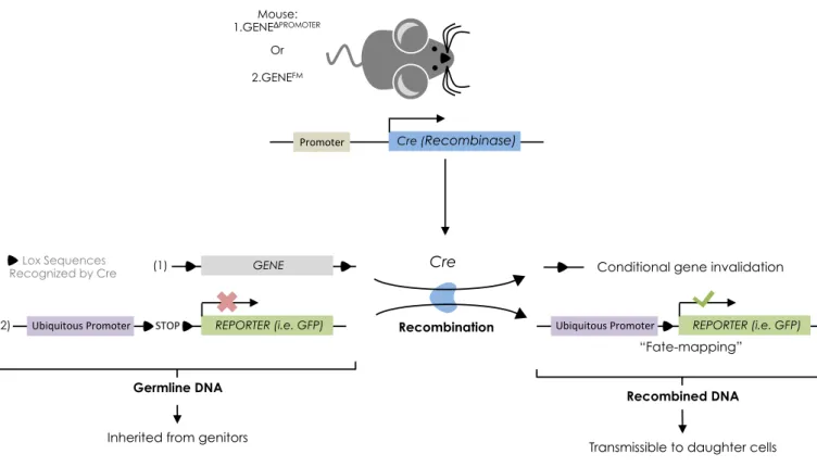
17 that the defect caused by gene invalidation affects that function of a bystander population. By generating BM chimera with KO mice as recipient, one could distinguish if the effect of gene invalidation is due to an intrinsic or extrinsic defect in hematopoietic cells, and evaluate the contribution of non-hematopoietic cells (i.e. recipient mice) for the function or development of the population of interest. As any cell could exert a bystander effect, the construction of mice that permit targeted gene invalidation based on the « Cre-lox » system facilitated the investigation of intrinsic gene requirement.
Cre-recombinase directly binds and recognizes Lox DNA sequences, which lead to the irreversible excision of the genetic fragment that is flanked by the Lox sequences. Transgenic mice that express Cre under a specific promoter (i.e. lineage specific surface marker or transcription factor), when crossed to transgenic mice bearing Lox sequences, allows for conditional gene targetting (Fig.5) . If Lox sequences flank a coding gene, Cre expression leads to the excision of the gene, thereby invalidating the gene in cells that activated the Cre-controlling promoter. As genetic recombination is transmissible during cellular division, daughter cells will inherit the invalidated gene.
This feature has proved itself crucial for the establishment of « fate-mapping » studies, in which the targeted genetic sequence is often a stop codon that prevents the expression of a reporter protein. Cre mediated recombination leads to the expression of the reporter protein (i.e. fluorescent protein), thereby genetically marking cells in a stable fashion, irrespectively of the transcriptional status of the Cre-controlling promoter, which supposedly reflects the expression of the gene of interest. By assessing whether cells marked with the reporter still express the gene of interest, one can assess the hierarchy between cell populations. If cells express both the gene of interest and the reporter, this reflects expression stability. However, if marked cells stopped expressing the gene of interest, and because Cre expression is mandatory for the labelling of these cells, this indicates that expression of the gene of interest had been lost.
Cre (Recombinase)
Promoter
Cre
GENE
REPORTER (i.e. GFP) Ubiquitous Promoter STOP
Lox Sequences
Recognized by Cre Conditional gene invalidation REPORTER (i.e. GFP) Ubiquitous Promoter Mouse: 1.GENEΔPROMOTER Or 2.GENEFM “Fate-mapping” (1) (2) Recombined DNA
Transmissible to daughter cells
Recombination
Germline DNA
Inherited from genitors
Chapter I
Paving the way for an ILC fate
Chapter I : Paving the way for an ILC fate
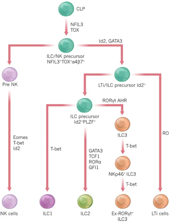
Figure 6 : Model for ILC development (2015) “The biology of innate lymphoid cells”
David Artis & Hergen Spits Nature 517, 293–301 (15 January 2015)
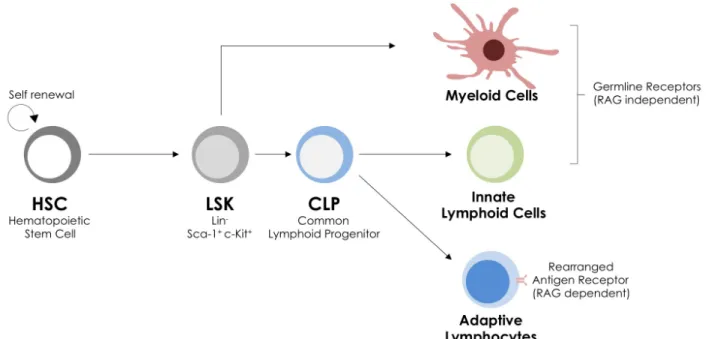
Around the 11th day of embryonic life (E11.5), murine HSC begin to colonize
the fetal liver (FL)6, an environment that fosters their self-renewal and onset their
differentiation into hematopoietic lineage. At E17.5, HSC migrate from the liver to the
bone marrow7 (BM) where they remain throughout life. In both organs, HSC eventually
give rise to common lymphoid progenitors (CLP)8, 9, which can differentiate into T and B
and cells, the two arms of adaptive immunity (Fig.7). CLPs were identified as cells negative for lineage surface markers (Lin-) bearing surface expression of the IL-7Rα chain (CD127+),
the Stem-Cell Factor (SCF) receptor c-kit (CD117+), the Stem cell antigen 1 (Sca-1+) and
Flt3 (Fms-Like tyrosine kinase 3 or CD135+)10. As discussed below, CLPs also retain the
potential to generate ILCs, and were used as a basis to identify an ILC-restricted progenitor.
ILC function and development share common features with those of TH cells. Most of the
molecules that will be mentionned tend to be better characterized in TH cells, I will often
introduce their role in TH cells before focusing on ILC.
Figure 7 : CLP give rise to RAG-independent ILC and RAG-dependent adaptive lymphocytes
TCF-1 : EILPing ILC
The Early ILC progenitor (EILP), was characterized according to the expression
of the transcription factor T cell factor 1 (TCF-1)11. Encoded by the gene Tcf7, TCF-1
acts at various stages in T cell biology.
T cell development occurs in the thymus, where thymocytes, the progenitors that mature into T cells, follow a well-defined pathway (for review, see Shah & Zuniga-Pflücker 201412). Briefly, CD4- CD8-, or double negative (DN) thymocytes begin to somatically rearrange their T-cell receptor genes in a RAG dependent manner. DN cells that successfully rearranged the T cell receptor (TCR) gene receive TCR-dependent survival signals, and acquire CD4 and CD8 expression. These Double Positive (DP) thymocytes subsequently acquire CD3 surface expression and a fully mature TCR. They concomitantly lose either CD4 or CD8 to become single positive (SP), therefore
specifying either into CD3+ CD8+ cytotoxic T cells that specialize in the destruction
of infected or transformed cells, or CD3+ CD4+ TH cells, which regulate other cellular partners during immune responses.
In thymocytes, TCF-1 cooperates with β-catenin to promote the transition
from DN stage to DP stage13 (Fig.9), while in mature TH cells, binds the promoter of
GATA3 and represses IFN-γ production14, thereby inducing TH2 cells development by
inhibiting TH1 cells-associated function.
With respect to ILC biology, TCF-1 deficiency dramatically affects the generation of ILC2 and NKp46+ ILC311, 15, 16 and to a lesser extent, ILC1a and ILC1b (Fig.10). EILP injected into lymphocyte deficient host (RAGKO/KO Il2rgKO/KO) further developed into all
ILC subsets (Fig.8), but no T and B, indicating that EILP only generate ILC11. In mice
that express a fluorescent protein under the control of the Tcf7 promoter (Tcf7GFP), EILP were identified as Lin- Sca-1+ CD117+ GFP+, and express the gut-homing associated
integrin α4β7. This surface phenotype overlaps with that of LSK (Lin- Sca-1+ CD117+
α4β7+)17, which are precursors downstream of HSC, as they lost self-renewal abilities,
but upstream of CLPs, as they generate both lymphoid and non-lymphoid cells, i.e. myeloid cells (Fig.7).
Out of commodity, I will now refer to Lin- Sca-1+ CD117+ CD127+α4β7+ cells as
KAS7 (for c-Kit, α4β7, Sca-1, IL-7rα), which do not represent any identified population
EILP lack CD127 expression, and are therefore phenotypically closer to LSK than CLPs. However, whether CLPs generate EILP remain unknown. If EILP are indeed downstream of CLPs, they would represent the “ILC/NK” precursor (Fig.6 versus 8). If CLPs fail to generate EILP, this would indicate the ILC commitment is marked before CD127 expression.
Intriguingly, DP thymocytes from transgenic mice in which β-catenin is rendered resistant
to ubiquitin-mediated degradation and subsequently more active, express higher amount
of CD12718, indicating that TCF-1 may require other molecular partner to maintain or
trigger CD127 expression in EILP.
EILP
ILC2
ILC3
ILC1
α4β7
Tox Nfil3 Tcf7CLP
?LSK
Nfil3 around the clock
Nuclear Factor, Interleukin 3 regulated (Nfil3) expression is readily detected
in EILP16 (Fig.10). Also known as E4BP419 (E4 promoter Binding protein 4), this basic
leucine zipper transcription factor is encoded by the Nfil3 gene, and operates both in hematopoietic and non-hematopoietic cells. For instance, hypothalamic neurons
contain mRNA transcripts for Nfil320. The hypothalamus regulates sleep, food intake
and hormone production, all of which are largely influenced by environmental cues such as exposure or absence of daylight and temperature. To define this internal clock, termed circadian rhythm, a set of genes, including Nfil3, cyclically modulates their expression21, 22.
In the hematopoietic compartment, cyclic expression of Nfil3 was reported to suppress
TH17 cells development through direct binding to the Rorc promoter, thereby linking
circadian rhythm and susceptibility to intestinal inflammation23. Nfil3 also directly controls cytokine expression, as it was originally identified through its ability to activate the Il3 promoter in human T cells19. In addition, overexpression of Nfil3 drives the
production of the TH2 associated-cytokine IL-13 and the anti-inflammatory cytokine
IL-10 by mature TH1 cells(24), reflecting its role in regulating lineage-associated TH cell function. Nfil3 is upregulated following chronic TCR stimulation of TH1 cells24, or upon
IL-3 stimulation of immature B Cells to promote their survival25.
Regarding the development of dendritic cells (DCs), which belong to the myeloid
lineage, the original study by Kashidawa report the absence of a subset of CD8α
expressing DCs in the spleen in Nfil3KO/KO mice26. In another report, Nfil3 deficiency
did not impact the generation of CD8α+ DCs in mice reconstituted with a mixture of
BM from wild-type (WT) mice and Nfil3 deficient mice27. These BM chimera stresses
the cell extrinsic role for Nfil3 in the generation of CD8α+ DCs, which therefore rely on
another Nfil3-dependent population for their development.
Remarkably, numerous reports testify that ILC intrinsically depend on Nfil328–36.
In our hands, Nfil3KO/KO mice are devoid ILC1a and ILC1ab, which contradicts published report identifying Nfil3-independent ILC1ab(32, 34) and ILC1a(37), an issue that will hopefully be clarified. Nonetheless, transfer of BM LSK from Nfil3 deficient mice failed
to generate the ILC compartment29, demonstrating that Nfil3 expression is intrinsically
required for ILC development. Conditional deletion of Nfil3 in mature ILC1b(30) or NKp46+ ILC331 had no effect on their generation, indicating that Nfil3 act at the early stages of ILC development and is dispensable for their homeostasis.
Interestingly, IL-7 stimulated CLP upregulated Nfil3 expression in vitro, which has also been reported in EILP that lack CD127. These conflicting results imply two outcomes. Either EILP are an intermediate stage between CLPs and mature ILC, and transiently modulate CD127 expression, therefore explaining the presence of Nfil3
“Tox Tox Tox”
Encoded by the Tox gene, Thymocyte selection-associated HMG box protein (TOX) is a transcription factor originally identified as a gene associated with thymocytes development38.
In DN thymocytes, TOX expression is TCR dependent. Indeed, RAG deficient thymocytes, which fail to rearrange their TCR and are subsequently blocked at an early DN stage, lack TOX expression38. Still, ToxKO/KO mice exhibit no defect in DP generation, indicating that although TOX expression is initiated early (Fig.9), it is not required for
the DN to DP transition39. Stimulation of DP thymocytes with phorbol
12-myristate13-acetate (PMA) and ionomycin, an ionophore that triggers Ca2+ influx used here to
mimic TCR signaling, triggered the upregulation of TOX38.
However, ToxKO/KO lack CD4+ SP thymocytes and mature TH cells, but display a
normal CD8+ T cell compartment, indicating that Tox expression is required for TH
cells commitment, and its absence lead to the generation of CD8+ T cells39. Remarkably, transgenic mice, in which TOX expression is driven by the Lymphocyte-specific protein
tyrosine kinase (lck) promoter, display biased generation toward CD8+ SP thymocytes,
although these cells failed to egress from the thymus and become mature40. These
results indicate that the level of intracellular TOX instructs CD4 versus CD8 cell fate decisions in the thymus (Fig.9).
Remarkably, mature ILC also express TOX, which expression was mandatory
for their generation. Indeed, pulmonary ILC1b and ILC2, splenic ILC1b and ILC3,
intestinal ILC1a, ILC1b, ILC2 were all affected by TOX deficiency41. Analysis of TOX/WT BM chimera revealed that this defect was cell intrinsic, as most of the ILC generated derived from the WT compartment.
Reminiscent of thymocytes development, Tox transcripts are detected early in the ILC
lineage, as EILP already express TOX11 (Fig. 8). In addition, retroviral transduction of
Nfil3 deficient CLPs with a vector expressing TOX partially rescued the generation of ILC,
indicating that TOX acts downstream of Nfil3 in ILC development36 (Fig. 10). Because ILC
do not rely on TCR expression for their generation, the question of whether Tox expression
is also controlled by Ca2+ influx or other factors such as Nfil3 will hopefully be answered
in future studies.
ID2 : CHILP vous plaît
Proteins of the Inhibitor of DNA Binding (Id) family encompass four members: Id1, Id2, Id3 and Id4. Id proteins are a peculiar form of transcription regulator as they are unable to directly bind DNA. As their name points out, Id proteins rather act by preventing the
DNA binding of the E-protein family of transcription factors (for reviews see Kee 2009)42.
E2A, a member of the E-protein family, is required for the generation of adaptive
lymphocytes43–45. Briefly, E2A deficient mice fail to rearrange B-cell receptor (BCR) and
subsequently lack B cells43, while thymocytes exhibit a partial block at the DN stage45.
The residual T cells that are generated in E2A deficient mice are highly susceptible to
transformation and generate lymphoma in older mice45. In cultured fetal liver HSC, E2A
deficiency led to the generation of ILC1ND and myeloid cells at the expense of T cells46. In immature B cells, invalidation of EBF1, an E2A target, led to the reprogramming into ILC2
and ILC347. Here, in the absence of signals require for an “adaptive fate”, commitment was
diverted toward innate lineages.
Consistent with this idea, Id proteins, which antagonize E-proteins, were shown to divert progenitors toward an “innate” fate48–50. Overexpression of Id1 in cultured LSK led to the extinction of B cell and promoted myeloid generation48. With respect to ILC1b and CD4+ ILC3, an early report, which preceded the discovery of other ILC subsets, demonstrated
that Id2 was crucial for their development 50. Retroviral transduction of fetal thymocytes
with Id2 abrogated T cell potential and only generated ILC1ND50. While ILC1b and CD4+
ILC3 are absent in Id2KO/KO mice, they developed normally in Id2KO/KO E2AKO/KO(49), pointing out that Id proteins acts by antagonizing E2A.
As a matter of fact, all ILC express and depend on Id23, 37, 49, 51–55. This finding was one of the basis for the current classification of ILC.
« We hypothesize that all ILCs develop from a common precursor that may depend on expression of the transcriptional repressor inhibitor of DNA binding 2 (ID2) »
Hergen Spits, David Artis, Marco Colonna, Andreas Diefenbach, James P. Di Santo, Gerard Eberl, Shigeo Koyasu, Richard M. Locksley, Andrew N.J. McKenzie, Reina E. Mebius, Fiona Powrie & Eric Vivier.
Subsequently, Id2 reporter mice allowed for the identification of an ILC precursor,
denominated CHILP (Common Helper-like ILC progenitor)56. In Id2GFP mice, CHILP were
defined as KAS7 CD135- GFP+. Transfer of CHILP into RAGKO/KOIl2rgKO/KO generated ILC1a,
ILC2, and ILC3, but failed to reconstitute the cytotoxic ILC1b compartment56. Analysis
of double Id2GFPToxTomato reporter mice revealed that all Lin-α4β7+ CD127+ coexpressed
Id2 and TOX41 (Fig.10). As CHILP were absent from Tox deficient mice41, Id2 expression
precede that of TOX. In a similar fashion, Nfil3 deficient mice also lacked CHILP29. Retroviral transduction of Nfil3 deficient LSK with Id2 restored the generation of ILC29. Chromatin immunoprecipitation with an anti-Nfil3 antibody revealed the enrichment for Id2 sequences, indicating that Nfil3 directly bind the Id2 gene29.
Ergo, CHILP requires Tox and Nfil3, and display a more restricted potential than
EILP, as they fail to generate cytotoxic ILC1b. CHILP appears therefore to be downstream
29
PLZF - Same-Same, but different.
NK T cells (NKT) are a family of lymphocyte that, like TH cells, originate from CD4+
SP thymocytes and are RAG dependent (for review see Constantinides & Bendelac 201357).
NKT effector functions largely rely on the transcription factor PLZF (Promyeolocytic Leukaemia Zinc Finger, encoded by Zbtb16), as PLZF deficient NKT fail to mature and
constituvely migrate to lymph nodes and spleen, from where they fail to egress58.
Although mature ILC are negative for PLZF59, fate-mapping analysis revealed that
a fraction of them had a prior history of PLZF expression. The majority of ILC2 from the
lung and the BM, as well as hepatic ILC1a and intestinal NKp46+ ILC3 originated from
PLZF expressing cells59. To a lesser extent, CD4- and CD4+ ILC3, as well as splenic and
hepatic ILC1b were also “fate-mapped” for PLZF expression. Consequently, BM KAS7
cells contained a fraction of PLZF expressing cells, which will be now referred to as PLZF+
ILCP(recursor)59. These cells expressed high amount of transcripts for Tox, Id2 and Tcf7,
features that are reminiscent of CHILP.
In vitro culture of PLZF+ ILCp could generate ILC1ND, ILC3ND and ILC2. In Zbtb16KO/
KO/WT BM chimera ILC2 and hepatic ILC1a were only derived from WT donor, indicating
that, although PLZF expression marked lymphoid progenitors with ILC potential, it was
only required for ILC2 and hepatic ILC1a development. Adoptive transfer of PLZF+ ILCP
in RAGKO/KO Il2rgKO/KO generated pulmonary ILC1ND and hepatic ILC1a, ILC2 in the lung and
the small intestine, and ILC3ND, but no T and B cells. As a matter of fact, CHILP contained
a fraction of PLZF+ and PLZF- cells56. PLZF+ ILCP numbers were also decreased in mice
deficient for Nfil329 and Tcf741, and therefore appear to be downstream of EILP, and closely related to CHILP (Fig.10).
COMMON GAMMA CHAIN : One to rule them all
Patients with Severe ImmunoDeficiency Syndrome (SCID) lack viable adaptive
immunity and are sometimes forced to live in sterile environment (for review see
Fischer et al. 2015)60. Several invalidating mutations cause SCID, including the ones
that affect the Il2rg gene. In human and mice, Il2rg encodes the common γ chain of the
IL-2 receptor, or CD132.
The first characterization of Il2rgKO/KO mice revealed that ILC1b were severely affected by lack of CD13261. As these mice still possess residual T and B cells, Il2rgKO/
KO were backcrossed to RAG to generate alymphoid RAGKO/KO Il2rgKO/KO(62).As mentioned
in the above sections, RAGKO/KO Il2rgKO/KO mice are commonly used as hosts for transfer
experiments. Indeed, organs of RAGKO/KO Il2rgKO/KO mice are devoid of lymphocytes, and
therefore provide an empty niche that can eventually be filled up by injected cells. Later, RAGKO/KO Il2rgKO/KO were shown to be also devoid of ILC23, 56, 63 and ILC364, 65,
such that common γ chain dependency is a common feature of ILC.
Cytokines for which cognate receptors associate with, and therefore require,
CD132 form the family of common γ chain cytokine, which includes 2, 4, 7,
IL-9, IL-15 and IL-21. As there are extensive reviews that discuss their role in TH cells (see
Rochman et al. 2009, Yamane & Paul. 2012)66, 67 , I will first briefly present the distinct dependency of ILC toward IL-7 and IL-15.
Acquisition of CD127 in hematopoietic cells progenitors is associated with
an increased potential to generate lymphocytes8,9. Encoded by the Il7ra gene, CD127
associates with CD132 to form a functioning receptor that permits IL-7 responsiveness (Fig.11). With respect to ILC precursor, only one report addressed IL-7 requirement
in vivo, showing that Lin- CD117+ α4β7+ Flt3- cells were unaffected in Il7ra deficient
mice56. Because CD127 could not be used as a discriminating marker in these mice,
whether these cells represent EILP, or downstream CHILP or PLZF+ ILCP, is still unknown. In addition, Thymic Stromal-derived Lymphopoietin (TSLP) signal through TSLPR that requires CD127 to transmit signal68 (Fig.11). Still, Mature ILC2 and ILC3 differ from ILC1 by their dependency on CD127, and are virtually absent in Il7KO/KO mice52, 64, 69 in Il7raKO/ KO(54, 70), while ILC1b numbers (except for
a subset that resides in the thymus)71
are unaffected in Il7raKO/KO(56, 70) or Il7KO/KO mice52, 71.
Figure 11 : Schematic rRptors involved in ILC survival
However, mice deficient for IL-15 are devoid of all ILC134, 52, 72, 73. As a matter of fact,
acquisition of CD122 expression is an early event that marks commitment to ILC1b fate.
Indeed, a fraction of Lin- CD122+ (refered to as NK cells precursor, or NKp) could generate ILC1b in vitro, and lacked myeloid, T and B cell potential74. Analysis of BM Lin- CD127+ cells
revealed that CD135, which identify CLP, and CD122 expression75, were mutually exclusive.
Remarkably, all Lin- CD127+ CD135-, either CD122- (referred to as pre-NKp) or for CD122+ (referred to as r(efined)NKp), generated splenic ILC1b, but no T and B cells upon transfer in RAGKO/KO Il2rgKO/KO(75).
Regarding the transcription factors involved in ILC commitment, Nfil3 expression is detected in BM preNKp, and increases from rNKp to ILC1b(29, 76). As Tcf7ko/ko mice lack NKp41,
they can be considered downstream of EILP. Finally, CHILP are described as Id2+ α4β7+
CD122- that can generate all ILC except for ILC1b(56). Although NKp are Id2 independent49, a fraction of NKp expresses Id277, and generated more ILC1b than their Id2- counterpart in
vitro, implying a role for Id2 in later stage of ILC1b development,.
Overall, CD122 and CD127 expression by ILC progenitor, and therefore IL-7 and
IL-15 responsiveness, appear to be important in the branching of ILC1b versus other ILC
I GATA3 my mind
GATA3 belongs to the GATA family of transcriptions factors, which binds
conserved WGATAR (W for A or T, and R for A or G) motif78. GATA3 deficient embryos
are non viable and die around E11.579. Therefore, most of the studies that investigated
GATA3 function in the immune system in vivo either relied on hematopoitic FL or conditional invalidation.
Briefly, GATA3 is required for T-cell development at several stages. Conditional invalidation of GATA3 driven by the lck promoter leads to a huge decrease in DP thymocytes, which reflects its role in the DN to DP transition, and driven by the Cd4 promoter, which expression is onset at the DP stage, lead to a marked decrease in
CD4 SP80. Although CD8 SP numbers are unaffected, CD8+ T cells further require
GATA3 to exhibit normal cytotoxic function driven by TCR signaling, as well as normal
proliferation81. Overall, GATA3 participates in the CD4/CD8 lineage determination,
and in the effector functions of mature CD8+ T cells (Fig.11). GATA3 also hallmarks TH2 responses, as it controls the expression and binds the promoters of Il4 and Il1382, 83.
GATA3 appears to instruct ILC in a similar way to T-cells. The generation of ILC1a, ILC2 and ILC3 was totally abrogated following transfer of GATA3 deficient FL cells or
in mice with conditional deletion in the hematopoietic compartment84, 85. The defect
was observed at an early stage, as Lin- CD127+, which includes ILC precursors, were
all absent in the FL in both models. GATA3 is therefore required for the development
of non-ILC1b ILC subsets. However, maturation of ILC1b was markedly affected, both
upon adoptive transfer of GATA3 deficient FL cells or in mice that conditionally lacked
GATA3 in the ILC1 compartment86, 87. Both studies demonstrated that GATA3 deficiency
reduced the number CD11b+ KLRG1+ ILC1b, which marks the final stage of maturation,
Figure 11 : GATA-3 in thymocytes development
and a defect in IFNγ production. In addition, GATA3 deficiency upregulated the chemokine
receptor CXCR4 expression by BM ILC1ND, known to be involved in the retention of ILC1ND
in the BM88. Blockade of CXCR4 with a chemical inhibitor allowed ILC1ND to efficiently
egress from the BM.
In Gata3GFP mice, a fraction of LSK are GFP+54, 89, while Gata3 transcripts are reduced
in Nfil3 deficient Lin- BM cells28, but present in PLZF+ ILCp59. Stainings for GATA3 protein
in CHILP or NKp were almost indistinguishable from the negative control56, 87, and the
report identifying EILP did not adress GATA3 expression. Still, analysis of ILC3 from mice in which GATA3 is conditionally deleted upon administration of doxycycline, revealed that Id2 and Tcf-7 were not affected by Gata3 deficiency, possibly excluding a role for GATA3 in
initiating Id2 and Tcf7 expression in precursor85. As such, Gata3 expression appears to be
initiated after Nfil3 and Tcf7, and is presumably downstream Id2 and upstream PLZF.
T-BET or not T-BET
T-bet belongs to the T-box family of transcription factors, which play diverse role
during embryonic development (for review, see Papaioannou 2014)90. Encoded by the gene
Tbx21, T-bet directs TH1 responses (for review, see Lazarevic et al. 2013)91. However, unlike GATA392, T-bet is absent in mature T-cells from the spleen93, and is first induced by IFNγ94 (Fig.12).
Upon activation, TCR signaling first blocks IL-12Rβ2 (encoded by Il12rb2)
expression therefore inhibiting IL-12 responsiveness. This transcriptional block is then relieved when TCR signaling stops, thereby allowing IL-2 mediated Il12rb2 transcription,
which permits IL-12 to induce T-bet expression94. Il12rb2 and Ifng are both direct T-bet
target genes, and signaling through their cognate receptors also induce T-bet expression, providing two amplifying loops for TH1 responses95, 96. In addition to Ifng and Il12rb2, T-bet
was shown to be required for the induction of TH1 associated genes such as chemokine
(Xcl1), chemokine receptors (Cxcr3, Ccr5) and cytokine receptors such as Il18r197.
In the early phase of TCR signaling, T-bet and GATA3 antagonize each other to determine Th1 versus Th2 polarization, as a phosphorylated form of T-bet directly interact with GATA3 and subsequently prevents TH2 gene transcription98. Tbx21 deficient
CD8+ T cells fail to acquire normal effector function upon antigen stimulation, display
reduced IFNγ production and cytotoxic potential, and fail to elicit recall response in
case of a second antigen encounter in vivo99. Overall, T-bet antagonize TH2 fate and
function, and promote TH1 responses.
As such, all ILC1 are T-bet+, although they display distinct dependency on Tbx21
expression. Tbx21 deficient mice completely lack intestinal and hepatic ILC1a(56, 100, 101),
while SG ILC1ab appear unaffected32. Remarkably, transgenic mice overexpressing T-bet
develop more ILC1a at the expense of ILC1b(101). However, splenic Tbx21 deficient ILC1b exhibit a defect in maturation, increased apoptosis and compensate by proliferating
more than their WT counterpart102. ILC1ND seem to accumulate in the BM of T-bet
deficient mice, which is linked to lower expression of S1pr5103, a T-bet-controlled
chemokine receptor involved in the egress of ILC1b from the BM103, 104, although T-bet
expression is not detected in preNKp, nor in proNKp, and unaffected by Nfil3 deficiency. Still, as they lack TCR, whether IFNγ or IL-12 is, like in T cells, responsible for the onset of T-bet expression in ILC precursor will probably be addressed in future studies.
EOMESsing around
Although IFNγ expression is controlled by T-bet, it can still be produced in
Tbx21 deficient mice99, suggesting that another transcription factor might compensate
for the loss of T-bet induced IFNγ expression. Eomes (encoded by Eomesodermin) also
belongs to the family of T-box proteins. In absence of Eomesodermin, embryos fail to develop beyond the blastocyst stage105.
In the hematopoietic lineage, Eomesodermin was originally reported to be expressed by activated or resting cytotoxic CD8+ T cells106. CD8+ T cells isolated from Eomes haplodeficient also exhibited reduced amount Granzyme B, a molecule involved in the cytotoxic program106. The finding that Tbx21KO/KO EomesKO/WT, in addition to
their lack of TH1 cells due to the loss of T-bet, had a marked defect in the cytotoxic
lymphocytes splenic compartment, i.e. CD8, NKT and ILC1b, suggested a role for Eomes
in their function107. Retroviral transduction of CD8+ T cells a dominant negative form
of Eomes led to reduced CD122 expression. Eomes was then shown to directly bind the
Il2rb promoter, and subsequently maintained homeostasis of IL-15 responsive cells107. Remarkably, CD8+ T cells that lacked both Eomes and T-bet totally lose the ability to
produce IFNγ, and display aberant IL-17 production108, indicating that they are redirected
toward the TH17 lineage. Overall, Eomes appear to control the establishment of the CD8
cytotoxic program while restricting a TH17 potential.
Conditional invalidation of Eomes in the hematopoietic compartment demonstrated that ILC1b generation critically relies on this transcription factor, while ILC1a, which lack Eomes expression, are unaffected100. In vitro, Eomes invalidation in ILC1b led to the loss
of ILC1b associated marker such as CD49b and the acquisition of ILC1a marker TRAIL and
fail to affect T-bet expression. Eomes ablation in Tbx21KO/KO ILC1b led to the complete loss of ILC1 markers, i.e. NK1.1 and NKp46100.
Total Lin- CD122+, which includes pre-NKp and rNKp, are still present in mice
lacking both T-bet and Eomes100. However, transcripts for Eomesodermin are detected in
CD122- preNKp and CD122+ rNKp76, Eomes therefore appear to precede CD122 expression
in ILC1b. In the BM, T-bet and Eomes expression are inversely correlated, at least in
ILC1b(101). Remarkably, retroviral transduction of Eomes in Lin- BM cells isolated from Nfil3
deficient mice rescued the generation of ILC1b(76), indicating that Eomes acts downstream
of Nfil3 in ILCp.
While T-bet controls ILC1a generation, Eomes directs ILC1b lineage program, and
might intervene early in ILCp in the branching between cytotoxic ILC1b and non-cytotoxic
ILC. Interestingly, Eomes has been shown to directly interact with and prevent GATA3
binding to the Il5 promoter in activated TH2 cells109, which may involve an antagonistic
functions role of these two transcription factors in cytotoxic/non cytotoxic ILC lineage determination. As such, Eomes appear to be tightly linked to CD122 expression, and its
expression should be onset before CD122- CD127+ CHILP and PLZF+ ILCp.
RORγt – Let him roar again
The Retinoic-acid related Orphan Receptors (ROR) are part of the Nuclear Receptors
(NRs) family (for rewiew, see Jetten 2009)110, which regroups transcription that
modulate their transcriptional activity through the binding of endogenous and/or exogenous ligands. The Rorc gene encodes two isoforms of the gamma member of
the RORs (RORγ)111, 112 through alternative splicing, among which transcripts for one
are highly enriched in the thymus, thereby naming the associated protein RORγt112,
113.
RORγt expression is readily detected in thymocytes, weaker in RAGKO/KO
thymocytes that lacks TCR, yet increases upon in vivo treatment of RAGKO/KO with
anti-CD3 activating antibody, a method used to mimic TCR signaling, therefore implying TCR
signaling for its induction113. In vitro overexpression of RORγt reduced the apoptosis
of a T-cell line following PMA-ionomycin or anti-CD3 stimulation, pointing out its
survival-promoting role112. As such, RORγt deficient mice exhibit a relative increase
in DN thymocytes, as the residual DP fail to rearrange TCR and subsequently undergo
massive apoptosis114. Still, some residual T-cells that escaped apoptosis develop
normally and egress the thymus to reach the periphery114. Consequently, transient
expression of RORγt actively participates to the generation of T cells.
As a matter of fact, sustained RORγt expression is mandatory for the generation
of pro-inflammatory TH17 cells115, 116. Overexpression of RORγt led to increase
production of IL-17115 and upregulation of the IL-23 receptor (Il23r) in undifferentiated TH cells in vitro116. However, isolated TH cells from RORγtGFP/GFP deficient mice display
a marked decrease in IL-17 production, and fail to upregulate Il23r expression115.
IL-23 stimulation of polarized TH17 cells further stabilized RORγt expression116, stressing
its role in controlling TH17 functions. As such, RORγt drive IL-23 responsiveness, and
participate in IL-17 expression, which hallmarks TH17 responses.
Several compounds were reported to activate RORγt transcriptional activity
through its ligand-binding domain117–120. Particularly, lipids that derive from cholesterol
metabolism, such as demosterol and zymosterol, which are precursors for cholesterol117,
120, or 7β,27-dihydroxy-cholesterol (7β,27-OHC)118, a cholesterol product. Remarkably,
TH cells deficient in CYP27A1, the enzyme that generate 7β,27-OHC, were less prone
to produce IL-17 compared to their WT counterpart118. Here, cholesterol derivatives
provided a cell-endogenous source of RORγt agonist, linking immune responses to
Remarkably, Retinoic Acid (RA), a vitamin A metabolite known to instruct development in vertebrates (for review, see Cunningham & Duester 2015)121, was reported to directly upregulate Rorc expression in isolated fetal CD4- and CD4+ ILC3122. In addition,
transgenic mice that expressed a truncated form of the RA receptor display lower RORγt
expression by fetal CD4- and CD4+ ILC3122. Whether RA induces RORγt in adult ILC3, which
include NKp46- and NKp46+ ILC3, has not been addressed. Nonetheless, this study still
provides important insight as to which stimulus control RORγt expression in ILC.
Most importantly, RORγtGFP/GFP deficient mice totally lack ILC3123, 124. However,
RORγt appear to be dispensable in mature ILC3 for their function125, and neither ILC1123,
124 nor ILC23, 4 require RORγt for their generation. Fate mapping experiment revealed that,
except for a subset of ILC152, 70, neither ILC1 nor ILC2 originated from RORγt expressing
cells52, 126. With respect to the interaction with other transcription factors, T-bet indirectly prevented RORγt-mediated transcriptional activity in TH cells91. As EILP, CHILP and PLZF+ ILCp are RORγt-, its expression is probably onset in situ in response to external stimuli.
Concluding remarks :
Some studies addressed other features associacted with ILC development (for
the effect of Notch signaling see Cherrier et al. 2010127, for CXCR6+ ILCp see Possot et al.
2011128, for fetal precursors see Bando et al. 2014129, for ILC1 precursor see Constantinides
et al. 2015130), but replacing them would render the scheme I presented more confusing,
and is not necessary for the points I want to address in my discussion. I also chose not to emphasize on factors involved in ILC2 commitment, as this manuscript focuses on ILC1 and ILC3.
Overall, commitment to ILC lineage occurs in a sequential manner. Early expression
of Tcf7 and Id2 allow for the extinction of B and T cell potential, respectively. Some
transcriptions factors like Nfil3 may act downstream to stabilize the ILC precursor pool, as their expression is dispensable in mature cells. Strikingly, the development of ILCs
branches with ILC1b cells fate decision, which could be attributed to Eomes expression.
Surely, the timely combination of transcription factors allows for the guiding to an ILC fate.
Tissue residency : Home Sweet Home
The above section presented evidences that BM hematopoietic precursors sequentially commit to an ILC fate. Intravenous injection of committed ILC precursor into recipient mice generates mature ILCs, indicating that, through the blood circulation, precursors are able to reach tissues where they ultimately differentiate into effectors. Nonetheless, in normal conditions, the pool of mature ILC could be either generated through constant influx of cells from the bone marrow, or maintained by cells that reside in the tissue and proliferate in situ.
To discriminate between these two situations, two mice, identifiable through the expression of distinct congenic markers, are surgically “joined” for them to share the same blood circulation. This experiment, called blood chimerism or parabiosis, allows to address whether molecules or cells generated in one mouse are transferable to another through the blood stream, and therefore also used to assess the anatomical provenance of cells inside tissues. If cells originate from the blood circulation, the proportion between cells originating from each mice eventually equilibrate to a 1:1 ratio, indicating that both mice contribute equally to replenish the pool of mature cells in tissues. However, for tissue-resident cells, circulating cells originating from one mouse would fail to replace cells that already reside in the other mouse.
Analysis of parabionts revealed that most ILCs are tissue residents72, 131, 132.
Indeed, thirty days post-surgical joining of CD45.1+ and CD45.2+ mice131, ILC1a, ILC1ab,
ILC2 and ILC3 were all CD45.1+ in CD45.1 mice, and CD45.2+ in CD45.2 mice, while
blood lymphocytes such as B and T cells were a mixture of CD45.1+ and CD45.2+.
Therefore, with the exception of ILC1b, which were equally derived from CD45.1+ and
CD45.2+ cells, all the other ILC subsets originated from the host mouse.
This finding is of paramount importance, as tissue-resident cells homeostasis, function and proliferation is therefore driven by tissue-signals received in situ, thereby increasing their reactivity and promptness toward harmful stimuli.
Chapter II
Activation & Function – Godspeed
Chapter II : Activation & Function – Godspeed you!
As part of the innate immune system, myeloid cells are engaged early during immune responses and, like ILCs, solely rely on germ-line encoded receptors for their generation, homeostasis and function. Sensing of microbial derived-products
through Toll Like Receptors (TLR, for reviews see Akira & Takeda 2004, Trinchieri
& Sher 2007, O’neill et al. 2013)133–135, most of which require the intracellular Myd88
adaptor, drive the production of cytokine that mediate ILC activation. As extensive reviews summarize the role of these cytokine in pathology and immune activation, I will briefly introduce them before addressing their relative role in instructing ILC1 and ILC3 function. The major cytokines that direct ILC1 and ILC3 responsiveness can be classified into two families, the IL-12 (see Gorieli et al. 2008, Teng et al. 2015)136, 137 and the IL-1 (see Dinarello 2009, see Sims & Smith 2010)138, 139 family.
In this chapter, I will present some of the ILC1 and ILC3 derived cytokines, and replace the two cytokines families (IL-12 and IL-1) in the context of ILC activation.
Figure 14 : Tissue-derived signals mediate ILC activation “The biology of innate lymphoid cells”
Gérard Eberl, Marco Colonna, James P. Di Santo, Andrew N. J. McKenzie Science 22 May 2015: Vol. 348, Issue 6237 DOI: 10.1126/science.aaa6566
IL-12 & IL-1 Family: Twelve plus one, not eleven plus two
IL-12 and IL-23 are both heterodimeric molecules, which share a common subunit IL-12p40140, and are therefore classified as member of the IL-12 family. IL-12p40 associate with IL-12p35 to form the IL-12, which binds its cognate heterodimeric receptor composed
of the two subunit IL-12Rβ1 and IL-12Rβ2. In
a similar fashion, association of IL-23R with
IL-12Rβ1 form the receptor for IL-23, which
itself comprise the subunits 12p40 and IL-23p19. IL-12 and IL-23 were mentioned in the
above sections as trigger for T-bet and RORγt
expression, respectively.
Unlike members of the IL-12 family, which are mainly produced by myeloid cells, both myeloid and non-hematopoietic cells, such as epithelial cells, can secrete
cytokines that belong to IL-1 family141–143. IL-1β and IL-18, like all members of the
IL-1 family, are first secreted in an inactive form, which is subsequently enzymatically
cleaved by caspase-1 to become bioactive. The main receptor for IL-1β is composed
of the two subunits IL1-R1 and IL1-R3, while IL-18 signals through the IL-18R
heterodimeric receptor, which comprises two subunits IL-18Rα and IL-18Rβ.
Fig 15 : IL-12 and IL-23 share the IL-12Rβ1 subunit
Fig 16 : IL-1 family members require enzymatic processing
Regarding T cells, an elegant study by Guo and colleagues highlighted the specific responsiveness of TH cells toward IL-1 family members144. Polarized TH1 expressed Il18r1
and accordingly responded to IL-18, while polarized TH17 expressed Il1r1 and responded to
IL-1β. Moreover, IL-1β increased IL-23-induced RORγt expression by TH17 cells, and IL-18
potentiated IL-12 induction of Tbx21 by TH1 cells. However, stimulation with IL-1 family
members only failed to elicit significant modulation of the aforementioned transcription factors, indicating that they are not the main driver for eliciting TH lineage programs, but rather synergize with IL-12 members to increase effector functions in mature cells.
As ILC1 and ILC3 share striking similarities with TH1 and TH17, respectively, one
can assume that they respond in a similar fashion to IL-12 and IL-1 family members.
IFNγ : An Army of one
Despite being the sole member of type II interferons, IFN-γ displays pleiotropic
activities as evidenced by combined immune defects in IFN-γ deficient mice145 (for reviews,
see Schroder et al. 2004)146. ILC1 constitutively express IL-12R and IL-18R, and produce IFN-γ production upon stimulation by IL-12 and IL-18101, 147.
Originally named Macrophage Activating Factor, IFN-γ potentiate the
antigen-presentation by myeloid cells by increasing the expression of both class I and class II major
histocompatibility complex (MHC-I and MHC-II)146, which are recognized by TCR-bearing
CD8+ and CD4+ T cells, respectively. TH1 cells also express the receptor for IFN-γ (IFNGR) 94,
which is required for their full maturation. Following LPS injection, Ifng deficient ILC1ND
efficiently migrate to lymph node, but fail to trigger TH1 cells activation and expansion148.
As such, ILC1-derived IFN-γ potentially promotes antigen-presentation by myeloid cells,
and expansion of adaptive TH1 cells, demonstrating its impact in the establishment of
adaptive responses (Fig. 12,17).
Moreover, IFN-γ increases iNOS (inducible Nitric Oxyde Synthase) expression by
myeloid cells146, which catalyzes the production toxic reactive nitrogen intermediates
(RINs)149. Indeed, mice, in which a myeloid specific promoter drives the expression of a
dominant negative form of the IFNGR, are unable to produce RINs, and subsequently fail
to survive infections with Trypanonosoma cruzi or Leishmania major150, highlighting the
role of IFN-γ in controlling myeloid-mediated clearance of these intracellular pathogens.
Although isolated myeloid cells from these mice exhibit normal IL-12p40 production,
pre-incubation, or “priming”, of myeloid cells with IFN-γ lead to their enhanced production
of IL-12p40, IL-12p35151 and IL23-p19152 production following stimulation with microbial
are the main innate source of IFN-γ, which drives the differentiation of inflammatory
monocytes into IL-12p40 producing DCs148. Here, IFN-γ signals instruct myeloid cells
to efficiently produce IL-12, either by “priming” or by inducing their differentiation. With respect to IL-1 family members, prolonged exposure of macrophages
to IFN-γ lead to decreased Il1b expression and IL-1β production in vitro153, while
stimulation of splenocytes with IFN-γ triggers IL-18 expression, although the cellular
source remains unclear.
IL-18 was originally named IFN-γ-inducing factor as it potentiated
IL-12-induced IFN-γ production by T cells154, 155. ILC1b isolated from IL-18 or IL-18R deficient
mice fail to elicit normal IFN-γ production ex vivo156. The defect in IFN-γ production
observed in Il18KO/KO could be restored to normal level only upon preculture of isolated
ILC1b with IL-18, but not with culture of IL-12 and IL-18, implying that IL-18 primes
ILC1b(156). Still, splenic and hepatic ILC1b compartment are normal in double deficient
mice for IL-12 and IL-18157, 158, indicating that these cytokines instruct the function,
but not the development of ILC1b (Fig.18). Moreover, Intestinal ILC1a were reported
to display higher expression of IL-12Rβ156, indicating that ILC1a potentially respond
better to IL-12 compared to ILC1b.
Overall, IL-12, IL-18, and IFN-γ are intertwined and provide amplifying loops in
promoting TH1 responses. As ILC1 are quick responder to IL-12 and IL-18, they provide
an innate source of IFN-γ, which impacts myeloid and T cell function.
MHC Peptide IFNγR IFNγ Intracellular pathogen (bacteria) IL-‐23p19 IL-‐12p40 IL-‐12p35 iNOS a b c
a. Increase antigen processing & presentation b. Increase production of IL-12 family members c. Increase digestion of intracellular pathogens
Perforin/Granzyme : Cytoxicity is my city
CD8+ T cell and ILC1b are cytotoxic lymphocytes, as they are both able to recognize infected or transformed cells, and subsequently trigger the apoptosis of the target
cells. Although the recognition mechanism differs, as CD8+ T cell activation requires
engagement of the somatically rearranged TCR while ILC1b only require germline encoded
receptors, both lymphocytes share common effector molecules to mediate their cytotoxic
effect, namely Perforin and Granzymes (for review see Voskoboinik et al. 2015)159. In this
section, I will concisely present these molecules before going back to ILC function.
Perforins were identified as molecule that could self-assemble to generate small pores (16-22um diameter) at the surface of cells. Encoded by the Prf1 gene, perforin is inactive at an acidic pH, and must be compartmentalized into granules to restrain its pore-forming activity. Mutation that affects the delivery of perforin into granules leads to the lysis of the lymphocytes that produces it, as the pores formed allows for liquid influx, which creates osmotic pressure and thereby disrupts the cellular membrane. Recognition of a target cells triggers Ca2+ influx in the cytotoxic lymphocytes, which in turn exocytose the granules content toward the target cells, a process also called degranulation.
Granules contained in cytotoxic lymphocytes comprise other molecules that are Granzymes, which are small enough to reach the cytosol of the target cells through the perforin-generated pores. Granzyme B (Gzmb) is the most potent pro-apoptotic molecule, as it induces apoptosis of the target-cell in less than 10 minutes through caspase activation160. However, Gzmb deficient ILC1b still exert cytotoxic function, which is then
solely ensured by Granzyme A (GzmA)160. Cell-death induced by Granzyme A differ from
both conventional apoptosis and cell death lysis, as target cells co-cultured with Gzmb
deficient ILC1b exhibited cell elongation, which is morphologically distinguishable from
the “bebbling” of cells that undergo conventional apoptosis. In addition, the rate of cellular
death was slower compared to cell cultured with Gzmb proficient ILC1b.
These findings demonstrate that ILC1b induces target cells death through at least
three mechanisms: perforin-induced cell lysis, which is a direct effect of the loss of membrane integrity of the target cells; granzyme B mediated apoptosis, which requires perforin to enter the cell and subsequently triggers caspase-induced apoptosis; and granzyme A, which induces a caspase-independent form of cell death.
ILC1b constituvely express Prf1 and Gzmb4, 100, 101, 147. Eomes appear to be key in
driving their expression, as it was reported to directly bind their promoter in TH2 cells