HAL Id: hal-02555520
https://hal-univ-paris.archives-ouvertes.fr/hal-02555520
Submitted on 27 Apr 2020
HAL is a multi-disciplinary open access archive for the deposit and dissemination of sci-entific research documents, whether they are pub-lished or not. The documents may come from teaching and research institutions in France or abroad, or from public or private research centers.
L’archive ouverte pluridisciplinaire HAL, est destinée au dépôt et à la diffusion de documents scientifiques de niveau recherche, publiés ou non, émanant des établissements d’enseignement et de recherche français ou étrangers, des laboratoires publics ou privés.
Development and internal validation of a diagnostic
score for gastric linitis plastica
J. Vivier-Chicoteau, J. Lambert, R. Coriat, P. Bonnot, D. Goéré, B. Roche,
M. Dior, G. Goujon, S. Morgant, Marc Pocard, et al.
To cite this version:
J. Vivier-Chicoteau, J. Lambert, R. Coriat, P. Bonnot, D. Goéré, et al.. Development and internal validation of a diagnostic score for gastric linitis plastica. Gastric Cancer, Springer Verlag (Germany), In press, �10.1007/s10120-020-01051-x�. �hal-02555520�
ORIGINAL ARTICLE
TITTLE:
Development and internal validation of a diagnostic score for gastric linitis plastica
AUTHORS:
J. VIVIER-CHICOTEAU1 (MD), J. LAMBERT2 (MD, PhD), R. CORIAT 3 (MD, PhD), P.E. BONNOT4 (MD), D. GOERE5 (MD, PhD), B. ROCHE6 (MD), M. DIOR7 (MD), G. GOUJON8 (MD), S.
MORGANT3 (MD), M. POCARD9 (MD, PhD), O. GLEHEN4 (MD, PhD), T. APARICIO1 (MD, PhD), J.M. GORNET1 (MD),
1 : Service de Gastroentérologie, Hôpital Saint Louis, Paris – France ; 2 : Service de Biostatistique, Hôpital Saint-Louis, Paris – France ; 3 : Service de Gastroentérologie, Hôpital Cochin, Paris – France ; 4 : Service de Chirurgie Digestive, Centre Hospitalier Lyon-Sud, Lyon – France; 5 : Service de Chirurgie Digestive, Hôpital Saint-Louis, Paris – France; 6 : Service d’Anatomopathologie, Hôpital Saint Louis, Paris – France 7 : Service de Gastroentérologie, Hôpital Louis Mourier, Colombes – France ; 8 : Service de Gastroentérologie, Hôpital Bichat, Paris – France ; 9 : Service de Chirurgie Digestive, Hôpital Lariboisière, Paris – France.
CORRESPONDING AUTHOR: Dr GORNET Jean-Marc (MD)
Dr Jean-Marc Gornet
Service de Gastroentérologie, Hôpital Saint Louis
1 Avenue Claude Vellefaux 75010 Paris, France
Phone +33 1 42 49 95 75 Fax +33 1 42 49 91 68
E-mail : [email protected]
SHORT RUNNING HEAD:
Diagnostic score for linitis plastica
ABSTRACT:
1
Background:
2
There is no consensual definition for gastric linitis plastica (GLP). We aim to construct a 3
diagnostic score to distinguish this rare tumor from usual gastric adenocarcinomas. 4
Methods:
5
In this retrospective study, all patients who had gastrectomy for cancer between 2007 and 6
2017 in French tertiary centers were included. The outcome was a diagnosis of GLP based on 7
pathological review of the surgical specimen. The diagnostic score was created by using 8
variables that were most frequently associated with GLP using penalized logistic regression 9
on multiply imputed datasets. We used discrimination measures to assess the performances 10
of the score. Internal validation was perfomed using bootstrapping methods to correct for 11
overoptimism. 12
Results:
13
220 patients including 71 linitis plastica (female 49%, median age 57 years) were analyzed. 14
The six parameters retained in the diagnosis score were the presence of large folds and/or 15
parietal thickening on at least one segment, pangastric infiltration and presence of gastric 16
stenosis on the upper endoscopy, circumferential thickening on at least one segment and 17
thickening of the third hyperechogenic layer on endoscopic ultrasound and the presence of 18
signet ring cells on endoscopic biopsies. The area under the ROC curve (AUC) was 0.967 with 19
a sensitivity of 94% [89.9-97.3] and a specificity of 88.7% [81.7-95.8] for a threshold of 2.75. 20
After internal validation, the corrected AUC was 0.959. 21
Conclusion:
It’s the first study validating a pre-therapeutic diagnostic score (Saint-Louis linitis score) with 23
an excellent ability to discriminate GLP from non-GLP adenocarcinomas. An external 24
validation is necessary to confirm our data. 25 26 KEYWORDS: 27 Linitis plastica 28 Diagnostic score 29 Gastrectomy 30
INTRODUCTION:
31
Gastric adenocarcinoma (GA) is the fifth most common cancer in the world (1). Despite 32
medico-surgical progress, its prognosis remains poor, ranking third among the most fatal 33
cancers (1,2). There are various classifications of GA, either purely histological (3–5), or 34
taking into account the macroscopic aspect (6), or the site of the tumor (7). Among the 35
different subtypes, gastric linitis plastica (GLP) represents a particular entity. It develops 36
from the submucosa and is characterized macroscopically by a major segmental or diffuse 37
thickening of the gastric wall and microscopically by the existence of poorly cohesive and/or 38
signet ring cells, within an abundant fibrous stroma infiltrating all the tunics (8,9). The terms 39
of poorly cohesive and/or signet ring cell carcinoma (SRC) and GLP are often indiscriminately 40
used leading to confusion in literature and difficulties to define the best therapeutic options 41
for this subtype of gastric tumor. GLP appears to have specific characteristics such as 42
younger age at diagnosis, female predominance, increased frequency of stages 3 and 4 and 43
lymph node invasion, and significantly decreased overall survival due to higher frequency of 44
R1 resection (10,11). Despite these specific features, there is to date no clear definition of 45
GLP. A recent consensus on the pathological definition and classification of poorly cohesive 46
gastric carcinoma propose that GA should be classified according to the WHO classification; 47
the term GLP being reserved for the description of the macroscopic characteristics of the 48
tumor (12). According to those discrepancies, the gold standard for GLP diagnosis is 49
currently based on histological examination of a surgical specimen (13,14). However in case 50
of locally advanced or metastatic disease which represents the vast majority of the patients, 51
surgery is rarely done. Thus, the diagnosis of GLP is mainly based on a simple set of 52
arguments (clinical, endoscopic, scannographic, histological). In case of a planned surgery for 53
localized GLP, the impact of preoperative chemotherapy remains uncertain and a total 54
gastrectomy is needed even in case of peroperative impression of free margin. The 55
development of a new diagnostic tool in order to make an early diagnosis of GLP remains 56
challenging and may lead to better understanding and significant therapeutic advances in 57
this field. The aim of this study is to construct a diagnostic score to discriminate GLP from 58
others GA. 59
METHODS:
60
All patients who underwent a gastrectomy for gastric cancer between 2007 and 2017 in 61
seven French tertiary centers were retrospectively identified either from a hospital database 62
known as Programme de Médicalisation des Systèmes d’Information (PMSI) or from 63
databases of the Gastroenterology departments. All the the files were reviewed by the same 64
person (JVC) to minimize missing data and control concordance; collected data included 65
information concerning demographic characteristics, case history, biological parameters, 66
description of endoscopic, endoscopic ultrasound and computed tomography scan findings, 67
type of surgery, histological analysis of surgical specimen and the treatments used. 68
Exclusion criteria were: genetic gastric cancer, history of gastric surgery for any reason, 69
history of endoscopic resection for superficial tumor prior to surgery (endoscopic mucosal 70
resection or submucosal dissection), gastro-esophageal junction cancer, non-71
adenocarcinomatous gastric tumor, adenocarcinoma infiltration of extra-gastric origin and 72
absence of tumor residue on the pathology report. We also excluded the files with at least 73
one major missing data (histological report of endoscopic biopsies or surgical specimen, 74
digestive endoscopy report). The large number of excluded patients is due to the 75
retrospective design of our study and the lack of computerization of medical data in some 76
centers (incomplete paper records). and non-available histological report. Patients were 77
treated in accordance with the Helsinki Declaration (World Medical Association Declaration 78
of Helsinki. Ethical principles for medical research involving human subjects. Bulletin of the 79
World Health Organization, 2001, ( ) , 373 - 374). All data were anonymously collected
80
and, according to the Loi Jardé (French law amended by Order No. 2016-800 and its 81
implementing decree No. 2016-1537 of 16/11/ 2016 relating to research involving the 82
human person), no patient consent was needed, as the treatment implemented in this study 83
was the standard recommended therapy. 84
To create the score, our study population was divided into 2 groups: GLP group and non-85
linitic adenocarcinoma (non-GLP or control group). 86
Definition of GLP and pathological analysis
87
The diagnosis of GLP was retained if the three following criteria were mentioned on the 88
pathology report of surgical specimen: 89
- Macroscopic examination of the surgical specimen showing segmental or pangastric 90
diffuse parietal thickening. 91
- Histological examination showing an abundant and diffuse fibrous stromal reaction 92
extended throughout the gastric lining to the sub serosa. 93
- Histological examination showing a carcinoma with more than 50% poorly cohesive 94
cells having classical SRC morphology. 95
96
The pathology reports of gastrectomy were all reviewed for validation by an expert 97
pathologist in the reference center of Assistance Publique des Hôpitaux de Paris (APHP) for 98
the treatment of oeso-gastric tumors. In some doubtful cases, a re-reading of the glass slides 99
was carried out. If one of the 3 criteria was absent, the patient was excluded from the GLP 100
group even if the clinical presentation and the morphological assessment appeared 101
compatible with the diagnosis. 102
Patients in the non GLP group were randomly selected from the same centers without 103
matching regardless of the presence or not of signet ring cells on surgical specimen. 104
All pathology reports of gastrectomy have been standardized according to the latest UICC 105
AJCC 2016 classification (15). 106
Parameters for diagnostic score
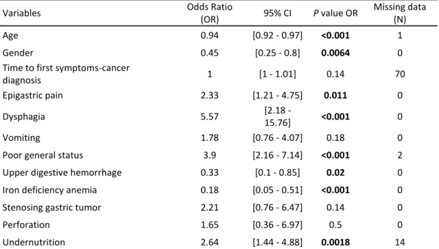
107
The parameters taken into account for the creation of the score were demographic 108
characteristics, symptoms at the diagnosis of GA, description of the initial upper 109
gastrointestinal endoscopy and if available of gastric endoscopic ultrasound, description of 110
pre-therapeutic abdominal computed tomography scan and histological description of 111
endoscopic biopsies at diagnosis (cf Table 1 and 2). The biological parameters included for 112
the creation of the score were: total blood count (anemia, increased neutrophils count, 113
thrombocytosis), high neutrophil-to-lymphocyte ratio, elevated C-reactive protein, 114
hypoalbuminemia, increased tumor markers (carcinoembryonic antigen, carbohydrate 115
antigen 19-9). Further details are provided in Supplementary Tables 1-5. 116
Statistical analysis
117
Patient characteristics are presented using medians and interquartile ranges for quantitative 118
data and counts with percentages for qualitative data. Characteristics of patients with and 119
without GLP were compared using Wilcoxon tests for quantitative data and Chi2 tests or 120
Fisher tests for qualitative data. 121
There were 12.6% of missing data among all the predictors considered, and only 5 % of 122
patients had no missing data. Under the hypothesis of missingness at random, we used 123
multiple imputations by chained equations to generate 20 imputed datasets. The diagnostic 124
score was constructed with the most frequently selected predictors on these 20 datasets. To 125
take into account for both the low variable/individual ratio with 71 patients having GLP and 126
49 candidate covariates and the high risks of collinearities between candidate covariates, we 127
used a generalized linear model with LASSO regularization (with 10-fold cross validation to 128
select λ parameter) to build the multivariable model and select predictors. Finally, due to the 129
near separation of some variables (no individuals in any of the modalities), a Firth penalized 130
logistic regression was performed to assess the respective importance of each predictor. The 131
Firth logistic model was applied to each of 20 imputed datasets and the resulting mean value 132
of each coefficient was used to construct the score. We rounded the coefficient to obtain an 133
easy to calculate diagnostic score. 134
Performances of this score were assessed through discrimination measures: ROC curve, Area 135
under the Curve (AUC), sensitivity and specificity at chosen threshold. 136
Finally, bootstrap resampling (200 bootstrap resampling for each of the 20 imputed 137
datasets) allowed us to obtain an internal validation to correct for over-optimism in the 138
discrimination measures. All statistical analyses were performed using R software 139
The methodology used is in agreement with the criteria defined by the TRIPOD checklist. 140
Further details are provided in Supplementary Table S6. 141
RESULTS:
142
Patients’ characteristics
143
The files of 457 patients aged over 18 years who underwent a gastrectomy for gastric cancer 144
were reviewed. Among them, 72 records were not analyzable due to major missing data. In 145
the remaining 385 patients, 165 presented an exclusion criteria. Therefore, a total of 220 146
patients (71 in the GLP group and 149 in the control group) met the inclusion criteria and 147
were included in the analysis (Figure 1). 148
The general characteristics of the study population at diagnosis are presented in Table 1. 149
Several statistical differences regarding epidemiological data and clinical presentation at 150
diagnosis were noted between GLP and non-GLP patients. In the GLP group, the population 151
was younger (p < 0.001) with a higher proportion of women (p = 0.007), a longer diagnostic 152
time (p=0.02) and the need to repeat iterative biopsy endoscopy more frequently (p < 153
0.001). Clinical presentation also differed with a higher proportion of patients with general 154
impairment (p < 0.001), undernutrition (p = 0.002), dysphagia (p < 0.001) and epigastralgia 155
(p = 0.01). In the non-GLP group, we noted a higher proportion of patients explored for 156
anemia or with externalized digestive hemorrhage (p = 0.03). Upfront surgery was 157
performed in 4 patients in the GLP group (surgical decision at baseline: n = 3, perforation: n 158
= 1) and one patient in the non-GLP group (perforation). 159
The description of the main additional examinations carried out for diagnostic purposes is 160
presented in Table 2. 161
Endoscopic findings
162
Several statistical differences were noted at endoscopic examination between GLP and non-163
GLP patients. In the GLP group, more patients with large folds or macroscopic tumor 164
infiltration on at least one segment (p < 0.001) with a higher frequency of multiple 165
ulcerations or erosions in the suspected area (p = 0.001). Difficulty with insufflation and the 166
presence of stenosis were also more frequently observed (p < 0.001). In the non-GLP group, 167
there was a higher proportion of patients with a single ulcer or ulcer-budding tumor (p < 168
0.001). On endoscopic ultrasound, there was more frequent thickening on at least one 169
segment (p < 0.04) or pangastric thickening in the GLP group (p < 0.001). This one was more 170
frequently circumferential with predominance over the third hyperechoic layer or fusion 171
aspect of the layers (p < 0.001). 172
Imaging
173
On contrast-enhanced computed tomography scan (13% with opacification with water or 174
contrast medium), we observed a higher proportion of diffuse parietal involvement and 175
circumferential thickening of the gastric wall. 176
Biology
177
Among all the biological parameters studied, only anemia was significantly more frequent in 178
the non-GLP group. No significant differences were observed on the other characteristics of 179
blood count and CRP, serum albumin and tumor marker elevation frequency (ACE and CA 180
19-9) between both groups. 181
Pathological findings
182
The comparative analysis of the histological characteristics of the gastrectomy specimens is 183
presented in Table 3. Again, we noted several statistically significant differences between 184
the two groups. In the GLP group, the number of total gastrectomy was higher with more 185
incomplete resection. The disease was more frequently pangastric with an increased 186
number of T4 status, positive lymph nodes, distant metastases and poorly cohesive and/or 187
SRC contingent. Among the patients with positive lymph nodes, we observed more 188
frequently a N3 status in the GLP group than in the non-GLP group (45% vs 18%). Among the 189
patients with metastatic location, peritoneal carcinomatosis only was known preoperatively 190
in 19 patients (14 in the GLP group and 5 in the non-GLP group) and found per-operatively in 191
15 patients (10 in the GLP group and 5 in the non-GLP group). In 5 patients of the GLP group, 192
the tumor was a mixed type (n = 4) or a majority mucinous type (n = 1), according to the 193
WHO 2010 classification. Of note, HER2 status positivity was low and the proportion of 194
patients with Helicobacter Pylori infection was similar in both groups. 195
Diagnostic score
196
The diagnosis score for gastric LP is presented in Table 4. Regarding first results, six variables 197
were selected to create the score. Three variables corresponding to uppergastrointestinal 198
endoscopic characteristics, the presence of large folds and/or gastric thickening on at least 199
one segment (1.5 points), pangastric infiltration (2 points) and presence of gastric stenosis (1 200
point). Two variables corresponding to endoscopic ultrasound characteristics, a 201
circumferential thickening on at least one segment (0.5 points) and predominance of the 202
lesion on the third hyperechoic layer (1 point). And one variable on histological report on 203
endoscopic biopsies, presence of poorly cohesive cells and/or signet ring cells (1.5 points). 204
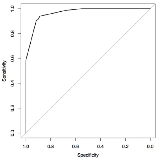
The score performance was evaluated by ROC curve (Figure 2), with an AUC of 0.967 [0.948 - 205
0.987], a sensitivity of 94% [89.9-97.3] and a specificity of 88.7% [81.7-95.8] for a threshold 206
of 2.75 points (observed performances on one of the 20 imputed datasets). 207
After internal bootstrap validation (resampling), the corrected AUC was 0.959. 208
DISCUSSION:
209
GLP: a clearly different entity
210
Firstly, our results confirmed that GLP tumor have to be considered as a different entity 211
from non-GLP tumors with different epidemiological, clinical, radiological and histological 212
presentation. The importance of the differences observed at diagnosis between these two 213
types of gastric tumors makes it necessary to use a reliable tool that clearly differentiates 214
them. 215
A new diagnostic score for GLP
216
To our knowledge, we are reporting the first diagnostic score to discriminate GLP from other 217
GA. This score has an excellent diagnostic performance to predict the existence of GLP with 218
an AUC of 0.967, a sensitivity of 94% and a specificity of 88.7% for a threshold of 2.75 points. 219
The resampling by boostrap allowed us to obtain an internal validation of the score 220
performances with a corrected AUC of 0.959 with reinforce its viability. The 2/1 ratio 221
between the GLP and the non-GLP group and the absence of a priori selection of the control 222
group allows a satisfactory validation sample to be obtained. 223
The six variables used to create the score include 3 endoscopic parameters: the presence of 224
large folds and / or parietal thickening on at least one segment (1.5 points), pangastric 225
infiltration (2 points) and the presence of gastric stenosis (1 point); 2 endoscopic ultrasound 226
parameters: circumferential thickening on at least one segment (0.5 points) and thickening 227
of the third hyperechoic layer (1 point) and a histological parameter: presence of poorly 228
cohesive and/or SRC (1.5 points). 229
GLP group was identified using 3 strict criteria based on histological analysis of the 230
gastrectomy specimen which is considered as the gold standard for the positive diagnosis of 231
GLP. In addition, this diagnosis was validated by a centralized review of histological reports 232
by a pathologist from a center specializing in the management of oesogastric tumors using a 233
keyword grid and in some cases a re-reading of the glass slides. As LP is a rare entity, 234
obtaining a group of 71 patients who were included using only the current gold standard can 235
be considered a large sample. Among the various parameters analyzed to create the score, 236
many differed significantly between the two samples. The differences in clinical, endoscopic, 237
scannographic and histological presentation observed in the GLP group have been previously 238
reported in the literature underlining the quality of our sampling (8,9,16). The percentage of 239
patients with SRC adenocarcinoma (22%) in the control group was also in agreement with 240
the literature (17–19). 241
GLP: a lack of a consensual definition to date
242
Histological analysis of the surgical specimen is not a tool that can be easily used in clinical 243
practice given the high frequency of GLP who will never be operated on, mainly because of 244
the greater aggressiveness of this pathology. Therefore, the definitions currently proposed 245
are mainly based on upper gastrointestinal endoscopy. Thus, Pedrazzani et al (20) defined 246
GLP as a thickening and stiffening of the gastric wall which involve circumferentially at least 247
one-third of the stomach, and Endo et al (21) more than two thirds of the stomach. More 248
recently, Agnes et al (9) proposed the following definition: thickening of the gastric wall, 249
with lack of distensibility, which involves more than one third of the gastric surface, both as 250
a circumferential involvement of more than one area, or a semi-circular involvement of 251
more than two areas. Finally Jung et al (16) proposed a decisional algorithm to diagnose GLP 252
based on macroscopic and microscopic data of the initial upper gastrointestinal endoscopy. 253
However, these definitions are not validated and the inter-observer reproducibility of the 254
description of endoscopic lesions is not known. 255
GLP: a new diagnostic score that uses routine exams
257
Endoscopic ultrasound is usually recommended for the diagnosis of GLP but its diagnostic 258
value in distinguishing it from classical GA has never been studied (22). Endoscopic 259
ultrasound puncture seems a useful tool in difficult cases but remains poorly evaluated and 260
most often useless (23). Although the presence of poorly cohesive cells and/or SRC is almost 261
constant in GLP, they can frequently be found in diffuse gastric adenocarcinomas and 262
therefore do not constitute a discriminant parameter. Our score allows the diagnosis of GLP 263
to be carried out with high sensitivity and specificity using usual explorations for the 264
diagnosis of GA. Even if the inter-observer reproducibility of the different examinations is 265
poorly known, the description of the macroscopic aspect on the upper gastrointestinal 266
endoscopy, the analysis of the different gastric wall layers in endoscopic ultrasound and the 267
histological description of gastric tumors represent routine procedures applicable in current 268
practice to establish a diagnostic score. Despite some differences, no discriminating clinical 269
parameters were found in the GLP sample. This is in agreement with the literature which 270
reports that gastric cancer symptoms are non-specific and that in the event of a positive 271
diagnosis there is no clinical sign to distinguish a particular tumor subtype. The CT scan has 272
been recently shown to be a useful tool for the diagnosis of GLP (24). This is confirmed by 273
our data which show a significantly increased frequency of circumferential and/or pangastric 274
parietal abnormalities. However, these two morphological parameters remain less 275
discriminating than those observed on the upper gastrointestinal endoscopy and the 276
endoscopic ultrasound. 277
GLP diagnostic score: a new tool to standardize its management
278
Despite the severity of this pathology, medico-surgical management of gastric LP remains 279
poorly codified. The rarity of this gastric tumor, the absence of a consensual definition and 280
the confusion created by the term signet ring cell carcinoma contribute to the absence of 281
therapeutic advances, although the GLP has different characteristics and a poorer prognosis. 282
Other scores have already been validated in GA in patients treated, notably to establish 283
survival predictive factors after gastrectomy (25–27) or in metastatic patients undergoing 284
chemotherapy (28,29). The validation of a diagnostic score specific to GLP provides a new 285
homogeneous pre-therapeutic definition that could standardize the management of this 286
pathology, which is considered chemoresistant (30). 287
Limitations
288
Nevertheless, our study has some limitations. The data were collected retrospectively with a 289
limited number of patients in the LP sample and no systematic centralized re-reading of all 290
the glass slides. The retrospective design of the study led to some missing data. However, all 291
the files including the pathology reports were centrally reviewed in order to reduce the 292
number of missing data. Some inaccuracies in upper gastrointestinal endoscopy, endoscopic 293
ultrasound and CT scan reports may also have led to misinterpretation of some data 294
however this parameter has been taken into account in the creation of our score by 295
performing multiple imputations using chain equations. Furthermore, our score was not 296
externally validated in an independent cohort. Nevertheless, the rarity of this pathology and 297
the difficulty of obtaining a homogeneous study group make it difficult to carry out such 298 work. 299 300 CONCLUSION: 301
We have constructed and validated the first score to diagnose GLP with high sensitivity and 302
specificity (Saint-Louis linitis score). This one is composed of six parameters easily applicable 303
in clinical practice and allows to determine a homogeneous group of patients in a pathology 304
where there is no consensual definition. The use of this score may help to improve the 305
therapeutic management of this subtype of GA, in particular the interest of preoperative 306
chemotherapy and extend of gastric resection if planned. However an external validation is 307
necessary in order to integrate this new score into clinical practice. 308
TABLES: 1 2 3
Variables GLP N = 71 Non-GLP N = 149 P value
Gender, n (%)
Female/Male 35/36 (49/51) 45/104 (30/70) 0.007
Age at diagnosis (years)
Median (IQR) 57 (45.5-63) 64 (56-71.5) <0.001
Time to first symptoms - cancer diagnosis (days)
Median (IQR) 103 (72-184) 67 (17-181) 0.02
Tumor stage at diagnosis, n (%)
Localized tumor 57 (80) 142 (95)
Metastatic tumor 14 (20) 7 (5)
Pre-operative treatment
Upfront surgery 24 (34) 69 (46)
Systemic chemotherapy 47 (66) 80 (54)
Systemic chemotherapy + PIPAC *, n (%) 2 (3) 0 (0) Clinical symptoms at diagnosis, n (%) **
Poor general status 41 (58) 39 (26) <0.001
Undernutrition 32 (45) 40 (27) 0.002 Dysphagia 14 (20) 6 (4) <0.001 Epigastric pain 58 (82) 97 (65) 0.01 Vomiting 11 (15.5) 14 (9.5) 0,255 Digestive hemorrhage 4 (5.5) 25 (17) 0.03 Occlusive syndrome 7 (10) 7 (5) 0,151 Perforation 3 (4) 4 (3) 0,684 Histological diagnosis, n (%)
Unique upper digestive endoscopy 49 (69) 140 (94) <0.001
Repeated upper digestive endoscopies 9 (12.5) 7 (5) <0.001
Upper endoscopic ultrasound 2 (3) 1 (0.5)
Exploratory coelioscopy 7 (10) 0 (0)
Inaugural surgery 4 (5.5) 1 (0.5)
4 5 6
Table 1: General characteristics of the study population 7
8 9
* Pressurized intraperitoneal aerosol chemotherapy 10
Variables GLP N = 71 Non-GLP N = 149 P value
Upper gastrointestinal endoscopy, n (%) *
Single ulcer 27 (38) 79 (53) 0.04
Ulcerations or multiple erosions 16 (22.5) 10 (6.5) 0.001
Ulcer-budding tumor 4 (5.5) 53 (35.5) <0.001
Large gastric folds or thickening on one segment 40 (56) 9 (6) <0.001
Large gastric folds or diffuse thickening 15 (21) 0 (0) <0.001
Difficulty of insufflation 13 (18) 0 (0) <0.001
Stenosis 21 (29.5) 18 (12) 0.001
Pangastric tumor infiltration 17 (24) 0 (0) <0.001
Tumor infiltration extending to the duodenal bulb 8 (11) 1 (1) <0.001
Tumor diagnosis not mentioned on the macroscopic aspect 16 (22.5) 4 (3) <0.001
Upper endoscopic ultrasound, n (%) *
Circumferential thickening 28 (39.5) 8 (5.5) <0.001
Pan gastric thickening 13 (18) 0 (0) <0.001
Thickening of an entire segment or a limited part 33 (46.5) 77 (52) 0.004
Wall thickening predominant on the 3rd hyperechoic layer 15 (21) 0 (0) <0.001
Layer fusion 14 (20) 11 (7.5) 0.008
Suspicious peri-gastric adenopathy 26 (36) 44 (29.5) 0,585
Scanner, n (%) **
Localized parietal abnormality 43 (60.5) 95 (64) 0,63
Diffuse parietal abnormality 19 (27) 4 (3) <0.001
Circumferential parietal abnormality 33 (46.5) 17 (11.5) <0.001
Suspicious peri-gastric adenopathy 21 (29.5) 66 (44) 0,128
1 2 3
Table 2: Characteristics of endoscopic findings and imaging in the study population
* Several possible lesions in the same patient
** Parietal abnormalities = thickening +/- parietal enhancement or endoluminal bud
4 5 6 7 8 9 10
Variables GLP N = 71 Non-GLP N = 149 P value Type of gastrectomy, n (%) Total 61 (86) 86 (58) <0.001 Partial 10 (14) 63 (42) <0.001 Resection, n (%) R0 46 (65) 141 (94) <0.001 R1 25 (35) 7 (5) R2 0 (0) 1 (1) Tumour site, n (%) <0.001 Pangastric 30 (42) 0 (0) Fundus 12 (17) 28 (19) Antrum/pylorus 24 (34) 93 (62)
Antro-fundic junction or body 5 (7) 28 (19)
AJCC TNM stage, n (%) * <0.001
pT1-T2 2 (3) 64 (43)
pT3-T4 69 (97) 85 (57)
Positive lymph nodes (any N) 55 (75.5) 85 (57) <0.001
M1 (metastatic location) 24 (34) 12 (8) <0.001
WHO classification, n (%)**
Poorly cohesive (including signet ring cells) 66 (93) 33 (22)
Tubular 0 (0) 60 (40.5) Papillary 0 (0) 6 (4) Mucinous 1 (1.5) 5 (3.5) Mixed 4 (5.5) 21 (14) Unknown 0 (0) 24 (16) Lymph node(s)
Number of lymph nodes analyzed, median (IQR) 26
(18-34) 22
(16-31) 0,054 Number of invaded lymph nodes, median (IQR) 8
(3-13) 4 (3-9) <0.001 HER2, n (%)*** Positive 1 (1.5) 12 (8) 0,112 Not determined 16 (22.5) 19 (13) 1 2 3
Table 3: histological characteristics of the gastrectomy specimens 4
5
* UICC/AJCC 2016 6
** WHO classification 2010 7
*** HER2 status has sometimes been determined on endoscopic biopsies 8
Items Point Upper gastrointestinal endoscopy
Large folds and / or gastric thickening on at least one segment 3.18 1.5
Pangastric infiltration 4.33 2
Stenosis 1.63 1
Upper endoscopic ultrasound
Circumferential thickening 0.18 0.5
Thickening of the gastric wall predominant on the third hyperechoic layer 2.49 1 Histology of gastric biopsies
Poorly cohesive and/or signet ring cells 3.14 1.5
1 2 3
Table 4: Diagnostic score of gastric linitis plastica 4
Each item scores 0 if absent or the tabulated value if present. The diagnostic score is the 5
sum of each items and ranges from 0 to 7.5; a higher score indicates a higher probability of 6
linitis plastic. The chosen threshold is 3: patients with a score<3 are considered not having a 7
gastric linitis plastica and patients with a score >=3 are considered having a gastric linitis 8
plastica 9
10
* coefficient are obtained with Firth penalized logistic regression model 11
12 13
Figure 1: Flow chart of the whole population 1 2 3 4 5 6 7 8 Excluded patients N=20
No confirmation after centralized proofreading N=10
Genetic mutation N=8
Sterilized gastrectomy specimen N=1 Mixed tumor contingent N=1
Excluded patients N=145
Oesogastric junction adenocarcinoma N=97 Previous endoscopic treatment N=14 Previous gastrectomy N=10 Sterilized gastrectomy specimen N=9 Mixed tumour contingent N=8 Genetic mutation N=4
Extra-gastric synchronous cancer N=3
Control group
N=149
Gastrectomy for gastric adenocarcinoma
N=385 Linitis Plastica N=91 Control group N=294 Linitis Plastica N=71 Study population N=220
Figure 2 : ROC curve 1
2 3 4
FIGURES LEGENDS 1 2
Figure 1: Flow chart 3
4
Figure 2: ROC curve. The AUC is 0,967 [0.948 - 0.987]; using a threshold of 2.75, the 5
sensitivity is 94% [89,9-97,3] and the specificity is 88,7% [81,7-95,8]. 6
7 8
All procedures followed were in accordance with the ethical standards of the 1
responsible committee on human experimentation (institutional and national) and 2
with the Helsinki Declaration of 1964 and later versions. 3
4
All the authors declare no conflict of interest for this article (ICMJE Form for 5
Disclosure of Potential Conflicts of Interest). 6
7
All data were anonymously collected and, according to the Loi Jardé, no patient consent was 8
needed, as the treatment implemented in this study was the standard recommended 9
therapy. 10
11 12 13 14
SUPPLEMENTARY TABLES S1 to S6: unadjusted association between each candidate 1
predictor and outcome. using Firth's bias-Reduced penalized-likelihood logistic 2
regression. 3
4 5
Table S1 : Clinical Variables 6
Variables Odds Ratio
(OR) 95% CI P value OR
Missing data (N)
Age 0.94 [0.92 - 0.97] <0.001 1
Gender 0.45 [0.25 - 0.8] 0.0064 0
Time to first symptoms-cancer
diagnosis 1 [1 - 1.01] 0.14 70
Epigastric pain 2.33 [1.21 - 4.75] 0.011 0
Dysphagia 5.57 [2.18 -
15.76] <0.001 0
Vomiting 1.78 [0.76 - 4.07] 0.18 0
Poor general status 3.9 [2.16 - 7.14] <0.001 2
Upper digestive hemorrhage 0.33 [0.1 - 0.85] 0.02 0
Iron deficiency anemia 0.18 [0.05 - 0.51] <0.001 0
Stenosing gastric tumor 2.21 [0.76 - 6.47] 0.14 0
Perforation 1.65 [0.36 - 6.97] 0.5 0
Undernutrition 2.64 [1.44 - 4.88] 0.0018 14
7 8
Table S2 Biological Variables 9
Variables Odds Ratio
(OR) 95% CI P value OR
Missing data (N)
Anemia 0.45 [0.24 -
0.81] 0.0084 15
Elevated C-reactive protein 1.33 [0.58 - 3.1] 0.5 99 Increased neutrophils count 0.7 [0.27 -
1.64] 0.42 37 Lymphopenia 0.52 [0.18 - 1.33] 0.18 54 Neutrophil-to-lymphocytes ratio 1 [0.91 - 1.07] 0.92 54 Thrombocytosis 0.23 [0.04 - 0.73] 0.011 35 Hypoalbuminemia 1.64 [0.82 - 3.27] 0.16 56 Increased CEA 0.36 [0.09 - 1.06] 0.064 38 Increased CA 19-9 0.76 [0.31 - 1.76] 0.53 47 10 11 12 13 14
1
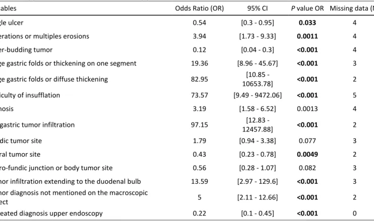
Table S3 Upper Gastrointestinal Endoscopy Variables 2
Variables Odds Ratio (OR) 95% CI P value OR Missing data (N)
Single ulcer 0.54 [0.3 - 0.95] 0.033 4
Ulcerations or multiples erosions 3.94 [1.73 - 9.33] 0.0011 4
Ulcer-budding tumor 0.12 [0.04 - 0.3] <0.001 4
Large gastric folds or thickening on one segment 19.36 [8.96 - 45.67] <0.001 3 Large gastric folds or diffuse thickening 82.95 [10.85 -
10653.78] <0.001 2
Difficulty of insufflation 73.57 [9.49 - 9472.06] <0.001 5
Stenosis 3.19 [1.58 - 6.52] 0.0013 4
Pangastric tumor infiltration 97.15 [12.83 -
12457.88] <0.001 2
Fundic tumor site 1.79 [0.94 - 3.38] 0.077 3
Antral tumor site 0.43 [0.23 - 0.78] 0.0049 2
Antro-fundic junction or body tumor site 0.56 [0.28 - 1.07] 0.082 3
Tumor infiltration extending to the duodenal bulb 13.59 [2.97 - 129.6] <0.001 3 Tumor diagnosis not mentioned on the macroscopic
aspect 5 [2.11 - 12.66] <0.001 2
Repeated diagnosis upper endoscopy 0.22 [0.1 - 0.45] <0.001 0
3 4
Table S4 Upper Endoscopic Ultrasound Variables 5
Variables Odds Ratio (OR) 95% CI P value OR Missing data (N)
Circumferential thickening 13.33 [5.56 - 34.98] <0.001 88 Pangastric thickening 68.91 [8.71 - 8908.76] <0.001 89 Thickening of an entire
segment or a limited part 0.24 [0.09 - 0.63] 0.0034 90 Wall thickening predominant
on the 3 rd hyperechoic layer 96.51 [12.24 - 12473.83] <0.001 97 Layer fusion 3.35 [1.38 - 8.36] 0.0076 99 Suspicious perigastric adenopathy 1.26 [0.62 - 2.59] 0.52 87 6
Table S5 CT Scan variables 7
Variables Odds Ratio
(OR) 95% CI P value OR
Missing data (N) Localized parietal abnormality 0.82 [0.45 - 1.54] 0.54 17 Diffuse parietal abnormality 12.02 [4.43 -
40.03] <0.001 15 Suspicious perigastric adenopathy 0.61 [0.33 - 1.11] 0.1 9
8 9
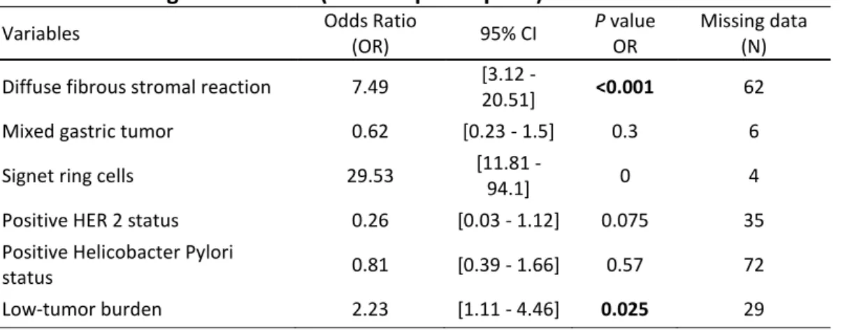
Table S6 histological variables (endoscopic biopsies) 1
Variables Odds Ratio
(OR) 95% CI
P value
OR
Missing data (N) Diffuse fibrous stromal reaction 7.49 [3.12 -
20.51] <0.001 62
Mixed gastric tumor 0.62 [0.23 - 1.5] 0.3 6
Signet ring cells 29.53 [11.81 -
94.1] 0 4
Positive HER 2 status 0.26 [0.03 - 1.12] 0.075 35 Positive Helicobacter Pylori
status 0.81 [0.39 - 1.66] 0.57 72 Low-tumor burden 2.23 [1.11 - 4.46] 0.025 29 2 3 4 5 6
Supplementary Table S7 (Tripod checklist) 7
8
Section/Topic Item Checklist Item Page
Title and abstract
Title 1 Identify the study as developing and/or validating a multivariable prediction model, the
target population, and the outcome to be predicted. 1 Abstract 2 Provide a summary of objectives, study design, setting, participants, sample size, predictors,
outcome, statistical analysis, results, and conclusions. 2 and 3 Introduction
Background and objectives
3a
Explain the medical context (including whether diagnostic or prognostic) and rationale for developing or validating the multivariable prediction model, including references to existing models.
4 and 5
3b Specify the objectives, including whether the study describes the development or
validation of the model or both. 5
Methods Source of data
4a Describe the study design or source of data (e.g., randomized trial, cohort, or
registry data), separately for the development and validation data sets, if applicable. 6 4b Specify the key study dates, including start of accrual; end of accrual; and, if
applicable, end of follow-up. 6
Participants
5a Specify key elements of the study setting (e.g., primary care, secondary care,
general population) including number and location of centres. 6 5b Describe eligibility criteria for participants. 6
5c Give details of treatments received, if relevant. Not
relevant
Outcome
6a Clearly define the outcome that is predicted by the prediction model, including how
and when assessed. 6 and 7
6b Report any actions to blind assessment of the outcome to be predicted. Not
relevant
Predictors
7a Clearly define all predictors used in developing or validating the multivariable prediction model, including how and when they were measured.
Table 1 and 2
7b Report any actions to blind assessment of predictors for the outcome and other
predictors. 7
Sample size 8 Explain how the study size was arrived at. 6
Missing data 9 Describe how missing data were handled (e.g., complete-case analysis, single
imputation, multiple imputation) with details of any imputation method. 8 Statistical
analysis methods
10a Describe how predictors were handled in the analyses. 8
10b Specify type of model, all model-building procedures (including any predictor
selection), and method for internal validation. 7 and 8 10d Specify all measures used to assess model performance and, if relevant, to
compare multiple models. 7 and 8
relevant
Results
Participants
13a
Describe the flow of participants through the study, including the number of participants with and without the outcome and, if applicable, a summary of the follow-up time. A diagram may be helpful.
10, figure 1
13b
Describe the characteristics of the participants (basic demographics, clinical features, available predictors), including the number of participants with missing data for predictors and outcome.
Table 1-3
Model development
14a Specify the number of participants and outcome events in each analysis. 10-12
14b If done, report the unadjusted association between each candidate predictor and outcome. Table 1 and Sup S1-S5 Model specification 15a
Present the full prediction model to allow predictions for individuals (i.e., all regression coefficients, and model intercept or baseline survival at a given time point).
Table 4
15b Explain how to the use the prediction model. Table 4
Model
performance 16 Report performance measures (with CIs) for the prediction model.
12, Figure 2
Discussion
Limitations 18 Discuss any limitations of the study (such as nonrepresentative sample, few events per
predictor, missing data). 16
Interpretation 19b Give an overall interpretation of the results, considering objectives, limitations, and
results from similar studies, and other relevant evidence. 13-16 Implications 20 Discuss the potential clinical use of the model and implications for future research. 15-17
Other information Supplementary information 21
Provide information about the availability of supplementary resources, such as study
protocol, Web calculator, and data sets. Not done Funding 22 Give the source of funding and the role of the funders for the present study. Not
relevant 1 2 3 4 5 6 7 8 9 10 11 12 13 14 15 16 17 18 19 20 21 22 23 24 25
REFERENCES: 1 2
1. Ferlay J, Soerjomataram I, Dikshit R, Eser S, Mathers C, Rebelo M, et al. Cancer 3
incidence and mortality worldwide: Sources, methods and major patterns in GLOBOCAN 4
2012. Int J Cancer. 2015;136(5):359–86. 5
6
2. Karimi P, Islami F, Anandasabapathy S, Freedman ND, Kamangar F. Gastric Cancer: 7
Descriptive Epidemiology, Risk Factors, Screening, and Prevention. Cancer Epidemiol 8
Biomarkers Prev. 2014;23(5):700–13. 9
10
3. Lauren P. The two histological main types of gastric carcinoma: diffuse and so-called 11
intestinal-type carcinoma. An attempt at a histo-clinical classification. Acta Pathol Microbiol 12
Scand. 1965;64:31–49. 13
14
4. Bosman, Fred T., Carneiro, Fatima, Hruban, Ralph H., Theise, Neil D. World Health 15
Organization (WHO) Classification of Tumours of the Digestive System. International Agency 16
for Research on Cancer (IARC) 4th Edition. 2010;45–79. 17
18
5. Japanese Gastric Cancer Association. Japanese classification of gastric carcinoma: 3rd 19
English edition. Gastric Cancer. 2011;14(2):101–12. 20
21
6. Borrmann R . Geschwülste Des Magens und Des Duodenums. Vol. I Berlin- Springer; 22
1926. 23
24
7. Siewert J, Stein H. Carcinoma of the gastroesophageal junction-Classification, 25
pathology and extent of resection. Dis Esophagus 1996;9-173-82. 26
27
8. Mastoraki A, Papanikolaou IS, Sakorafas G, Safioleas M. Facing the challenge of 28
managing linitis plastica–review of the literature. Hepatogastroenterology. 29
2009;56(96):1773–8. 30
31
9. Agnes A, Estrella JS, Badgwell B. The significance of a nineteenth century definition in 32
the era of genomics: linitis plastica. World J Surg Oncol. 2017;15(1). 33
34
10. Chang JM, Lara KA, Gray RJ, Pockaj BA, Wasif N. Clinical Outcomes after Surgery for 35
Linitis Plastica of the Stomach: Analysis of a Population Cancer Registry. Am Surg. 36
2017;83(1):23–9. 37
38
11. Blackham AU, Swords DS, Levine EA, Fino NF, Squires MH, Poultsides G, et al. Is Linitis 39
Plastica a Contraindication for Surgical Resection: A Multi-Institution Study of the U.S. 40
Gastric Cancer Collaborative. Ann Surg Oncol. 2016;23(4):1203–11. 41
42
12. Mariette C, Carneiro F, Grabsch HI, van der Post RS, Allum W, de Manzoni G, et al. 43
Consensus on the pathological definition and classification of poorly cohesive gastric 44
carcinoma. Gastric Cancer Off J Int Gastric Cancer Assoc Jpn Gastric Cancer Assoc. 2018 Aug 45
25. 46
13. Palli D, Bianchi S, Cipriani F, Duca P, Amorosi A, Avellini C, et al. Reproducibility of 1
histologic classification of gastric cancer. Br J Cancer. 1991;63(5):765. 2
3
14. Flucke U, Mönig SP, Baldus SE, Zirbes TK, Bollschweiler E, Thiele J, et al. Differences 4
between biopsy- or specimen-related Laurén and World Health Organization classification in 5
gastric cancer. World J Surg. 2002;26(2):137–40. 6
7
15. Amin MB, Edge SB, Greene FL, et al, eds. AJCC Cancer Staging Manual. 8th ed. New 8
York: Springer; 2017. 9
10
16. Jung K, Park MI, Kim SE, Park SJ. Borrmann Type 4 Advanced Gastric Cancer: Focus on 11
the Development of Scirrhous Gastric Cancer. Clin Endosc. 2016;49(4):336–45. 12
13
17. Pernot S. Signet-ring cell carcinoma of the stomach: Impact on prognosis and specific 14
therapeutic challenge. World J Gastroenterol. 2015;21(40):11428. 15
16
18. Golembeski CP, Genta RM. Signet-ring cell carcinoma in gastric biopsies: expecting 17
the unexpected. J Clin Pathol. 2013;66(2):136–9. 18
19
19. Piessen G, Amielh D, Messager M, Vinatier E, Leteurtre E, Triboulet JP, et al. Is 20
Pretreatment Endoscopic Biopsy a Good Predictor of Signet Ring Cell Histology in Gastric 21
Carcinoma? World J Surg. 2012;36(2):346–54. 22
23
20. Pedrazzani C, Marrelli D, Pacelli F, Di Cosmo M, Mura G, Bettarini F, et al. Gastric 24
linitis plastica: which role for surgical resection? Gastric Cancer. 2012;15(1):56–60. 25
26
21. Endo K, Sakurai M, Kusumoto E, Uehara H, Yamaguchi S, Tsutsumi N, et al. Biological 27
significance of localized Type IV scirrhous gastric cancer. Oncol Lett. 2012;3(1):94–9. 28
29
22. Mocellin S, Pasquali S. Diagnostic accuracy of endoscopic ultrasonography (EUS) for 30
the preoperative locoregional staging of primary gastric cancer. Cochrane Database Syst Rev. 31
2015;(2). 32
33
23. Liu Y, Chen K, Yang X-J. Endoscopic Ultrasound Guided Fine Needle Aspiration used in 34
diagnosing Gastric Linitis Plastica: Metastatic Lymph Nodes can be valuable targets. J 35
Gastroenterol Hepatol. 2018. 36
37
24. Morgant S, Artru P, Oudjit A, Lourenco N, Pasquer A, Walter T, et al. Computed 38
tomography scan efficacy in staging gastric linitis plastica lesion: a retrospective multicentric 39
French study. Cancer Manag Res. 2018;10:3825–31. 40
41
25. Han D-S, Suh Y-S, Kong S-H, Lee H-J, Choi Y, Aikou S, et al. Nomogram predicting long- 42
term survival after d2 gastrectomy for gastric cancer. J Clin Oncol Off J Am Soc Clin Oncol. 43
2012;30(31):3834–40. 44
45
26. Hirabayashi S, Kosugi S, Isobe Y, Nashimoto A, Oda I, Hayashi K, et al. Development 46
negative, locally advanced gastric cancer. Ann Oncol Off J Eur Soc Med Oncol. 1
2014;25(6):1179–84. 2
3
27. Zheng Z-F, Lu J, Wang W, Desiderio J, Li P, Xie J-W, et al. Development and External 4
Validation of a Simplified Nomogram Predicting Individual Survival After R0 Resection for 5
Gastric Cancer: An International, Multicenter Study. Ann Surg Oncol. 2018. 6
7
28. Narita Y, Kadowaki S, Oze I, Kito Y, Kawakami T, Machida N, et al. Establishment and 8
validation of prognostic nomograms in first-line metastatic gastric cancer patients. J 9
Gastrointest Oncol. 2018;9(1):52–63. 10
11
29. Kim SY, Yoon MJ, Park YI, Kim MJ, Nam B-H, Park SR. Nomograms predicting survival 12
of patients with unresectable or metastatic gastric cancer who receive combination 13
cytotoxic chemotherapy as first-line treatment. Gastric Cancer Off J Int Gastric Cancer Assoc 14
Jpn Gastric Cancer Assoc. 2018;21(3):453–63. 15
16
30. Messager M, Lefevre JH, Pichot-Delahaye V, Souadka A, Piessen G, Mariette C, et al. 17
The Impact of Perioperative Chemotherapy on Survival in Patients With Gastric Signet Ring 18
Cell Adenocarcinoma: A Multicenter Comparative Study. Ann Surg. 2011;254(5):684–93. 19
20 21
22