HAL Id: cea-00268875
https://hal-cea.archives-ouvertes.fr/cea-00268875
Submitted on 1 Apr 2008
HAL is a multi-disciplinary open access archive for the deposit and dissemination of sci-entific research documents, whether they are pub-lished or not. The documents may come from teaching and research institutions in France or abroad, or from public or private research centers.
L’archive ouverte pluridisciplinaire HAL, est destinée au dépôt et à la diffusion de documents scientifiques de niveau recherche, publiés ou non, émanant des établissements d’enseignement et de recherche français ou étrangers, des laboratoires publics ou privés.
HLA-G*0105N null allele encodes functional HLA-G
isoforms.
Magali Le Discorde, Caroline Le Danff, Philippe Moreau, Nathalie
Rouas-Freiss, Edgardo D Carosella
To cite this version:
Magali Le Discorde, Caroline Le Danff, Philippe Moreau, Nathalie Rouas-Freiss, Edgardo D Carosella. HLA-G*0105N null allele encodes functional HLA-G isoforms.. Biology of Reproduction, Society for the Study of Reproduction, 2005, 73 (2), pp.280-8. �10.1095/biolreprod.104.037986�. �cea-00268875�
280 DOI 10.1095/biolreprod.104.037986
HLA-G*0105N Null Allele Encodes Functional HLA-G Isoforms
1Magali Le Discorde,2Caroline Le Danff, Philippe Moreau, Nathalie Rouas-Freiss, and Edgardo D. Carosella
Service de Recherches en He´mato-Immunologie, CEA-DSV-DRM, Institut d’He´matologie, Hoˆpital Saint-Louis, Paris 75010, France
ABSTRACT
Expression of the nonclassical HLA class I antigen,HLA-G, is associated with immune tolerance in view of its role in main-taining the fetus in utero, allowing tumor escape, and favoring graft acceptance. Expressed on invasive trophoblast cells, HLA-G molecules bind inhibitory receptors on maternal T lympho-cytes and NK cells, thereby blocking their cytolytic activities and protecting the fetus from maternal immune system attack. The G gene consists of 15 alleles, including a null allele, HLA-G*0105N. HLA-G*0105N presents a single base deletion, pre-venting translation of both membrane-bound (HLA-G1) and full-length soluble isoforms (G5) as well as of the spliced HLA-G4 isoform. The identification of healthy subjects homozygous for thisHLA-G null allele suggests that the HLA-G*0105N allele may generate other HLA-G isoforms, such as membrane-bound HLA-G2 and -G3 and the soluble HLA-G6 and -G7 proteins, which may substitute for HLA-G1 and -G5, thus assuming the immune tolerogeneic function of HLA-G. To investigate this point, we cloned genomicHLA-G*0105N DNA and transfected it into an HLA-class I-positive human cell line. The results ob-tained indicated that HLA-G proteins were indeed present in HLA-G*0105N-transfected cells and were able to protect against NK cell lysis. These findings emphasize the role of the other HLA-G isoforms as immune tolerogeneic molecules that may also contribute to maternal tolerance of the semiallogenic fetus as well as tumor escape and other types of allogeneic tissue acceptance.
embryo, gene regulation, immunology, implantation, pregnancy
INTRODUCTION
Protection of the semiallogenic fetus against maternal immune recognition and attack has been attributed princi-pally to the high and almost unique expression of human leukocyte antigen-G (HLA-G) on trophoblast cells at the fetal-maternal interface [1]. The nonclassical HLA-G gene is characterized by alternative splicing that yields seven proteins, four membrane bound (HLA-G1 through -G4) and three soluble (HLA-G5 through -G7). Full-length HLA-G mRNA encodes the HLA-G1 protein, which has three ex-tracellular globular domains, one membrane-anchored do-main, and one intracytoplasmic domain. Exons 3 and/or 4
1Supported by the Commissariat a` l’Energie Atomique.
2Correspondence: Magali Le Discorde, Service de Recherches en
He´-mato-Immunologie, CEA-DSV-DRM, Institut d’He´matologie, Hoˆpital Saint-Louis, Paris 75010, France. FAX: 33 1 48 03 19 60;
e-mail: [email protected] Received: 19 November 2004. First decision: 22 December 2004. Accepted: 17 March 2005.
Q 2005 by the Society for the Study of Reproduction, Inc. ISSN: 0006-3363. http://www.biolreprod.org
may be deleted from the primary transcript, yielding the alternative mRNA HLA-G2, HLA-G3, and HLA-G4 forms. In addition, the insertion of introns 2 or 4 may generate soluble isoforms, such as HLA-G5 (the soluble full-length HLA-G1 counterpart), HLA-G6 (the soluble HLA-G2 counterpart), and HLA-G7 (the soluble HLA-G3 counter-part) (reviewed in [2]). The structures of both membrane-bound HLA-G1 and soluble HLA-G5 proteins are similar to those of classical class I proteins, consisting of three extracellular domains linked tob2-microglobulin (b2m).
Functional assays have demonstrated that both mem-brane-bound and soluble HLA-G proteins are able to inhibit NK cell and antigen-specific T-cell cytotoxicity [3, 4] and the proliferation of allogeneic T cells [5–7]. Moreover, sol-uble HLA-G is able to induce apoptosis of both activated CD81 T and NK cells [8, 9].
The identification of these seven HLA-G protein iso-forms suggested that HLA-G provides multiple functions at the immune-privileged sites where they are expressed, such as the fetal-maternal interface, the thymus, and the cornea [10–12]. On the other hand, in the absence of a given HLA-G isoform, this redundancy may serve to maintain the im-mune tolerogenic function provided by one or more of the other HLA-G isoforms.
The HLA-G gene consists of 15 alleles, including the
HLA-G*0105N null allele, which is characterized by a
sin-gle base-pair deletion in exon 3 [13]. This deletion of a single cytosine at codon 130 results in a gap in the open reading frame, causing a premature stop at either codon 189 (TGA) near the beginning of exon 4, which blocks trans-lation of HLA-G1 and -G5, or codon 297 (TAG) in exon 5, blocking the translation of G4. However,
HLA-G*0105N is able to maintain translation of both the
mem-brane-bound HLA-G2 and -G3 proteins and the soluble HLA-G6 and -G7 proteins, in all of which exon 3, con-taining the deletion, is removed by alternative splicing (Fig. 1).
The HLA-G*0105N null allele has been described in healthy adults whose own gestations and deliveries were normal (without complications). The detection of individ-uals who are genetically homozygous for the
HLA-G*0105N allele suggests that the HLA-G isoforms encoded
by this allele possess functions able to compensate for the absence of both the HLA-G1 and -G5 proteins and to matain the immune privileged status of the fetal-maternal in-terface [14, 15].
Based on the previously mentioned observations and on a previous study showing that selection may have increased the frequency of the HLA-G*0105N allele [16], the goal of the present study was to demonstrate that in the absence of the HLA-G1, -G5, and -G4 isoforms, the other HLA-G iso-forms present in HLA-G*0105N can indeed provide a pro-tective function.
281
HLA-G∗0105N ENCODES FUNCTIONAL PROTEINS
In the present work, we investigate the structural and functional aspects of HLA-G*0105N proteins. We con-structed the HLA-G*0105N null allele genomic DNA in the pcDNA3.1 expression vector, which enabled us to study the expression and function of its proteins. The aim of this work was to acquire more data on spliced HLA-G isoforms in general and to understand how they could compensate for the absence of full-length HLA-G proteins.
MATERIALS AND METHODS
In Vitro Site-Directed Mutagenesis
In vitro site-directed mutagenesis was carried out using the Stratagene QuickChange XL site-directed mutagenesis kit, which allows site-specific mutation in double-stranded plasmids. The plasmid DNA template (50 ng) HLA-G*010102-pcDNA3.1, isolated from the dam1JM109 Escherichia
coli strain, was replicated by PfuTurbo DNA polymerase (2.5 U) using two synthetic primers, G.Null S and G.Null AS (125 ng each), consisting of 15 bases on both sides of theDC mutation. The oligonucleotide primers, each complementary to opposite strands of the HLA-G*010102 insert, were extended during temperature cycling (958C for 1 min; 18 cycles at 958C for 50 sec, 608C for 50 sec, 688C for 15 min) and terminated by 7 min at 688C. Following temperature cycling, amplification reactions were cooled and digested for 1 h at 378C using Dpn I restriction enzyme to eliminate the nonmutated parental supercoiled double-stranded DNA. At this step, genomic HLA-G*010102-pcDNA3.1 was replaced by genomic HLA-G*0105N-pcDNA3.1. The mutated DNA was then transformed in XL10-Gold ultracompetent cells.
Cell Lines, Transfection, and Cultures
Melanoma M8 (kindly provided by F. Jotereau, INSERM U211, Nantes, France) and histiocytic lymphoma U937 (American Type Culture Collection) human cell lines are HLA-class II negative; HLA-A, -B, -C, and -E positive (HLA-A1, -A2, -B12, and B40/male); but HLA-G nega-tive. The NKL NK cell line (kindly provided by E.H. Weiss, Department of Anthropology and Human Genetics, Munich, Germany) [17, 18] and the YT2C2 NK [19, 20] subclone (kindly provided by P. Paul, Hospital Saint-Louis, Paris, France) have been previously described. All cells were maintained in RPMI 1640 plus Glutamax medium supplemented with 1
mg/ml gentamicin, fungizone, and 20% inactivated fetal calf serum. NKL
cells were concurrently cultured in the presence of 50 U/ml of interleukin-2 (ILinterleukin-2) (Sigma-Aldrich, St. Quentin Fallavier, France) and boosted to 100 U/ml IL2 the day before the cytotoxicity assays. M8 and U937 transfec-tants were selected using 200 mU/ml hygromycin B (Invitrogen, Cergy Pontoise, France). HLA-G*0105N inserted into the pcDNA3.1/Hygro (-) expression vector was transfected into M8 and U937 cells. We worked with bulk transfectants since absence of the HLA-G1 membrane-bound isoform prevents the selection of positive cloned cells by cell sorting. The other transfectants, M8-HLA-G1 through -G4 and M8-HLA-G*010102, have been previously characterized [21, 22].
Classical and Real-Time RT-PCR Analysis
Reverse transcription-polymerase chain reaction (RT-PCR) was carried out to validate HLA-G expression and to determine which isoforms were expressed in our new M8-HLA-G*0105N cell line. Total RNA was purified by RNA-WIZ reagent (Ambion, Huntingdon, U.K.) according to the man-ufacturer’s instructions. The cDNA was synthesized on 5mg RNA using oligo (dT) primers (Invitrogen) and MMLV-reverse transcriptase (Life Technologies, Cergy-Pontoise, France) for 1 h at 428C. Heating for 8 min at 958C stopped the reaction.
Real-time PCR (ABI PRISM 7000 SDS) was used to determine the quantities of HLA-G transcripts in transfected cell lines. Duplex PCR was carried out for 40 amplification rounds in the presence of TaqMan Uni-versal PCR Master Mix, using the TaqMan Assay Reagent and GAPDH as an endogenous control (probe with VIC reporter and TAMRA er), HLA-G-specific probe (200 nM, FAM reporter, and TAMRA quench-er), and HLA-G-specific primers (300 mM, Q biogen).
Classical PCR was carried out according to the 13th HLA Workshop [21] procedure, using a Perkin-Elmer DNA thermal cycler in a total vol-ume of 100ml containing 2 ml of the RT reaction product, 200 mM of each dNTP (Invitrogen), 100 ng of each primer, 10ml of 103 Taq buffer, and 2.5 U of Taq polymerase (Applied Biosystems, Courtabœuf, France). All G isoforms were amplified with G.257F/G.1004R primers,
HLA-G2 with G.-3/G.1216 primers, HLA-G3 with G.-3-4/G.1216 primers, and HLA-G5 with G.257/G.i4. These PCR products were revealed by radio-active hybridization with G.927 oligonucleotide, except for HLA-G5 am-plification, which was revealed using the G.R probe.
Vector Construction and Oligonucleotides
In vitro site-directed mutagenesis was accomplished from the genomic HLA-G*010102 sequence inserted into pcDNA3.1/Hygro (-) expression vector (Invitrogen). The HLA-G*010102 sequence begins in the promoter at the XbaI site (460 nucleotides upstream from ATG) and continues to the EcoRV site in the 39UT [21]. The cytosine deletion was introduced using two primers, G.Null S (59-GCCCTGAACGAGGACTGCGCTCCT GGACCG) and G.Null AS (59-CGGTCCAGGAGCGCAGTCCTCGTTC AGGGC).
Real-time PCR was carried out with the G.948 forward primer located in exon 5 (59-CTGGTTGTCCTTGCAGCTGTAG), the G.1002 reverse primer located in both sides of exon 5 and exon 6 (59-CCTTTTCAATCTG AGCTCTTCTTTCT), the G.971F internal probe (59-CACTGGAGCTGC GGTCGCTGCT), the FAM reporter, and the TAMRA quencher.
For classical PCR, all HLA-G transcripts were amplified from exon 2 by G.257 forward (59-GGAAGAGGAGACACGGAACA) to exon 5/6 by G.1004 reverse (59-CCTTTTCAATCTGAGCTCTTCTTT). HLA-G2 and HLA-G3 transcripts were amplified, respectively, from exons 2/4 by G.-3 forward (59-ACCAGAGCGAGGCCAACCCC) and from exons 2/5 by G.-3-4 forward (59-ACCAGAGCGAGGCCAAGCAG) to the 39UT sequence by G.1216 reverse (59-GACGGAGACATCCCAGCCCC). Transcripts cor-responding to soluble forms were amplified from exon 2 by G.257 up to intron 4 by G.i4 reverse (59- CTGGGAAAGGAGGTGAAGGT). Internal probes were localized in exon 5 with G.927 reverse (5 9-CCAGCAAC-GATACCCATGAT) or in exon 2 with G.R (5 9-GGTCTGCAGGTT-CATTCTGTC).
Monoclonal Antibodies
The following murine antibodies were used: 4H84, IgG1 recognizing
a1 domain common to all HLA-G isoforms (provided by Michael
Mc-Master [23]); MEM-G/04, IgG1 recognizing the denatured form of HLA-G1, -G2, -G5, and -G6 (provided by Vaclav Horejsi, Prague, Czech Re-public); MEM-G/09, IgG1 recognizing the HLA-G molecules associated with beta2-microglobulin (HLA-G1 and -G5) [24] (Exbio, Prague, Czech Republic); 5A6G7, IgG1 antibody against intron 4 of soluble HLA-G5 and -G6 isoforms made in our laboratory [25].
Immunocytochemical Analysis
Cells were grown on LabTech slides (Dutscher, Brumath, France), fixed in acetone, and stored at2808C until evaluation of the expression of HLA-G and class I protein by immunohistochemistry, using the Ultra-Tech HRP Streptavidin-Biotin Universal Detection System (Immunotech Coulter, Roissy, France) as previously described [12]. Briefly, endogenous perox-idase was blocked with 3% H2O2, and antibodies were diluted in buffer
containing 0.1% saponin.
Flow Cytometry Assays
The flow cytometry technique was used to analyze cell-surface HLA-G protein expression. Transfected cells were phenotyped according to stan-dard procedure by incubating them with the primary anti-HLA-G antibody MEM-G/09 in PBS 2% heat-inactivated fetal calf serum for 30 min at 48C. Cells were subsequently stained with an F(ab9)2goat anti-mouse IgG
an-tibody conjugated with phycoerythrin (Immunotech Coulter).
Cell-Surface Protein Biotinylation
Cell-surface proteins of viable M8 transfected cells were biotinylated as previously described [4]. Cells were first washed twice with PBS and treated with sulfo-NHS-LC-biotin (Pierce, Rockford, IL; 200 mg/ml of PBS) for 4 min at room temperature. The cells were washed twice with PBS, treated with 50 mM glycine for 5 min, and again washed twice with PBS. Dead or dying (nonadherent) cells were removed by washing. Cells were then detached and collected in tubes, then washed in PBS five times at 48C. Cells in the last pellet were readied for analysis by immunopre-cipitation.
Immunoprecipitation of Cell-Surface Proteins
Cells were lysed in 1 ml of lysis buffer (0.5% CHAPS, 50 mM Tris-HCl). After centrifugation at 14 000 rpm for 30 min at 48C, biotinylated
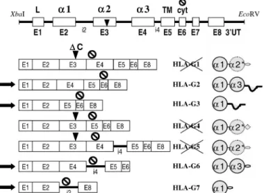
FIG. 1. Schematic drawing of theXbaI - EcoRV genomic DNA sequence of G. The G*0105N sequence corresponds to that of HLA-G*010102, except for the deletion of cytosine, indicated by an arrow in exon 3 (DC). The HLA-G gene consists of eight exons (E1–E8), correspond-ing, respectively, to the peptide leader domains (L),a1, a2, and a3, the transmembrane domain (TM), and the intracytoplasmic domain (cyt). Ex-ons were separated by intrEx-ons (i). TheHLA-G*0105N gene putatively en-codes the membrane-bound HLA-G2, -G3, and soluble HLA-G6 and -G7 proteins, in all of which exon 3, which contains the deletion, is deleted. HLA-G1, -G4, and -G5 might consist of the leader peptide, the complete a1 domain, the first half of the a2 domain, and a premature end in the following exon.
FIG. 2. DNA sequencing electropherogram of theHLA-G*010102 allele (A) and before deletion of the cytosine in exon 3 and after directed mu-tagenesis, yielding theHLA-G*0105N allele (B).
proteins were precipitated from the supernatant by precipitation with 100
ml of 50% streptavidin-agarose beads (Bio-Rad, Marnes la Coquette,
France) for 1 h at 48C. Nonbiotinylated proteins in the supernatant of the first centrifugation were taken to be the intracellular material and retained for Western blot analysis. Bead-bound biotinylated proteins were washed in three buffers (buffer 1: 0.1% CHAPS, 150 mM NaCl 40 mM, 0.05% NaN3, and 20 mM Tris-HCl pH7.5; buffer 2: 0.05% CHAPS, 0.1% SDS,
300 mM NaCl, and 10 mM Tris-HCl pH 8; buffer 3: 0.1% CHAPS and 20 mM Tris-HCl pH 7.4). Biotinylated proteins were then resuspended in 60ml Laemmli buffer 23, and 40 ml of the nonbiotinylated proteins were mixed with 20ml Laemmli buffer 63. These two samples were boiled for 5 min and analyzed by Western blot.
Deglycosylation Treatment
Forty microliters of M8 transfectants lysed in 0.5% CHAPS; 50 mM Tris-HCl pH 7.5 were deglycosylated, using peptide-N-glycosidase F from flavobacterium, according to Sigma’s instructions (Sigma-Aldrich). The deglycosylated proteins were examined by Western blot analysis.
Western Blot Analysis
Proteins were loaded and separated on 12% Tris-glycine-SDS poly-acrylamide gels and electroblotted to nitrocellulose membranes (Hybond, Amersham Biosciences, Orsay, France). The membranes were probed with an HLA-G-specific antibody (4H84, MEM-G/04), then revealed with per-oxidase-conjugated sheep anti-mouse IgG Ab (Sigma-Aldrich). Western blots were developed by chemiluminescence (Amersham).
Cytotoxicity Assays
The cytolytic activity of the NKL cells and YT2C2 cells used as ef-fectors (E) was assessed in 4-h51Cr release assays in which the effector
cells were mixed with 53 103 51Cr-labeled transfected-M8 or U937 (T)
(100mCi51Cr sodium chromate; 1 Ci5 37 Gbq; Amersham) at various
E:T ratios, as previously described [4]. The percentage specific lysis was calculated as follows: % specific lysis5 [(cpm experimental 2 cpm spon-taneous release)/(cpm maximum release2 cpm spontaneous release)] 3 100. Spontaneous release was determined by incubation of labeled target cells in RPMI 1640 medium supplemented with 10% FCS. Maximum release was determined by solubilizing target cells in 0.1 N HCl. In all experiments, spontaneous release was less than 10% of maximum release.
RESULTS
Construction of HLA-G*0105N Null Allele Genomic DNA
To study proteins encoded by the HLA-G null allele, we generated a DNA sequence identical to HLA-G*0105N in the pcDNA 3.1 expression vector. In vitro site-directed mu-tagenesis of HLA-G*010102 genomic DNA allowed us to delete the cytosine at position 815 from ATG (exon 1), yielding the genomic DNA of the HLA-G*0105N allele. We used the HLA-G*010102 rather than the HLA-G*010101 reference sequence since HLA-G*010102 and -G*0105N have identical DNA sequences with the exception of the cytosine deletion at codon 130 [14]. The XbaI-EcoRV
HLA-G*010102 fragment linked into the pcDNA3.1 vector
used as the template is represented in Figure 1, and the electropherogram of DNA sequences before and after di-rected mutagenesis demonstrating deletion of the cytosine in the newly synthesized DNA is shown Figure 2, the re-maining sequence being identical. The genomic
HLA-G*0105N DNA presented a deletion at codon 130 (CTG→
TGC), which generated a stop codon 171 nucleotides fur-ther on at codon 189 (GTG → TGA) in exon 4 or 495 nucleotides further on at codon 297 (GTA→TAG) in exon 5. At this step, we had obtained a copy of the
HLA-G*0105N allele of 3932 nucleotides, beginning 460 bp
up-stream from exon 1 and ending 541 bp downup-stream from exon 8.
Pattern of mRNA Expression in HLA-G*0105N-Transfected Cells
Real-time RT-PCR was carried out on mRNA prepared from M8 cells transfected with both genomic
HLA-G*0105N and HLA-G*010102 as well as mRNA from
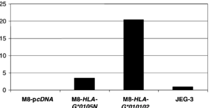
JEG-3 cells (Fig. JEG-3). These results revealed that higher levels of HLA-G transcripts were produced in both
M8-HLA-G*0105N and M8-HLA-G*010102 than in the JEG-3 cells
(JEG-3 is taken as a reference since it is one of the rare cell lines that constitutively expresses HLA-G proteins). HLA-G transcript levels measured in the M8-transfected cells varied according to the particular HLA-G sequence
283
HLA-G∗0105N ENCODES FUNCTIONAL PROTEINS
FIG. 3. Results of real-time reverse transcription-polymerase chain re-action (RT-PCR) analysis showing relative quantities of HLA-G transcripts in M8-HLA-G*0105N, M8-HLA-G*010102, and M8-pcDNA compared with that of JEG-3 cells (assigned a value of 1).
FIG. 4. Reverse transcription-polymerase chain reaction (RT-PCR) and Southern blot characterization ofHLA-G mRNA isoforms in M8-HLA-G*0105N (a–d). PCR amplifications are carried out using pan-HLA-G primers G.257F/G.1004R (a, e, f); primer set G.-3F/G.1216R, specific for HLA-G2 (b); primer set G.-3–4F/G.1216R, specific forHLA-G3 (c); and primer set G.257F / G.i4R, specific for both HLA-G5 and -G6 (d). HLA-G*010102 (e) and M8-pcDNA (f) are, respectively, the positive and negative controls. Southern blots a, b, c, e, and f were hybridized with G.927R (exon 5), and blot d was hybridized with G.R (exon 2) probes.
transfected, lower levels being found for
M8-HLA-G*0105N than for M8-HLA-G*010102 (Fig. 4). To
deter-mine the mRNA pattern of M8-HLA-G*0105N, we carried out HLA-G-specific RT-PCR and radioactive hybridization reactions comparing G*0105N with
M8-HLA-G*010102. The specific G.927 probe, located in exon 5,
was used to hybridize all PCR products except those of G.257/G.i4, which were hybridized using the G.R probe, located in exon 2. The G.257F/G.1004R primers disclosed four bands, corresponding to the principal mRNAs:
HLA-G5 (889 bp), -G1 (767 bp), -G2 and -G4 (491 bp), and -G3 (215 bp) (Fig. 4a). HLA-G2 was amplified using the
G.-3/G.1216 primers (627 bp) (Fig. 4b) and HLA-G3 with the G.-3-4/G.1216 primers (341 bp) (Fig. 4c). The G.257/ G.i4 primers allowed amplification of HLA-G5 (765 bp) and HLA-G6 (489 bp) (Fig. 4d) . We thus confirmed that all HLA-G mRNA had been transcribed in
M8-HLA-G*0105N cell line.
HLA-G Proteins Are Translated in HLA-G*0105N-Transfected Cells
The proteins synthesized by the HLA-G*0105N allele have been deduced from its nucleotide sequence [14]. Ac-cording to these authors, HLA-G isoform amino-acid se-quences deduced from the HLA-G*0105N DNA sequence would yield the complete membrane-bound HLA-G2 and -G3, as well as the soluble HLA-G6 and -G7 isoforms.
HLA-G1, -G4, and -G5 undergo a reading frame shift, and the corresponding proteins are prematurely truncated in the null allele.
To detect HLA-G proteins that were expressed by
M8-HLA-G*0105N in our experiments, we carried out
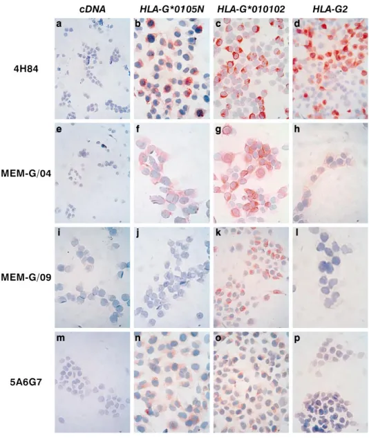
immu-nocytochemistry assays on permeabilized cells. As may be seen in Figure 5, M8-HLA-G*0105N cells were positively stained by 4H84, an antibody that targets the a1 domain present in all HLA-G isoforms (Fig. 5b); by MEM-G/04, an antibody that recognizes the a3 domain (Fig. 5f); and by 5A6G7, an antisoluble antibody against HLA-G5 and -G6) (Fig. 5n). As expected, the presence of HLA-G1 or -G5 was not detected in M8-HLA-G*0105N with the MEM-G/09 antibody (Fig. 5j). All these anti-HLA-G antibodies had positively bound M8-HLA-G*010102, a genomic HLA-G sequence in which HLA-G1 is predominantly ex-pressed, but one would expect the other isoforms to be present (Fig. 5, c, g, k, and o). The HLA-G2 isoform was stained by both the 4H84 and the MEM-G/04 antibodies in M8-HLA-G2 cells (Fig. 5, d and h). Collectively, these im-munocytochemical analyses revealed the presence of HLA-G2 and -G6 isoforms in M8-HLA-G*0105N.
Using flow cytometry and the MEM-G/09 antibody, we confirmed that whereas M8-HLA-G*0105N did not express HLA-G1 protein at the cell surface, M8-HLA-G*010102 did (Fig. 6). Because of the unavailability of antibodies able to bind HLA-G2 and HLA-G3 in the flow cytometry assay, we were unable to identify these membrane-bound iso-forms, which could have been translated by
M8-HLA-G*0105N cells.
Characterization of HLA-G Proteins in HLA-G*0105N-Transfected Cells
In succeeding experiments, we attempted to characterize the HLA-G proteins that had been translated in
G*0105N cells, using G*010102 and M8-HLA-G2 as positive controls. We used immunoprecipitation of
biotinylated cell-surface proteins to achieve separation of membrane-bound and intracytoplasmic proteins. The results revealed membrane-bound HLA-G2 protein on
HLA-G2 (bands at 30 and 27 kDa) and HLA-G1 protein on M8-HLA-G*010102 (one band at 43 kDa), but no HLA-G
mol-ecules could be detected in M8-HLA-G*0105N by this method (Fig. 7A). However, study of the intracellular
pro-FIG. 5. Immunocytostaining of M8 trans-fectants with anti-HLA-G antibodies 4H84 (a–d), MEM-G/04 (e–h), MEM-G/09 (i–l), and 5A6G7 (m–p). All antibodies are neg-ative in the control M8-pcDNA (a, e, i,
m). The HLA-G1, -G2, -G5, -G6
anti-body MEM-G/04 is positive in allHLA-G transfected cells (f–h). The antiHLAG1, -G5 antibody MEM-G/09 stained M8-HLA-G*010102 (k) but not M8-HLA-G*0105N (j) or M8HLAG2 (l). The antiHLAG5, -G6 antibody 5A6G7 stained M8-HLA-G*0105N (n) and M8-HLA-G*010102 (o) but not M8-HLA-G2 (p). Original magnifi-cation3200.
FIG. 6. Detection by flow cytometry analysis of the HLA-G1 isoform on M8-HLA-G*010102 but not on M8-HLA-G*0105N. Cells were labeled by indirect immunofluorescence with MEM-G/09 mAb (bold profiles) and an isotope-matched control Ab (light profiles). M8-pcDNA and M8-HLA-G1 cells were used as controls.
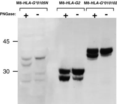
teins revealed the presence of HLA-G isoforms. We ob-tained three bands, one at 30 kDa and one at 27 kDa, that were identical to those produced by M8-HLA-G2, plus one located above them, at 35 kDa, that has never been char-acterized (Fig. 7B). To determine whether this latter band corresponded to hyperglycosylated HLA-G2, we treated the M8-HLA-G*0105N cells with PNGase F, an enzyme that catalyzes the removal of N-linked oligosaccharide chains from glycoproteins (Fig. 8). This enzyme treatment
re-vealed a smaller band just below that of the mature protein, corresponding to the deglycosylated protein. This finding indicated that M8-HLA-G*0105N had indeed translated some glycoproteins, but the molecular weight obtained for the 35-kDa protein did not correspond to any known HLA-G isoform. Staining with 4H84 and MEM-HLA-G/04 antibodies allowed us to determine that at least thea1 and a3 domains were present in this protein, which remains to be charac-terized.
285
HLA-G∗0105N ENCODES FUNCTIONAL PROTEINS
FIG. 7. The HLA-G*0105N allele produced intracytoplasmic HLA-G proteins. Cell-surface proteins are biotinylated and separated from the nonbiotinylated inside proteins. Immunostaining with both 4H84 and MEM-G/04 mAbs revealed the intracellular presence of HLA-G proteins (B) but not on the cell surface (A). The mechanism providing the addi-tional smaller band for HLA-G2 (i.e., band at 27 kDa) remains to be
determined. FIG. 8. HLA-G*0105N isoforms are translated as glycoproteins. Lysate from M8 transfectants were treated with PNGase F allowing visualization of mature and deglycosylated proteins. Western blots were stained with 4H84 mAb. PNGase F treatment removed oligosaccharides from the gly-coproteins. Revelation of two bands (glycosylated and not) indicated that this treatment was not complete. M8-HLA-G*0105N presents two weak bands, at 30 and 27 kDa (representing glycosylated and nonglycolysated HLA-G2) and one unidentified band at 35 kDa, which migrated at 31 kDa after PNGase F treatment.
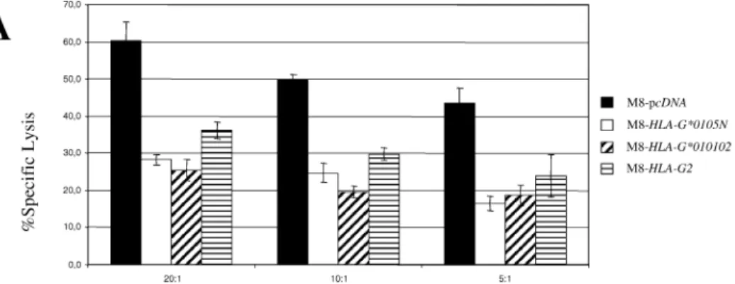
HLA-G*0105N-Transfected Cells Are Protected from NK Cell-Mediated Cytotoxicity
To determine whether the HLA-G*0105N allele translat-ed functional HLA-G isoforms, cytotoxic assays were car-ried out confronting either the NKL or the YT2C2 cell line [17, 19] used as the effector with the M8 or U937 trans-fectant cells used as the target (Fig. 9). When stimulated with interleukin-2, the NKL human NK cell line retained most of the original features of NK cells, including very similar proliferative responses and natural killing function. While YT2C2 express only KIR2DL4, NKL cells express both ILT2 and KIR2DL4 inhibitory receptors [18, 26], which are implicated in the NK-cell cytotoxicity pathways, by binding to HLA-G molecules. The lytic activity of NKL against M8-HLA-G*0105N was compared with its lytic ac-tivity against M8-HLA-G*010102 cells, which at least ex-press HLA-G1, as well as against M8-HLA-G2 cells, which express only the HLA-G2 isoform, and against M8-pcDNA, used as the HLA-G-negative control. Cytotoxicity experi-ments showed that in addition to M8-HLA-G2, both geno-mic M8-HLA-G*0105N and M8-HLA-G*010102 were pro-tected from NKL lysis. Lysis of M8-transfectants was re-duced by 50% compared with that of the M8-pcDNA con-trol cell line. Inhibition of cytotoxicity was also obtained using YT2C2 cells, which only express the HLA-G-specific receptor against U937-transfectants.
DISCUSSION
After our study of the tolerogeneic role of HLA-G in maternal-fetal tolerance [27, 28], we designed experiments to identify the structural and functional characteristics of proteins generated by the HLA-G*0105N allele. First, we used site-directed mutagenesis to construct the
HLA-G*0105N from the nucleotide sequence of HLA-G*010102
and transfected it into the M8 human cell line. Then we characterized the transcripts and proteins generated by the
HLA-G*0105N gene before analyzing the functional
capac-ity of the isoforms produced.
Although the deletion of one base in exon 3 of
HLA-G*0105N disrupts the reading frame, it should have no
ef-fect on transcription of the primary transcript or on its
al-ternative splicing [14]. Indeed, in our RT-PCR analysis, the
HLA-G mRNA profile was the same in both M8-HLA-G*0105N- and M8-HLA-G*010102-transfected cells,
indi-cating the presence of all HLA-G mRNA transcripts nor-mally found. Thus, deletion of the cytosine in codon 130 did not block the transcription and splicing mechanisms in the null allele. Nevertheless, real-time and classical RT-PCR showed that HLA-G mRNA levels were lower in
M8-HLA-G*0105N than in M8-HLA-G*010102 cells, and
clas-sical PCR revealed that the HLA-G1/HLA-G2 mRNA ratio was lower in G*0105N than in
M8-HLA-G*010102. This discrepancy in the expression level of
HLA-G genotypes and isoforms has already been reported for alleles other than the null allele in pathological preg-nancies in which altered HLA-G transcription was associ-ated with certain HLA-G genotypes [29, 30] as well as in a study of the role of HLA-G alleles in determining the soluble HLA-G protein plasma level [31]. Another expla-nation is that nonsense-mediated decay, a eukaryotic reg-ulatory process that degrades mRNA with premature ter-mination codons, could be responsible for the higher deg-radation of HLA-G1 transcripts in cells expressing the null allele compared with other alleles [32].
Furthermore, the higher levels of mRNA in transfected cells compared with JEG-3 cells could be explained by the fact that 1) in transfected cells, HLA-G is ligated in the pcDNA3.1 plasmid, which possesses the CMV promoter and is thus able to influence and perhaps increase HLA-G mRNA expression, and 2) HLA-G*010102 has been de-scribed as a much higher ‘‘HLA-G secreting’’ allele than either HLA-G*0105N or HLA-G*010103 (carried by JEG-3 cells [JEG-3JEG-3]) [JEG-31].
Finally, although mRNA levels are higher in M8-trans-fected cells than in JEG-3 cells, this cannot be correlated with either the quality or the quantity of the corresponding
FIG. 9. M8-HLA-G*0105N is able to in-hibit NKL cell cytolytic activity. Chromium release assays were carried out using M8-or U937-pcDNA, M8- M8-or U937-HLA-G*0105N, M8- or U937-HLA-G*010102, or M8-HLA-G2 as target cells and NKL cell line (A) or YT2C2 cell line (B) as ef-fectors. Results are expressed as the per-centage lysis recorded in a 4-h51Cr-release
assay. Values represent means of triplicate 6 SD. This experiment is representative of five distinct experiments.
translated isoform proteins. Thus, the mRNA level is lower in M8-HLA-G*0105N cells than in M8-HLA-G010102 cells (Fig. 3), and both M8 transfectants are equally pro-tected from NKL-mediated cytolysis (Fig. 9A).
Since the presence of HLA-G mRNA had been demon-strated in M8-HLA-G*0105N, we also looked for HLA-G proteins in it. For this purpose, HLA-G*0105N-transfected cells were immunostained with specific HLA-G antibodies targeting either all HLA-G proteins or specific isoforms. As expected from the deletion in HLA-G*0105N, neither the HLA-G1 nor the HLA-G5 isoform was detected in
M8-HLA-G*0105N since these cells were not stained by the
MEM-G/09 mAb, which is specific for G1 and HLA-G5. The absence of HLA-G5 and of shed HLA-G1 was confirmed by a specific ELISA (data not shown). Interest-ingly, the other mAbs that recognized either all HLA-G isoforms (4H84), both G5 and -G6 (5A6G7), or HLA-G1, -G2, -G5, and -G6 (MEM-G/04) were positive in
M8-HLA-G*0105N, demonstrating that in the absence of both
HLA-G1 and -G5, other HLA-G isoforms are produced. These isoforms may correspond to at least the HLA-G2 and/or HLA-G6 proteins. In support of this observation, previous immunohistochemical analyses of placentas ho-mozygous for HLA-G*0105N presented negative staining with an anti-HLA-G1, -G5 antibody (87G) and positive staining with an anti-HLA-G1, -G2 antibody (15C6) [15]. These results confirm that HLA-G proteins are indeed translated in HLA-G*0105N cells.
To further characterize the HLA-G isoforms produced by M8-HLA-G*0105N cells, we carried out
immunoprecip-itation and Western blot experiments. Under our experi-mental conditions, proteins at 35 and 30 kDa were detected within such cells but not on the cell surface. However, we cannot exclude that HLA-G proteins are secreted by these cells and that they exercise biological effects. To address this point, we carried out experiments aimed at analyzing the role of the HLA-G proteins generated by
M8-HLA-G*0105N with respect to NK function and tested their
ca-pacity to inhibit NK cell lysis.
Considered together, these results demonstrate that non-HLA-G1/-G5 isoforms of the HLA-G*0105N allele partic-ipate in the inhibition of NK cells. This inhibition is per-haps not mediated by HLA-E or other HLA class I mole-cules, since HLA-G*0105N-transfected cells were protected from lysis by the NK-like clone Y2T2C2. Indeed Y2T2C2 expresses the HLA-G-specific receptor KIR2DL4, but not HLA-A-, -B-, -C-, and -E-specific KIRs.
The HLA-G*0105N allele is estimated to have appeared 18 000 yr ago, leading to the conclusion that it had been positively selected during evolutionary history [16]. A high incidence of the null allele has notably been reported in Africa [34]. A possible explanation for this selection is that it may be due to the high incidence of intrauterine patho-gens among the Africa population, which therefore has to maintain an efficient immune system to eliminate uterine infections [16]. One would expect that the absence of HLA-G1 and -G5 in HLA-G*0105N individuals would serve to maintain the immunocompetence of maternal NK and T cells at the maternal-fetal interface [15, 16]. Weighing the trade-off between increased risk of miscarriage due to the
287
HLA-G∗0105N ENCODES FUNCTIONAL PROTEINS
absence of HLA-G1 and -G5 but low risk of uterine infec-tion and successful outcome of pregnancy but high risk of womb infection, G*0105N was selected over HLA-G1, -G5, thus eradicating intrauterine pathogen contami-nation despite the fact that the null genotype is a contrib-uting factor in spontaneous abortion [35, 36]. Our results suggest that HLA-G*0105N would be of interest for pur-poses other than merely preventing interactions between HLA-G1, -G5 and inhibitory NK receptors since HLA-G2 has maintained that function. The advantages of the selec-tion of this allele must be further investigated.
The emergence of the HLA-G*0105N homozygous gene is not the only condition under which HLA-G protein ex-pression has been found to be impaired. As for classical HLA-class I molecules, both HLA-G1 and -G5 contain three globular domains associated with b2m and present peptides. Their conformation renders these HLA molecules TAP andb2m dependent. Therefore, in TAP- or b2m-de-ficient individuals, NK-mediated reactions may still occur, and NK cells may retain the ability to reject allogeneic and autologous HLA-class I-negative cells. However, studies of such individuals showed that HLA-class I-deficient cells remain protected from NK lysis. Notably, there are other situations in which expression of both HLA-G1 and -G5 isoforms would also be altered without negatively affecting the outcome of pregnancy. One example is the survival of homozygous TAP-negative children in whom one would expect HLA-G1/-G5 placental expression to be impaired. Indeed, expression of these two isoforms as a trimolecular
a-chain/b2m/peptide complex is TAP dependent. In the
fe-tuses of such TAP-negative children, the other HLA-G iso-forms, whose expression may be independent of peptide loading via TAP, may substitute for the loss of HLA-G1/-G5, thus contributing to the survival of these fetuses [37]. In another study, functional analyses indicated that TAP2/2 NK cells were unable to kill autologous class
I-negative cells. This inhibition was attributed to an unknown inhibitory receptor capable of binding ligand expressed by targeted autologous class I-negative cells, thereby down-regulating NK cell cytotoxicity [38]. Considered together, these studies suggest that still-unknown mechanisms pre-vent an attack against autologous normal cells that express insufficient quantities of HLA-class I molecules. Therefore, the HLA-G2, -G3, -G6, and -G7 isoforms, in which cell processing and transport are probably peptide independent, might be candidate molecules to compensate for the loss of HLA class I antigens, including HLA-G1 and -G5. The fact that TAP2/2 NK cells express an unidentified receptor
sug-gests that this unidentified inhibitory receptor is specific for spliced HLA-G isoforms. These interpretations could be ex-trapolated to tumor cells, which often present down-regu-lation of classical and nonclassical HLA class I molecules. Expression of the TAP andb2m-independent spliced HLA-G isoforms (HLA-HLA-G2 through -HLA-G7) observed in some tu-mors may constitute a new escape mechanism from NK cell-mediated lysis [39].
Finally, our study provided information indicating that
HLA-G*0105N does not encode the complete HLA-G1 or
HLA-G5 isoforms but does encode functional HLA-G pro-teins able to inhibit NK cell cytolysis. Thus, although the biological functions of HLA-G1 and HLA-G5 proteins are abrogated, the other isoforms can assume their roles. Two different effectors were used against M8-transfectant cells: the NKL and YT2C2 cells. Since NKL cells expressed ILT2, KIR2DL4, and CD94/NKG2A receptors, lysis of
HLA-G*0105N cells could be due to a direct interaction
between HLA-G and ILT2 or KIR2DL4 receptors and/or to a indirect effect through HLA-E (whose surface expression is up-regulated in presence of HLA-G leader peptide) and CD94/NKG2A. Nevertheless, results obtained with the YT2C2 effector cells demonstrate that at least HLA-G pro-teins expressed by HLA-G*0105N inhibit NK lysis through direct interaction with KIR2DL4. Our results showing that
HLA-G*0105N cells retained the capacity to inhibit NK
cy-totoxicity activity agree with the previously described in-hibitory role played by the HLA-G2 and -G3 [4]. In support of a biological role for other HLA-G isoforms than -G1 and -G5, two recent reports found that 1) HLA-G2 and -G6 isoforms are expressed by specific subpopulations of tro-phoblast cells [40] and that 2) recombinant HLA-G6 in-duces TGF-b production by monocytic lineage cells [41].
Most classical HLA-class I genes are translated into one isoform. Therefore, a mutation in the gene yields a null allele, which results in the disappearance of this HLA mol-ecule. A feature of the HLA-G gene is that it encodes seven isoforms, including some that are not altered in the null genotype (HLA-G2/-G3/-G6/-G7). Thus, HLA-G*0105N could maintain the presence of functional HLA-G proteins. In this respect, it would be important to determine the func-tion of each HLA-G isoform and whether a specific role may be attributed to each of them in pregnancy as well as in tumors and transplants.
ACKNOWLEDGMENTS
We thank Sylvie Sousa and De´borah Sangrouber for technical assis-tance, Ire`ne Krawice-Radanne for adapting the photographs, and Noah Hardy for editing the text.
REFERENCES
1. Kovats S, Main EK, Librach C, Stubblebine M, Fisher SJ, DeMars R. A class I antigen, HLA-G, expressed in human trophoblasts. Sci-ence 1990; 248:220–223.
2. Carosella ED, Moreau P, Le Maoult J, Le Discorde M, Dausset J, Rouas-Freiss N. HLA-G molecules: from maternal-fetal tolerance to tissue acceptance. Adv Immunol 2003; 81:199–252.
3. Marchal-Bras-Goncalves R, Rouas-Freiss N, Connan F, Choppin J, Dausset J, Carosella ED, Kirszenbaum M, Guillet J. A soluble HLA-G protein that inhibits natural killer cell-mediated cytotoxicity. Trans-plant Proc 2001; 33:2355–2359.
4. Riteau B, Rouas-Freiss N, Menier C, Paul P, Dausset J, Carosella ED. HLA-G2, -G3, and -G4 isoforms expressed as nonmature cell surface glycoproteins inhibit NK and antigen-specific CTL cytolysis. J Im-munol 2001; 166:5018–5026.
5. Bainbridge DR, Ellis SA, Sargent IL. HLA-G suppresses proliferation of CD4(1) T-lymphocytes. J Reprod Immunol 2000; 48:17–26. 6. Lila N, Rouas-Freiss N, Dausset J, Carpentier A, Carosella ED.
Sol-uble HLA-G protein secreted by allo-specific CD41 T cells suppress-es the allo-proliferative rsuppress-esponse: a CD41 T cell regulatory mecha-nism. Proc Natl Acad Sci U S A 2001; 98:12150–12155.
7. LeMaoult J, Krawice-Radanne I, Dausset J, Carosella ED. HLA-G1-expressing antigen-presenting cells induce immunosuppressive CD41 T cells. Proc Natl Acad Sci U S A 2004; 101:7064–7069.
8. Fournel S, Aguerre-Girr M, Huc X, Lenfant F, Alam A, Toubert A, Bensussan A, Le Bouteiller P. Cutting edge: soluble HLA-G1 triggers CD95/CD95 ligand-mediated apoptosis in activated CD81 cells by interacting with CD8. J Immunol 2000; 164:6100–6104.
9. Contini P, Ghio M, Poggi A, Filaci G, Indiveri F, Ferrone S, Puppo F. Soluble HLA-A, -B, -C, and -G molecules induce apoptosis in T and NK CD81 cells and inhibit cytotoxic T cell activity through CD8 ligation. Eur J Immunol 2003; 33:125–134.
10. Ellis SA, Sargent IL, Redman CW, McMichael AJ. Evidence for a novel HLA antigen found on human extravillous trophoblast and a choriocarcinoma cell line. Immunology 1986; 59:595–601.
11. Crisa L, McMaster MT, Ishii JK, Fisher SJ, Salomon DR. Identifica-tion of a thymic epithelial cell subset sharing expression of the class Ib HLA-G molecule with fetal trophoblasts. J Exp Med 1997; 186: 289–298.
12. Le Discorde M, Moreau P, Sabatier P, Legeais JM, Carosella ED. Expression of HLA-G in human cornea, an immune-privileged tissue. Hum Immunol 2003; 64:1039–1044.
13. Castro MJ, Morales P, Catalfamo M, Fernandez-Soria V, Suarez B, Varela P, Perez-Blas M, Alvarez M, Jaraquemada D, Arnaiz-Villena A. Lack of HLA-G soluble isoforms in Graves-Basedow thyrocytes and complete cDNA sequence of the HLA-G*01012 allele. Eur J Im-munogenet 1998; 25:311–315.
14. Castro MJ, Morales P, Rojo-Amigo R, Martnez-Laso J, Allende L, Varela P, Garcia-Berciano M, Guillen-Perales J, Arnaiz-Villena A. Ho-mozygous HLA-G*0105N healthy individuals indicate that mem-brane-anchored HLA-G1 molecule is not necessary for survival. Tis-sue Antigens 2000; 56:232–239.
15. Ober C, Aldrich C, Rosinsky B, Robertson A, Walker MA, Willadsen S, Verp MS, Geraghty DE, Hunt JS. HLA-G1 protein expression is not essential for fetal survival. Placenta 1998; 19:127–132.
16. Aldrich C, Wambebe C, Odama L, Di Rienzo A, Ober C. Linkage disequilibrium and age estimates of a deletion polymorphism (1597DeltaC) in HLA-G suggest non-neutral evolution. Hum Immu-nol 2002; 63:405–412.
17. Robertson MJ, Cochran KJ, Cameron C, Le JM, Tantravahi R, Ritz J. Characterization of a cell line, NKL, derived from an aggressive human natural killer cell leukemia. Exp Hematol 1996; 24:406–415. 18. Menier C, Riteau B, Carosella ED, Rouas-Freiss N. MICA triggering
signal for NK cell tumor lysis is counteracted by HLA-G1-mediated inhibitory signal. Int J Cancer 2002; 100:63–70.
19. Yoneda N, Tatsumi E, Kawano S, Teshigawara K, Oka T, Fukuda M, Yamaguchi N. Detection of Epstein-Barr virus genome in natural-kill-er-like cell line, YT. Leukemia 1992; 6:136–141.
20. Riteau B, Menier C, Khalil-Daher I, Martinozzi S, Pla M, Dausset J, Carosella ED, Rouas-Freiss N. HLA-G1 co-expression boosts the HLA class I-mediated NK lysis inhibition. Int Immunol 2001; 13:193– 201.
21. Paul P, Rouas-Freiss N, Moreau P, Cabestre FA, Menier C, Khalil-Daher I, Pangault C, Onno M, Fauchet R, Martinez-Laso J, Morales P, Villena AA, Giacomini P, Natali PG, Frumento G, Ferrara GB, McMaster M, Fisher S, Schust D, Ferrone S, Dausset J, Geraghty D, Carosella ED. HLA-G, -E, -F preworkshop: tools and protocols for analysis of non-classical class I genes transcription and protein ex-pression. Hum Immunol 2000; 61:1177–1195.
22. Riteau B, Moreau P, Menier C, Khalil-Daher I, Khosrotehrani K, Bras-Goncalves R, Paul P, Dausset J, Rouas-Freiss N, Carosella ED. Char-acterization of HLA-G1, -G2, -G3, and -G4 isoforms transfected in a human melanoma cell line. Transplant Proc 2001; 33:2360–2364. 23. McMaster M, Zhou Y, Shorter S, Kapasi K, Geraghty D, Lim KH,
Fisher S. HLA-G isoforms produced by placental cytotrophoblasts and found in amniotic fluid are due to unusual glycosylation. J Immunol 1998; 160:5922–5928.
24. Menier C, Saez B, Horejsi V, Martinozzi S, Krawice-Radanne I, Bruel S, Le Danff C, Reboul M, Hilgert I, Rabreau M, Larrad ML, Pla M, Carosella ED, Rouas-Freiss N. Characterization of monoclonal anti-bodies recognizing HLA-G or HLA-E: new tools to analyze the ex-pression of nonclassical HLA class I molecules. Hum Immunol 2003; 64:315–326.
25. Le Rond S, Le Maoult J, Creput C, Menier C, Deschamps M, Le Friec G, Amiot L, Durrbach A, Dausset J, Carosella ED, Rouas-Freiss N. Alloreactive CD41 and CD81 T cells express the immunotolerant HLA-G molecule in mixed lymphocyte reactions: in vivo implications in transplanted patients. Eur J Immunol 2004; 34:649–660.
26. Saverino D, Fabbi M, Ghiotto F, Merlo A, Bruno S, Zarcone D, Tenca C, Tiso M, Santoro G, Anastasi G, Cosman D, Grossi CE, Ciccone E. The CD85/LIR-1/ILT-2 inhibitory receptor is expressed by all hu-man T lymphocytes and down regulates their functions. J Immunol 2000; 65:3742–3755.
27. Rouas-Freiss N, Marchal RE, Kirszenbaum M, Dausset J, Carosella ED. The alpha1 domain of HLA-G1 and HLA-G2 inhibits cytotoxicity induced by natural killer cells: is HLA-G the public ligand for natural killer cell inhibitory receptors? Proc Natl Acad Sci U S A 1997; 94: 5249–5254.
28. Rabreau M, Rouas-Freiss N, Landi M, Le Danff C, Carosella ED. HLA-G expression in trophoblast cells is independent of embryonic development. Hum Immunol 2000; 61:1108–1112.
29. O’Brien M, McCarthy T, Jenkins D, Paul P, Dausset J, Carosella ED, Moreau P. Altered HLA-G transcription in pre-eclampsia is associated with allele specific inheritance: possible role of the HLA-G gene in susceptibility to the disease. Cell Mol Life Sci 2001; 58:1943–1949. 30. Hviid TV, Hylenius S, Rorbye C, Nielsen LG. HLA-G allelic variants
are associated with differences in the HLA-G mRNA isoform profile and HLA-G mRNA levels. Immunogenetics 2003; 55:63–79. 31. Rebmann V, van der Ven K, Passler M, Pfeiffer K, Krebs D,
Grosse-Wilde H. Association of soluble HLA-G plasma levels with HLA-G alleles. Tissue Antigens 2001; 57:15–21.
32. Stamm S, Ben-Ari S, Rafalska I, Tang Y, Zhang Z, Toiber D, Thanaraj TA, Soreq H. Function of alternative splicing. Gene 2005; 344:1–20. 33. Hiby SE, King A, Sharkey A, Loke YW. Molecular studies of tro-phoblast HLA-G: polymorphism, isoforms, imprinting and expression in preimplantation embryo. Tissue Antigens 1999; 53:1–13. 34. Matte C, Lacaille J, Zijenah L, Ward B, Roger M. G and
HLA-E polymorphisms in an indigenous African population. The ZVITAM-BO Study Group. Hum Immunol 2000; 61:1150–1156.
35. Aldrich CL, Stephenson MD, Karrison T, Odem RR, Branch DW, Scott JR, Schreiber JR, Ober C. HLA-G genotypes and pregnancy outcome in couples with unexplained recurrent miscarriage. Mol Hum Reprod 2001; 7:1167–1172.
36. Pfeiffer KA, Fimmers R, Engels G, van der Ven H, van der Ven K. The HLA-G genotype is potentially associated with idiopathic recur-rent spontaneous abortion. Mol Hum Reprod 2001; 7:373–378. 37. de la Salle H, Hanau D, Fricker D, Urlacher A, Kelly A, Salamero J,
Powis SH, Donato L, Bausinger H, Laforet M, Jeras M, Spehner D, Bieber T, Falkenrodt A, Cazanave J-P, Trowsdale J, Tongio MM. Ho-mozygous human TAP peptide transporter mutation in HLA class I deficiency. Science 1994; 265:237–241.
38. Vitale M, Zimmer J, Castriconi R, Hanau D, Donato L, Bottino C, Moretta L, de la Salle H, Moretta A. Analysis of natural killer cells in TAP2-deficient patients: expression of functional triggering recep-tors and evidence for the existence of inhibitory receptor(s) that pre-vent lysis of normal autologous cells. Blood 2002; 99:1723–1729. 39. Tajima A, Tanaka T, Ebata T, Takeda K, Kawasaki A, Kelly JM, Darcy
PK, Vance RE, Raulet DH, Kinoshita K, Okumura K, Smyth MJ, Yagita H. Blastocyst MHC, a putative murine homologue of HLA-G, protects TAP-deficient tumor cells from natural killer cell-mediated rejection in vivo. J Immunol 2003; 171:1715–1721.
40. Morales PJ, Pace JL, Platt JS, Phillips TA, Morgan K, Fazleabas AT, Hunt JS. Placental cell expression of HLA-G2 isoforms is limited to the invasive trophoblast phenotype. J Immunol 2003; 171:6215–6224. 41. McIntire RH, Morales PJ, Petroff MG, Colonna M, Hunt JS. Recom-binant HLA-G5 and -G6 drive U937 myelomonocytic cell production of TGF-{beta}1. J Leukoc Biol 2004; 76:1220–1228.