HAL Id: tel-03122688
https://tel.archives-ouvertes.fr/tel-03122688
Submitted on 27 Jan 2021HAL is a multi-disciplinary open access
archive for the deposit and dissemination of sci-entific research documents, whether they are pub-lished or not. The documents may come from teaching and research institutions in France or abroad, or from public or private research centers.
L’archive ouverte pluridisciplinaire HAL, est destinée au dépôt et à la diffusion de documents scientifiques de niveau recherche, publiés ou non, émanant des établissements d’enseignement et de recherche français ou étrangers, des laboratoires publics ou privés.
Peripheral and intestinal microbiota alterations in
Autism Spectrum Disorders : The specific role of
p-Cresol
Patricia Bermudez-Martin
To cite this version:
Patricia Bermudez-Martin. Peripheral and intestinal microbiota alterations in Autism Spectrum Dis-orders : The specific role of p-Cresol. Molecular biology. Université Côte d’Azur, 2020. English. �NNT : 2020COAZ6011�. �tel-03122688�
P
erturbations de la Périphérie et du
Microbiote Intestinal dans les
Troubles du Spectre Autistique
Le Rôle Spécifique du p-Crésol
Patricia BERMUDEZ MARTIN
Institut de Pharmacologie Moléculaire et Cellulaire, Valbonne, France
Présentée en vue de l’obtention du grade de docteur en
Sciences de la Vie et de la Santé – mention : Interactions Moléculaires et Cellulaires d’Université Côte d’Azur
Dirigée par : Laetitia DAVIDOVIC Soutenue le : 9 Septembre 2020
Devant le jury, composé de : Dr. Jöelle CHABRY,
D.R. INSERM, Université Côte d’Azur
Dr. Hélène BOUDIN,
C.R. INSERM, Université de Nantes
Dr. Sylvie RABOT,
C.R. INRAE, Université Paris-Saclay
Dr. Julie Le MERRER
D.R. CNRS, Université de Tours
Pr. Nicolas GLAICHENHAUS, P.U. Université Côte d’Azur
Dr. Pierre-Yves MOUSSET,
D.R. NovoBiome SAS
1
Perturbations de la Périphérie et du
Microbiote Intestinal dans les Troubles
du Spectre Autistique
Le Rôle Spécifique du p-Crésol
Devant le jury composé de :
Présidente du jury :
Jöelle CHABRY, Directeur de Recherche INSERM, Université Côte d’Azur
Rapporteurs :
Hélène BOUDIN, Chargée de Recherche INSERM, Université de Nantes Sylvie RABOT, Chargée de Recherche INRAE, Université Paris-Saclay
Examinateurs :
Julie LE MERRER, Directeur de Recherche CNRS, Université de Tours Nicolas GLAICHENHAUS, Professeur d’Université, Université Côte d’Azur
Invités :
Pierre-Yves MOUSSET, Directeur Général, NovoBiome, Latresne
Directrice de thèse :
2
En Français
Titre de la thèse : Perturbations de la Périphérie et du Microbiote Intestinal dans
les Troubles du Spectre Autistique
Sous-titre de la thèse : Le Rôle Spécifique du p-Crésol
Mots-clés : Autisme, comportement, p-Crésol, microbiote, dysbiose, circuit
de la récompense
Résumé
Ce travail de thèse porte principalement sur les anomalies périphériques associées aux Troubles du Spectre Autistique (TSA), un groupe de pathologies du neurodéveloppement fréquentes.
La première section est consacrée au travail principal de ma thèse et porte sur l'influence du microbiote et des métabolites bactériens dans les TSA. Chez les patients autistes, les déficits d’interaction sociale et les comportements répétitifs ou stéréotypies sont fréquemment associés à des symptômes gastro-intestinaux, des anomalies de composition de la flore intestinale et des niveaux anormaux de métabolites produits par le microbiote. Ceci laisse suggérer que des perturbations de l’axe microbiote-intestin-cerveau pourraient contribuer au développement des TSA. Néanmoins, le lien de causalité entre dysbiose, métabolites microbiens et comportements autistiques reste à démontrer. Notre hypothèse de travail est que certains métabolites microbiens sont les médiateurs des effets délétères de la flore intestinale sur les comportements impactés dans les TSA. Nous avons étudié le para-Crésol (p-Crésol), un métabolite microbien anormalement élevé chez les patients TSA et notamment produit par les Clostridies, des bactéries surabondantes chez ces patients. Nous avons exploré chez la souris le lien de causalité entre l’exposition au p-Crésol et le développement de comportements autistiques. Nous avons ainsi montré que les souris exposées au p-Crésol pendant 4 semaines présentent des stéréotypies et des déficits d’interaction sociale. Ces symptômes comportementaux propres aux TSA sont accompagnés d’une diminution de l’excitabilité des neurones dopaminergiques dans l’aire tegmentaire ventrale (ATV), une région du cerveau clé pour la récompense sociale, connue pour être dérégulée chez les patients TSA. Nous observons également des anomalies de la composition du microbiote, avec les abondances de certains taxa microbiens corrélées aux déficits d’interactions sociales. Enfin, la restauration d’un microbiote sain par un transfert de microbiote de souris normales à des souris exposées au p-Crésol nous a permis de restaurer les déficits d’interaction sociale et l'activité dopaminergique. Cette étude démontre, d’une part, un lien de cause à effet entre l’exposition à un métabolite microbien et le développement des comportements autistiques, et d’autre part, que la manipulation du microbiote est une voie thérapeutique possible pour les TSA.
3
La seconde section de la thèse se compose de trois articles, auxquels j’ai contribué, et qui portent sur l’exploration des phénotypes périphériques dans le Syndrome de l'X Fragile (FXS), la première cause génétique de TSA. Le premier article porte sur l'étude de la dérégulation du métabolisme du glucose et des lipides dans le FXS et repose sur la souris
Fmr1-KO, le modèle murin du FXS, ainsi que des échantillons de patients FXS. Dans le
second article, nous avons analysé l'impact de l’inactivation du gène Fmr1 sur la composition corporelle et la structure osseuse dans le modèle murin du FXS. Dans le troisième article, nous avons mis en évidence des anomalies des profils de chimiokines dans le sérum de patients FXS, laissant supposer des dysfonctions immunitaires.
Dans l’ensemble, mon travail de thèse a permis de mettre en évidence une contribution périphérique dans les TSA, et en particulier le rôle clé de métabolite microbien p-Crésol dans le développement des comportements autistiques.
4
In English
Thesis title: Peripheral and Intestinal Microbiota Alterations in Autism
Spectrum Disorders
Thesis sub-title: The Specific Role of p-Cresol
Keywords: Autism, behaviour, p-Cresol, microbiota, dysbiosis, reward
circuit
Abstract
This thesis mainly focuses on the peripheral anomalies associated with Autism Spectrum Disorders (ASD), a group of frequent neurodevelopmental pathologies.
The first section is devoted to my main thesis work and focuses on the influence of the microbiota and bacterial metabolites in ASD. In autistic patients, deficits in social interaction and repetitive behaviours (stereotypies) are frequently associated with gastrointestinal symptoms, abnormal composition of the intestinal flora and abnormal levels of metabolites produced by the microbiota. This suggests that disturbances in the microbiota-gut-brain axis could contribute to the development of ASD. However, the causal link between dysbiosis, microbial metabolites and autistic behaviour remains to be demonstrated. Our working hypothesis is that certain microbial metabolites are mediators of the deleterious effects of the intestinal flora on the behaviours impacted in ASD. We have studied the microbial metabolite
para-Cresol (p-Cresol) that is abnormally elevated in ASD patients and produced in particular
by Clostridia bacteria that are overabundant in these patients. We have explored in mice the causal relationship between exposure to p-Cresol and the development of autistic behaviours. We have thus shown that mice exposed to p-Cresol for 4 weeks display social interaction deficits and stereotypies. These behavioural symptoms specific to ASD are accompanied by a decrease in the excitability of dopaminergic neurons in the Ventral Tegmental Area (VTA), a brain region key to social reward and known to be deregulated in ASD patients. We also highlighted anomalies in the composition of the microbiota upon p-Cresol exposure, with the abundances of specific microbial taxa correlated with social behaviour deficits. Finally, the restoration of a healthy microbiota by a transfer of microbiota from normal mice to mice exposed to p-Cresol allowed us to restore social behaviour deficits and VTA dopaminergic activity. This study demonstrates a causal relationship between exposure to the microbial metabolite p-Cresol and the development of ASD core behaviours and also a possible therapeutic avenue for ASD through the manipulation of microbiota.
The second section of the thesis consists of three articles, to which I have contributed, that explore peripheral phenotypes in Fragile X Syndrome (FXS), the leading genetic cause of ASD. The first article addresses the deregulation of glucose and lipid metabolism in FXS relying on the Fmr1-KO mouse, the mouse model of FXS, as well as on samples from FXS
5
patients. In the second article, we analysed the impact of Fmr1 gene inactivation on body composition and bone structure in the mouse model of FXS. In the third article, we highlighted anomalies in the profiles of chemokines in the serum of FXS patients, suggesting immune dysfunctions.
Overall, my thesis work highlighted peripheral contributions in ASD, and in particular the key role of the microbial metabolite p-Cresol in the development of autistic behaviour.
Acknowledgements
I would particularly like to thank my supervisor Laetitia for teaching me everything I know as a scientist, and helping me both professionally and personally. For trusting me to start this project 5 years ago and encouraging me when things didn't go as we expected. I also want to thank Nicolas for accepting me in his team. Thanks to both of you for this opportunity, I will always be grateful to you.
I also wanted to thank Antoine, for being a great lab partner. I have missed working with you during these years. Thank you for your help and for teaching me what it means to work as a team. Thank to Cristina for those long conversations and for encouraging me.
I also wanted to thank Julie and Jerome, for making me feel at home when I talk to them, for their sympathy, for teaching me everything I know about behavior, for their advice in the experiments, for their sympathy, help, thank you very much. Thank you for being part of my follow-up committee. Thank Xavi for his participation in my follow-up committee, his sympathy and thank you for your support. I also want to thank Jacques and Sebastian for all their support, their conversations and advices, their great help in the project and always welcome me in their lab with a smile.
Special thanks to Helene Boudin for her extraordinary welcome in Nantes and her sympathy. Thank you very much for agreeing to be part of my thesis committee.
I want to thank Jöelle Chabry for helping me in the beginning with the behaviour and always greeting me with a big smile. Thank you for accepting to be the president of the jury for my thesis.
I would also like to thank Sylvie Rabot and Pierre-Yves Mousset for accepting to be part of my thesis jury.
I also want to thank Thomas Lorivel for his enormous help in Animex and always being reactive and helping me. Thanks to Lucien for taking care of my animals and the long conversations during the long days in Animex.
I want to thank my family for always supporting and encouraging me over the years. Thanks to my mother Marimar for all her help and encouragement. I wouldn't be here without all your help, patience and dedication. Gracias Mama, no estaría hoy aquí si no fuese por ti, toda tú ayuda, tú apoyo y toda tú dedicación. Thanks to my father Teo for encouraging me in difficult times and supporting me. Thank to my sister and brother Cristina and Diego for bringing a smile to my face on the bad days and for listening to me and giving me good advice. Thanks to Luca, because if it wasn't for you, this would never have started, thanks for supporting me and giving me advice.
I want to thanks Ester for always being there since our adventure began in Nice 7 years ago, thanks for supporting me and always encouraging me. Thanks to Nathaly for teaching me to see things differently and for our long infinite talks on the promenade.
6
Table of Contents
LIST OF ABBREVIATIONS ... 8 LIST OF FIGURES ... 11 LIST OF TABLES ... 11 CHAPTER 1: INTRODUCTION ... 12 NEURODEVELOPMENTAL DISORDERS ... 12AUTISM SPECTRUM DISORDERS ... 13
Generalities ... 13
A. Prevalence, Core Symptoms of ASD and Diagnosis ... 13
1. Associated Peripheral Symptoms ... 14
2. a. Immune Dysfunction ... 14 b. Gastrointestinal Symptoms ... 15 ASD Causes ... 16 B. Genetic Causes ... 16 1. a. Genes Linked to ASD ... 16
b. Functions of the Genes Involved ... 17
c. One Example of a Syndromic Form of ASD: Fragile X Syndrome ... 17
Environmental Factors ... 19
2. a. In utero Exposure to Drugs Consumed by the Mother ... 19
b. In utero Exposure to Maternal Immune Activation ... 20
c. Mode of Delivery ... 21
Disturbed Neuronal Circuits in ASD ... 21
C. ASD Animal Models ... 23
D. Introduction ... 23
1. Genetic and Environmental Animal Models in ASD ... 24
2. a. Genetic Models ... 24
i. Fmr1-knockout (KO) mice ... 24
ii. Shank3b-KO mice ... 25
iii. BTBR T+ Itpr3tf/J (BTBR) mice ... 26
b. Environmentally-Induced ASD Models ... 26
i. In utero Exposure to Valproic Acid ... 26
ii. In utero Exposure to MIA ... 27
iii. In utero Exposure to Diet-Induced Maternal Obesity (DIO) ... 28
THE MICROBIOTA-GUT-BRAIN AXIS IN ASD ... 29
The Microbiota-Gut-Brain Axis: a Bidirectional Communication Axis ... 29
A. Introduction ... 29
1. The Microbiota-Gut-Brain-Axis ... 30
2. a. Involvement of the Peripheral Nervous System ... 30
i. Enteric Nervous System (ENS) ... 30
ii. Vagus Nerve (Parasympathetic System) ... 31
b. Involvement of Neuroendocrine Signals ... 32
i. Involvement of the Hypothalamus-Pituitary-Adrenocortical Axis (HPA) ... 32
ii. Involvement of Intestinal Epithelial Cells ... 33
c. Involvement of Immune Signals ... 34
d. Involvement of Microbial Metabolites ... 34
Microbiota Can Impact Brain Function and Behaviour ... 36
3. a. GF Animals Exhibit Neurobiological and Behavioural Impairments ... 36
b. Developmental Windows for the Brain and the Microbiota ... 37
Evidences for a Disruption of the Microbiota-Gut-Brain Axis in ASD ... 39
B. Microbiota Dysbiosis in ASD ... 39
1. a. ASD Risk Factors Can Target the Microbiota ... 39
b. Microbiota Dysbiosis in ASD Patients and ASD Models ... 40
c. Manipulations of the Microbiota Can Improve ASD Symptoms ... 42
i. Faecal Microbiota Transplantation (FMT) ... 42
ii. Probiotic ... 42
Abnormal Microbial Metabolites Patterns in ASD Patients and ASD Models ... 43
2. a. SCFAs Altered in ASD ... 43
7
c. Phenylalanine-Tyrosine-Derived Metabolites Altered in ASD ... 46
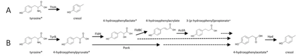
The Specific Case of p-Cresol in ASD ... 46
3. a. General Introduction ... 47
b. p-Cresol Metabolism in the Host ... 47
c. Clinical Evidence Connecting p-Cresol to ASD ... 48
i. Increased Excretion of p-Cresol in ASD ... 48
ii. Correlation with ASD GI Symptoms and ASD Core Behaviours ... 48
iii. Increased Abundances of Bacterial Taxa Synthetizing p-Cresol in ASD ... 49
d. p-Cresol and Behaviour in Mice ... 49
CHAPTER 2: ROLE OF THE MICROBIAL METABOLITE P-CRESOL IN ASD ... 50
I. CONTEXT ... 50
II. RESEARCH HYPOTHESIS AND OBJECTIVES OF THE STUDY ... 51
III. RESULTS ... 52
IV. MANUSCRIPT ... 53
CONCLUSIONS AND PERSPECTIVES ... 114
Conclusions ... 114
A. Perspectives in the p-Cresol Mouse Model ... 114
B. Understanding the Cellular and Molecular Pathways Impacted by p-Cresol ... 114
1. Possible Therapeutic Strategies to Explore in the p-Cresol Model ... 116
2. Possible Therapeutic Intervention Strategies Targeting the Microbiota in ASD ... 117
3. a. FMT ... 117
b. Probiotic and Prebiotic Treatment ... 118
CHAPTER 3: PERIPHERAL PHENOTYPES IN FRAGILE X SYNDROME ... 120
I. CONTEXT ... 120
METABOLIC ANOMALIES IN FXS:PUBLICATION #1 ... 120
Research Hypothesis and Objectives of the Study ... 120
A. Results ... 121
B. Manuscript ... 122
C. SKELETAL ANOMALIES IN FXS: PUBLICATION #2 ... 172
Research Hypothesis and Objectives of the Study ... 172
A. Results ... 172
B. Manuscript ... 173
C. IMMUNE ANOMALIES IN FXS: PUBLICATION #3 ... 187
Research Hypothesis and Objectives of the Study ... 187
A. Results ... 187 B. Manuscript ... 188 C. CONCLUSIONS ON FXSWORK ... 201 TAKE-HOME MESSAGE ... 202 BIBLIOGRAPHY ... 203
8
List of Abbreviations
3,4-Dihydroxyphenylacetic Acid DOPAC
4-Ethylphenylsulfate 4-EPS
5-Aminovaleric Acid 5AV
5-Hidroxi-Triptamina 5-HT
Acyl-CoA Dehydrogenase AcdA
Attention-Deficit/Hyperactivity Disorder ADHD
Autism Spectrum Disorder ASD
Blood-Brain Barrier BBB
Body Mass Index BMI
Brain-Derived Neurotrophic Factor BDNF
Cadherin-1 CDH1
Caesarean Section CS
Calcium voltage-gated channel CACN
Central Nervous System CNS
Choline Acetyltransferase ChAT
Cluster of Differentiation 3 CD3
Coeliac Disease CD
Complement 4B C4B
Corticotrophin-Releasing Hormone CRH
Dendritic Cells DC
Diagnostic and Statistical Manual of Mental Disorders DSM-V
Diet-Induced Maternal Obesity DIO
Dorsal Raphe Nucleus dRN
Dorsal Vagal Complex DVC
Enteric Nervous System ENS
Enterocromaffin Cells EC
Eotaxin-1 CCL11
Faecal Microbiota Transplantation FMT
Fragile X Mental Retardation 1 gene FMR1
Fragile X Mental Retardation Protein FMRP
Fragile X Related 2 Fxr2
Fragile X Syndrome FXS
Free Fatty Acid Receptor 1-2 FFAR1-2
Functional Magnetic Resonance Imaging fMRI
G Protein-Coupled Receptor 142 GPR142
G Protein-Coupled Receptor 35 GPR35
G Protein-Coupled Receptor 41 GPR41
G Protein-Coupled Receptor 43 GPR43
Gamma-Aminobutyric Acid type A Receptor GABR
Gastro-Intestinal GI
Gestational Diabetes Mellitus GDM
Glial Fibrillary Acidic Protein GFAP
Glutamate Ionotropic Receptor AMPA type subunit 1 GRIA1
Glutamate Ionotropic Receptor NMDA type subunit 1 GRIN1
Granulocyte-Macrophage Colony Stimulating Factor GM-CSF
Gut-Associated Lymphoid Tissue GALT
9
Hydroxycarboxylic acid receptor 2 Hca2
Hydroxyphenylacetate Decarboxylase Hpd
Hypothalamus-Pituitary-Adrenocortical HPA
Indole-3-Lactic Acid ILA
Innate Lymphoid Cells ILC
Innate Lymphoid Cells ILCs
Intellectual Disability ID
InterFeron-γ IFN-γ
InterLeukin-1 Receptor Antagonist IL-1R
InterLeukin-10 IL-10
InterLeukin-6 IL-6
InterLeukin-8 IL-8
InterLeukine 1 Receptor Accessory Protein Like 1 IL1RAPL1
Intraepithelial Lymphocytes IEC
Intraepithelial Lymphocytes IECs
kiloDalton kDa
Lipopolysaccharides LPS
Long-Term Depression LTD
Long-Term Potentiation LTP
Macrophage Migration Inhibitory Factor MIF
Major Histocompatibility Complex MHC
Maternal Immune Activation MIA
Medium Spiny Neurons MSN
Methyl-CpG Binding Protein 2 MECP2
Microfold Epithelial Cells M
Miniature Excitatory Postsynaptic Currents mEPSC
Modified Checklist for Autism in Toddlers – Revised M-CHAT-R
Monocyte Chemoattractant Protein-1 MCP-1
Neurexins NRXN
NeuroDevelopmental Disorders NDD
Neuroligin 3 NLGN3
Neuroligin 4 NLGN4
Nitric Oxide Synthase nNOS
NonObese Diabetic NOD
Nuclear factor-κB NF-κB
Nucleus Accumbens NAc
Nucleus of Solitary Tract NTS
Olfactory Receptor 78 OLFR78
ParaVentricular Nucleus PVN
Pathogen-Associated Molecular Patterns PAMP
Phenyllactate Dehydratase FldBC
Phenyllactate Dehydrogenase FldH
Polyinosinic-Polycytidylic Acid poly Poly (I:C)
Postsynaptic Density Protein 95 PSD95
Potassium voltage-gated channel KCN
PreFrontal Cortex PFC
Propionic Acid PPA
Protein Kinase C Beta PRKCB
Psoriasis PS
10
Quantitative – Checklist for Autism in Toddlers Q-CHAT
Restricted Repetitive Behaviour RRB
Rheumatoid Arthritis RA
SH3/Ankyrin Domain gene 3 SHANK3
Short Chain Fatty Acid SCFA
Single Nucleotide Polymorphisms SNP
Sodium voltage-gated Channel SCN
Synapsin-1 and -2 SYN1-2
Thiazole Synthase H ThiH
Transforming Growth Factor Beta 1 TGF-β1
Tryptophan Hydroxylase 1 Tph1
Tuberous Sclerosis Complex 2 TSC2
Tumor Necrosis Factor-α TNF-α
Type-1 Diabetes TD1
Tyrosine Aminotransferase B TyrB
Ubiquitin Protein Ligase E3A UBE3A
Vaginal Delivery VD
Valproic Acid VPA
Ventral Tegmental Area VTA
α-Amino-3-Hydroxy-5-Methyl-4-Isoxazolepropionic Acid AMPA
11
List of Figures
Figure 1: In utero exposure to maternal environmental factors increases the risk of ASD in offspring. ... 19 Figure 2: Maternal immune activation risk factor for ASD in offspring (Estes and McAllister, 2016) ... 21 Figure 3: Brain regions involved in the Mesolimbic Reward Circuit. ... 22 Figure 4: Mesolimbic reward pathway: white matter tracts connecting the NAc and the VTA (Supekar et al., 2018). ... 23 Figure 5: Communication pathways from the gut to the brain... 30 Figure 6: Microbiota and neurodevelopmental windows: implications for brain disorders by (Borre et al., 2014) . ... 38 Figure 7: Factors shaping the neonatal microbiome (Tamburini et al., 2016) ... 39 Figure 8: Direct (A) and Indirect (B) pathway for p-Cresol synthesis using tyrosine as a precursor by gut bacteria (Saito et al., 2018). ... 47
List of Tables
Table 1: Epithelial cells of the GI system. ... 33 Table 2: Effects of bacterial metabolites on the host’s immune and GI function. ... 36 Table 3: Behavioural phenotypes and biological processes in GF rodents. ... 37
12
Chapter 1: Introduction
Neurodevelopmental Disorders
Central Nervous System (CNS) disorders with early onset are commonly termed Neurodevelopmental Disorders (NDD). NDD include a large number of pathologies in which abnormal neurodevelopment leads to neurological and/or behavioural impairments. These pathologies affect four times more males than females (Rutter et al., 2003). Intellectual Disability (ID), Autism Spectrum Disorders (ASD), Attention-Deficit/Hyperactivity Disorder (ADHD), motor disorders and epilepsy are common NDD (Thapar et al., 2017). NDD are frequently associated with communication and social behaviour deficits, sensory disorders, learning disabilities, hyperactivity, anxiety, depression, mood instability, sleep problems, stereotypies, self-injurious behaviours, etc. The prevalence and intensity of the different symptoms will determine the diagnosis class and the specific needs for each NDD patient (Hansen et al., 2018). The symptomatology and severity of symptoms vary greatly among patients, even within a specific diagnosis class, and the frequent comorbidities among NDD underline the great heterogeneity in the clinical spectrum of NDD.
The differential manifestations of NDD can be explained by complex interactions between genetic predisposition and exposure to different environmental NDD risk factors (Jones and Szatmari, 2002). Several environmental factors -such as nutrition, pollution, immune changes, stress, environmental enrichment- are known to influence neurodevelopment (Hertz-Picciotto et al., 2018). In addition, the existence of critical time-windows in neurodevelopment, as well as the limited time-window for recovery or compensation of abnormal neurodevelopment modulates the risk to later develop NDD (Krol and Feng, 2018). One important neurodevelopmental time-window is the prenatal period. In utero, the foetus can be exposed to large number of factors that can compromise neurodevelopment at various stages. NDD environmental risk factors include in utero exposure to drugs consumed during pregnancy, maternal immune activation, maternal obesity or pollution (Hertz-Picciotto et al., 2018). Even the birth delivery mode can affect the child’s behavioural outcome during childhood (Hertz-Picciotto et al., 2018).
This Thesis focuses on a specific class of NDD, ASD, and encompasses studies related to i) the role played by the environment, and more specifically the microbiota in the development of ASD and ii) a genetic form of ASD, Fragile X Syndrome.
13
Autism Spectrum Disorders
Generalities
A.
Prevalence, Core Symptoms of ASD and Diagnosis
1.
ASD are characterized by impairment in communication and social behaviour, as well as the
presence of restricted repetitive behaviours and atypical sensory processing (Green et al.,
2016; Lai et al., 2013). ASD are often accompanied by comorbidities such as ADHD, ID, anxiety and depressive disorders (Cremone-Caira et al., 2019; Mazurek et al., 2013). Epidemiological studies report an incidence of 1 in 68 children with ASD in 2012 in the United States, with an estimated male: female sex ratio of 4:1 (Christensen et al., 2019). In recent years, the prevalence of ASD has increased. This may be due to a higher exposure to environmental factors that increase the risk of the onset of ASD (Lai et al., 2013). In fact, recent improvements in ASD diagnosis criteria also allow reducing the diagnosis bias of ASD in women, which used to be under-recognised based on anterior diagnostic criteria (Lai et al., 2013).
Most of ASD cases are detected from 24 to 36 months of age (Bejarano-Martín et al., 2019). ASD screening and diagnosis is performed when family members, paediatrician or caregivers suggest that the infant has developmental difficulties. These infants are screened using either of these two questionnaires: Modified Checklist for Autism in Toddlers, Revised (M-CHAT-R) (Robins and Casagrande, 2014) or the Quantitative Checklist for Autism in Toddlers (Q-CHAT) (Allison et al., 2008). These screening tools are designed to identify difficulties in young children and determine whether the toddler should be referred for full ASD diagnosis evaluation (Baird et al., 2000). ASD clinical diagnosis relies on the Diagnostic and Statistical Manual of Mental Disorders (DSM-V) criteria (Doernberg and Hollander, 2016). DSM-V diagnostic criteria for ASD rely on the evaluation of two behavioural domains: social communication impairments and restricted repetitive behaviours (RRBs) (Lord and Bishop, 2015). Social and communication problems encountered in ASD include deficits in social-emotional reciprocity and non-verbal behaviours involved in social interaction, and maladaptative social behaviour. The restricted/repetitive behaviours associated with ASD include repetitive movements or stereotypies, perseverative behaviours, inflexible routines, restrictive interests and hypo- or hyper-reactivity to sensory stimuli (Lord and Bishop, 2015). Once diagnosed with ASD, the child will be redirected to specialized mental health services.
14
Professionals will inform families of the child's specific needs, highlighting the educational needs (Bejarano-Martín et al., 2019).
Associated Peripheral Symptoms
2.
a. Immune Dysfunction
In addition to behavioural symptoms, ASD patients frequently present immune dysfunction. ASD patients are at increased risk of developing immune disorders such as asthma, food allergies or autoimmune disorders (e.g. coeliac disease, Crohn’s disease, type 1 diabetes, rheumatoid arthritis, psoriasis…) (Ashwood et al., 2006; Atladóttir et al., 2009). Furthermore, peripheral alterations in cytokine levels are also observed in the blood of ASD patients. Recent meta-analysis show that increased plasma levels of Interferon (IFN)-γ, Interleukin (IL)-1β, IL-6, Tumor Necrosis Factor (TNF)-Monocyte Chemoattractant Protein-1
(MCP-1), Eotaxin-1 (CCL1(MCP-1), IL-8 and reduced levels of Transforming Growth Factor Beta 1 (
TGF-β1), IL-10, Interleukin-1 Receptor Antagonist (IL-1R are associated with ASD (Masi et al., 2015; Saghazadeh et al., 2019).
Furthermore, some studies suggest that some ASD patients exhibit neuroinflammation. Neuroinflammation is an inflammatory process that occurs within the CNS (DiSabato et al., 2016) which involves resident cells in the brain -such as astrocytes, microglia, endothelial cells- and immune cells migrating from the periphery. Neuroinflammation is accompanied by an increased production of cytokine, chemokines and second messengers driving the inflammatory process (DiSabato et al., 2016). Several studies conducted on post mortem brains of ASD patients revealed microglial and astroglial activation, which is a hallmark of neuroinflammation. The expression of the astrocyte marker Glial Fibrillary Acidic Protein (GFAP) was increased in the cortex of ASD patients (Laurence and Fatemi, 2005). Also, the cortex of ASD patients exhibit an increased microglial density and signs of microglial activation such as increases in microglial somatic volume, retraction, thickening and extension of prolongations (Morgan et al., 2010; Tetreault et al., 2012). One study showed increased cytokine levels including TNF-α, IL-6, Granulocyte-Macrophage Colony Stimulating Factor (GM-CSF), IFN-γ and chemokine Interleukin-8 (IL-8) in post mortem ASD brain tissues (Li et al., 2009).
15
In addition, signs of inflammation have been detected in the gastrointestinal (GI) tract of ASD patients. Some studies have shown abnormalities in the proinflammatory cytokines levels
expressed by CD3+ lymphocytes (e.g. increased level of TNF-α and INF-γ, IL-4 and IL-5 with
reduction of IL-10) in intestinal biopsies from ASD children (Ashwood and Wakefield, 2006; Jyonouchi, 2009; Luna et al., 2017).
b. Gastrointestinal Symptoms
A considerable number of studies have shown that ASD patients are more prone to GI functional disorders (McElhanon et al., 2014). Further, ASD patients are 4.4 times more likely to exhibit GI symptoms than neurotypical individuals (McElhanon et al., 2014). The most common symptoms are abdominal pain, diarrhoea, constipation and abdominal bloating (Chaidez et al., 2014). Furthermore, ASD children are 5 times more susceptible to develop feeding problems as compared to neurotypical children (Sharp et al., 2013), and often present anorexia and anhedonia (Mazefsky et al., 2014). In ASD patients, the GI problems are associated with increased severity of social/affective problem, social withdrawal, repetitive behaviours, irritability, hyperactivity, anxiety and aggressiveness (Adams et al., 2011; Buie et al., 2010; Gorrindo et al., 2012; Mazefsky et al., 2014; Nikolov et al., 2009).
Moreover, some ASD patients also present alterations of intestinal permeability (D’Eufemia et al., 1996; Esnafoglu et al., 2017; De Magistris et al., 2010). In physiological conditions, the intestinal epithelium strictly controls the passage of particles, toxins and microorganism. In pathological conditions, the intestinal epithelium can become more permeable or ―leaky‖ and allow passage of potentially harmful molecules and microorganisms (Camilleri et al., 2012). This can also be accompanied by intestinal inflammation, that may eventually become
systemic (Mu et al., 2017). This inflammation can also contribute to exacerbate the intestinal
damages and allow the translocation of intestinal bacteria and other harmful substances (Mu et al., 2017). Studies in subsets of ASD patients have highlighted a reduction in the expression of tight junctions proteins, which regulate the permeability of the intestinal barrier (Fiorentino et al., 2016; Luna et al., 2017).
Both GI problems and immune system dysfunction in ASD patients may be closely related to anomalies of microbiota composition, or dysbiosis, found in ASD patients (Fung et al., 2017). This topic will be further developed in section 3 of the Thesis, which addresses the possible contribution of perturbations of the microbiota-gut-brain axis in ASD.
16
ASD Causes
B.
The causes for ASD are complex and multifactorial. As most NDD, ASD likely result from strong interaction between genetic susceptibility and exposure to environmental factors which perturb neurodevelopment. The genetic contribution in ASD appears to be approximately 45% for monozygotic twins and 16% for dizygotic twins (Bourgeron, 2016). Twin studies have also revealed that genetics and environmental factors share equal influence on ASD risk (Bourgeron, 2016).
Genetic Causes
1.
a. Genes Linked to ASD
Approximately 10-25% of ASD cases can be explained by mutation in specific genetic loci. ASD is associated with more than 913 genes, as summarized in the SFARI database (https://www.sfari.org/resource/sfari-gene/). We can distinguish non-syndromic and syndromic forms of ASD.
Non-syndromic forms are generally of unknown genetic aetiology and accounts for the majority of ASD cases. Non-syndromic forms are usually related to the presence of several allelic variants corresponding to single nucleotide polymorphisms (SNP). These variants usually have slight effects and can be found in the general population (Bourgeron, 2016). Allelic variants play an important role in the susceptibility to ASD. In some individuals, a specific genetic background will be able to buffer or compensate the impact of the rare genetic variations. In contrast, in some individuals, the buffering capacity of the genetic background will not be sufficient to compensate the impact of the deleterious mutations and they will develop ASD.
Syndromic forms of ASD account for a small percentage of ASD cases and are linked to the deletion, duplication or silencing of a specific genetic locus (Rylaarsdam and Guemez-Gamboa, 2019). Usually, syndromic forms of ASD are accompanied with a clinically defined pattern of somatic abnormalities (such as dysmorphic features, gastrointestinal problems) and a neurobehavioral phenotype that include ASD and often learning and ID.
17
b. Functions of the Genes Involved
The genes associated with ASD operate multiple functions. However, there is a high incidence of genes with synaptic function or immune function.
Among the genes affecting synaptic function and plasticity lie genes encoding adhesion proteins (e.g. neuroligin3, 4 (NLGN3/4), neurexins (NRXN), and cadherins (CDH1)), synaptic vesicles proteins (e.g. synapsin-1 and -2 (SYN1-2)), channels (e.g. sodium, calcium and potassium voltage-gated channel (SCN, CACN and KCN) and receptors (e.g. glutamate and GABA receptors (GRIA1, GRIN1 and GABR)) that are critical for synaptic functions (Rylaarsdam and Guemez-Gamboa, 2019). Genes involved in signal transduction such as
Tuberous sclerosis complex 2 (TSC2), and protein degradation such as Ubiquitin protein ligase E3A (UBE3A) are also associated with ASD (Rylaarsdam and Guemez-Gamboa, 2019).
Finally, genes encoding regulators of mRNA translation -such as the Fragile X Mental
Retardation 1 gene encoding the RNA-binding protein FMRP linked to Fragile X Syndrome-
or transcription -such as the Methyl-CpG binding protein 2 encoding the transcription factor MECP2 linked to Rett Syndrome- are also associated with ASD (Rylaarsdam and Guemez-Gamboa, 2019). Loss-of-function of these genes was shown to impact synaptic development and function in animal models and these are thought to contribute to the behavioural impairments in ASD patients.
Another class of genes associated with ASD is related to immune components such as: Macrophage Migration Inhibitory Factor (MIF), Human Leukocyte Antigen Complex
(HLA-A2, HLA-DRB1), Major Histocompatibility Complex (MHC class I and MHC class II),
elements involved in complement cascade (C4B) and Interleukin 1 Receptor Accessory Protein Like 1 (IL1RAPL1) (Estes and McAllister, 2015). In patients bearing mutations in these genes, perturbations of immune processes could impair neurodevelopment and contribute to behavioural alterations (Estes and McAllister, 2015).
c. One Example of a Syndromic Form of ASD: Fragile X Syndrome
Clinical features of FXS. Fragile X syndrome (FXS) is the first genetic cause of ASD
diagnosis, with up to 50% of Fragile X patients diagnosed with ASD (Penagarikano et al., 2007). FXS is also the most common form of inherited ID mainly associated with mild to strong cognitive impairments, attention deficits and hyperactivity. Besides presenting behavioural alterations, FXS is also accompanied by physical abnormalities (Kidd et al.,
18
2014). In subsets of FXS patients, morphometric studies have highlighted increased stature and height (Butler et al., 1993) and a general overgrowth in prepubertal boys affected by FXS (Kidd et al., 2014; De Vries et al., 1995). FXS patients present connective tissue dysplasia (Hagerman et al., 1984), dental and mandibular anomalies (Sabbagh-Haddad et al., 2016), orthopedic anomalies such as scoliosis (Davids et al., 1990), and abnormal metacarpophalangeal pattern profile (Butler et al., 1988). In addition, FXS patients display specific craniofacial abnormalities with reduced facial depth, hypoplasticity of the nasal bone–cartilage interface and narrow mid-facial width exaggerating ear prominence (Heulens et al., 2013). Also, subtle craniofacial anomalies with morphometric changes in the mandible and skull are observed in FXS patients (Heulens et al., 2013).
Functions of the gene mutated in FXS. FXS is caused by the silencing of the FMR1 gene,
located in a ―fragile‖ site on the X chromosome in the 27q3 region (Harrison et al., 1983). Given that the FMR1 gene is X-linked, random inactivation of the mutated allele in females yields a biased sex-ratio of 2 males (1:4000 males) for 1 female affected (1:8000 females), with a global incidence rate of 1:6000 in the general population. In FXS patients, abnormal expansions of CGG trinucleotide repeats in the 5′ untranslated region of the FMR1 gene lead to a hypermethylation of the CpG island upstream to the gene promotor. As a result, the
FMR1 gene is silenced and the encoded protein, FMRP is absent (Penagarikano et al., 2007).
FMRP is an RNA binding protein involved in the regulation of mRNA translation, transport and stability (Penagarikano et al., 2007). The FMR1 gene is expressed in the CNS and in all peripheral tissues, to the exception of adult skeletal and cardiac muscle (Davidovic et al., 2006; Khandjian et al., 1998). Loss of FMRP in the brain induces anomalies in the formation of synapses, with an increased density of immature dendritic spines thought to be at the basis of the ID (Hinton et al., 1991).
As a reflection of the expression of FMRP in the periphery, some FXS patients exhibit changes in metabolic or immune markers with decreased cholesterol levels, increase of triacylglycerols levels and changes in the levels of cytokines and chemokines (Ashwood et al., 2006; Berry-Kravis et al., 2015; Lisik et al., 2016), although these changes cannot be considered a patient-specific trait. These findings suggest that the effect of FMR1-deficiency generates physiological dysfunction in the CNS and in the periphery.
In the 4th sections of this Thesis, we will present three studies carried out on Fragile X
patients and the mouse model of FXS addressing the peripheral consequences of FMR1-deficiency.
19
Environmental Factors
2.
Environmental influences in combination with individual genetic susceptibility modulate the risk of developing ASD. In recent years, the importance of environmental factors during pregnancy has been pointed out as possible triggers for the development of ASD in the offspring. The nature of the environmental factors and the time-window of exposure during gestation are important elements that can modulate the genetic predisposition of an individual to NDD (Hertz-Picciotto et al., 2018). Following the ―multiple hit‖ model for ASD, the repeated exposure to detrimental environmental factors can result in the appearance of ASD in genetically predisposed individuals (Miles, 2011). Some of the factors that increase the risk
of the appearance of ASD in the offspring are summarized in Figure 1.
Figure 1: In utero exposure to maternal environmental factors increases the risk of ASD in
offspring.
Abbreviations: TD1, Type-1 Diabetes; RA, Rheumatoid Arthritis; CD, Coeliac Disease; PS,
Psoriasis; BMI, obesity based in Body Mass Index; GDM, Gestational Diabetes Mellitus; CS, Caesarean Section; VD, Vaginal Delivery.
a. In utero Exposure to Drugs Consumed by the Mother
Epidemiological studies have shown that exposure to teratogens substances such as valproic acid -an anticonvulsant drug and mood stabilizer, used to treat migraines, epilepsy and bipolar
20
disorder- (Cotariu and Zaidman, 1991; Veroniki et al., 2017), thalidomine -an anti-emetic medication- (Jones and Szatmari, 2002) or misoprostol -used to treat gastric ulcers- (Ornoy et al., 2015) during the prenatal and early postnatal periods can alter neurodevelopment and lead to ASD in the offspring (Dufour-Rainfray et al., 2011).
b. In utero Exposure to Maternal Immune Activation
There are three important risk factors for ASD linked to maternal immune activation (MIA). First, diagnosis of maternal autoimmune diseases (e.g. TD1, RA, CD, PS and Crohn’s disease…) increases the risk of developing ASD in the offspring (Atladóttir et al., 2009). Second, the presence of maternal antibodies against foetal brain proteins (IgG antibodies) also increases the risk of ASD in the offspring (Hsiao, 2013). These antibodies would access the foetal brain at a developmental stage at which the blood-brain barrier (BBB) is still immature. This could contribute to generate brain abnormalities that may favour the emergence of later neurological problems in the offspring. Third, maternal infections and notably viral infection during the first trimester of pregnancy or unspecific infections during the second trimester are associated with an increased ASD risk in the offspring (Atladóttir et al., 2010). Frequent episodes of fever during pregnancy, linked to an infectious state, are also associated with increased ASD risk in the offspring (Hornig et al., 2018). Studies in mouse models have highlighted that the gestational period of exposure, the type of MIA (viral, bacterial or chronic) and the duration and intensity of the MIA will determine the appearance of neurodevelopmental and behavioural problems in the progeny (Estes and McAllister, 2015). It is hypothesised that maternal pro-inflammatory cytokines released upon MIA reach the foetal brain and perturb neurodevelopmental processes, increasing the susceptibility to later develop
21
Figure 2: Maternal immune activation risk factor for ASD in offspring (Estes and McAllister,
2016)
c. Mode of Delivery
The birth delivery mode can also modulate ASD risk. Several studies suggests that delivery by caesarean section increases the risk of the offspring to develop ASD (Curran et al., 2015; Yip et al., 2017). Meta-analyses have also identified other environmental risk factors for ASD such as advanced parental age, birth complications (e.g. trauma, ischemia, hypoxia), pregnancy-related risk factors (e.g. foetal distress, multiple births), deficiencies in essential nutrients (e.g. fatty acids, vitamin D and folate) and prenatal exposure to environmental toxins (e.g. heavy metals, pesticides, endocrine-disrupting chemicals) (Modabbernia et al., 2017; Wang et al., 2016).
Disturbed Neuronal Circuits in ASD
C.
The connection between behavioural abnormalities and deregulation in neural circuits and synaptic function in ASD is the subject of active research to understand the neurobiological correlates of ASD behavioural impairments. Several brain circuits have been identified as altered in ASD patients. Notably, the reward circuit could be involved in ASD-related social behaviour deficits (Supekar et al., 2018).
Social interactions are pleasurable events for humans and animals, as shown notably by the activation of the reward circuit by social stimuli – activation that is blunted in ASD patients (Pellissier et al., 2018). The mesolimbic dopamine reward circuit controls notably the social
reward and the literature suggests that ASD patients experience a lower social reward as
compared to neurotypical subjects (Chevallier et al., 2012). The interconnection between brain regions involved in the reward circuit have been mostly described in rodents and encompass: the Ventral Tegmental Area (VTA), Nucleus Accumbens (NAc), Paraventricular
Nucleus (PVN) and Prefrontal Cortex (PFC) (Figure 3). To dissect out the connections
22
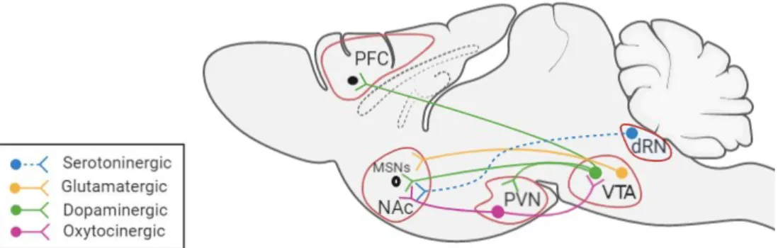
Figure 3: Brain regions involved in the Mesolimbic Reward Circuit.
Abbreviations: PFC, Prefrontal Cortex; MSNs, Medium Spiny Neurons; NAc, Nucleus
Accumbens; PVN, Paraventricular Nucleus; VTA, Ventral Tegmental Area; dRN, dorsal Raphe Nucleus.
The VTA is a critical region for the mesolimbic reward circuit and is involved in the modulation of responses to reward stimuli. The VTA is localised in the brainstem and is composed mainly of GABA, dopaminergic and glutamatergic neurons (Morales and Margolis, 2017). VTA glutamate neurons are important for the socialisation of mice (Krishnan et al., 2017). The reduction of VTA glutamatergic transmission to Medium Spiny Neurons (MSNs) in the NAc, decreased the sniffing time in mice (Krishnan et al., 2017). On the other hand, VTA dopamine neurons project notably towards the NAc and the PFC. VTA dopamine neurons are regulated by oxytocinergic projection from the PVN (Hung et al., 2017). Oxytocin enhances the excitability of VTA dopamine neurons projecting to the NAc, which reinforces social interactions (Hung et al., 2017). In the NAc, MSNs express the D1 and D2 dopamine receptors (Dölen et al., 2013). At the same time oxytocinergic neurons of the PVN and serotoninergic neurons of the dRN project to the NAc and both are important for the synaptic plasticity in the NAc that is essential for the social reward process (Dölen et al., 2013).
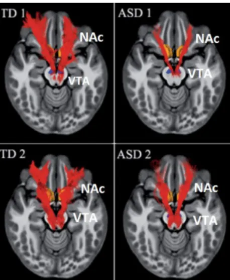
Functional neuroimaging, a non-invasive technique to monitor the activity and connectivity of brain regions in the resting state or during a task, was used to analyse the social reward system in ASD patients. A functional magnetic resonance imaging (fMRI) study reported a decrease in the density of the fibres which originate in the VTA and project to the NAc in
young ASD patients (Figure 4) (Supekar et al., 2018). Another study has also shown a
decreased activity in the NAc in ASD patients during reward-related motivation (Assaf et al., 2013).
23
Figure 4: Mesolimbic reward pathway: white matter tracts connecting the NAc and the VTA
(Supekar et al., 2018).
Finally, one study combined electroencephalography and high-resolution eye-tracking and
reported that ASD patients present decreased activation of the PFC and the anterior cingulate cortex during neural processing of dynamic cartoons related to human-like social interactions (Jan et al., 2019). Also, using positron emission tomographic scanning, one study highlighted that ASD patients displayed a decreased dopaminergic activity in the anterior medial PFC during a story comprehension paradigm (Ernst et al., 1997).
Altogether, these studies suggest that social behaviour deficits in ASD patients could result from dysfunctions in the circuitry processing social reward, which modulate notably social cognition, perception and motivation (Mundy, 2003).
ASD Animal Models
D.
Introduction
1.
ASD animal models have been developed to better understand the pathophysiological processes leading to ASD in human. There are three criteria established by Willner in 1984 that an animal model must fulfil in order to be considered a good model for neuropsychiatric disorders (Belzung and Lemoine, 2011). First, the construct validity criterion is met when the behavioural phenotypes of the model and the symptoms and manifestation of the disease in human are homologous (Belzung and Lemoine, 2011). Second, the face validity criterion is reached when the biological dysfunctions observed in the animal model reflects the currently understood disease aetiology and the identified biomarkers in humans (similarity of mechanisms and neurobiological substrates). Third, predictive validity is achieved when there
24
are similarities in the response of the animal model to therapies and notably pharmacological interventions used in human to treat the disease.
The mouse is an adequate organism to model neuropsychiatric diseases such as ASD (Crawley, 2012; Silverman et al., 2010). In mice, behavioural testing enables to analyse the behavioural dimensions related to ASD core symptoms (social behaviour, repetitive movements/stereotypies, behavioural flexibility and sensory processing) and comorbidities (cognition, emotional processing with anxiety/depression features…). Similarly, neurodevelopmental, neurobiological and physiological processes are very similar between mice and human, enabling to evaluate the construct validity of the models. Finally, mice can be treated with drugs or subjected to different interventional strategies as it is done in humans to treat ASD, enabling to assess their predictive validity.
Along these criteria, a large number of mouse models have been validated to study ASD. The
SFARI base (https://gene.sfari.org/database/animal-models/genetic-animal-models/)
recapitulates the available models, classified along 3 categories: 1) 277 genetic models are associated with mutation in a gene or a genetic region that is known to be associated with ASD, 2) 45 induced environmental models are obtained by in utero exposure to environmental factors known to increase the risk of the onset of ASD in the offspring, and 3) 8 inbred models of idiopathic ASD are described.
Genetic and Environmental Animal Models in ASD
2.
a. Genetic Models
There are several genetic models in mice, that are usually generated by the deletion, duplication, mutation or loss of function of a gene or a genetic locus that is known to be associated with ASD in humans (Carmichael and Lockhart, 2012; Patel et al., 2018). Hereafter, we review several commonly used models of ASD.
i. Fmr1-knockout (KO) mice
Two mouse models for FXS have been developed (Dutch Belgium consortium 1991, (Mientjes et al., 2006)). Fmr1-KO mice recapitulate a number of the behavioural abnormalities observed in Fragile X patients: cognitive impairments, autistic-like features, such as social interaction deficits and stereotypies, and hyperactivity (Bakker and Oostra, 2003). On the neurobiological level, it has been observed that there is an increase in the
25
number of immature dendritic spines and an abnormal increase in local protein synthesis at the synapse (Bakker and Oostra, 2003; Nimchinsky et al., 2001). Furthermore, several studies have described alterations of various forms of synaptic plasticity in Fmr1-KO mice, which could explain the behavioural defects observed (Bakker and Oostra, 2003). In particular, the
Fmr1-KO mice display deficits in long-term depression (LTD) and long-term potentiation
(LTP) in the hippocampus and cortex (Bagni and Zukin, 2019; Huber et al., 2002; Li et al., 2002). In addition, abnormalities in cortical-cerebellar circuits have also been described, which perturb VTA dopamine release in the PFC (Rogers et al., 2013).
Fmr1 gene is widely expressed not only in the CNS but also in peripheral tissues (liver,
adipose tissue, skin) (Davidovic et al., 2006; Khandjian et al., 1998). When I started my PhD, very few studies had addressed the peripheral effects of Fmr1 gene silencing. The double
Fmr1/Fxr2 KO mouse obtained by double inactivation of the Fmr1 and Fxr2 gene (Fxr2
being an autosomal homolog 2 of Fmr1 gene) displayed an increase in glucose tolerance and insulin sensitivity, a reduction in adiposity and circulating glucose (Lumaban and Nelson, 2015). In addition, cranioencephalic abnormalities have been described in Fmr1-KO mice, such as an increase in the size of the mandible-skull complex similar, to what was observed in Fragile X patients (Heulens et al., 2013).
I contributed to two studies which identified metabolic and body composition anomalies in
Fmr1-KO mice. These studies are presented in the 4th section of this Thesis.
ii. Shank3b-KO mice
Mutations in the SH3/ankyrin domain gene 3 (SHANK3) are typically associated with Phelan-McDermid syndrome, a syndromic form of ASD (Balaan et al., 2019).
Shank3b-KO mice present social interactions impairments with reduced number of nose
contacts and an increase in self-injurious repetitive grooming (Peça et al., 2011). At the neurobiological level, the Shank3b gene encodes the PostSynaptic Density protein of 95 kDa (PSD95) expressed at glutamatergic synapses. This protein regulates the trafficking and localization of glutamate receptors. The deletion of the Shank3b gene in mice alters postsynaptic molecular composition in the striatum with consequences on the glutamatergic system (Peça et al., 2011). Shank3b-KO mice also display anomalies in the oxytocinergic and dopaminergic system. More specifically, Shank3b-KO mice present a decrease of dopamine neurons excitability in the VTA, an important region involved in the social reward circuit (Bariselli et al., 2016). As a consequence, Shank3b-KO mice present social interaction
26
impairments and notably deficits in the social preference for a novel congener (Bariselli et al., 2016). These effects are accompanied by an increase in the expression of α-amino-3-hydroxy-5-methyl-4-isoxazolepropionic acid (AMPA) receptors and the consequent disturbance in VTA synaptic plasticity (Bariselli et al., 2016, 2018). Furthermore, Shank3b-KO GABAergic MSNs, which receive dopaminergic inputs from the VTA, present an increase in dendritic arborisation while dendritic spines density is reduced. This is accompanied by a decrease in the frequency and amplitude of their spontaneous activity or miniature Excitatory Postsynaptic Currents (mEPSC). The deletion of the Shank3b gene in mice alters glutamatergic (Peça et al., 2011) as well as dopaminergic transmission (Bariselli et al., 2016).
iii. BTBR T+ Itpr3tf/J (BTBR) mice
The BTBR mouse is considered as an idiopathic model of ASD. BTBR mice exhibit impaired social interactions, increased repetitive behaviours, compulsive cleaning, alterations in behavioural flexibility and learning deficits, compared to C57BL6/J control mice (Amodeo et al., 2012). The BTBR model is also characterized by the complete absence of the corpus
callosum, and a significant reduction of the hippocampal commissure. A reduction in the corpus callosum volume is also observed in some ASD patients (Pagnozzi et al., 2018).
Moreover, cerebral ventricles are reduced and this is accompanied by a decrease in neurogenesis (Meyza and Blanchard, 2017). Alterations in the dopaminergic system are also observed, contributing to an hypoactivation of the reward system (Squillace et al., 2014). Finally, this model also presents an increase of proinflammatory cytokines such as 33, IL-18 and IL-1 (Meyza and Blanchard, 2017) and an increase of autoantibodies (IgG) directed against brain proteins in the cortex, hippocampus, striatum and cerebellum (Kim et al., 2016). Finally, BTBR mice display increased intestinal permeability (Golubeva et al., 2017).
b. Environmentally-Induced ASD Models
In this section we will describe the different environmental models of ASD. All of them are developed during the prenatal period.
i. In utero Exposure to Valproic Acid
Valproic acid (VPA) has been associated with an increased risk of developing ASD in the offspring (Christensen et al., 2014). An animal model has been developed in which pregnant dams are injected intraperitoneally with VPA at embryonic day 12.5. The progeny of these
27
dams, exposed in utero to VPA, display social interaction impairments, repetitive/compulsive movements, anxiety-like behaviour and memory deficits (Kataoka et al., 2013; Wagner et al., 2006). VPA mice also exhibit signs of neuroinflammation in the brain and the male offspring displays decreased levels of serotonin in the PFC and amygdala (De Theije et al., 2014). In addition, VPA mice display increased intestinal permeability and decreased serotonin production in the gut (De Theije et al., 2014).
ii. In utero Exposure to MIA
Another risk factor for ASD in the offspring is in utero exposure to MIA (Estes and McAllister, 2016). An animal model has been generated in which MIA is triggered by either exposure to the Influenza virus or to pathogen-associated molecular patterns (PAMPs) via injection of lipopolysaccharides (LPS), which mimic bacterial infection or the synthetic double-stranded RNA polyinosinic-polycytidylic acid poly (I:C), which mimic viral infection. The most commonly used model is obtained by intraperitoneal injection of poly (I:C) at E12.5 (Hsiao et al., 2013; Zuckerman et al., 2003). However, in this model, the dose and developmental window targeted, i.e. the precise timing of poly (I:C) is a critical element for the appearance of behavioural abnormalities in the offspring.
The behavioural features of MIA offspring are social behaviour impairments, decrease in ultrasonic vocalizations, repetitive movements and deficits in sensorimotor gating, highlighting reduced behavioural flexibility, resembling the core symptoms of ASD (Hsiao et al., 2013; Smith et al., 2007). MIA offspring also display enhanced anxiety and short-term memory impairment (Luchicchi et al., 2016), resembling the frequent comorbidities in ASD of anxiety disorder and ID. At the molecular level, the MIA offspring present alterations in the reward circuit, notably due to reduced activity of dopamine neurons in the VTA (Luchicchi et al., 2016).
At the systemic level, this model also has GI disturbances, with an increase in intestinal permeability (Hsiao et al, 2013). The integrity of the intestinal barrier is compromised in this model, with a decrease in tight junction proteins expression and an increase in inflammatory markers expression (cytokines).
28
iii. In utero Exposure to Diet-Induced Maternal Obesity (DIO)
Maternal obesity during pregnancy is a risk in the development of neurological disorders in the offspring (Connolly et al., 2016). Thus, an animal model has been developed, where pregnant dams are fed on a high-fat diet (Buffington et al., 2016). Mice born from DIO mothers display social behaviour impairments, increased repetitive behaviours and anxiety (Buffington et al., 2016). These animals also present anomalies in the reward circuit (Buffington et al., 2016). Concretely, the DIO offspring exhibit impairments in the synaptic plasticity of dopamine neurons (long term potentiation, LTP) in the VTA (Buffington et al., 2016).
In conclusion, all these genetic and environmental models of ASD exhibit behavioural abnormalities similar to those observed in ASD patients, and notably social interactions impairments and stereotypies. This is accompanied by alterations in dopaminergic neurotransmission, and deficits in the social reward circuit appear as one of the neurobiological features correlates for the behavioural alterations. Finally, some ASD models display neuroinflammation and increased intestinal permeability revealing that immune and gut dysfunctions could also be linked to microbiota dysbiosis, as developed in the next section.
In this Thesis, we will present a new environmental mouse model of ASD obtained by the manipulation of its microbiota.
29
The Microbiota-Gut-Brain Axis in ASD
The Microbiota-Gut-Brain Axis: a Bidirectional
A.
Communication Axis
Introduction
1.
The term microbiota refers to the ecological communities of commensal, symbiotic and pathogenic microorganisms that live in symbiosis with the human body. Microbiota reside in a number of tissues and biofluids: skin, mammary glands, placenta, saliva, oral mucosa, vagina, uterus and GI tract (Cho and Blaser, 2012). The microbiota includes bacteria, bacteriophages, fungi, protozoa and viruses that live inside and on the human body (Fattorusso et al., 2019). The human gut hosts more than 100 trillion organisms. Microorganisms outnumber by 1.3-fold the average number of somatic and germ cells of an adult human, according to recent revised estimates (Sender et al., 2016). Strikingly, the collective genome of the microbiota, termed the microbiome, contains 100 times more genes than the human genome. This gene richness confers unique functional properties to the microbiota, notably in terms of metabolic and enzymatic reactions which cannot be performed by the host (Stilling et al., 2014).
In a way, the gut microbiota can be assimilated to a human organ that can be transplanted and accomplishes specific functions, notably digestion and metabolism of food (David et al., 2014), regulations of intestinal barrier functions (Hooper et al., 2001), gut immune maturation and homeostasis (Chung et al., 2012), regulation of hormonal and neurotransmitters secretion and of host’s brain function and behaviour (Goulet et al., 2019).
Specific alterations in microbiota composition, notably in terms of microbial abundances and diversity or metabolic output are termed dysbiosis. Microbiota dysbiosis influences health outcomes and when microbiota homeostasis is compromised, several pathological conditions may arise and notably ASD. In this Section, we will review the evidence in the literature supporting the hypothesis that dysregulation of the Microbiota-Gut-Brain axis contributes to the development of ASD symptoms.
30
The Microbiota-Gut-Brain-Axis
2.
The Microbiota-Gut-Brain axis refers to the bidirectional communication between the microbiota and the brain. The main routes of communication involve the peripheral nervous system (mainly the enteric nervous system and parasympathetic system), the neuroendocrine system and the immune system (Carabotti et al., 2015; Fülling et al., 2019; Sherwin et al., 2019; Vuong and Hsiao, 2017). All these systems are interconnected and can mutually influence each other. Also, these systems are modulated by environmental factors and neuroendocrine systems such as the hypothalamus-pituitary axis and the limbic system (Carabotti et al., 2015).
Figure 5: Communication pathways from the gut to the brain.
Abbreviations: ENS, Enteric nervous system; EC, Enterocromaffin cells; IECs, Intraepithelial
lymphocytes; ILCs, Innate lymphoid cells; DC, Dendritic cells.
a. Involvement of the Peripheral Nervous System
i. Enteric Nervous System (ENS)
The ENS is a part of the autonomous nervous system. The ENS is composed of enteric neurons and glial cells that form two plexus (i.e. myenteric and submucosal plexus), located between the circular and longitudinal muscle layers of the of GI tract (Nagy and Goldstein,
31
2017). The ENS is mostly composed of two neuronal types: cholinergic neurons (which account for 60% of the neurons in the GI tract), sensory and excitatory neurons and nitrinergic inhibitory interneurons and motoneurons (Rao and Gershon, 2016). Enteric neurons express cell-surface G-protein-coupled receptors (GPCRs) which can detect microbial metabolites, such as GPR41-43 for SCFAs and GPR142-35 for aromatic amino acids (Tryptophan, L-Phenylalanine and Kynurenic acid) (Husted et al., 2017). The ENS operates independently of the CNS even if it connected to the spinal cord and brain (Carabotti et al., 2015). Also, the ENS also has close connections with epithelial cells and immune cells in the intestinal barrier and integrates their signals (Carabotti et al., 2015). The function of the ENS is to regulate the neuroendocrine secretion and motility of the digestive tract as well as the maintenance of the mucosa and the regulation of intestinal immunity (Rao and Gershon, 2016).
ii. Vagus Nerve (Parasympathetic System)
The vagus nerve connects the ENS to the CNS. Sensory information coming from the GI tract is relayed by vagal afferent fibres to the nucleus of the solitary tract (NTS). The NTS is an integrative centre of sensory information in the CNS. Via efferent projections, the NTS is connected to several regions of the brain, including the limbic system (Browning and Travagli, 2014). The vagus nerve can detect microbial signals and relay information from the GI tract to the CNS (Cryan and Dinan, 2012). Vagal afferent fibres do not cross the epithelial layer and are therefore not in direct contact with the gut luminal microbiota. Therefore, these fibres can detect signals from the microbiota either by directly sensing bacterial compounds which have diffused through the epithelium or by integrating signals communicated by epithelial cells (Bonaz et al., 2018). Recent studies suggest that the vagus nerve is an important route by which the gut microbiota can send signals to the brain and induce behavioural changes. There is some evidence on how the microbiota can use this pathway to mediate behavioural and physiological changes in the brain (Bonaz et al., 2018). There are three examples of how different probiotics produce changes in behaviour using the vagus nerve as a pathway and the effects are suppressed after vagotomy, a surgical procedure in which a sub-diaphragmatic section of the vagus nerve is performed. The first example is the administration of Lactobacillus reuteri increases oxytocin release from the hypothalamus and improves social behaviour (Sgritta et al., 2019). This behavioural effects are blocked in vagotomized animals (Poutahidis et al., 2013; Sgritta et al., 2019). The second example is the administration of Bifidobacterium longum NCC3001 which decreases anxiety levels in mice