HAL Id: hal-02262558
https://hal-amu.archives-ouvertes.fr/hal-02262558
Submitted on 10 Dec 2019
HAL is a multi-disciplinary open access
archive for the deposit and dissemination of
sci-entific research documents, whether they are
pub-lished or not. The documents may come from
teaching and research institutions in France or
abroad, or from public or private research centers.
L’archive ouverte pluridisciplinaire HAL, est
destinée au dépôt et à la diffusion de documents
scientifiques de niveau recherche, publiés ou non,
émanant des établissements d’enseignement et de
recherche français ou étrangers, des laboratoires
publics ou privés.
Distributed under a Creative Commons Attribution| 4.0 International License
description of Raoultibacter massiliensis gen. nov., sp.
nov. and Raoultibacter timonensis sp. nov, two new
bacterial species isolated from the human gut
Sory Ibrahima Traore, Melhem Bilen, Mamadou Beye, Awa Diop, Maxime
Descartes Mbogning Fonkou, Mamadou Tall, Caroline Michelle, Muhammad
Yasir, Esam Ibraheem Azhar, Fehmida Bibi, et al.
To cite this version:
Sory Ibrahima Traore, Melhem Bilen, Mamadou Beye, Awa Diop, Maxime Descartes Mbogning
Fonkou, et al.. Noncontiguous finished genome sequence and description of Raoultibacter massiliensis
gen. nov., sp. nov. and Raoultibacter timonensis sp. nov, two new bacterial species isolated from the
human gut. MicrobiologyOpen, Wiley, 2019, 8 (6), pp.e00758. �10.1002/mbo3.758�. �hal-02262558�
MicrobiologyOpen. 2019;8:e758.
|
1 of 18 https://doi.org/10.1002/mbo3.758www.MicrobiologyOpen.com Received: 4 April 2018
|
Revised: 30 September 2018|
Accepted: 1 October 2018DOI: 10.1002/mbo3.758
O R I G I N A L A R T I C L E
Noncontiguous finished genome sequence and description of
Raoultibacter massiliensis gen. nov., sp. nov. and Raoultibacter
timonensis sp. nov, two new bacterial species isolated from the
human gut
Sory Ibrahima Traore
1*| Melhem Bilen
1,2*| Mamadou Beye
3| Awa Diop
3|
Maxime Descartes Mbogning Fonkou
1| Mamadou Lamine Tall
1| Caroline Michelle
1|
Muhammad Yasir
4| Esam Ibraheem Azhar
4,5| Fehmida Bibi
4| Fadi Bittar
1|
Asif Ahmad Jiman‐Fatani
6| Ziad Daoud
6| Fréderic Cadoret
1|
Pierre‐Edouard Fournier
3| Sophie Edouard
1This is an open access article under the terms of the Creative Commons Attribution License, which permits use, distribution and reproduction in any medium, provided the original work is properly cited.
© 2019 The Authors. MicrobiologyOpen published by John Wiley & Sons Ltd.
*The authors contributed equally to this article. 1UMR MEPHI, IRD, APHM, IHU
Méditerranée‐Infection, Aix‐Marseille Université, Marseille, France
2Clinical Microbiology Department, Faculty
of Medicine and Medical sciences, University of Balamand, Amioun, Lebanon
3UMR VITROME, IRD, AP‐HM, SSA, IHU
Méditerranée‐Infection, Aix‐Marseille Université, Marseille, France
4Special Infectious Agents Unit, King Fahd
Medical Research Center, King Abdulaziz University, Jeddah, Saudi Arabia
5Medical Laboratory Technology
Department, Faculty of Applied Medical Sciences, King Abdulaziz University, Jeddah, Saudi Arabia
6Department of Medical Microbiology and
Parasitology, Faculty of Medicine, King Abdulaziz University, Jeddah, Saudi Arabia Correspondence
Sophie Edouard, UMR MEPHI, IRD, APHM, IHU Méditerranée‐Infection, Aix‐Marseille Université, Marseille, France.
Email: sophie.edouard@univ‐amu.fr Funding information
National Plan for Science, Technology and Innovation (MAARIFAH), Grant/Award Number: 12MED3108‐03; “Investissements d'avenir”, Grant/Award Number: 10‐IAHU‐ 03; Région Provence Alpes Côte d'Azur; European funding FEDER PRIMI
Abstract
As part of the culturomics project aiming at describing the human microbiota, we report in this study the description of the new bacterial genus Raoultibacter gen. nov. that includes two new species, that is, R. massiliensis sp. nov. and R. timonensis sp. nov. The R. massiliensis type strain Marseille‐P2849T was isolated from the fecal specimen
of a healthy 19‐year‐old Saudi Bedouin, while R. timonensis type strain Marseille‐ P3277T was isolated from the feces of an 11‐year‐old pygmy female living in Congo.
Strain Marseille‐P2849T exhibited 91.4% 16S rRNA sequence similarity with Gordonibacter urolithinfaciens, its phylogenetic closest neighbor with standing in no‐
menclature. As well, strain Marseille‐P3277T exhibited 97.96% 16S rRNA similarity
with strain Marseille‐P2849T. Both strains were Gram‐positive, motile, nonspore‐
forming rod and form transparent microcolonies on blood agar in both anaerobic and microaerophilic atmospheres. The genome sizes of strain Marseille‐P2849T and
strain Marseille‐P3277T were 3,657,161 bp and 4,000,215 bp, respectively. Using a
taxono‐genomic approach combining the phenotypic, biochemical, and genomic characteristics, we propose the genus Raoultibacter gen. nov., which contains strains Marseille‐P2849T (= CSUR P2849T, = DSM 103407T) and Marseille‐P3277T (=CCUG
70680T, =CSUR P3277T) as type strains of the species R. massiliensis sp. nov., and R. timonensis sp. nov., respectively.
K E Y W O R D S
culturomics, human gut microbiota, new bacterial species, Raoultibacter massiliensis,
1 | INTRODUCTION
The human microbiota is a highly diverse consortium of microbes colonizing different regions of the human body. The role of the microbiota took an important interest in the scientific and medi‐ cal communities as it was demonstrated to be involved in human health (Alegre, Mannon, & Mannon, 2014; Glenwright et al., 2017; Honda & Littman, 2016; Round & Mazmanian, 2009). For instance, a dysbiosis of the microbiota has been proven to be implicated in a growing number of pathologies and its modulation can have beneficial impacts on the host (Smits, Bouter, de Vos, Borody, & Nieuwdorp, 2013; Zak‐Gołąb, Olszanecka‐Glinianowicz, Kocełak, & Chudek, 2014). Over the past decade, great advances have been achieved by the development of next‐generation DNA se‐ quencing technologies, which led to a considerable progress in the study of different ecosystems including the intestinal micro‐ biota (Margulies et al., 2005). However, many drawbacks appeared when using these molecular methods, such as the inability to dis‐ tinguish between dead or living bacteria and the depth bias that neglects a minority but important bacterial species (Greub, 2012). Consequently, a new approach “culturomics” was developed in our laboratory in order to exhaustively explore the microbial ecosys‐ tems and to increase the chance of isolating previously uncultured bacteria (Lagier et al., 2012, 2015, 2016 ). Culturomics relies on the
multiplication of culture conditions (including the variation of tem‐ perature, media, atmosphere…) and is coupled by a rapid bacterial identification method, the matrix‐assisted laser desorption/ioniza‐ tion time‐of‐flight mass spectrometry (MALDI‐TOF‐MS). The latter proved its efficiency in describing the human gut microbiota by reporting a significant number of previously uncultured and novel bacterial species (Lagier et al., 2016). Nevertheless, we are still far from understanding the human microbiome since only around 2,776 human bacterial species have been isolated, knowing that up to 1,012 bacteria are estimated to be present in only 1 g of stool (Bilen et al., 2018; Hugon et al., 2015). In the present work, the two studied organisms, strains Marseille‐P2849T and Marseille‐P3277T,
were isolated from the stool samples of a 19‐year‐old healthy Saudi Bedouin and an 11‐year‐old Congolese pygmy female, re‐ spectively. These bacteria were not identified using MALDI‐TOF‐ MS. The sequencing and phylogenetic analysis of their 16S rRNA genes classified them as members of a new genus within the family
Eggerthellaceae (Gupta, Chen, Adeolu, & Chai, 2013). This family
contains the type genus Eggerthella and the genera Adlercreutzia,
Asaccharobacter, Cryptobacterium, Denitrobacterium, Enterorhabdus, Gordonibacter, Paraeggerthella, Enteroscipio, Rubneribacter, and Slackia (Gupta et al., 2013). Among its members, Eggerthella lenta
is commonly detected in humans and has been associated with bacteremia in patients with intraabdominal or gastrointestinal tract
F I G U R E 1 Gel view comparing Raoultibacter massiliensis gen. nov., sp. nov. strain Marseille‐P2849T and strain Raoultibacter timonensis
gen. nov., sp. nov. strain Marseille‐P3277T with other closely related species present in our matrix‐assisted laser desorption/ionization time‐
of‐flight mass spectrometry spectrum database. The gel view displays the raw spectra of loaded spectrum files arranged in a pseudo‐gel like look. The x‐axis records the m/z value. The left y‐axis displays the running spectrum number originating from subsequent spectra loading. The peak intensity is expressed by a gray scale scheme code. The color bar and the right y‐axis indicate the relation between the color of the peak and its intensity, in arbitrary units. Displayed species are indicated on the left
pathologies, bacteremia complicated by spondylodiscitis, psoas ab‐ scess, and meningitis (Gardiner et al., 2015; Gardiner, Korman, & Junckerstorff, 2014; Wong, Aoki, & Rubinstein, 2014). We herein describe the new genus Raoultibacter gen. nov. within the family
Eggerthellaceae using the taxono‐genomic approach (Fournier &
Drancourt, 2015). Strain Marseille‐P2849T (= CSUR P2849, = DSM
103407) is the type strain of the new species Raoultibacter massil‐
iensis sp. nov and Marseille‐P3277T is the type strain of the species
Raoultibacter timonensis sp. nov (=CCUG 70680, =CSUR P3277).
2 | METHODS AND MATERIALS
2.1 | Ethical requirements and sample collection
Strain Marseille‐P2849T was isolated in April 2016 from the stool sam‐ple of a 19‐year‐old healthy Bedouin male living in Saudi Arabia. As for strain Marseille‐P3277T, it was isolated in June 2016 from the stool
specimen of an 11‐year‐old healthy Pygmy female living in Congo. The
fecal specimens were preserved at 4°C and sent to Marseille, where they were stored at −80°C in 2015. The donors gave a signed informed consent, and the study was validated by the ethics committee of the Institut Federatif de Recherche 48 under number 09‐022.
2.2 | Isolation of the strains
Stool samples were diluted with phosphate‐buffered saline (Life Technologies, Carlsbad, CA, USA) and multiple culture conditions were applied as previously described (Jean‐Christophe Lagier et al., 2016). Bacterial growth assessment was done by directly culturing samples from the blood culture bottles on Columbia blood agar (Biomerieux, France). Strain Marseille‐P2849T was isolated after stool sample’s
incubation in an anaerobic blood culture bottle (Becton‐Dickinson, BACTEC Plus anaerobic/F Media, Le pont de Claix, France) supple‐ mented with 5 ml filtered rumen for 7 days at 37°C. Similarly, strain Marseille‐P3277T was isolated after 2 days of stool sample incubation
in an anaerobic blood culture bottle supplemented with 5 ml sterile
F I G U R E 2 Phylogenetic tree highlighting the position of Raoultibacter massiliensis strain gen. nov., sp. nov. strain Marseille‐P2849T
and Raoultibacter timonensis gen. nov., sp. nov. strain Marseille‐P3277T relative to other closely related species. Strains and their GenBank
accession numbers of 16S rRNA gene are indicated in brackets. Sequences were aligned using ClustalW, with default parameters and phylogenetic inferences obtained using the neighbor‐joining method with 500 bootstrap replicates, within MEGA7 software. The scale bar represents a 2% nucleotide sequence divergence
sheep blood and 5 ml filtered rumen at 37°C. Colonies were purified by selecting independent colonies directly from the plate and subcul‐ turing it.
2.3 | Strain identification by MALDI‐TOF‐MS and
16S rRNA gene sequencing
Identification of bacterial colonies was done using matrix‐as‐ sisted laser desorption/ionization time‐of‐flight mass spectrometry (MALDI‐TOF‐MS) analysis as previously described (Seng et al., 2010). When MALDI‐TOF‐MS failed to identify the new organisms (score <1.7), 16S rRNA gene sequencing was performed using the fD1 and rP2 primers as formerly done (Drancourt, Berger, & Raoult, 2004). Each 16S rRNA sequence was compared with the nr database of the National Center for Biotechnology Information using the BLAST software (https://blast.ncbi.nlm.nih.gov). Compared to its phylo‐ genetically closest species with standing in nomenclature, a 95% similarity threshold was used to define a new genus and a 98.65% similarity threshold was used to define a new species (Kim, Oh, Park, & Chun, 2014). The mass spectrum and 16S rRNA sequence of the newly isolated species were submitted in the URMITE (https://www.
mediterranee‐infection.com/article.php?laref=256&titre=urms‐ database) and EMBL‐EBI databases, respectively.
2.4 | Phylogenetic tree
For phylogenetic analysis, sequences of the phylogenetically closest species were obtained after performing a BLASTn search within the 16S rRNA database of “The All‐Species Living Tree" Project of Silva (The SILVA and “All‐species Living Tree Project (LTP)” taxonomic frameworks, 2013). Alignment was performed using CLUSTALW (Thompson, Higgins, & Gibson, 1994) and MEGA software (Kumar, Tamura, & Nei, 1994) was used for phylogenetic inferences genera‐ tion using the maximum likelihood method.
2.5 | Morphologic observation and
growth conditions
Following Gram staining, bacterial cells were observed using a Leica DM 2500 photonic microscope (Leica Microsystems, Nanterre, France) with a 100X oil immersion lens. A wet mount was performed to determine motility of both bacteria and a Leica DM 1000 photonic
F I G U R E 3 Gram staining of (a) Raoultibacter massiliensis gen. nov., sp. nov. strain Marseille‐P2849T and (b) Raoultibacter timonensis gen.
nov., sp. nov strain Marseille‐P3277T. Transmission electron microscopy images of R. massiliensis gen. nov., sp. nov. strain Marseille‐P2849T
(c) and R. timonensis gen. nov., sp. nov strain Marseille‐P3277T (d) using a Tecnai G20 transmission electron microscope (FEI Company). The
scale bar represents 200 nm
(a) (b)
microscope (Leica Microsystems) at a 1,000× total magnification. A Tecnai G20 (FEI company, Limeil‐Brevannes, France) electron micro‐ scope was used for bacterial cell imaging at an operating voltage of 60 kV, as previously described (Elsawi et al., 2017).
Culture of strains Marseille‐P2849T and Marseille‐P3277T was
attempted using several growth conditions in order to determine the optimal ones. Culture assays were performed on 5% sheep blood‐en‐ riched Columbia agar (bioMerieux) under different atmosphere including aerobic, anaerobic (GENbag Anaer, BioMerieux, France), and microaero‐ philic (GENbag Microaer, bioMerieux, Marcy‐l’Étoile, France) conditions. GENbag is commercially available, disposable sachet containing different chemical compounds (activated charcoal, sodium ascorbate, and others) used in the production of an anaerobic environment free of elemental oxygen gas (O2) or microaerophilic environment with 5% of elemental
oxygen gas. Different growth temperatures (25, 28, 37, 45, 55°C), pH values (6–8.5), and NaCl concentrations (5–100 g/L) were also tested.
2.6 | Biochemical analysis, fatty acid methyl ester
analysis, and antibiotic susceptibility testing
Biochemical characteristics of the strains were investigated using API ZYM, 20A and 50CH strips (BioMérieux) according to the
manufacturer’s instructions. A 20‐min‐thermic shock of fresh colonies at 80°C was done in order to test sporulation. Catalase (BioMerieux) activity was determined in 3% hydrogen peroxide so‐ lution and oxidase activity was assessed using an oxidase reagent (Becton‐Dickinson).
Cellular fatty acid methyl ester (FAME) analysis was performed by gas chromatography/mass spectrometry (GC/MS). Two sam‐ ples were prepared with approximately 17 mg of bacterial biomass per tube for strain Marseille‐P2849T and 5 mg per tube for strain
Marseille‐P3277T. Briefly, fatty acid methyl esters were separated
using an Elite 5‐MS column and monitored by mass spectrometry (Clarus 500—SQ 8 S, Perkin Elmer, Courtaboeuf, France) as pre‐ viously described (Dione et al., 2016). Spectral database search was performed using MS Search 2.0 operated with the Standard Reference Database 1A (NIST, Gaithersburg, USA) and the FAMEs mass spectral database (Wiley, Chichester, UK).
Antibiotic susceptibility was tested using the E test gradient strip method (BioMerieux) to determine the minimal inhibitory concen‐ tration (MIC) of each tested antibiotic on blood Colombia agar media (BioMerieux, France).
2.7 | DNA extraction, genome
sequencing, and assembly
Genomic DNA (gDNA) of strains Marseille‐P2849T and Marseille‐
P3277T was extracted in two steps. A mechanical treatment was
first performed using acid‐washed glass beads (G4649‐500g Sigma) and a FastPrep BIO 101 instrument (Qbiogene, Strasbourg, France) at maximum speed (6.5) for 90 s. Then after a 2 hr lysozyme incu‐ bation at 37°C, DNA was extracted on the EZ1 biorobot (Qiagen) with EZ1 DNA tissue kit according to the manufacturer’s recom‐ mendations. Each gDNA was quantified by a Qubit assay with the high sensitivity kit (Life Technologies, Carlsbad, CA, USA) and was sequenced using the MiSeq technology (Illumina Inc, San Diego, CA, USA) with the Mate‐Pair strategy. Both gDNAs were barcoded in order to be mixed with 10 other projects with the Nextera Mate‐Pair sample prep kit (Illumina).
Each Mate‐Pair library was prepared with 1.5 µg of gDNA using the Nextera Mate‐Pair Illumina guide. Both gDNAs were si‐ multaneously fragmented and tagged with a Mate‐Pair junction adapter. The fragmentation patterns were validated on an Agilent 2100 BioAnalyzer (Agilent Technologies Inc, Santa Clara, CA, USA) using a DNA 7500 labchip. DNA fragments size ranged between 1.5 and 11 kb. Strain Marseille‐P2849T DNA fragments had an opti‐
mal size of 8.345 Kb, while strain Marseille‐P3277T had an optimal
size of 6.291 kb. No size selection was performed and 600 ng of tagmented fragments was circularized for strain Marseille‐P2849T
and 404.1 ng for strain Marseille‐P3277T. The circularized DNAs
were mechanically sheared to small fragments with an optimal size at 960 bp on the Covaris device S2 in T6 tubes (Covaris, Woburn, MA, USA). The library profiles were visualized on a High Sensitivity Bioanalyzer LabChip (Agilent Technologies Inc, Santa Clara, CA, USA) and the final concentrations were measured at 12.3 and
TA B L E 1 Classification and general features of Raoultibacter
massiliensis strain Marseille‐P2849T and Raoultibacter timonensis
strain Marseille‐P3277T
Properties Term
Current classification
Domain: Bacteria Domain: Bacteria
Phylum: Actinobacteria Phylum:
Actinobacteria
Class: Coriobacteriia Class: Coriobacteriia Order: Eggerthellales Order: Eggerthellales Family: Eggerthellaceae Family:
Eggerthellaceae
Genus: Raoultibacter Genus: Raoultibacter Species: R. massiliensis Species: R.
timonensis
Type strain:
Marseille‐P2849T Type strain: Marseille‐P3277T
Gram‐stain Positive Positive
Cell shape Rod Rod
Motility Motile Motile
Sporulation Nonsporulating Nonsporulating
Temperature range 25–45°C 25–4°C Optimum temperature 37°C 37°C Oxygen
requirement Anaerobic or microaerophilic Anaerobic or microaerophilic Biotic
relationship
Free living Free living
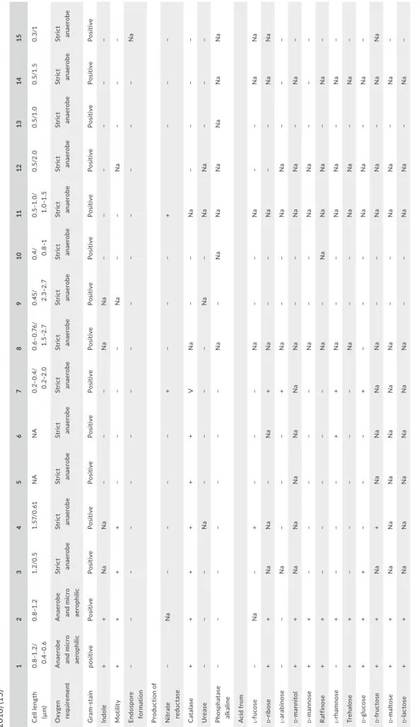
T A B LE 2 Di ff er en tia l c ha ra ct er is tic s o f R aou ltib ac ter m as sili en sis s tr ain M ar sei lle‐P 28 49 T (1 ), Raou ltib ac ter tim on en sis s tr ain M ar sei lle‐P 32 77 T (2 ), G or donib ac ter p am el ae ae s tr ai n 7 ‐1 0‐1 ‐b T (W ür de m an n e t a l., 2 00 9) ( 3) , G or donib ac ter u ro lit hin fac ien s C EB A S 1/ 15 P T (S el m a, T om ás ‐B ar be rá n, B el tr án , G ar cí a‐ V ill al ba , & E sp ín , 2 01 4) (4 ), Eg ge rt he lla si ne nsi s H K U 14 T (L au e t a l., 2 00 4) (5 ), Pa rae gg er th ell a h ong kong en sis s tr ai n H K U 10 T (L au e t a l., 2 00 4; W ür de m an n e t a l., 2 00 9) ( 6) , E gg er th ell a l en ta J CM 9 97 T (W ad e e t a l., 1 99 9) ( 7) , Ad ler cr eu tz ia e qu oli fac ien s s tr ai n D SM 19 45 0 T (M ar uo , S ak am ot o, It o, T od a, & B en no , 2 00 8) (8 ), A sac cha rob ac ter c el at us s tr ain d o0 3 T (M in am id a e t a l., 2 00 8) ( 9) , C ry pt ob ac te riu m c ur tu m s tr ain 1 2‐ 3 T (N ak az aw a et a l., 1 99 9) (1 0) , D en itr ob ac ter iu m d et ox ifi ca ns s tr ain N PO H 1 T (A nder so n, R as m us sen , Jen sen , & A lli so n, 2 00 0) (1 1) , E nt er or ha bdu s m uc os ic ol a s tr ain M t1 B 8 T (C lav el e t a l., 2 00 9) ( 12 ), Sl ack ia e xi gu a st ra in S ‐7 T (W ad e e t a l., 1 99 9) ( 13 ), Ell ag ib ac ter is ou ro lit hini fac ien s C EB A S 4A T (B el tr án , R om o‐ Va qu er o, E sp ín , T om ás ‐B ar be rá n, & S el m a, 2 01 8) (1 4) , R ub ner ib ac ter b ad eni en sis R es AG ‐8 5 T (D an yl ec e t a l., 20 18 ) ( 15 ) 1 2 3 4 5 6 7 8 9 10 11 12 13 14 15 C el l l en gt h (µ m ) 0. 8–1 .2 / 0.4– 0. 6 0. 8–1 .2 1. 2/0 .5 1. 57/ 0. 61 NA NA 0. 2– 0. 4/ 0. 2– 2.0 0. 6– 0. 76 / 1. 5–2 .7 0. 45/ 2.3– 2. 7 0. 4/ 0.8–1 0. 5– 1.0 / 1. 0– 1. 5 0. 5/ 2.0 0. 5/ 1.0 0.5 /1 .5 0. 3/1 O xy ge n re qu ire m en t A na er ob e an d micr o ae ro ph ili c A na er ob e an d micr o ae ro ph ili c St ric t an ae rob e St ric t an ae rob e St ric t an ae rob e St ric t an ae rob e St ric t an ae rob e St ric t an ae rob e St ric t an ae rob e St ric t an ae rob e St ric t an ae rob e St ric t an ae rob e St ric t an ae rob e St ric t an ae rob e St ric t an ae rob e G ra m‐ st ai n pos iti ve Pos iti ve Pos iti ve Pos iti ve Pos iti ve Pos iti ve Pos iti ve Pos iti ve Pos iti ve Pos iti ve Pos iti ve Pos iti ve Pos iti ve Pos iti ve Pos iti ve In dole + + N a N a − − − N a N a − − − − − − M ot ili ty + + + + − − − − N a − − N a − − − En dos po re for m at ion − − − − − − − − − − − − − − N a Pr odu ct ion o f N itr ate re du ct as e − N a − − − − + − − − + − − − C at al as e + + + + + + V N a − − N a − − − − U re as e − − − N a − − − − N a − N a N a − − − Ph os pha ta se al ka lin e − − − − − − − N a − N a N a N a N a N a N a A cid fro m l ‐f uc os e − N a − + − − − N a − − N a − − N a N a d ‐r ib os e + + N a N a − N a + N a − − N a − − N a N a l ‐a ra bi no se − − N a − − − + N a − − N a N a − − − d ‐m ann itol + + N a N a N a N a N a N a − − N a N a − N a − d ‐m ann os e + + − − − − − N a − − N a N a − − − Raf fin os e + + − − − − − N a − N a N a N a − N a − l ‐r ha mn ose − + − − − + + N a − − N a N a − N a − Tre ha los e + + − − − − − N a − − N a N a − N a − d ‐g lu co se + + + − − − + − − − N a N a − N a − d ‐f ru ct os e + + N a + N a N a N a N a − − N a N a − N a N a d ‐m al to se + + N a N a N a N a N a N a − − N a N a − N a − d ‐la ct os e + + N a N a N a N a N a N a − − N a N a − N a − (Con tinues )
3.9 nmol/L for strains Marseille‐P2849T and Marseille‐P3277T,
respectively.
The libraries were normalized at 2 nM and pooled. After a dena‐ turation step and dilution at 15 pM, the pool of libraries was loaded onto the reagent cartridge and then onto the instrument along with the flow cell. Automated cluster generation and sequencing run were performed in a single 39‐hr run in a 2 × 151‐bp.
For strain Marseille‐P2849T, total information of 4.5 Gb was ob‐
tained from a 477 K/mm2 cluster density with a cluster passing qual‐
ity control filters of 94.8% (8,444,000 passing filter paired reads). Within this run, the index representation for strain Marseille‐P2849T
was determined to be of 8.34%. For strain Marseille‐P3277T, total in‐
formation of 6.3 Gb was obtained from a 673 K/mm2 cluster density
with a cluster passing quality control filters of 95.4% (12,453,000 clusters). Within this run, the index representation for this strain was determined to be of 7.29%. The 769,472 and 907,611 paired reads of strains Marseille‐P2849T and Marseille‐P3277T, respectively, were
trimmed, assembled, annotated, and analyzed using the same pipe‐ line adapted in our previous studies (Elsawi et al., 2017).
2.8 | Genome annotation and analysis
Prodigal was used for open reading frame (ORF) prediction (Hyatt et al., 2010) with default parameters. We excluded predicted ORFs spanning a sequencing gap region (containing N). The bacterial pro‐ teome was predicted using BLASTP (E‐value of 1e03, coverage of 0.7 and identity percent of 30) against the clusters of orthologous groups (COGs) database. If no hit was found, we searched against the nr database (Clark, Karsch‐Mizrachi, Lipman, Ostell, & Sayers, 2016) using BLASTP with an E‐value of 1e03, coverage 0.7, and an identity percent of 30. An E‐value of 1e05 was used if the length of sequences was smaller than 80 amino acids. PFam conserved domains (PFAM‐A and PFAM‐B domains) were searched on each protein with the hhmscan tools analysis. RNAmmer (Lagesen et al., 2007) and tRNAScanSE tool (Lowe & Chan, 2016) were used to find ribosomal rRNAs genes and tRNA genes, respectively. ORFans were identified if all the BLASTP performed had negative results (E‐value inferior to 1e03 for ORFs with sequence size above 80 aa or E‐value inferior to 1e05 for ORFs with sequence length smaller than 80 aa). For data management and visualization of genomic features, Artemis (Carver, Harris, Berriman, Parkhill, & McQuillan, 2012) was used. We used the MAGI in‐house software to analyze the mean level of nu‐ cleotide sequence similarity at the genome level. It calculated the average genomic identity of gene sequences (AGIOS) among com‐ pared genomes (Ramasamy et al., 2014). This software combines the Proteinortho software (Lechner et al., 2011) for detecting or‐ thologous proteins in pairwise genomic comparisons. Then, the corresponding genes were retrieved and the mean percentage of nucleotide sequence identity among orthologous ORFs was deter‐ mined using the Needleman–Wunsch global alignment algorithm.
We also used the Genome‐to‐Genome Distance Calculator web service to calculate digital DNA:DNA hybridization estimates (dDDH) with confidence intervals under recommended settings
1 2 3 4 5 6 7 8 9 10 11 12 13 14 15 D N A G +C co nte nt (m ol%) 59. 01 59. 6 66 .4 66 .4 64 .9 61 .1 63. 8 63. 5 63 50 .9 59. 5 64. 2 62 .1 59. 6 65 .1 Is ol at ion sou rc e H uma n fec es H uma n fec es H uma n col on H uma n fec es B lo od cu ltu re B lo od cu ltu re H uma n fec es H uma n fec es Ra t c ec um H uma n or al cav iti es B ovi ne ru m en Ile al muc os a of mic e H uma n or al le sio ns H uma n fec es H uma n fec es N ote . N A : d at a n ot a va ila bl e; v : v ar ia bl e. T A B LE 2 (Co nti nue d)
(Formula 2, BLASTþ) (Auch, Klenk, & Göker, 2010; Meier‐Kolthoff, Auch, Klenk, & Göker, 2013).
3 | RESULTS
3.1 | Strain identification by MALDI‐TOF‐MS and
16S rRNA sequencing
Matrix‐assisted laser desorption/ionization‐TOF‐MS failed to iden‐ tify strains Marseille‐P2849T and P3277T at the genus and spe‐
cies levels (score <1.7). The spectra of strain Marseille‐P2849T and
Marseille‐P3277T were added to our URMS database (Supporting
Information Figure S1). A gel view comparing the available mass spectrum of the new isolated species to the mass spectrum of its phylogenetically close species was done (Figure 1). Mass spectrum of each organism was unique and did not match any other spectrum, confirming the novelty of both studied strains.
Strain Marseille‐P2849T exhibited a 91.4% 16S rRNA gene se‐
quence similarity with Gordonibacter urolithinfaciens strain CEBAS 1/15 PT (GenBank accession number HG000667), the phylogenet‐ ically closest species with standing in nomenclature (Figure 2), sug‐ gesting it as a new genus within the family Eggerthellaceae, namely
Raoultibacter. As for strain Marseille‐P3277T, it exhibited a 97.96%
sequence similarity with strain Marseille‐P2849T, suggesting it as
a new species within the Raoultibacter genus. The 16S rRNA se‐ quences of strains Marseille‐P2849T and Marseille‐P3277T were
deposited in EMBL‐EBI under accession numbers LT576395 and LT623894, respectively.
3.2 | Phenotypic characteristics and
biochemical features
Strains Marseille‐P2849T and Marseille‐P3277T form translucent
microcolonies on 5% sheep blood‐enriched Columbia agar (bio‐ Mérieux) with a mean diameter ranging from 0.1 to 0.4 mm. The growth of both strains was observed in anaerobic and microaero‐ philic atmospheres at 28, 37, and 45°C but optimally under an‐ aerobic conditions at 37°C after 48 hr of incubation. No growth was obtained at 55°C or in aerobic atmosphere. Bacterial cells were motile, Gram‐positive (Figure 3a,b), and nonsporeforming rod. Strain Marseille‐P2849T cells had a length ranging between
0.8 and 1.2 μm with a mean diameter ranging from 0.4 to 0.6 μm (Figure 3c,d). As for strain Marseille‐P3277T, its cells were 1–2 μm
long with a mean diameter ranging from 0.35 to 0.44 μm. Both strains were catalase positive, oxidase negative, tolerated pH levels ranging between 6 and 8.5 and could not sustain NaCl
TA B L E 3 Cellular fatty acid composition (%) of strain Marseille‐P2849T and strain Marseille‐P3277T compared with other type strains of
closely related species: 1, strain Marseille‐P2849T; 2, strain Marseille‐P3277T; 3, Gordonibacter urolithinfaciens strain CEBAS 1/15PT; 4,
Gordonibacter pamelaeae strain 7‐10‐1‐bT; 5, Paraeggerthella hongkongensis DSM 16106T; 6, Eggerthella lenta DSM 2243T; 7, Eggerthella
sinensis DSM 16107T
Fatty acids 1 2 3 4 5 6 7
C18:1n9 9‐octadecenoic acid 36.4 38.1 27 6.8 55.1 42.3 36.6
C16:0 Hexadecanoic acid 18.2 25.4 4.4 4.5 7.1 6.7 7.6
C14:0 Tetradecanoic acid 12.7 10.9 5.2 16.3 6.9 12.5 7.7
C15:0 anteiso 12‐methyl‐tetradecanoic acid 7.3 1.4 22.7 36.9 1.1 16.3 21.2
C18:2n6 9,12‐octadecadienoic acid 6.7 9 ND ND 1.4 ND ND
C18:0 Octadecanoic acid 3.4 5.7 5.6 1.5 4.7 1.4 1.5
C18:1n7 11‐octadecenoic acid 3.2 3.7 1.4 ND 4.3 2.6 2.3
C15:0 iso 13‐methyl‐tetradecanoic acid 2.8 2.8 3.6 5.5 0 1.1 0
C12:0 Dodecanoic acid 1.8 1.8 TR 5 7.7 2.9 1.1
C13:0 iso 11‐methyl‐dodecanoic acid 1.5 ND TR 2 ND ND ND
C14:0 iso 12‐methyl‐tridecanoic acid 1.4 ND 13.4 18.3 0 7.5 17.1
C15:0 Pentadecanoic acid 1.2 1.1 ND ND ND ND ND
13:0 anteiso 10‐methyl‐dodecanoic acid 1.1 ND ND ND ND ND 1
C20:4n6 5,8,11,14‐eicosatetraenoic acid TR 1.2 ND ND ND ND ND
C20:5n3 5,8,11,14,17‐eicosapentaenoic
acid
ND TR ND ND ND ND ND
C5:0 iso 3‐methyl‐butanoic acid TR ND ND ND ND ND ND
C13:0 Tridecanoic acid TR ND ND ND ND ND ND
C16:1n7 9‐hexadecenoic acid TR ND 2 3.2 8.8 4.4 2.6
Note. Values represent the percentage of total identified fatty acid methyl esters only (aldehydes, dimethyl acetals and unidentified “summed features”
described previously were not included). Data of the close species were taken as reported by Selma et al. (2014). ND: not detected; TR: trace amounts <1%.
concentration >5 g/L. The classification and general features of strains Marseille‐P2849T and Marseille‐P3277T are summarized in
Table 1.
Using an API® 50CH strip (bioMérieux), positive reactions were observed for both strains for glycerol, d‐ribose, d‐galac‐
tose, d‐glucose, d‐fructose, d‐mannose, d‐mannitol, d‐arabitol,
N‐acetylglucosamine, amygdaline, arbutin, esculin ferric citrate,
salicin, d‐maltose, d‐lactose, d‐saccharose, d‐trehalose, d‐melezi‐
tose, gentiobiose, d‐tagatose, and potassium gluconate. In addi‐
tion, positive reactions were observed for strain Marseille‐P2849T
with amidon and potassium 5‐ketogluconate, and for strain Marseille‐P3277T with methyl‐αd‐glucosamine, d‐cellobiose, and d‐turanose (Table 2). Negative reactions were observed for both
strains for erythritol, A‐arabinose, l‐Arabinose, d‐Xylose, l‐xylose, d‐adonitol, methyl‐βd‐xylopyranoside, l‐sorbose, l‐rhamnose, dul‐
citol, inositol, methyl‐αd‐mannopyranoside, methyl‐αd‐glucopy‐ ranoside, d‐cellobiose, d‐melibiose, inulin, d‐raffinose, glycogen,
xylitol, d‐turanose, d‐xylose, d‐fucose, l‐fucose, l‐arabitol, and po‐
tassium 2‐ketogluconate.
Using an API® 20A strip (bioMérieux), both strains produced indole. In addition, positive reactions were observed for d‐glucose, d‐mannitol, d‐lactose, d‐saccharose, d‐maltose, salicin, l‐arabinose,
gelatine, d‐mannose, esculin ferric citrate, d‐cellobiose d‐melezitose, d‐raffinose, d‐sorbitol, and d‐trehalose for both strains. Positive re‐
action was observed for strain Marseille‐P3277T, but not Marseille‐
P2849T, with l‐rhamnose. No reaction was obtained for urease and d‐xylose for both strains.
Using an API® ZYM strip (bioMérieux), both strains exhibited esterase (C4), esterase lipase (C8), lipase (C14), leucine arylamidase, valine arylamidase, cystine arylamidase, phosphatase acid, and naphthol phosphohydrolase activities but no phosphatase alkaline was observed. In addition, positive reactions were observed for strain Marseille‐P3277T with trypsin, α‐chymotrypsin, α‐galactosi‐
dase, β‐galactosidase, β‐glucuronidase, α‐glucosidase, β‐glucosidase,
N‐acetyl‐β‐glucosaminidase, and α‐mannosidase. An α‐fucosidase
activity was observed only for strain Marseille‐P2849T.
The major fatty acids identified for strains Marseille‐P2849T and
Marseille‐P3277T were 9‐octadecenoic acid (Cl8:ln9, 36% and 38%,
Raoultibacter massiliensis Raoultibacter timonensis
Number Percent (%) Number Percent (%)
Size (bp) 3,657,161 100 4,000,215 100
Number of G+C 2,158,456 59 2,396,128 59.9
Number total of genes 3,073 100 3,284 100
Total number of protein‐coding genes
3,025 98.4 3,232 99.33
Total number of RNA Genes
48 1.56 52 1.58
Total number of tRNA Genes
45 1.6 48 1.46
Total number of rRNA (5S, 16S, 23S) Genes
3 0.1 3 0.12
Coding sequence gene
protein size 3,156,910 86.3 3,498,188 87.45 Number of proteins associated with clusters of ortholo‐ gous groups 2,365 77 2,562 78.01 Number of proteins associated with orfan
253 8,23 323 9.83
Number of proteins
with peptide signal 385 12,5 512 15.59
Number of genes associated with PKS or NRPS 6 0.18 14 0.45 Number of genes associated with virulence 470 15.3 481 14.64 Number of proteins with TMH 855 27.8 940 28.62 Notes. The total is based on either the size of the genome in base pairs or the total number of pro‐
tein‐coding genes in the annotated genome.
TA B L E 4 Nucleotide content and gene
count levels of the genome of strain
Raoultibacter massiliensis Marseille‐P2849T
and Raoultibacter timonensis strain Marseille‐P3277T
respectively), hexadecanoic acid (C16:0, 18% and 25%), and tetrade‐ canoic acid (Cl4:0, 13% and 11%; Table 3). Strain Marseille‐P3277T
exhibited unusually long chain fatty acids (C20:4n6 and C20:5n3). Among tested antibiotics, strains Marseille‐P2849T and Marseille‐
P3277T were susceptible to amoxicillin (MIC 0.50 and 1 µg/ml, re‐
spectively), imipenem (0.047 and 0.047 µg/ml), metronidazole (0.023 and 0.064 µg/ml), rifampicin (0.003 and 0.008 µg/ml), and erythro‐ mycin (0.32 and 0.016 µg/ml). Both strains were resistant to dapto‐ mycin, minocycline, amikacin, vancomycin, and cefotaxime.
3.3 | Genomic properties
The draft genome of strain Marseille‐P2849T was 3,657,161‐bp long
with a G+C content of 59.02 mol% (Table 4; Figure 4a). It was com‐ posed of nine scaffolds (35 contigs). Of the 3,073 predicted genes, 3,025 were protein‐coding genes and 48 were RNAs (one complete rRNA operon and 45 tRNA genes). A total of 2,365 proteins (76.86%) were assigned to COGs and 253 genes were identified as ORFans (8.23%). Six genes were associated with polyketide synthases (PKS) or nonribosomal peptide synthetases (NRPS; 0.18%) and 470 genes were associated with virulence (15.29%). As for strain Marseille‐ P3277T, the genome size was 4,000,215‐bp long with a 59.9 mol%
G+C content (Figure 4b). It was composed of 21 scaffolds (composed of 84 contigs). Of the 3,284 predicted genes, 3,232 were protein‐ coding genes and 52 were RNAs (one complete rRNA operon and 49 tRNA genes). A total of 2,562 proteins (78.01%) were assigned to COGs and 323 genes were identified as ORFans (9.83%). The genome of strain Marseille‐P3277T contained 14 genes associated with PKS
or NRPS (0.45%) and 481 genes associated with virulence (14.64%). The genome statistics are presented in Table 4, and the distribution of genes into COGs functional categories is summarized in Table 5.
3.4 | Genomic comparison
The draft genome sequence structure of strains Marseille‐P2849T
and Marseille‐P3277T is summarized in Figure 4. The draft ge‐
nome sequence of strain Marseille‐P2849T was larger than that of
G. urolithinfaciens, Atopobium fossor, Denitrobacterium detoxificans, Atopobium parvulum, Olsenella profusa, Olsenella uli, E. lenta, and Gordonibacter pamelaeae (3.29, 1.66, 2.45, 1.54, 2.72, 2.05, 3.63,
and 3.61 Mb, respectively) but smaller than that of strain Marseille‐ P3277T (3.94 Mb, Table 6). The G+C content of strains Marseille‐
P2849T and Marseille‐P3277T was larger than those of A. fossor
and A. parvulum (59.02 and 59.9 vs. 45.4 and 45.7, respectively), but smaller than those of G. urolithinfaciens, D. detoxificans, G. pamelaeae,
E. lenta, O. profusa, and O. uli (66.1 59.5%, 64.0%, 64.2%, 64.2%, and
64.7%, respectively). The gene content of strain Marseille‐P2849T
was smaller than that of strain Marseille‐P3277T (3,073 and 3,284,
respectively), but larger than that of G. urolithinfaciens, A. fossor,
G. pamelaeae, D. detoxificans, A. parvulum, O. profusa, and E. lenta
(2,836, 1,487, 2,027, 1,762, 1,353, 2,650, and 3,070, respectively). The distribution of functional classes of predicted genes of strains Marseille‐P2849T and Marseille‐P3277T according to the COGs of
proteins is summarized in Figure 5.
Strain Marseille‐P2849T shared 1,542, 1,370, 555, 571,
1,069, 693, 683, 1,084, 1,404, and 911 orthologous proteins with strain Marseille‐P3277T, G. urolithinfaciens, A. parvulum, A. fossor,
Adlercreutzia equolifaciens, Olsenella umbonata, O. profusa, G. pa‐ melaeae, E. lenta, and D. detoxificans, respectively. The AGIOS val‐
ues among the eight most closely related species ranged between 58.12% and 81.35%. When compared to these eight species, strain Marseille‐P2849T AGIOS values ranged from 58.97% with A. fos‐
sor to 73.75% with G. pamelaeae. Similarly, strain Marseille‐P3277T
F I G U R E 4 Graphical circular map of the genome of (a) Raoultibacter massiliensis gen. nov., sp. nov. strain Marseille‐P2849T and (b) strain
Raoultibacter timonensis gen. nov., sp. nov. strain Marseille‐P3277T. From the outside to the center, contigs (red/gray), clusters of orthologous
groups (COGs) category of genes on the forward strand (three circles), genes on the forward strand (blue circle), genes on the reverse strand (red circle), COG category of genes on the reverse strand (three circles), G+C skew (purple indicates positive values and olive negative values)
exhibited AGIOS values ranging from 58.95% with A. fossor to 74.19% with G. pamelaeae (Table 7). The AGIOS values obtained for strains Marseille‐P2849T and Marseille‐P3277T, between 58.12%
and 81.35%, support their new species status.
In addition, dDDH values obtained between strain Marseille‐ P2849T, strain Marseille‐P3277T, G. urolithinfaciens, A. parvulum,
A. fossor, A. equolifaciens, O. umbonata, O. profusa, G. pamelaeae, E. lenta, and D. detoxificans were of 25.2% (22.9–27.7), 22.4%
Code Raoultibacter massiliensis Raoultibacter timonensis Description
Value % of total Value % of total
[J] 134 4.43 142 4.39 Translation
[A] 0 0 0 0 RNA processing and
modification
[K] 264 8.73 291 9.01 Transcription
[L] 102 3.37 95 2.94 Replication, recombination and
repair
[B] 0 0 0 0 Chromatin structure and
dynamics
[D] 23 0.76 16 0.5 Cell cycle control. mitosis and
meiosis
[Y] 0 0 0 0 Nuclear structure
[V] 64 2.12 57 1.76 Defense mechanisms
[T] 181 5.98 214 6.62 Signal transduction mechanisms
[M] 121 4 115 3.56 Cell wall/membrane biogenesis
[N] 8 0.26 9 0.28 Cell motility
[Z] 0 0 0 0 Cytoskeleton
[W] 0 0 0 0 Extracellular structures
[U] 18 0.6 20 0.62 Intracellular trafficking and
secretion
[O] 83 2.74 86 2.66 Posttranslational modification,
protein turnover, chaperones
[X] 5 0.17 2 0.06 Mobilome: prophages,
transposons
[C] 409 13.52 477 14.76 Energy production and
conversion
[G] 118 3.9 132 4.08 Carbohydrate transport and
metabolism
[E] 160 5.29 171 5.29 Amino acid transport and
metabolism
[F] 55 1.82 58 1.79 Nucleotide transport and
metabolism
[H] 65 2.15 69 2.13 Coenzyme transport and
metabolism
[I] 49 1.61 55 1.7 Lipid transport and metabolism
[P] 120 3.97 139 4.3 Inorganic ion transport and
metabolism
[Q] 18 0.6 21 0.65 Secondary metabolites
biosynthesis, transport and catabolism
[R] 214 7.07 226 6.99 General function prediction only
[S] 154 5.09 167 5.18 Function unknown
– 660 21.82 670 20.73 Not in COGs
Note. The total is based on either the size of the genome in base pairs or the total number of protein‐
coding genes in the annotated genome.
TA B L E 5 Number of genes associated
with the 25 general clusters of orthologous group (COG) functional categories
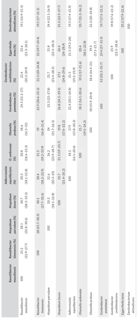
(20.2–24.9), 28.1% (25.8–30.6), 30.7% (28.3–33.2), 20.3% (18.1– 22.8), 20.8% (18.6–23.3), 18.6% (16.5–21), 24.5% (22.2–27), 23.6% (21.3–26.1), and 19.1% (16.9–21.5), respectively (Table 8). These dDDH values were lower than the 70%, value threshold for species demarcation, thus confirming that the two studied strains are repre‐ sentative of two new species (Meier‐Kolthoff et al., 2013).
4 | DISCUSSION
Culturomics is a high‐throughput culture approach that enabled the isolation of approximately 1,057 bacterial species including 247 new species from the human gut in our laboratory (Jean‐ Christophe Lagier et al., 2016). Along with the development of culturomics, a new polyphasic approach, taxonogenomics, was developed in order to describe novel bacterial species using their biochemical, proteomic, and genomic properties (Fournier & Drancourt, 2015). This approach has the advantage of exhibiting a higher inter and intralaboratory reproducibility when compared
to DNA‐DNA hybridization and chemotaxonomic methods. Based on MALDI‐TOF‐MS analysis, 16S rRNA gene sequence compari‐ son (<95% similarity), genome comparison, AGIOS and dDDH values, we propose the creation of the new genus Raoultibacter gen. nov within the family Eggerthellaceae that belongs to the phylum Actinobacteria. Members of this family belong to the class Coriobacteriia. Many revisions have been made to the clas‐ sification of this group by using various molecular techniques and Gupta et al. (2013 proposed the taxonomic division of this class into two orders (Coriobacteriales and Eggerthellales) and three families including Coriobacteriaceae, Atopobiaceae, and
Eggerthellaceae (Stackebrandt, Rainey, & Ward‐Rainey, 1997).
Members of the Eggerthellaceae are predominantly anaerobic, nonsporeforming, catalase and Gram‐positive, rods or cocci. As well, strains Marseille‐P2849T and Marseille‐P3277T are
Gram‐positive. Most of the species closely related to the genus
Raoultibacter gen. nov. were isolated from the human gut micro‐
biota and, to date, exhibited a low pathogenicity (Gardiner et al., 2014; Lee et al., 2012).
F I G U R E 5 Distribution of functional classes of predicted genes according to the clusters of orthologous groups of proteins of
Raoultibacter massiliensis gen. nov., sp. nov. strain Marseille‐P2849T and strain Raoultibacter timonensis gen. nov., sp. nov. strain Marseille‐
5 | CONCLUSION
The biochemical, proteomic, genetic, and genomic characteristics of strains Marseille‐P2849T and Marseille‐P3277T confirmed that
they belong to two distinct species within a new genus in the fam‐ ily Eggerthellaceae, for which we propose the names Raoultibacter gen. nov., R. massiliensis sp. nov., and R. timonensis sp. nov. The type strain of R. massiliensis sp. nov., Marseille‐P2849T, was isolated from
the feces of a 19‐year‐old healthy male Saudi Bedouin, whereas the type strain of R. timonensis sp. nov., Marseille‐P3277T, was iso‐
lated from the feces of a healthy 11‐year‐old Pygmy female living in Congo.
6 | TA XONOMIC AND NOMENCL ATUR AL
PROPOSALS
6.1 | Description of Raoultibacter gen. nov.
Raoultibacter (ra.ou.l.ti.bac'ter. N.L. masc. n, “Raoultibacter,” composed
of Raoult, in the honor of the French microbiologist Didier Raoult, founder of the IHU Mediterranée‐Infection in Marseille and inventor of culturomics, the culture strategy that has enabled the discovery of more than 250 bacterial species, and bacter, for rod).
Raoultibacter forms transparent microcolonies on blood agar
with a mean diameter of 0.1–0.4 mm. Cells are Gram‐positive, non‐ sporeforming, motile rod that grow in microaerophilic and anaerobic atmospheres, with an optimal growth at 37°C after 48 hr of incuba‐ tion. The pH tolerance ranges from 6 to 8.5. The type species of the genus is R. massiliensis sp. nov. The type strain of the genus is strain Marseille‐P2849T.
6.2 | Description of Raoultibacter massiliensis
sp. nov.
Raoultibacter massiliensis (mas.si.li.en'sis. L. masc. adj. massiliensis,
from Massilia, the Latin name of Marseille, where the type strain was first isolated).
Raoultibacter massiliensis is a Gram‐positive and motile rod whose
individual cells measure 0.8–1.2 µm in length and 0.4–0.6 µm in di‐ ameter. Transparent microcolonies obtained on 5% sheep blood‐en‐ riched Columbia agar exhibit a diameter of 0.1–0.4 mm. The optimal growth is observed at 37°C after 48 hr of incubation. It is oxidase negative but catalase positive. Indole is produced. Using API strips, positive reactions are observed with glycerol, d‐ribose, d‐galactose, d‐glucose, d‐fructose, d‐mannose, d‐mannitol, N‐acetylglucos‐
amine, amygdaline, arbutin, esculin ferric citrate, salicin, d‐maltose, d‐lactose, d‐saccharose, d‐trehalose, d‐melezitose, gentiobiose, d‐tagatose, potassium gluconate, l‐arabinose, gelatine, d‐cellobiose, d‐melezitose, d‐raffinose, d‐sorbitol, amidon, and potassium 5‐ke‐
togluconate. Fucosidase, esterase (C4), esterase lipase (C8), lipase (C14), leucine arylamidase, valine arylamidase, cystine arylamidase, acid phosphatase, and naphthol phosphohydrolase activities are present but no reaction is obtained for urease and alkaline phos‐ phatase. The major fatty acids are 9‐octadecenoic acid (36%), hexa‐ decanoic acid (18%), and tetradecanoic acid (13%). The genome is 3,657,161 bp long with a DNA G+C content of 59.02 mol%. The 16S rRNA and genome sequences were both deposited in EMBL/ EBI under accession numbers LT576395 and FZQX00000000, re‐ spectively. The habitat of this bacterium is the human gut. The type strain Marseille‐P2849T (= CSUR P2849 = DSM 103407) was iso‐
lated from a stool specimen of a healthy 19‐year‐old male Bedouin living in Saudi Arabia.
TA B L E 6 Genome comparison of species closely related to Raoultibacter massiliensis strain Marseille‐P2849T and Raoultibacter timonensis
strain Marseille‐P3277T
Species INSDC identifier Size (Mb) G+C (mol %) Gene Content
Raoultibacter massiliensis strain
Marseille‐P2849T FZQX00000000 3.65 59.01 3,021
Raoultibacter timonensis strain
Marseille‐P3277T OEPT00000000 3.94 59.6 3,277
Eggerthella lenta strain DSM 2243T NC_013204.1 3.63 64.2 3,146
Denitrobacterium detoxificans strain
NPOH1T NZ_CP011402.1 2.45 59.5 2,023
Gordonibacter pamelaeae strain 7‐10‐1‐bT NC_021021.1 3.61 64 3,352
Atopobium fossor strain ATCC 43386T AXXR00000000.1 1.66 45.4 1,505
Atopobium parvulum strain DSM 20469T NC_013203.1 1.54 45.7 1,406
Olsenella profusa strain DSM 13989T AWEZ00000000.1 2.72 64.2 2,707
Olsenella uli strain ATCC 49627T CP002106.1 2.05 64.7 1,822
Adlercreutzia equolifaciens strain
DSM19450T NC_022567.1 2.86 63.5 2,326
Gordonibacter urolithinfaciens strain
CEBAS 1/15PT NZ_LT900217.1 3.29 66.1 2,836
6.3 | Description of Raoultibacter timonensis sp. nov.
Raoultibacter timonensis (ti.mo.nen'sis, N.L. masc. adj., timonensis per‐
taining to La Timone, the name of the university hospital in Marseille, France, where the strain was first isolated).
Raoultibacter timonensis is a Gram‐positive and motile rod whose
individual cells measure 1–2 µm in length and 0.35–0.44 µm in diam‐ eter. Transparent microcolonies grown on 5% sheep blood‐enriched Columbia agar have a diameter of 0.1–0.4 mm with an optimal growth at 37°C after a 48 hr incubation period in anaerobic conditions. It is oxidase negative and catalase positive. Using API strips, positive re‐ actions are observed with glycerol, d‐ribose, d‐galactose, d‐glucose, d‐fructose, d‐mannose, d‐mannitol, N‐acetylglucosamine, amygdaline,
arbutin, esculin ferric citrate, salicin, d‐maltose, d‐lactose, d‐saccha‐
rose, d‐trehalose, d‐melezitose, gentiobiose, d‐tagatose, methyl‐ αd‐
glucosamine, d‐cellobiose, d‐turanose, l‐rhamnose, glycerol, potassium
gluconate, l‐arabinose, gelatin, d‐cellobiose, d‐melezitose, d‐raffinose,
and d‐sorbitol. Trypsin, α‐chymotrypsin, α‐galactosidase, β‐galactosi‐
dase, β‐glucuronidase, α‐glucosidase, β‐glucosidase, N‐acetyl‐β‐glu‐ cosaminidase, α‐mannosidase, exhibited esterase (C4), esterase lipase (C8), lipase (C14), leucine arylamidase, valine arylamidase, cystine arylamidase, acid phosphatase, and naphthol phosphohydrolase ac‐ tivities are present. No reactions are obtained for urease and phos‐ phatase alkaline. The major fatty acids are 9‐octadecenoic acid (38%), hexadecanoic acid (25%), and tetradecanoic acid (11%). The genome is 4,000,215‐bp long with a DNA G+C content of 59.9 mol%. The 16S rRNA and genome sequences were deposited in EMBL/EBI under accession numbers LT623894 and OEPT00000000, respectively. The habitat of this bacterial strain is the human gut. The type strain Marseille‐P3277T (= CSUR P3277 = CCUG 70680) was isolated from
the human stool of a 11‐year‐old healthy Pygmy female.
ACKNOWLEDGMENTS
This study was supported by IHU Méditerranée Infection, Marseille, France, and by the French Government under the “Investissements d’avenir” (Investments for the Future) program managed by the Agence Nationale de la Recherche (ANR, fr: National Agency for Research), (reference: Méditerranée Infection 10‐IAHU‐03). This work was supported by Région Provence Alpes Côte d’Azur and European funding FEDER PRIMI.
CONFLIC T OF INTEREST
The authors declare no conflict of interest.
AUTHORS CONTRIBUTION
Sory Ibrahima Traore and Melhem Bilen isolated the bacteria and drafted the manuscript. Sory Ibrahima Traore, Melhem Bilen, Maxime Descartes Mbogning Fonkou, and Fréderic Cadoret par‐ ticipated to experiment for phenotypic characterization of these strains. Caroline Michelle performed the genomic sequencing. Fadi
T A B LE 7 N um be r o f o rt ho lo go us p ro te in s sh ar ed b et w ee n ge no m es (u pp er ri gh t) an d A G IO S va lu es (% ) o bt ai ne d (lo w er le ft ) Ra ou lti ba cte r m as si lie ns is Ra ou lti ba cte r timon en si s Ato po bi um par vu lum Ato po bi um fo ss or A dl ercre ut zi a equ oli fa ci en s O ls enel la umbon at a O ls enel la pr of us a G or don ib ac te r pa mel ae ae G or don ib ac te r ur ol ith in fa ci en s Eg ger thel la le nta D en itr ob ac te riu m de to xi fic an s Raou ltib ac ter m as si lien si s 3, 025 1, 542 555 571 1, 069 69 3 683 1, 08 4 1, 37 0 1,4 04 911 Raou ltib ac ter tim on en si s 81 .2 5 3, 23 2 52 9 552 1, 029 647 643 1, 08 6 1, 057 1, 37 3 86 3 At op ob iu m p ar vu lu m 59. 35 59. 27 1, 36 3 70 6 52 3 772 76 9 41 2 43 4 576 53 4 At op ob iu m fo ss or 58 .9 7 58 .9 5 66 .76 1, 487 546 774 75 4 42 5 50 0 605 54 1 Ad le rc re ut zi a e qu oli fa ci en s 69 .69 70.0 9 58 .3 58 .1 2 2, 27 8 649 621 770 60 9 1, 094 861 O lse nel la u m bo nat a 64 .29 64 .82 63 .57 62 .6 6 66 .2 2, 059 90 9 496 40 9 719 645 O lse nel la p ro fu sa 63 .8 1 64 .37 62 .9 5 62 .7 3 65 .9 7 74 .2 1 2, 59 3 501 483 70 4 62 8 G or don ib ac ter p am el ae ae 73 .75 74 .1 9 58 .9 5 58 .7 3 74 .4 6 67. 76 66 .8 4 3, 228 1, 42 6 1, 056 64 4 G or don ib ac ter ur oli th in fa ci en s 72 .8 5 73 .5 8 55 .47 56 .14 74 .0 4 66 .7 66 .1 91 .6 2,7 93 987 74 5 Eg ge rt he lla le nt a 72 .9 2 73 .3 5 58 .39 58 .0 6 73 .45 67 66 .14 81 .3 5 80 .4 8 3, 11 6 921 D eni tr ob ac te riu m de to xi fic an s 68 .4 6 68 .75 60 .29 60 .14 68.8 4 64 .9 56 64. 84 70 .75 71 .0 5 69. 92 1,9 60 N ote . T he n um be r o f p ro te in s p er g en om e i s i nd ic at ed i n b ol d. T he s tr ai ns o f t he s pe ci es i nc lu de d i n t he g en om ic a na ly si s w er e g iv en i n T ab le 6 .
T A B LE 8 D ig ita l D N A– D N A h yb rid iz at io n va lu es (% ) o bt ai ne d by c om pa ris on o f R aou ltib ac ter m as sili en sis s tr ain M ar sei lle‐P 28 49 T a nd Raou ltib ac ter tim on en sis s tr ain M ar sei lle‐P 32 77 T w ith ot he r c lo se ly re la te d sp ec ie s us in g th e G G D C fo rm ul a 2 so ft w ar e (D D H e st im at es b as ed o n id en tit ie s/H SP le ng th ) a, u pp er r ig ht Ra ou lti ba cte r m as si lie ns is Ra ou lti ba cte r timon en si s ( % ) Ato po bi um par vu lum (% ) Ato po bi um fo ss or (% ) A dl ercre ut zi a equ oli fa ci en s (% ) O . u m bo na ta (% ) O ls enel la pr of us a (% ) G or don ib ac te r pa mel ae ae (% ) G or don ib ac te r ur ol ith in fa ci en s (% ) Eg ger thel la le nt a (% ) D en itr ob ac te riu m de to xi fic an s ( % ) Raou ltib ac ter m as sili en si s 10 0 25 .2 (2 2. 9– 27. 7) 28 .1 (2 5. 8–3 0. 6) 30 .7 (2 8. 3–3 3. 2) 20 .3 (1 8. 1– 22 .8 ) 20 .8 (1 8. 6–2 3. 3) 18 .6 (1 6. 5–2 1) 24 .5 ( 22 .2 –27 ) 22 .4 (20 .2 –2 4. 9) 23 .6 (2 1. 3–2 6. 1) 19. 1 ( 16 .9 –2 1. 5) Raou ltib ac ter tim on en si s 10 0 28 (2 5. 7– 30 .5 ) 30 .1 (2 7. 7– 32 .6 ) 20 .4 (1 8. 2–2 2. 9) 21 .5 (1 9. 2–2 3. 9) 19 (1 6. 8–2 1. 4) 22 .9 (2 0. 6– 25 .3 ) 22 .3 (20 –2 4. 8) 22 (1 9. 7–2 4. 4) 19. 1 ( 17 –2 1. 5) At op ob iu m p ar vu lu m 10 0 20 .3 (1 8. 1– 22 .8 ) 22 .6 (2 0. 3–2 5) 26 .2 (2 3. 9– 28 .7 ) 24 (2 1. 7– 26 .5 ) 25 .3 ( 23 –27 .8 ) 25 .7 (2 3. 4– 28 .2 ) 25 .8 (2 3. 5– 28 .3 ) 24 .4 (2 2.1 –2 6. 9) At op ob iu m fo ss or 10 0 23 .7 (2 1.4– 26 .2 ) 21 .3 ( 19 –2 3. 7) 19. 8 (17 .6 –2 2. 2) 26 .8 (2 4. 5–2 9. 3) 27 .1 (2 4. 8–2 9. 6) 26 .4 (2 4– 28 .9 ) 25 .2 ( 22 .9 –27 .7 ) Ad le rc re ut zi a eq uoli fa ci en s 10 0 18 .2 (1 6. 1– 20 .6 ) 17. 9 (1 5. 8–2 0. 3) 22 .4 (2 0.1 –24 .8 ) 21 .5 (1 9. 2–2 3. 9) 21 .5 (1 9. 3–2 4) 19. 5 ( 17 .4 –2 1. 9) O lse nel la u m bo nat a 10 0 21 .7 (1 9. 5–2 4. 2) 18 .2 (1 6. 1– 20 .6 ) 19. 2 ( 17 –2 1. 6) 20 .4 (1 8. 1– 22 .8 ) 33 .7 (3 1. 3–3 6. 2) O lse nel la p ro fu sa 10 0 18 (1 5. 9–2 0. 4) 18 .6 ( 16 .4 –21 ) 19. 3 (17 .1 –2 1. 7) 22 .3 (20 –2 4. 8) G or don ib ac ter pa m ela ea e 10 0 53 (5 0. 3–5 5. 7) 29 .4 ( 27 –3 1. 9) 19. 7 ( 17 .5 –2 2. 1) G or don ib ac ter ur oli th in fa ci en s 10 0 25 .9 (2 3. 5– 28 .4 ) 19. 8 ( 17 .6 –2 2. 2) Eg ge rt he lla le nt a 10 0 20 .2 (17 .9 –2 2. 6) D eni tr ob ac te riu m de to xi fic an s 10 0 a Th e co nf id en ce in te rv al s in di ca te t he in he re nt u nc er ta in ty in e st im at in g D N A h yb rid iz at io n es tim at es (D D H ) v al ue s fr om in te rg en om ic d is ta nc es b as ed o n m od el s de riv ed f ro m e m pi ric al t es t da ta s et s (w hi ch a re a lw ay s lim ite d in s ize ).