HAL Id: hal-02165970
https://hal.sorbonne-universite.fr/hal-02165970
Submitted on 26 Jun 2019
HAL is a multi-disciplinary open access archive for the deposit and dissemination of sci-entific research documents, whether they are pub-lished or not. The documents may come from teaching and research institutions in France or abroad, or from public or private research centers.
L’archive ouverte pluridisciplinaire HAL, est destinée au dépôt et à la diffusion de documents scientifiques de niveau recherche, publiés ou non, émanant des établissements d’enseignement et de recherche français ou étrangers, des laboratoires publics ou privés.
lymphoma in the blood of patients with Q fever
Clea Melenotte, Soraya Mezouar, Amira Ben Amara, Simon Benatti, Jacques
Chiaroni, Christian Devaux, Régis Costello, Guido Kroemer, Jean-Louis
Mege, Didier Raoult
To cite this version:
Clea Melenotte, Soraya Mezouar, Amira Ben Amara, Simon Benatti, Jacques Chiaroni, et al.. A transcriptional signature associated with non-Hodgkin lymphoma in the blood of patients with Q fever. PLoS ONE, Public Library of Science, 2019, 14 (6), pp.e0217542. �10.1371/journal.pone.0217542�. �hal-02165970�
A transcriptional signature associated with
non-Hodgkin lymphoma in the blood of
patients with Q fever
Cle´a Melenotte1☯, Soraya Mezouar1☯, Amira Ben Amara1, Simon Benatti2,
Jacques Chiaroni3,4, Christian Devaux1, Re´gis Costello5, Guido Kroemer6,7,8,9,10,11,12,13, Jean-Louis Mege1, Didier Raoult1*
1 Aix-Marseille Univ, IRD, APHM, MEPHI, IHU-Me´diterrane´ e Infection, Marseille, France, 2 Infectious Diseases, ASST Papa Giovanni XXIII, Bergamo, Italy, 3 E´ tablissement Franc¸ais du Sang Alpes
Me´diterrane´e, Marseille, France, 4 Aix-Marseille University, CNRS, ADES, "Biologie des Groupes Sanguins", Marseille, France, 5 Service d’he´matologie de la Conception, Assistance publique des hoˆpitaux de Marseille, Marseille, France, 6 Gustave Roussy Cancer Campus, Villejuif, France, 7 INSERM, U1138, Paris, France, 8 Equipe 11 labellise´ e par la Ligue Nationale contre le Cancer, Centre de Recherche des Cordeliers, Paris, France, 9 Universite´ Paris Descartes, Sorbonne Paris Cite´, Paris, France, 10 Metabolomics and Cell Biology Platforms, Gustave Roussy Cancer Campus, Villejuif, France, 11 Universite´ Pierre et Marie Curie, Paris, France, 12 Poˆle de Biologie, Hoˆpital Europe´en Georges Pompidou, AP-HP, Paris, France, 13 Karolinska Institute, Department of Women’s and Children’s Health, Karolinska University Hospital, Stockholm, Sweden
☯These authors contributed equally to this work.
*didier.raoult@gmail.com
Abstract
Coxiella burnetii, the agent causing Q fever, has been associated with B-cell non-Hodgkin
lymphoma (NHL). To better clarify this link, we analysed the genetic transcriptomic profile of peripheral blood leukocytes from patients with C. burnetii infection to identify possible links to lymphoma. Microarray analyses revealed that 1189 genes were expressed differently (p<.001 and fold change�4) in whole blood of patients with C. burnetii infection compared to controls. In addition, 95 genes expressed in patients with non-Hodgkin lymphoma (NHL) and in patients with C. burnetii persistent infection have allowed us to establish the ‘C.
bur-netii-associated NHL signature’. Among these, 33 genes previously found modulated in C. burnetii-associated -NHL by the microarray analysis were selected and their mRNA
expres-sion levels were measured in distinct C. burnetii-induced pathologies, namely, acute Q fever, focalized persistent infection, lymphadenitis and C.burnetii-associated NHL. Specific genes involved in anti-apoptotic process were found highly expressed in leukocytes from patients with C. burnetii associated-NHL: MIR17HG, REL and SP100. This signature dif-fered from that found for NHL-control group. Patients with C. burnetii lymphadenitis pre-sented significant elevated levels of BCL2 and ETS1 mRNAs. Altogether, we identified a specific transcriptionnal signature for NHL during C. burnetii infection reflecting the up-regu-lation of anti-apoptotic processes and the fact that lymphadenitis might constitute a critical step towards lymphomagenesis.
a1111111111 a1111111111 a1111111111 a1111111111 a1111111111 OPEN ACCESS
Citation: Melenotte C, Mezouar S, Ben Amara A, Benatti S, Chiaroni J, Devaux C, et al. (2019) A transcriptional signature associated with non-Hodgkin lymphoma in the blood of patients with Q fever. PLoS ONE 14(6): e0217542.https://doi.org/ 10.1371/journal.pone.0217542
Editor: Riccardo Dolcetti, Fondazione IRCCS Istituto Nazionale dei Tumori, ITALY Received: September 6, 2018 Accepted: May 14, 2019 Published: June 10, 2019
Copyright:© 2019 Melenotte et al. This is an open access article distributed under the terms of the Creative Commons Attribution License, which permits unrestricted use, distribution, and reproduction in any medium, provided the original author and source are credited.
Data Availability Statement: All relevant data are within the manuscript and its Supporting Information files.
Funding: Soraya Mezouar was supported by a“Fondation pour la Recherche Me´dicale” (FRM) postdoctoral fellowship (SPF20151234951). This work was supported by the French Government under the « Investissements d’avenir »
(Investments for the Future) program managed by the Agence Nationale de la Recherche (ANR, fr: National Agency for Research), (reference:
Introduction
NHL is the most common type (~90%) of lymphoma in humans. Many risk factors have been identified for malignant transformation: immune disorders, infection, lifestyle, genetics, family history and profession [1]. The most common type of lymphoma is diffuse large B-cell lym-phoma (DLBCL) which manifests as fast-growing nodal or extranodal tumors [2]. Neoplasic transformation from germinal center B cells results from the accumulation of genetic modifi-cations. Some indolent follicular lymphomas (FL) can evolve into more aggressive cancers, such as DLBCL or Burkitt lymphoma (BL) [2]. Gene expression profiling using microrrays or RNAseq has revealed the huge diversity of mechanisms accounting for neoplasic transforma-tion [3]. This technology has identified two molecularly distinct forms of DLBCL, one charac-teristic of germinal center B-cells (GCB) and one of activated-B cells (ABC) [3].
It is estimated that one fifth of all human cancers are linked to viral and bacterial infections, and this also applies to NHL [4].Helicobacter pylori causes most gastric mucosa-associated
lymphoid tissues (MALT) lymphomas and is the only bacterium to be classified by the WHO as a class I carcinogen [4]. In addition, Epstein Barr virus (EBV) is associated with Burkitt and nasal NK T-cell lymphoma, hepatitis C virus with splenic marginal zone lymphoma and DLBCL,Borrelia burgdorferi and Chlamydia psittacii with marginal zone lymphoma [4–6].
Coxiella burnetii is an intracellular bacterium causing Q fever, a symptomatic disease in
40% of the cases. The clinical manifestations of acute Q fever are pneumonia and hepatitis, while persistent focalizedC. burnetii infection is dominated by cardiovascular infections and is
more rare (5%) [7].C. burnetii infection has recently been described as being involved in
lym-phomagenesis. To date, 45 cases ofC. burnetii associated NHL have been reported in the
litera-ture [8–11].C. burnetii persistent focalized infection was associated with a 5 and 14 fold
increased risk of NHL in the Netherlands and in France respectively.In situ, C. burnetii was
detected in the tumoral microenvironment within macrophages and plasmacytoid dendritic cells, but not in B-cell. Interleukin 10 was described as an immunomodulatory cytokine favor-ing B-cell proliferation [12]. Lymphadenitis is considered a critical step to lymphomagenesis associated withC. burnetii [13]. Of note,C. burnetii replicates in a parasitophorous vacuole
within human cells and must repress apoptosis of its host to optimally accomplish its life cycle [14]. This is achieved by increasing the expression of the anti-apoptotic BCL2 protein family member Bcl-2-related protein A1 (BCL2A1) and by reducing the expression of the pro-apo-ptotic BCL2 protein family members BAX and BAK [14,15].
Here, we evaluated the role ofC. burnetii infection in B-cell NHL by analysing the whole
blood transcriptomic profile of patients withC. burnetii infection. As NHL is a multifactorial
disease, we wanted to know whether the expression of genes involved in DLBCL and FL sug-gested by Flowerset al. was involved in cases of C. burnetii associated NHL (EXOC2, MYC, NCOA1, PTV1, CXCR5, ETS1, LPP and BCL2) [1]. Our data strongly suggest that persistent infection withC. burnetii causes alterations in the transcription of genes involved in anti
apo-ptotic and proliferative process.
Materials and methods
Patients and case definition
Q fever definition. In the French National Reference Center (NRC) for Q fever, patients
with positive serology consistent withC. burnetii infection according to the recently updated
criteria were screened from 1991 to 2016. Acute Q fever infection was defined by the associa-tion of clinical symptoms (fever and/or hepatitis and/or pneumonia) with serologic criteria for phase II IgG levels � 200 and phase II IgM levels � 50 or seroconversion.Coxiella burnetii Me´diterrane´e Infection 10-IAHU-03). -This work
was also supported by Re´gion Provence Alpes Coˆte d’Azur and European funding FEDER PRIMMI (Fonds Europe´en de De´veloppement Re´gional -Plateformes de Recherche et d’Innovation Mutualise´es Me´diterrane´e Infection) -GK is supported by -GK is supported by the Ligue contre le Cancer Comite´ de Charente-Maritime (e´quipe labellise´e); AgenceNational de la Recherche (ANR) – Projets blancs; ANR under the frame of E-Rare-2, the ERA-Net for Research on Rare Diseases; Association pour la recherche sur le cancer (ARC); Cance´ropoˆle Ile-de-France; Chancelerie des universite´s de Paris (Legs Poix), Fondation pour la Recherche Me´dicale (FRM); a donation by Elior; the European Commission (ArtForce); European Research Area Network on Cardiovascular Diseases (ERA-CVD, MINOTAUR); the European Research Council (ERC); Fondation Carrefour; Institut National du Cancer (INCa); Inserm (HTE); Institut Universitaire de France; LeDucq Foundation; the LabEx Immuno-Oncology; the RHU Torino Lumière; the Seerave Foundation; the SIRIC Stratified Oncology Cell DNA Repair and Tumor Immune Elimination (SOCRATE); the SIRIC Cancer Research and Personalized Medicine (CARPEM); and the Paris Alliance of Cancer Research Institutes (PACRI).
Competing interests: The authors have declared that no competing interests exist.
persistent infections, such as persistent hepatitis, endocarditis and vascular infection were determined as the recent update criteria [16][7]. In the French National reference center for Q fever, patients screened forC. burnetii serology are also screened for other intracellular
bacte-ria such asChlamydia, Bartonella and Rickettsia. Moreover, patients included in the group of C. burnetii associated with NHL were screened for EBV reactivation. All patients included in
our study were found negative for these pathogen.
Lymphoma diagnosis. Diagnosis of lymphoma based on cytologic analysis was confirmed
by a centralized reviewing by an expert haematologist as previoulsy described, and was typed according to criteria updated by the World health organization [17].
Study population. Among patients followed in the French NRC for Q fever, we selected
61 patients with acute Q fever,C. burnetii persistent infection, C. burnetii lymphadenitis or C. burnetii with lymphoma (Tables1&2). For each patients, clinical information including med-ical past history, clinmed-ical symptoms, immunodepression as well as antibiotic and antitumoral treatment were collected. Healthy donor were used as control (Etablissement Franc¸ais du sang). Twenty-one samples (5 controls, 8 acute Q fever and 8C. burnetii persistent infection)
were investigated with microarrays and 49 samples (10 controls, 11 acute Q fever, 11C. burne-tii persistent infection, 4 C. burneburne-tii lymphadenitis, 4 C. burneburne-tii associated-NHL and a control
group of 10 patients with NHL withoutC. burnetii infection) were analyzed by real time
quan-titative reverse transcription-polymerase chain reaction (q-RTPCR). Among the 4 patients withC. burnetii associated NHL included in this study, one had follicular lymphoma, one had
diffuse and large B-cell lymphoma, one a marginal zone lymphoma and one had a mucosa associated Gastric lymphoma [7]. Characteristics of patients selected for microarray and q-RTPCR experiments are summarized in Tables1and2respectively.
An informed written consent was obtained from each subject and blood test was performed with the approbation of the ethical comitee of “Aix-Marseille University”. The study was con-ducted according to the principle of the Helsinki’s declaration.
Table 1. Patients withC. burnetii infection characteristic for microarray analysis.
Patients characteristics Acute Q fever C. burnetii persistent infection
N = 8 8
Sex (men/women) 6/2 6/2
Age (mean±SD) 45±15 56±24
C. burnetii infectious focus
Endocarditis 0 6
Vascular infection 0 1
Hepatitis 5 1
Pneumonia 2 0
Flu-like syndrome 1 0
IgG I mediane [IQR] 75 [12.5–300] 6400 [25600–24800] IgG II mediane [IQR] 800 [400–2000] 18 000 [6400–19200] IgM II mediane [IQR] 300 [150–1200] 25 [0–425] Outcome
Doxycycline alone 8
-Doxycycline-Hydroxychloroquine - 8
Surgery 0 2
Remission 8 8
IQR: interquartile range
Samples collection and ribonucleic acid (RNA) preparation
RNAs were extracted from whole blood (PAXgene tubes, Qiagen, Courtaboeuf, France) or PBMCs as previously isolated from EDTA tubes (Sigma Aldrich, Saint-Quentin Fallavier, France) using Ficoll (Eurobio, Lees Ulis, France) gradient and centrifugation as previously described [18] with RNeasy Mini Kit with a DNase I treatment to eliminate DNA contami-nants and according to the manufacturer instructions (Qiagen, Courtaboeuf, France).
Microarray and data analysis
The quantity and the quality of extracted RNAs were assessed using a Nanodrop 1000 spectro-photometer (Thermo Fisher Scientific, Eclubens, Switzerland) and 2100 Bioanalyzer (Agilent Technologies, Montpellier, France). Microarray experiments used chips containing 45.000 probes (4x44K Whole Human Genome) and One-Color Microarray-Based Gene Expression Analysis provided by Agilent Technologies. Four hundred ng of extracted RNAs were labeled using Cyanin-3 CTP using a low input Quick Amp Labeling kit, one color (Agilent technolo-gies). This step lead to amplify and label target RNA to generate cRNA (Cy3-) for further oligo microarrays used in gene expression profiling. Labelled RNAs were hybridized for 17 hours at 65˚C using QIAmp labeling kit according to the manufacturer’s recommendations. Slides were washed and scanned with a pixel size of 5μm using a DNA Microarray scanner G2505C. The raw data were extracted using feature Extraction Software 10.5.1. Data were processed using GeneSpring GX 14.9 software (Agilent Technologies, Montpellier, France). Modulated genes were analyzed using Ingenuity Pathway Analysis (IPA, Qiagen, Courtaboeuf, France) or ClustVis software. The data have been deposited in NCBI’s Gene Expression Omnibus and are accessible via GEO series accession number GSE (GSE112086).
Table 2. Patients characteristics for qRT-PCR analysis.
Patients characteristics Acute Q fever C. burnetii persistent
infection C. burnetii lymphadenitis C. burnetii and lymphoma Controls NHL without C. burnetii infection N = 11 11 4 4 10 Sex (men/women) 7/4 8/3 4/0 2/2 3/7 Age (mean±SD) 43±16 58±13 56±16 73±11 63±18
C. burnetii infectious focus NoC. burnetii infectious focus
Endocarditis 0 10 1 2
Vascular infection 0 1 - 1
Hepatitis 9 - 1
-Pneumonia 1 - 1 1
Flu-like syndrome 1 - -
-IgG I mediane [IQR] 100 [0–200] 1600 [800–12800] 3400 [250–9600] 500 [150–800] Negative IgG II mediane [IQR] 1600 [800–
2400]
3200 [1600–9400] 850 [1600–4200] 1000 [400–1600] IgM II mediane [IQR] 400 [100–800] 0 [0–100] 50 [0–100] 0 [0–100]
Outcome NA Doxycycline alone 11 - - - Doxycycline-Hydroxychloroquine - 11 4 1 Surgery 0 0 - -Remission 11 11 4 2
IQR: interquartile range, NA: not applicable https://doi.org/10.1371/journal.pone.0217542.t002
Reverse transcription-polymerase chain reaction
RT-PCR was performed in order to obtain cDNA using the Moloney murine leukemia virus-reverse transcriptase (MMLV-RT) kit (Life Technologies. Marseille. France) as previously described [18]. Specific genes found modulated by microarray or strongly involved in lym-phoma according to prior work2were then selected and validated by q-PCR using specific primers listed in theS1 Tableas well as the SYBR Green Fast Master Mix (Roche Diagnostics, Meylan, France). The results were normalized to housekeeping geneβ-actin and expressed as fold change (FC) = 2−ΔΔCt, whichΔΔCt = (CtTarget—CtActin)assay—(CtTarget—CtActin)control. The
threshold cycle (Ct) was defined as the number of cycles required to detect the fluorescent sig-nal. The expression of genes was considered as modulated when FC � 1.5.
Statistical analysis
One-way ANOVA test was used to compare several groups of normally distributed variables. A post hoc Tukey’s honestly significant difference test was subsequently applied when ANOVA showed a p-value <0.05. A Kruskal-Wallis test by ranks was performed to compare non-normally distributed variables. A two-by-two comparison of non-parametric data was performed using a two-tailed non parametric Mann-Withney test. All tests were 2-sided and p <0.05 was considered significant.
Results
Relevant shift in gene transcription associated with Q fever
We used whole genome microarrays to study the whole blood transcriptional profile of Q fever patients (including patients with acute Q fever and patients with persistentC. burnetii
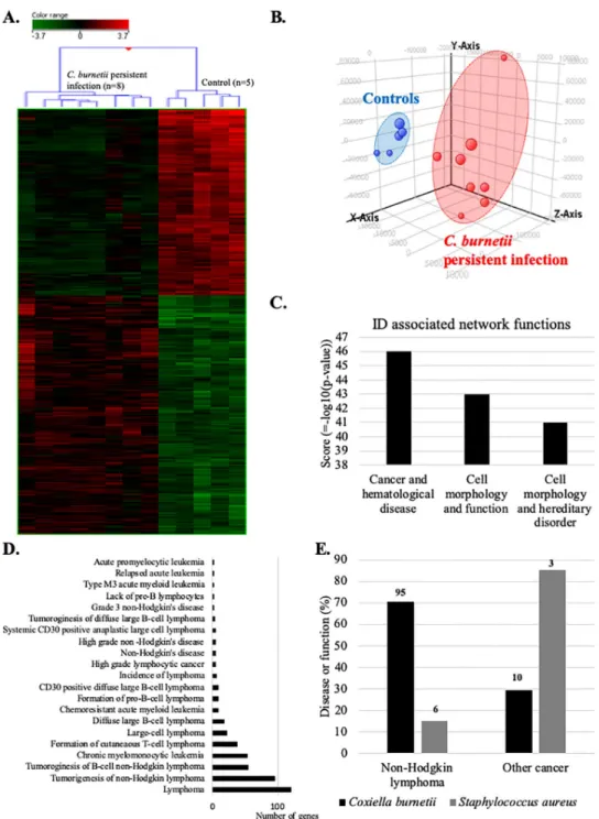
infection) versus healthy donors. We identified 5588 genes differentially expressed between these two groups with a statistical significance (p � 0.01) with 2189 up- and 3399 down-modu-lated genes (Fig 1). Because of the relatively large number of differentially expressed genes, we focused on genes that exhibit a >4 fold change between both groups and found 476 modulated genes (317 up- and 159 down-modulated genes. Non-supervised hierarchical clustering sepa-rated the transcriptional profiles of patients and controls (Fig 1A and 1B). Gene Ontology (GO) analysis of the transcriptional profiles underscored an association of Q fever-associated gene expression alterations with metabolic processes (18%), cardiovascular diseases (16%), cancers (10%), immune disorders (8%), infections (6%) and renal disorders (6%) (Fig 1C).
Persistent
C. burnetii infection induces transcriptional changes involved in
non-Hodgkin lympoma
We next compared the blood transcriptome of patients with persistentC. burnetii infection
with that of controls by microarray. Hierarchical clustering (Fig 2A) and principal component analysis (Fig 2B) clearly discriminated the gene expression profiles of patients and controls. 1687 genes were differentially expressed (p �0.01 and fold change �1.5). Among them, we found 739 up- and 948 down-modulated genes. Then, we used IPA software to inspect diseases or functions linked to persistentC. burnetii infection. The investigation of identifiers (ID)
associated network functions (Fig 2C) revealed that “cancer and hematological disease” gene categories was at the top of this list (z-score = 46). The IPA “disease or function” ontology revealed 21 significant terms associated with lymphoma (Fig 2D). 76% of these terms was associated with NHL and included expression changes affecting 95 genes. Given the specific objective of this study, we decided to focus on this group of genes that we referred to as the
We then analyzed genes of Cb-NHL signature by means of GO annotation. The GO term analysis revealed a regulation of apoptotic process, a positive regulation of NF-κB signaling, a cellular response to mechanical stimulus, an innate immune response, cell adhesion, cell migration, extrinsic apoptotic signaling pathway and an inflammatory response and apoptotic process (S1 Fig).
Fig 1. Transcritpional profile of patients withC. burnetii infection. Microarray transcriptional profile using whole
blood from patients withC. burnetii infection and healthy donors. Differential gene expression contrasting patients
with Q fever and healthy controls was analysed by (A) hierarchical clustering with samples in row and genes in column. Gene expression was colored from green (down-regulated) to red (up-regulated). (B) Graphic representation of the principal component analysis indicating patients with Q fever (in blue) and controls (in red). (C) Differentially expressed genes in Q fever patients were subjected to gene ontology (GO) annotation for biological processes. The percentages of GO terms are classified by groups of diseases and concern all patients with Q fever.
To determine whether acute Q fever might be a risk factor for lymphoma, we used IPA soft-ware to explore categories of network functions in microarray data (S2A and S2B Fig). This analysis led to a more diffuse association with multiple disease categories, including multiple different types of cancer, not just lymphoma (gastrointestinal disease, organismal injury and
Fig 2. Transcriptional profile of patients withC. burneti persistent infection links to lymphoma. Microarray
analyses were performed on whole blood from patients with persistentC. burnetii infection and healthy controls.
Transcriptional profiles of patients with persistentC. burnetii infection were analyzed by (A) hierarchical clustering,
(B) principal component analysis, (C) using IPA software to identify associated networks, and (D) according to the “disease or function” ontology of this software. (E) Comparison of the transcripitional effects ofC. burnetii and S. aureus on genes linked to NHL or other cancers. The numbers at the top of each column indicates the number of genes
incriminated.
Table 3.C. burnetii—Non-Hodgkin lymphoma signature.
Gene Fold change Gene description
ADAMTSL3 5.880324 ADAMTS like 3
AIF1 -2.9805992 Allograft inflammatory factor 1 ALOX5 -2.834 Arachidonate 5-lipoxygenase
ANKIB1 4.8102517 Ankyrin repeat and IBR domain containing 1 ANKRD36 2.2068887 Ankyrin repeat domain 36
ASAP2 -4.2549253 ArfGAP with SH3 domain Ankyrin repeat and PH domain 2
ATP10A -4.458795 ATPase phospholipid transporting 10A (putative) B2M -3.521 Beta-2-microglobulin
BAG1 -2.899064 BCL2 associated athanogene 1 BCL2L11 2.6857266 BCL2 like 11
CASP10 -1.7338927 Caspase 10
CASP2 3.9026406 Caspase 2
CASP4 -2.6195037 Caspase 4
CASP8 3.4592438 Caspase 8
CCDC97 -2.008394 Coiled-coil domain containing 97 CD44 -2.1155155 CD44 molecule
CD63 -2.6959426 CD63 molecule
CEBPB -1.9058212 CCAAT/enhancer binding protein beta
CNTRL 2.5811584 Centriolin
CREBBP 3.0448112 CREB binding protein
CTAGE1 3.4368377 Cutaneous T-cell lymphoma-associated antigen 1 CTAGE5 3.5890672 CTAGE family member 5, export factor CTNNB1 4.6916003 Catenin beta 1
CUL9 -2.9435835 Cullin 9
DCLRE1C 1.8426613 DNA cross-link repair 1C DNMT3B -3.6707778 DNA methyltransferase 3 beta
DYNLL2 -2.3400176 Dynein light chain LC8-type 2 EGR1 -3.795406 Early growth response 1 FBXO11 2.1503875 F-box protein 11
FYN 3.284 FYN proto-oncogene, src family tyrosine kinase G6PD 4.5535316 Glucose-6-phosphate dehydrogenase
GNB1 1.9694024 G protein subunit beta 1
GP1BA -3.7912498 Glycoprotein Ib platelet alpha subunit GRK3 3.358 G protein-coupled receptor kinase 3
HIC1 3.1705003 Hypermethylated in cancer 1
HLA-G 5.7384872 Major histocompatibility complex, class I, G HUWE1 2.3448784 HECT, UBA and WWE domain containing 1
IKZF1 2.0111005 IKAROS family zinc finger 1 IL13RA1 -2.7759755 Interleukin 13 receptor subunit alpha 1
IL2RG 3.2423832 Interleukin 2 receptor subunit gamma ITGAX -2.8220086 Integrin subunit alpha X
JAK3 -3.003575 Janus kinase 3
KIAA1551 3.162272 KIAA1551
KLHDC3 2.2549937 Kelch domain containing 3 KLHL6 -3.3247852 Kelch like family member 6 KMT2C 2.083985 Lysine methyltransferase 2C
Table 3. (Continued)
Gene Fold change Gene description
LIG3 -2.9250553 DNA ligase 3
MAP3K8 2.3423563 Mitogen-activated protein kinase kinase kinase 8 MAP4K4 1.5909171 Mitogen-activated protein kinase 4
MED1 -4.125631 Mediator complex subunit 1 MICALCL -2.5829322 MICAL C-terminal like MIR17HG 3.2123563 miR-17-92a-1 cluster host gene
MKNK2 -2.5849397 MAP kinase interacting serine/threonine kinase 2
NF1 2.524433 Neurofibromin 1
NFKBIA -2.8016303 Nuclear factor of Kappa light polypeptide gene enhancer in B cells inhibitor alpha NIPBL 3.1714787 NIPBL, cohesin loading factor
NOTCH2 -2.1726353 Notch 2
OGG1 2.5774076 8-oxoguanine DNA glycosylase
PBX1 -3.845063 PBX homeobox 1
PLCG1 1.9097688 Phospholipase C gamma 1
PLEKHG3 2.318101 Pleckstrin homology and RhoGEF domain containing G3 PPP6R3 3.2296784 Protein phosphatase 6 regulatory subunit 3
PTPN11 -3.1850703 Protein tyrosine phosphatase, non-receptor type 11 QARS 3.6468294 Glutaminyl-tRNA synthetase
RAPGEF1 3.936472 Rap guanine nucleotide exchange factor 1 RARA -2.5248845 Retinoic acid receptor alpha
REL 1.7198911 REL proto-oncogene, NF-kB subunit RELB -2.859732 RELB proto-oncogene, NF-kB subunit RPS2 -3.7340784 Ribosomal protein S2
RPSA -3.64 Ribosomal protein SA
SERPINA1 -2.598 Serpin family A member 1 SLC16A7 2.801318 Solute carrier family 16 member 7
SMARCA4 4.9222918 SWI/SNF related actin dependent regulator of chromatin, subfamily a, member 4 SMARCB1 3.59208 SWI/SNF related actin dependent regulator of chromatin, subfamily b, member 1
SP100 2.505 SP100 nuclear antigen
SPECC1 -2.0302236 Sperm antigen with calponin homology and coiled-coil domains 1 TDRD1 3.4261622 Tudor domain containing 1
TET2 5.2559843 Tet methylcytosine dioxygenase 2 TGFB1 2.3384285 Transforming growth factor beta 1
TLR7 4.3670783 Toll like receptor 7
TNFRSF8 -1.8059382 TNF receptor superfamily member 8 TNIK -2.174313 TRAF2 and NCK interacting kinase TPM1 -4.584908 Tropomyosin 1 (alpha)
TRAF1 1.6773132 TNF receptor associated factor 1 TRIM38 2.384246 Tripartite motif containing 38
TRIP12 2.4232147 Thyroid hormone receptor interactor 12 TTC21B 2.1786764 Tetratricopeptide repeat domain 21B TUBA1C -3.2958088 Tubulin alpha 1c
TUBA3C -2.4673393 Tubulin alpha 3c TUBB3 -8.211237 Tubulin beta 3 class III TUBB4B -2.5735104 Tubulin beta 4B class IVb
UBE2F -2.2094207 Ubiquitin conjugating enzyme E2 F (putative) UIMC1 -2.096621 Ubiquitin interaction motif containing 1
abnormalities, neurological and dermatological disease,S2C and S2D Fig). Four genes overex-pressed in Q fever were previously implicated in NHL, namely,BCL2, BCL7A, BCL9 and BCL11 (S2 Table). Altogether, these results confirm the suspicion that acute Q fever is much less associated with NHL than persistentC. burnetii infection.
Finally, to determine whether the correlation found with NHL was specific toC. burnetii
infection, we compared theC. burnetii–related dataset to published [19] microarray analyses of patients withS. aureus and persistent endocarditis previously performed from whole blood
(S3 Fig). In patients withS. aureus endocarditis, only 15% terms of “disease or function”
ontol-ogy, including 6 genes, were associated with NHL (Fig 2E). This comparison confirms the exis-tence of a specific link between persistentC. burnetii infection and lymphoma/NHL, which is
not observed in case ofS. aureus endocarditis.
The Cb-NHL signature in patients with different types of
C. burnetii
infection
We used qRT-PCR to investigate the Cb-NHL signature in a cohort ofC. burnetii-infected
patients including 11 with acute Q fever, 11 patients with persistent infection, 4 patients with lymphadenitis and 4 patients withC. burnetii infection and lymphoma and 10 with NHL
withoutC. burnetii infection included in the NHL-control group (Table 2). Among the large number of genes (95) listed in the Cb-NHL signature, we selected 24 genes based on their functional links to cancer and lymphoma [1]. In addition, we included 9 genes specifically associated with lymphoma based on Flowers’ signature [1]. This type of PCA revealed particu-lar Cb-NHL signature in Q fever patients (log2FC) normalized to 10 healthy donors (Fig 3A). In this PCA, the group ofC. burnetii-infected patients with lymphoma, and that with C. burne-tii lymphadenitis largely overlapped. In contrast, no such kind of superposition was observed
when the Flowers’ signature is applied (S4 Fig). Both PCA and further analysis of Cb-NHL sig-nature by unsupervised hierarchical clustering (Fig 3B) suggest that patients with persistentC. burnetii infection can be separated from patients with C. burnetii lymphadenitis or C. burnetii
associated NHL. Again, these distinctions were not observed using Flowers’s signature (S4 Fig). Three genes of the Cb-NHL signature were strongly up-regulated (fold change between 100 and 1000) in patients withC. burnetii and lymphoma and significantly increased when
compared toC. burnetii persistent infection and to NHL-control: MIR17HG (p = 0.0026), SP100 (p = 0.026) and REL (p = 0.0024) (Fig 3C). In the group of patients withC. burnetii
infection and lymphoma,BAG1, OGG1 and NCOA1-v1 were also significantly up-regulated,
as compared to other groups, namely, acute Q fever and persistentC. burnetii infection
(Table 4).
The specificity of theC. burnetii associated NHL transcriptomic signature was supported
by gene expression analysis of the NHL control group, in whichMIR17HG, SP100, REL, and ETS1 were not upmodulated, whereas BCL2 was significantly highly expressed (Fig 3C and 3D). Interestingly, antibiotic treatment with doxycycline and hydroxychloroquine (the stan-dard medication to treatC. burnetii infection) did not affect the gene expression profiles found
in patients (S5 Fig).
Table 3. (Continued)
Gene Fold change Gene description
WWC3 -1.7466848 WWC family member 3
XRCC5 -2.593 X-ray repair cross complementing 5 https://doi.org/10.1371/journal.pone.0217542.t003
Some similarities were observed between the group of patients carryingC. burnetii and
lymphoma, and those withC. burnetii lymphadenitis. Both groups exhibited similar gene
expression profiles, and the aforementioned lymphoma-associated genes (MIR17HG, REL, BAG1, OGG1, SP100 and NCOA1-v1) were up-regulated (Fig 3CandS6 Fig). It is interesting
Fig 3. qRT-PCR analysis of disease-associated alterations in gene expression within the Cb-NHL signature. PBMC samples from patients with acute Q fever, persistentC. burnetii infection, C. burnetii associated NHL, C. burnetii
lymphadenitis and NHL-controls were compared to healthy controls by using qRT-PCR. The expression of genes from the Cb-NHL signature were evaluated as fold change of investigated gene/β-actin mRNA. (A) Principal component analysis reveals the overlap between the modulated genes (log2 fold change) of the three groups of patients with persistentC. burnetii infection (blue), C. burnetii associated NHL (purple) or C. burnetii lymphadenitis (green) and
NHL-control (red). (B) Modulated genes from Cb-NHL (log2 fold change) were represented as a heatmap with samples in columns and genes in rows. Gene expression was colored from blue (down-regulated) to red (up-regulated). (C) Comparison of patients affected by persistentC. burnetii infection to patients with lymphoma and
lymphadenitis led to the identification ofMIR17HG, REL and SP100 as significantly up-regulated in patients with
lymphoma and lymphadenitis. (D) In patients with lymphadenitis,BCL2 and ETS1 were significantly up-regulated.
to note that in addition to theSP100 and REL genes, the lymphadenitis group has significantly
up-regulatedETS1 (p = 0.0046) and BCL2 genes (p = 0.021), both genes involved in the
inhibi-tion of apoptosis, tumorigenesis and cell proliferainhibi-tion, while these patient groups had no known occurrence of lymphoma. (Fig 3D). This latter result supports the idea that lymphade-nitis might constitute a step towards lymphomagenesis.
Discussion
The present study identifies a specific transcriptomic signature of Cb-NHL among patients with persistentC. burnetii infection strengthening the link between Q fever and the
develop-ment of NHL.
Three genes,REL, MIR17HG and SP100, constitute the core of this transcriptomic
signa-ture. This is intriguing because these genes have already been involved in cell survival, B-cell proliferation and tumorigenesisvia anti-apoptotic process. MIR17HG is involved in cell
sur-vival proliferation and differentiation both in solid tumor and in lymphoma, and is associated with poor prognosis in Burkitt lymphoma and DLBCL [20].SP100 stimulates the
transcrip-tional activity of ETS1, a protooncogene ubiquitously expressed in the lymph nodes and in bone marrow, which is involved in apoptosis, invasion and metastasis (Table 4) [21].REL is a
proto-oncogene involved in survival and proliferation of B-lymphocytes and its mutation is associated with B-cell lymphoma [22]. REL-BCL11A fusion or gain ofREL, has been
previ-ously linked to the transformation of FL to DLBCL, and this may be a genetic marker for tumor progression [22]. In the lymphadenitis group, as in the lymphoma group, two genes of the core of the transcriptomicC. burnetii-NHL signature, REL and SP100, were significantly
upmodulated when compared to the control group.
Regarding the suggested genetic risk factors proposed by Flowerset al., only NCOA1, was
significantly up-regulated in two groups: lymphoma and lymphadenitis, whereasBCL2 was
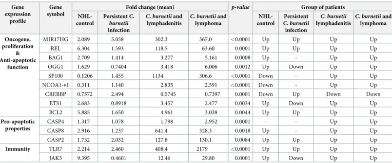
significantly expressed in cases ofC. burnetii lymphadenitis [1].NCOA1, nuclear receptor Table 4. Quantitative-RT-PCR analysis.
Gene expression
profile
Gene symbol
Fold change (mean) p-value Group of patients
NHL-control PersistentC. burnetii infection C. burnetii and lymphadenitis C. burnetii and lymphoma NHL-control Persistent C. burnetii infection C. burnetii lymphadenitis C. burnetii and lymphoma Oncogene, proliferation & Anti-apoptotic function MIR17HG 2.089 5.058 302.3 567.0 <0.0001 Up Up Up Up REL 6.304 1.593 118.5 63.60 0.0001 Up Up Up Up BAG1 2.709 1.414 3.277 5.161 0.0008 Up - Up Up OGG1 1.629 0.7404 3.418 6.006 0.0012 Up Down Up Up SP100 0.1206 1.455 1134 306.6 <0.0001 Down - Up Up NCOA1-v1 0.311 1.140 2.835 2.591 <0.0001 Down - Up Up
CREBBP 0.7572 2.494 0.5745 0.7397 0.0001 Down Up Down Down
ETS1 2.683 0.8918 3.457 2.477 0.0034 Up Down Up Up BCL2 5.885 1.650 4.961 3.038 0.0044 Up Up Up Up Pro-apoptotic properties CASP4 1.317 1.078 1.798 2.952 0.0001 - - Up Up CASP8 2.916 1.237 641.4 328.3 0.0018 Up - Up Up CASP2 1.732 2.032 127.8 130.1 0.0084 Up Up Up Up Immunity TLR7 2.214 2.460 408.4 2179 <0.0001 Up Up Up Up JAK3 9.395 0.4601 12.46 29.80 0.0001 Up Down Up Up
P value is calculated as a fold change in comparison to the control group (healthy donors) Up = fold change>1.5; down = fold change<1; -: fold change between 1 and 1.5 https://doi.org/10.1371/journal.pone.0217542.t004
co-activator 1, acts as a transctiptional coactivator for steroid and nuclear hormones receptors and may increase the induction of the T-cell receptor endocytosis [23].BCL2 gene expression
is well known to block apoptosis and promote B-cell lymphoma and is even the target of anti-tumoral therapy [24]. Its high expression observed in case ofC. burnetii lymphadenitis
sup-ports lymphadenitis as a critical step in lymphomagenesis, whereas its high expression in case of NHL control group is concordant with previous reports [25]. While Flowerset al. suggested
that a preexisting genetic risk factor could play a role in the development of NHL in patients withC. burnetii infection, our results rather indicate that C. burnetii infection triggers the
expression of genes implicated in anti apoptotic and proliferative mechanisms.
Regarding the Cb-NHL signature and using pubmed data base, we identified 42 genes asso-ciated with tumor development in patients carrying viral or bacterial pathogens.REL is among
the common genes modulated in infection by EBV, HTLV,C. jejuni, H. pylori, B. burgdorferi
orC. psittaci (Fig 4A). During EBV infection, REL and STAT3 have a potent effect on cell growth and in lymphomagenesis [26]. On the contrary, REL deficiency leads to decreased pro-liferation, decreased survival of EBV transformed cells and could even lead to their necrotic death [27]. In HTLV infection, REL proteins are involved in the canonical NF-κB pathway,
which induces the transcription of anti-apoptoticBCL2A1 and BCL2L1 [28]. InBorrelia burg-dorferi infection, REL is responsible for the ppGpp synthesis that facilitates the bacterium
growth and virulence [29]. Nevertheless, the precise role ofREL in infections by C. jejuni and C. psittacii has not been yet clarified.
There is a vast literature on the effects of EBV andH. pylori on gene expression. Epstein
barr virus latency gene products drive viral persistence in memory B-cells and malignancy by controling cell growth, division, differentiation and apoptosis [30,31]. Latent transcipts, such as Epstein-Barr virus nuclear antigen 1 (EBNAs) and latent membrane proteins (LMPs) were detected in EBV-associated B-cell lymphoma [30,31]. EBV-positive and EBV-negative BL could even be differentiated by their apoptosis-related gene expression profile [31]. Interest-ingly, the transcriptomic profile of EBV-positive benign lymphadenophathy was described as closer to BL than to post-transplant lymphoproliferative disease [32], supporting EBV-positive lymphadenopathy as a critical step leading to lymphomagenesis, similar to what we suggest here forC. burnetii infection [32].
C. burnetii modulates the apoptotic pathway through Beclin 1/BCL2 to establish succesfull
infection of the host cell [33]. Here, we report thatBCL2 was significantly up-regulated in the
blood of patients withC. burnetii lymphadenitis. The activity of PKB/AKT and MAPK1/ERK2
or MAPK3/ERK1 seems to be required for the apoptosis-suppressive effect ofC. burnetii
infec-tion [34]. More recently, the anti-apoptotic protein myeloid cell leukemia-1 (MCL1) has been found to be increased in neutrophils infected by avirulentC. burnetii and this effect was linked
to the inhibition of caspase-3 cleavage and the activation of the MAPK survival pathway [34]. Furtherin vitro experimentation is necessary to unravel the mechanims through which
infec-tion of certain leukocyte subtypes (such as monocytes/macrophages) may be coupled with the inhibition of apoptosis in B cells.
In this study, the transcriptomic profile was obtained from the whole blood. In blood,BCL2
gene expression was proved to be significantly up-regulated in patients with chronic lymphoid leukemia [35]. In patients with NHL, a concordant expression between bone marrow and peripheral blood BCL2/JH expression was reported [25]. The identification of the high-level expression of BCL2 in the blood of patients with lymphadenitis might constitute a biomarker of a prelymphomatous grade requiring close monitoring.
The up-regulation of genes coding for caspases (CASP2, CASP4, CASP8) that we have
Fig 4. (A) Link between genes contained in Cb-NHL signature and infection by cancer-associated microorganisms. Venn diagram showing overlap between genes contained in the Cb-NHL signature that are also deregulated in non-Hodgkin lymphoma associated with Epstein-Barr virus (EBV), human T lymphocyte virus (HTLV),Campylobacter jejuni, Helicobacter pylori, Borrelia burgdorferi or Chlamydia psittaci. (B) Pathophysiological mechanism. Fom persistent C. burnetii infection (endocarditis, vascular infection), to C. burnetii lymphadenitis and lymphoma. Peripheral blood monocyte cells genomic transcriptional profiles indicated an overexpression of BCL2 and ETS1 mRNAs in cases of C. burnetii lymphadenitis and an overexpression of MIR17HG, REL and SP100 genes in cases of C. burnetii associated lymphoma. All these gene are related to the inhibition of apoptosis and/or cellular proliferation.
infection insofar, as the regulation of caspase activation essentially occurs at the post-transla-tional level [36].
In conclusion, we have identified a transcriptomic signature from PBMC of patients with
C. burnetii-associated lymphoma that consists of the overexpression of several genes including MIR17HG, REL and SP100. Patients with C. burnetii lymphadenitis presented high level of BCL2 mRNA in blood, shared a similar transcriptomic signature with patients infected with C. burnetii and were diagnosed with lymphoma. This argues in favor of the hypothesis that C. burnetii lymphadenitis may facilitate subsequent lymphomagenesis leading to NHL.
Supporting information
S1 Fig. Cb-NHL signature by using the GO annotation for biological process. GO terms
related to regulation of apoptotic process, positive regulation of NF-κB signaling, cellular response to mechanical stimulus, innate immune response, cell adhesion, cell migration, extrinsic apoptotic signaling pathway, inflammatory response and apoptotic process. (TIFF)
S2 Fig. Gene expression signature between patients with acute Q fever (n = 8) and patients withC. burnetii persistent infection (n = 8). Differential gene expression between acute Q
fever and persistentC. burnetii infection was analysed by hierarchical clustering (A) and
bioin-formatic plots results (B). In panel B the bioinbioin-formatic plots results according to 3 axis shows a segregation gene modulation between patients with acute Q fever and patients withC. burnetii
persistent infection as compared with the control group. Differentially expressed genes from Cb-NHL signature were subjected to GO annotation to identify the biological process. The his-togram graphs (C) and (D) show the repartition of dieases associated with acuteC. burnetii
infection. (TIFF)
S3 Fig. Microarray investigation betweenC. burnetii and S. aureus induced endocarditis.
Differential gene expression between persistentC. burnetii infection and S. aureus endocarditis
was analysed by (A) hierarchical clustering and (B) bioinformatic plots results. In panel B, the results of the bioinformatics plots along 3 axes show a modulation of the segregation gene between patients with persistentC. burnetii infection and S. aureus endocarditis compared to
the control group. (TIFF)
S4 Fig. q-RTPCR analysis of genes modulation from Flowers’ signature. PBMC samples of
patients with acute Q fever,C. burnetii persistent infection, C. burnetii associated NHL, C. bur-netii lymphadenitis and NHL-control, were investigated by q-RTPCR and normalized to
con-trols for the expression of genes from Flowers’ signature. (A) Generated by ClustVis software, principal composant analysis reveals the overlaping of the modulated genes (log2FC) from the three groupsC. burnetii persistent infection (blue), C. burnetii and lymphoma (purple) and C. burnetii lymphadenitis (green) and NHL-control (red). (B) Modulated gene from
Flower-s’signature were represented as heatmap with samples in column and gene in row. Gene expression was colored from blue (down-modulated) to red (up-modulated).
(TIFF)
S5 Fig. Investigation of the NHL genes expression according to treatment. Among the large
number of genes (95) listed in the Cb-NHL signature, we selected 24 genes based on their functional links to cancer and lymphoma. In addition, we included 9 genes specifically associ-ated with lymphoma based on Flowers’ signature. A total of 33 genes were analysed according
to antibiotic treatment with doxycycline and hydroxyplaquenil (the standard medication to treatC. burnetii infection). 0. Samples from patients never treated (red). 1. Samples from
patients with ongoing doxycycline and hydroxyplaquenil treatment (blue). 2. Samples from patients before receiving doxycycline and hydroxyplaquenil treatment (green). 3. Samples from patients after doxycycline and hydroxyplaquenil treatment. 4. Samples from patients after doxycycline treatment (purple). 5. Samples from patients with ongoing doxycycline treat-ment (brown). Using q-RTPCR, we investigated the Flowers’ signature (log2FC) which (A) principal composant analysis and (B) hierarchical clustering with samples from persistent Q fever (red), lymphoma (green) or remission (blue) periods from index case. Up- and down-modulated genes were represented in red and blue respectively. The analysis shows that gene expression was not affected by the antibiotic treatment received from patients.
(TIFF)
S6 Fig. Gene expression analysis. PBMC samples of patients withC. burnetii persistent
infec-tion,C. burnetii associated-NHL, C. burnetii lymphadenitis and NHL-control were
investi-gated by q-RTPCR, normalized to controls to investigate the expression of genes from Cb-NHL and Flowers’ signatures: (A)OGG1, (B) BAG1 and (C) NCOA1-v1.
(TIFF)
S1 Table. Specific primers used to performed qRT-PCR.
(PDF)
S2 Table. List of genes modulated in acute Q fever.
(PDF)
Author Contributions
Conceptualization: Cle´a Melenotte, Soraya Mezouar, Re´gis Costello. Data curation: Cle´a Melenotte, Soraya Mezouar.
Formal analysis: Cle´a Melenotte, Soraya Mezouar. Funding acquisition: Re´gis Costello.
Methodology: Soraya Mezouar, Amira Ben Amara, Simon Benatti, Jacques Chiaroni. Supervision: Christian Devaux, Guido Kroemer, Jean-Louis Mege, Didier Raoult. Writing – original draft: Cle´a Melenotte, Soraya Mezouar.
Writing – review & editing: Cle´a Melenotte, Soraya Mezouar, Christian Devaux, Guido
Kroe-mer, Jean-Louis Mege.
References
1. Flowers CR, Skibola CF. Identifying risk factors for B-cell lymphoma. Blood. 2016; 127: 10–11.https:// doi.org/10.1182/blood-2015-11-677203PMID:26744436
2. Lossos IS, Gascoyne RD. Transformation of follicular lymphoma. Best Pract Res Clin Haematol. 2011; 24: 147–163.https://doi.org/10.1016/j.beha.2011.02.006PMID:21658615
3. Alizadeh AA, Eisen MB, Davis RE, Ma C, Lossos IS, Rosenwald A, et al. Distinct types of diffuse large B-cell lymphoma identified by gene expression profiling. Nature. 2000; 403: 503–511.https://doi.org/10. 1038/35000501PMID:10676951
4. de Martel C, Ferlay J, Franceschi S, Vignat J, Bray F, Forman D, et al. Global burden of cancers attribut-able to infections in 2008: a review and synthetic analysis. Lancet Oncol. 2012; 13: 607–615.https://doi. org/10.1016/S1470-2045(12)70137-7PMID:22575588
5. Peek RM, Blaser MJ. Helicobacter pylori and gastrointestinal tract adenocarcinomas. Nat Rev Cancer. 2002; 2: 28–37.https://doi.org/10.1038/nrc703PMID:11902583
6. Saha A, Robertson ES. Epstein-Barr virus-associated B-cell lymphomas: pathogenesis and clinical out-comes. Clin Cancer Res Off J Am Assoc Cancer Res. 2011; 17: 3056–3063.https://doi.org/10.1158/ 1078-0432.CCR-10-2578PMID:21372216
7. Melenotte C. Clinical Features and Complications of Coxiella burnetii Infections From the French National Reference Center for Q Fever. JAMA Network Open. 24 Aug 2018: 1–15.
8. Melenotte C, Million M, Audoly G, Gorse A, Dutronc H, Roland G, et al. B-cell non-Hodgkin lymphoma linked to Coxiella burnetii. Blood. 2016; 127: 113–121.https://doi.org/10.1182/blood-2015-04-639617
PMID:26463422
9. van Roeden SE, van Houwelingen F, Donkers CMJ, Hogewoning SJ, de Lange MMA, van der Hoek W, et al. Exposure to Coxiella burnetii and risk of non-Hodgkin lymphoma: a retrospective population-based analysis in the Netherlands. Lancet Haematol. 2018;https://doi.org/10.1016/S2352-3026(18) 30038-3
10. van Roeden SE, Melenotte C, Hermans MHA, Sinnige HAM, Nooijen PTGA, Audoly G, et al. Case report: Coxiella burnetii vascular infection and lymphoma in the Netherlands. Infection. 2018; 46: 131– 134.https://doi.org/10.1007/s15010-017-1061-9PMID:28840502
11. van Roeden SE, Hermans MHA, Nooijen PTGA, Herbers A, Bleeker-Rovers CP, Hoepelman AIM, et al. Coxiella burnetii in non-Hodgkin lymphoma tissue samples: Innocent until proven otherwise? Immuno-biology. 2018;https://doi.org/10.1016/j.imbio.2018.11.012PMID:30638649
12. Melenotte C, Million M, Audoly G, Gorse A, Dutronc H, Roland G, et al. B-cell non-Hodgkin lymphoma linked to Coxiella burnetii. Blood. 2015;https://doi.org/10.1182/blood-2015-04-639617PMID:
26463422
13. Melenotte C, Million M, Audoly G, Gorse A, Dutronc H, Roland G, et al. B-cell non-Hodgkin lymphoma linked to Coxiella burnetii. Blood. 2016; 127: 113–121.https://doi.org/10.1182/blood-2015-04-639617
PMID:26463422
14. Melenotte C, Raoult D. Pro-apoptotic effect of doxycycline and hydroxychloroquine on B-cell lymphoma induced by C. burnetii. Oncotarget. 2017; 9: 2726–2727.https://doi.org/10.18632/oncotarget.23397
PMID:29416805
15. Voth DE, Howe D, Heinzen RA. Coxiella burnetii inhibits apoptosis in human THP-1 cells and monkey primary alveolar macrophages. Infect Immun. 2007; 75: 4263–4271. https://doi.org/10.1128/IAI.00594-07PMID:17606599
16. Eldin C, Me´lenotte C, Mediannikov O, Ghigo E, Million M, Edouard S, et al. From Q Fever to Coxiella burnetii Infection: a Paradigm Change. Clin Microbiol Rev. 2017; 30: 115–190.https://doi.org/10.1128/ CMR.00045-16PMID:27856520
17. Swerdlow SH, Campo E, Pileri SA, Harris NL, Stein H, Siebert R, et al. The 2016 revision of the World Health Organization classification of lymphoid neoplasms. Blood. 2016; 127: 2375–2390.https://doi. org/10.1182/blood-2016-01-643569PMID:26980727
18. Ka MB, Mezouar S, Ben Amara A, Raoult D, Ghigo E, Olive D, et al. Coxiella burnetii Induces Inflamma-tory Interferon-Like Signature in Plasmacytoid Dendritic Cells: A New Feature of Immune Response in Q Fever. Front Cell Infect Microbiol. 2016; 6.https://doi.org/10.3389/fcimb.2016.00070PMID:
27446817
19. Benoit M, Thuny F, Le Priol Y, Lepidi H, Bastonero S, Casalta J-P, et al. The Transcriptional Programme of Human Heart Valves Reveals the Natural History of Infective Endocarditis. PLoS ONE. 2010; 5.
https://doi.org/10.1371/journal.pone.0008939PMID:20126625
20. Tavakoli R, Vakilian S, Langroudi L, Arefian E, Sahmani M, Soleimani M, et al. The role of miR-17-92 cluster in the expression of tumor suppressor genes in unrestricted somatic stem cells. Biol J Int Assoc Biol Stand. 2017; 46: 143–147.https://doi.org/10.1016/j.biologicals.2017.02.006PMID:28222938
21. He C, Wu S, Gao A, Su Y, Min H, Shang Z-F, et al. Phosphorylation of ETS-1 is a critical event in DNA polymerase iota-induced invasion and metastasis of esophageal squamous cell carcinoma. Cancer Sci. 2017; 108: 2503–2510.https://doi.org/10.1111/cas.13399PMID:28905458
22. Hunter JE, Leslie J, Perkins ND. c-Rel and its many roles in cancer: an old story with new twists. Br J Cancer. 2016; 114: 1–6.https://doi.org/10.1038/bjc.2015.410PMID:26757421
23. Peng M, Zhao G, Yang F, Cheng G, Huang J, Qin X, et al. NCOA1 is a novel susceptibility gene for mul-tiple myeloma in the Chinese population: A case-control study. PLOS ONE. 2017; 12: e0173298.
https://doi.org/10.1371/journal.pone.0173298PMID:28264017
24. Ruefli-Brasse A, Reed JC. Therapeutics targeting Bcl-2 in hematological malignancies. Biochem J. 2017; 474: 3643–3657.https://doi.org/10.1042/BCJ20170080PMID:29061914
25. Yuan R, Dowling P, Zucca E, Diggelmann H, Cavalli F. Detection of bcl-2/JH rearrangement in follicular and diffuse lymphoma: concordant results of peripheral blood and bone marrow analysis at diagnosis. Br J Cancer. 1993; 67: 922–925.https://doi.org/10.1038/bjc.1993.171PMID:8494725
26. Transcriptome Changes Induced by Epstein-Barr Virus LMP1 and LMP2A in Transgenic Lympho-cytes and Lymphoma [Internet]. [cited 12 May 2018].https://www.ncbi.nlm.nih.gov/pmc/articles/ PMC3448168/
27. Valentı´n-Acevedo A, Sinquett FL, Covey LR. c-Rel deficiency increases caspase-4 expression and leads to ER stress and necrosis in EBV-transformed cells. PloS One. 2011; 6: e25467.https://doi.org/ 10.1371/journal.pone.0025467PMID:21984918
28. Macaire H, Riquet A, Moncollin V, Bie´mont-Trescol M-C, Duc Dodon M, Hermine O, et al. Tax protein-induced expression of antiapoptotic Bfl-1 protein contributes to survival of human T-cell leukemia virus type 1 (HTLV-1)-infected T-cells. J Biol Chem. 2012; 287: 21357–21370.https://doi.org/10.1074/jbc. M112.340992PMID:22553204
29. Gilmore TD. Role of rel family genes in normal and malignant lymphoid cell growth. Cancer Surv. 1992; 15: 69–87. PMID:1451115
30. Greijer AE, Ramayanti O, Verkuijlen S a. WM, NovalićZ, Juwana H, Middeldorp JM. Quantitative multi-target RNA profiling in Epstein-Barr virus infected tumor cells. J Virol Methods. 2017; 241: 24–33.
https://doi.org/10.1016/j.jviromet.2016.12.007PMID:27993616
31. Ghosh Roy S, Robertson ES, Saha A. Epigenetic Impact on EBV Associated B-Cell Lymphomagenesis. Biomolecules. 2016; 6.https://doi.org/10.3390/biom6040046PMID:27886133
32. Ramos J-C, Sin S-H, Staudt MR, Roy D, Vahrson W, Dezube BJ, et al. Nuclear factor kappa B pathway associated biomarkers in AIDS defining malignancies. Int J Cancer. 2012; 130: 2728–2733.https://doi. org/10.1002/ijc.26302PMID:21792887
33. Va´zquez CL, Colombo MI. Coxiella burnetii modulates Beclin 1 and Bcl-2, preventing host cell apoptosis to generate a persistent bacterial infection. Cell Death Differ. 2010; 17: 421–438.https://doi.org/10. 1038/cdd.2009.129PMID:19798108
34. Cherla R, Zhang Y, Ledbetter L, Zhang G. Coxiella burnetii Inhibits Neutrophil Apoptosis by Exploiting Survival Pathways and Antiapoptotic Protein Mcl-1. Infect Immun. 2018; 86.https://doi.org/10.1128/IAI. 00504-17PMID:29311244
35. Vucicevic K, Jakovljevic V, Colovic N, Tosic N, Kostic T, Glumac I, et al. Association of Bax Expression and Bcl2/Bax Ratio with Clinical and Molecular Prognostic Markers in Chronic Lymphocytic Leukemia. J Med Biochem. 2016; 35: 150–157.https://doi.org/10.1515/jomb-2015-0017PMID:28356875
36. Gross A, McDonnell JM, Korsmeyer SJ. BCL-2 family members and the mitochondria in apoptosis. Genes Dev. 1999; 13: 1899–1911.https://doi.org/10.1101/gad.13.15.1899PMID:10444588