HAL Id: hal-00552563
https://hal.archives-ouvertes.fr/hal-00552563
Submitted on 6 Jan 2011
HAL is a multi-disciplinary open access archive for the deposit and dissemination of sci-entific research documents, whether they are pub-lished or not. The documents may come from teaching and research institutions in France or abroad, or from public or private research centers.
L’archive ouverte pluridisciplinaire HAL, est destinée au dépôt et à la diffusion de documents scientifiques de niveau recherche, publiés ou non, émanant des établissements d’enseignement et de recherche français ou étrangers, des laboratoires publics ou privés.
CIRRHOTIC PATIENTS: A CASE-CONTROL STUDY
Geoffroy Vanbiervliet, Sarah Giudicelli-Bornard, Thierry Piche, Frédéric
Berthier, Eve Gelsi, Jérôme Filippi, Rodolphe Anty, Kamel Arab,
Pierre-Michel Huet, Xavier Hebuterne, et al.
To cite this version:
Geoffroy Vanbiervliet, Sarah Giudicelli-Bornard, Thierry Piche, Frédéric Berthier, Eve Gelsi, et al.. PREDICTIVE FACTORS OF BLEEDING RELATED TO POSTBANDING ULCER FOLLOW-ING ENDOSCOPIC VARICEAL LIGATION IN CIRRHOTIC PATIENTS: A CASE-CONTROL STUDY. Alimentary Pharmacology and Therapeutics, Wiley, 2010, 32 (2), pp.225. �10.1111/j.1365-2036.2010.04331.x�. �hal-00552563�
For Peer Review
PREDICTIVE FACTORS OF BLEEDING RELATED TO POSTBANDING ULCER FOLLOWING ENDOSCOPIC VARICEAL
LIGATION IN CIRRHOTIC PATIENTS: A CASE-CONTROL STUDY
Journal: Alimentary Pharmacology & Therapeutics Manuscript ID: APT-0155-2010.R1
Manuscript Type: Original Scientific Paper Date Submitted by the
Author: 14-Apr-2010
Complete List of Authors: Vanbiervliet, Geoffroy; Hôpital L'Archet 2, Endoscopie Digestive Giudicelli-Bornard, Sarah; CHU Nice, Hépato-Gastroentérologie Piche, Thierry; CHU Nice, Hépato-Gastroentérologie
Berthier, Frédéric; CHU Nice, DIM
Gelsi, Eve; CHU Nice, Hépato-Gastroentérologie Filippi, Jérôme; CHU Nice, Hépato-Gastroentérologie Anty, Rodolphe; CHU Nice, Hépato-Gastroentérologie Arab, Kamel; CHU Nice, Hépato-Gastroentérologie Huet, Pierre-Michel; CHU Nice, Hépato-Gastroentérologie Hebuterne, Xavier; CHU Nice, Hépato-Gastroentérologie Tran, Albert; CHU Nice, Hépato-Gastroentérologie Keywords: Portal hypertension < Hepatology, Varices < Hepatology,
Endoscopy < Topics, Acidity (oesophageal) < Topics
For Peer Review
Original article
PREDICTIVE FACTORS OF BLEEDING RELATED TO
POSTBANDING ULCER FOLLOWING ENDOSCOPIC VARICEAL
LIGATION IN CIRRHOTIC PATIENTS: A CASE-CONTROL STUDY
Geoffroy Vanbiervliet1 2, Sarah Giudicelli-Bornard1 2, Thierry Piche1 2 3, Frédéric Berthier2 4,Eve Gelsi1 2, Jérôme Filippi1 2, Rodolphe Anty1 2 3, Kamel Arab1 2, Pierre Michel Huet1 2, Xavier Hébuterne1 2, Albert Tran1 2 3
1
Université de Sophia-Antipolis, Faculté de Médecine, Nice, F-06107, France
2
Centre Hospitalier de Nice, Pôle Digestif, Nice, F-06202, France
3
Institut National de la Santé et de la Recherche Médicale (INSERM), U895, Nice, F-06204, Cedex 3, France
4
Centre Hospitalier de Nice, Département d’information médicale, Nice, F-06000, France
Short Title: Postbanding bleeding after variceal ligation
A part of this work has been presented at the 16th of the United European gastroenterology Week (UEGW) in Vienna (October 2008)
Electronic word count: 2613
Number of figure: 1
Number of Table: 6
Corresponding author :
Docteur VANBIERVLIET Geoffroy Pôle Digestif
Endoscopie Digestive Hôpital l’Archet 2
151, route de Saint-Antoine de Ginestiére BP 3079 06202 NICE Cedex 3 FRANCE Tél. : + 33 4 92 03 63 85 Fax : + 33 4 92 03 59 28 E-mail : vanbiervliet.g@chu-nice.fr 3 4 5 6 7 8 9 10 11 12 13 14 15 16 17 18 19 20 21 22 23 24 25 26 27 28 29 30 31 32 33 34 35 36 37 38 39 40 41 42 43 44 45 46 47 48 49 50 51 52 53 54 55 56 57 58 59 60
For Peer Review
ABSTRACT
Background: Life-threatening bleeding due to early spontaneous slippage of rubber bands
have been described after variceal ligation in cirrhotic patients.
Aims: To determine the predictive factors of this complication in cirrhotic patients.
Methods: Among 605 patients, 21 patients (mean age 56.6 ± 13.5 years) developed 23
spontaneous band slippages with bleeding on postbanding ulcer, as confirmed by endoscopy.
Cirrhosis was alcoholic in 13 patients (62%), post viral hepatitis in 3 (14%) and from other
causes in 5 (24%). A case-control study was performed comparing 17 from these patients who
presented the complication after a first ligation with 84 of the 584 controls who underwent
first endoscopic variceal ligation without bleeding complication.
Results: Bleeding occurred 13.5 days ± 7.3 [2-29] following ligation. Eleven patients died
following the bleeding complication (52%). Using a multivariate analysis, previous upper
variceal digestive bleeding (OR 12.07, 95%CI [2.3-63.43]), peptic oesophagitis (OR 8.9,
95%CI [1.65-47.8]), high APRI score (OR 1.54, 95%CI [1.11-2.16]) and low prothrombin
index (OR 0.54, 95% CI [0.31-0.94]) were independent predictive factors of bleeding.
Conclusions: Bleeding related to postbanding ulcer is a rare but severe complication. The
proposed predictive factors should be looked for and minimized before variceal ligation.
Electronic word count: 195 words
Key words: oesophageal variceal ligation, bleeding, cirrhosis, spontaneous slippage, rubber
bands, postbanding ulcer. 3 4 5 6 7 8 9 10 11 12 13 14 15 16 17 18 19 20 21 22 23 24 25 26 27 28 29 30 31 32 33 34 35 36 37 38 39 40 41 42 43 44 45 46 47 48 49 50 51 52 53 54 55 56 57 58 59 60
For Peer Review
INTRODUCTION
In spite of a more efficient approach to upper digestive bleedings over the past three
decades, acute oesophageal variceal bleeding remains a severe and deadly complication in the
cirrhotic patient life which marks a turning point in the evolution of their liver disease 1. In this context, endoscopic variceal band ligation (EVL) has been proposed as one of the best
emergency as well as prophylactic haemostatic treatment. This approach has been
recommended now by the consensus workshop in these different situations 2, 3. EVL is considered to be safe with few treatment-related complications 4. Indeed, most frequently reported complications are minors with transient dysphagia or post ligature pain 5. On the other hand, oesophageal stricture formations have only been reported in 1.9% of cases 6.
Following EVL, a local ulcer is commonly found with a ensuing well described
sequence of the pathological changes 7, 8. After strangulation, variceal thrombosis occurs with varying degrees of ischemic necrosis inducing detachment of the rubber band. Then a shallow
ulcer usually occurs that heals within 2 to 3 weeks, allowing the development of fibrosis in
the sub mucosa. In case of premature detachment of the rubber band, before variceal
thrombosis, marked alterations of the mucosa can be seen with dilated variceal vessels in
necrotic areas 8. These pathological findings may explain the rare rebleeding episodes from oesophageal ulcerations following EVL 7. Up to now only few reports of life-threatening bleeding due to early spontaneous slippage of rubber bands and post banding ulcer have been
described 9-11. These reports were only dealing with case reports and no large study of this complication is available in cirrhotic patients treated by EVL.
The aim of this case-control study was to determine the predictive factors of bleeding
from spontaneous slippage of rubber bands and post-EVL ulcer in cirrhotic patients. 3 4 5 6 7 8 9 10 11 12 13 14 15 16 17 18 19 20 21 22 23 24 25 26 27 28 29 30 31 32 33 34 35 36 37 38 39 40 41 42 43 44 45 46 47 48 49 50 51 52 53 54 55 56 57 58 59 60
For Peer Review
MATERIALS AND METHODS
Study population
The characteristics of patients enrolled in this case-control study, are summarized in Table 1.
Six hundred five cirrhotic patients who underwent 837 EVL in our unit from 2002 to 2007
were evaluated and their chart reviewed. “Cases” were defined as cirrhotic patients who
developed a digestive bleeding related to ulceration after spontaneous slippage of rubber band
following a first EVL performed in emergency or prophylactic situation. This bleeding was an
active one (spurting or oozing) due to the ulceration induced by the previous EVL 5-7, confirmed by an gastroscopy performed within 12 hours after the haemorrhage and without
other causes of digestive bleeding (such as non variceal upper gastrointestinal bleeding,
gastric antral vascular ectasia, gastro oesophageal variceal bleeding or rebleeding site on
oesophageal varices). Using this definition, twenty-one (3%) patients (17 males and 4
females, mean age: 56.6 years ± 13.5) presented 23 bleeding episodes following EVL in
relation to ulceration after sliding of rubber band but only in 17 patients (81%) the
haemorrhage developed following a first ligation (and in four patients (19%) following a
second session of ligation) (Table 1). Two patients presented 2 episodes of bleeding.
“Controls” were defined as patients who underwent a first EVL without any complication
(EVL performed in emergency or prophylactic situations). Eighty-four controls (60 males and
24 females, mean age: 58.5 years ± 13.1) were then selected from the 605 patients with an
EVL in our unit, using a string of computer-generated random numbers in a 4:1 ratio and
compared with cases. For the case – control study and statistical analysis, we only kept and
compare the 17 “cases” who presented a bleeding episode after the first EVL with the
“controls”. Patients gave written informed consent to the EVL procedure and authorized the
use of their personal data. The variables were retrospectively collected on the computer data
base and the medical records of the institution. 3 4 5 6 7 8 9 10 11 12 13 14 15 16 17 18 19 20 21 22 23 24 25 26 27 28 29 30 31 32 33 34 35 36 37 38 39 40 41 42 43 44 45 46 47 48 49 50 51 52 53 54 55 56 57 58 59 60
For Peer Review
Endoscopic procedure for EVL
Five senior endoscopists from our unit (experience of more than 250 ligations) performed the
EVL under general propofol-induced anaesthesia with or without tracheal intubation. The
endoscopes used were adult gastroscope type Olympus GIF 160 (Olympus Medical Systems,
Hamburg, Germany) or Fujinon EG-450WR5 (Fujinon Medical Systems, Willich, Germany).
The same procedure of ligation was used for each patient: 1) grade 2 and 3 varices were
identified during withdrawal of the endoscope following a complete gastric aspiration.; 2)
bands were launched on varices (mean of 5.2 ± 2, maximum 12) with the same device (4 or 6
shooter Saeed multiband ligator, Cook Medical Endoscopy, Limerick, Ireland) in an
ascending way through the oesophagus. In case of EVL for emergency treatment (acute
bleeding), a rubber band was launched on the bleeding varices (protocol used in our unit).
Following EVL, proton pump inhibitors (PPI) (esomeprazole, 40 mg per day) were administrated during at least 3 days in all patients. Food intake was allowed 12 hours
following the endoscopic procedure in case of prophylactic EVL and at the discretion of the
physician in case of EVL for acute bleeding.
Treatment of the bleeding related to postbanding ulcer after spontaneous slippage of
rubber band
In all “case” patients octreotide (25 µg per hour) and PPI dose infusion (esomeprazole – 40
mg per day) during five days after the bleeding complication were given. All patients received
prophylactic antibiotherapy (ofloxacine) and the gastroscopy was performed in the six first
hours after admission. Cyanoacrylate endoscopic injections were performed in 10 patients
(43%), five patients (24%) had oesophageal balloon tamponade (for uncontrolled and massive
bleeding with haemodynamic deficiency) and two patients (14%) had an emergency
transjugular intrahepatic portosystemic shunts (TIPSS). 3 4 5 6 7 8 9 10 11 12 13 14 15 16 17 18 19 20 21 22 23 24 25 26 27 28 29 30 31 32 33 34 35 36 37 38 39 40 41 42 43 44 45 46 47 48 49 50 51 52 53 54 55 56 57 58 59 60
For Peer Review
Statistical analysis
In univariate analysis, we compared cases and controls with Chi-square distribution or
Fisher’s exact test to assess significance between proportions, and with two samples Student t
test to assess significance with quantitative data. In multivariate analysis, binary logistic
regression models were applied with bleeding after the EVL as dependant variable. Variables
significantly associated with bleeding (with a significance level of p < .05) and with a clinical
relevance were selected for possible inclusion in multivariate logistic regression models.
Relationships were assessed by adjusted odds ratio (OR) with 95% confidence interval (CI).
Two-sided p values < .05 were considered statistically significant. We used SAS software
(Enterprise Guide 4.1 for Windows) for all statistical analyses (S.A.S institute Inc., Cary, NC,
USA). 3 4 5 6 7 8 9 10 11 12 13 14 15 16 17 18 19 20 21 22 23 24 25 26 27 28 29 30 31 32 33 34 35 36 37 38 39 40 41 42 43 44 45 46 47 48 49 50 51 52 53 54 55 56 57 58 59 60
For Peer Review
RESULTS
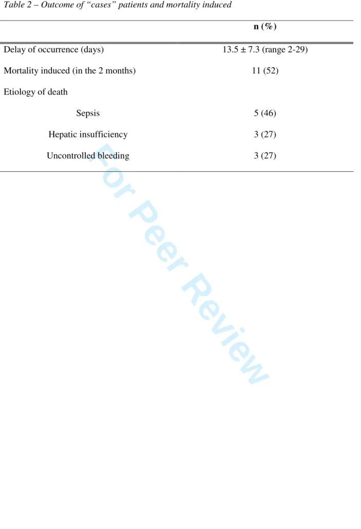
Outcome of patient after bleeding related to postbanding ulcer following EVL
Results are summarized in table 2. The prevalence of the bleeding complication was of 2.74%
(23 episodes among 837 EVL) that occurred with a mean delay of 13.5 ± 7.3 days (range
2-29). This complication was quite severe, 11 of the 21 patients (52%) dying within a few days
and, in the majority of cases (5 patients), secondary to a sepsis (one spontaneous bacterial
peritonitis, two pneumonia and two septicemia). Seven of the 11 patients who died, did not
receive prophylactic antibiotherapy during the EVL session inducing the postbanding ulcer. In
univariate analysis, the development of a sepsis during the evolution was the only predictive
factor of the fatal outcome (p = .03).
Comparison between "cases" and controls
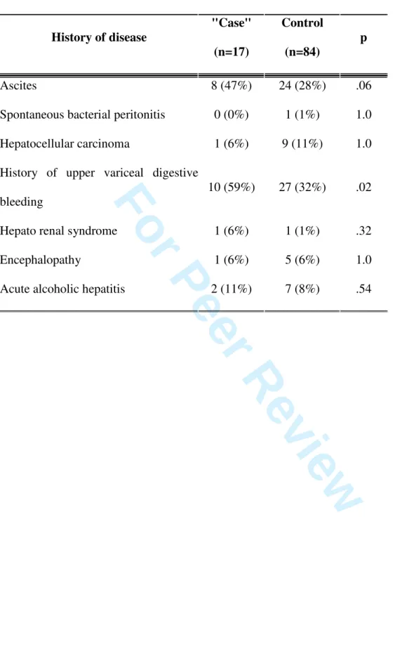
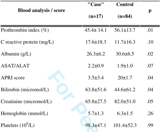
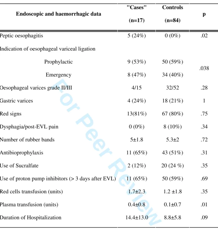
The clinical, biological and endoscopic data as well as the medical history between "cases"
and controls are summarized in Tables 1, 3, 4 and 5. The severity of the liver disease was
significantly higher in "cases" than in controls with a higher Child Pugh class (p=. 04) and
score (9.1 ± 2.6 versus 7.7 ± 2.2, p = .02). The presence of peptic oesophagitis (p = .02),
previous upper variceal digestive bleeding (p = .02), the prophylactic EVL condition (p =
.038), a higher platelet ratio index (APRI score) (p = .04), fresh plasma transfusion (p = .01),
a lower total bilirubin level (p = .04) and a lower prothrombin index (p = .01) and albumin (p
= .02) were significantly associated with the occurrence of the bleeding complication in
"cases".
There was a trend of reaching significant difference for ascites (p = .06) and higher ratio
aspartate aminotransferase / alanine aminotransferase (p = .07) in "cases".
There was no difference between "cases" and controls regarding: the previous use of
ß-blockers, the post EVL PPI use during more than 3 days and post EVL sucralfate use; the
presence of gastro oesophageal varices; the size of varices treated; the presence of red signs; 3 4 5 6 7 8 9 10 11 12 13 14 15 16 17 18 19 20 21 22 23 24 25 26 27 28 29 30 31 32 33 34 35 36 37 38 39 40 41 42 43 44 45 46 47 48 49 50 51 52 53 54 55 56 57 58 59 60
For Peer Review
the number of rubber bands launched; the occurrence of post EVL pain; the
antibioprophylaxis; the performance of EVL in emergency or in primary or secondary
prophylactic situation; neither with the presence of symptomatic anaemia and the duration of
hospitalization. 3 4 5 6 7 8 9 10 11 12 13 14 15 16 17 18 19 20 21 22 23 24 25 26 27 28 29 30 31 32 33 34 35 36 37 38 39 40 41 42 43 44 45 46 47 48 49 50 51 52 53 54 55 56 57 58 59 60
For Peer Review
Multivariate analysis
Variables significantly linked in univariate analysis to the occurrence of the bleeding
complication following EVL, were assessed by multivariate analysis (Data included in the
model are presented in table 6). Logistic regression identified four independent variables
predictive of bleeding on ulceration after spontaneous slippage of rubber band: previous upper
variceal digestive bleeding (OR 12.07, 95%CI [2.3-63.43]), the presence of peptic
oesophagitis(OR 8.9, 95%CI [1.65-47.8]), a high APRI score (OR 1.54, 95%CI [1.11-2.16])
and a low prothrombin index (OR 0.54, 95% CI [0.31-0.94])(Table 4).
Because the prothrombin index appeared to be a predictive variable of the occurrence
of the bleeding complication, a Receiver-Operator Characteristics (ROC) curve was
constructed to determine if a threshold value could be calculated allowing the discrimination
of patients with or without risk of developing bleeding on post EVL ulcer and the slippage of
the rubber band (Figure 1). A threshold value below 45% had a sensitivity, a specificity, a
positive predictive value (PPV) of 47%, 81%, and 12% respectively and a negative predictive
value (NPV) of 67% (area under curve of 0.7). 3 4 5 6 7 8 9 10 11 12 13 14 15 16 17 18 19 20 21 22 23 24 25 26 27 28 29 30 31 32 33 34 35 36 37 38 39 40 41 42 43 44 45 46 47 48 49 50 51 52 53 54 55 56 57 58 59 60
For Peer Review
DISCUSSION
This large case control study shows for the first time that bleeding on ulcer after early
spontaneous slippage of rubber band post EVL is a serious complication. Four independent
factors (previous upper variceal digestive bleeding, the presence of peptic oesophagitis, a high
APRI score and a low prothrombin index) are predictive of this life threatening complication.
Only a few case reports have been reported 9-11. Most of the previous studies on bleeding following EVL in cirrhotic patients focalized on the rebleeding condition and risks without
specifying the site of the bleeding by endoscopy. In these studies the rate of rebleeding within
5 days was 15% and the mortality was 14% within 6 weeks 12, 13. In the present study, we have analysed only patients in whom an endoscopic evaluation confirmed that of the post
EVL bleeding was due to an ulceration induced by the rubber band and its spontaneous
slippage. The prevalence of this complication is low (2.74%) but the study confirms the
severe outcome of these bleedings with a high rate of death (52%), mainly due to the
development of a sepsis responsible for the majority of death of the patients. Bacterial
infection has already been reported as a risk factor of rebleeding following EVL in a previous
retrospective study, but the source of bleeding was not documented 14. In the present study, the high mortality rate is certainly explained by the low rate of prophylactic antibiotherapy
performed during the initial EVL procedure (62%), particularly at the beginning of the study:
seven out of eleven patients who died did not receive this prophylactic antibiotherapy which
is actually recommended 2.
The severity of the liver disease is an important factor in the occurrence of this
bleeding complication. Low prothrombin index, high APRI score and previous upper variceal
digestive bleeding appeared to be independent predictive factors of bleeding related to post
EVL ulcerations. The poor liver condition (Child Pugh C class, high MELD score) have been
previously well identified as predictive factors of rebleeding in cirrhotic patients 12, 14. The 3 4 5 6 7 8 9 10 11 12 13 14 15 16 17 18 19 20 21 22 23 24 25 26 27 28 29 30 31 32 33 34 35 36 37 38 39 40 41 42 43 44 45 46 47 48 49 50 51 52 53 54 55 56 57 58 59 60
For Peer Review
reduced coagulation ability, an increased vascular fragility and a large extension of
sub-mucosal oesophageal varices induced by the portal hypertension might explain the importance
of bleeding from the post-banding induced ulcer without effective local thrombosis.
The present study also stresses the importance of a local factor: the presence of peptic
oesophagitis as an independent predictive factor of this complication. These findings strongly
suggest that the mucosal damages induced by the acid exposure of the lower part of the
oesophagus might promote the early slippage of the rubber band or the bleeding from the
post-banding ulcer. When considering healing of post EVL ulcers, the use of PPI (pantoprazole) has been reported to induce smaller lesions in comparison to placebo, but the
effect on bleeding was not conclusive 15, 16. In our study, all patients were treated with a mono dose PPI during at least the first three days following EVL. Using higher doses of PPI might
have better prevented bleeding related to postbanding ulcer. Nevertheless there was no
difference regarding the use of extended PPI treatment (> 3 days) between cases and control
patient. Moreover it have been suggested that PPI therapy could be associated with a higher
risk of community-acquired pneumonia and spontaneous bacterial peritonitis in cirrhotic
patients 17, 18. Indeed, the acid gastric environment is considered an important barrier to prevent colonization of the stomach and small intestine by ingested bacteria. Low-acid states
may compromise this barrier, increasing bacterial colonization and making the host more
susceptible to enteric infections and bacterial translocation 19. Conversely, a recent meta-analysis of clinical trials failed to show a conclusive association between PPI and respiratory
infection and a retrospective case control study including 116 cirrhotic patients did not find
any association between PPI use and spontaneous bacterial peritonitis 20, 21. The use of sucralfate, known for its experimental mucosal cyto-protective action, has been proposed to
improve the ulcer healing following variceal sclerotherapy and ligation 22-24. Compared to antacid, sucralfate allowed a faster post EVL ulcer healing in a group of 45 randomized 3 4 5 6 7 8 9 10 11 12 13 14 15 16 17 18 19 20 21 22 23 24 25 26 27 28 29 30 31 32 33 34 35 36 37 38 39 40 41 42 43 44 45 46 47 48 49 50 51 52 53 54 55 56 57 58 59 60
For Peer Review
cirrhotic patients, but the association of both drugs was not assessed 25. In our study, we failed to find any benefit in the use of sucralfate for the prevention of bleeding related to post
banding ulcer, but the number of treaded patients treated was very low.
The independent predictive factors found would allow a better anticipation and
prevention of the occurrence of post EVL ulcer bleedings. Alternatives to EVL should be
evaluated in the prophylaxis of the bleeding induced by portal hypertension. The monitoring
of beta-blockers using measurement of the hepatic venous pressure gradient has been reported
to significantly reduce the risk of a first haemorrhage in patients with large varices and to
improve survival 26, 27. In addition, two recent prospective, randomized studies have shown a long-term benefit and enhanced survival in hepatic venous pressure gradient-guided
pharmacological treatment over EVL in cirrhotic patients for the prevention of rebleeding 28,
29
. If confirmed, these data suggest that EVL might not be the first choice in the treatment of
variceal bleeding, being reserved for emergency bleeding conditions or pharmacological
nonresponders.
Finally several different endoscopic approaches have been proposed in order to
substitute the EVL, clipping and detachable endoloops. In a preliminary study, variceal
clipping has been shown to be effective in the management of bleeding oesophageal varices,
without producing ulcer on attached site, as observed with band ligation, therefore avoiding
the risk of haemorrhage 30. Similar observations have been reported with detachable endoloops, but without evaluation of the slippage of the snare and ulcer formation following
the endoscopic treatment 31.
In conclusion, the bleeding of ulcer due to spontaneous slippage of rubber band after
EVL is a serious complication in which a fatal outcome seems to be influenced in the majority
of cases by a sepsis. This present study pointed out at four independent predictive factors for
the occurrence of this bleeding complication: presence of peptic oesophagitis, previous upper 3 4 5 6 7 8 9 10 11 12 13 14 15 16 17 18 19 20 21 22 23 24 25 26 27 28 29 30 31 32 33 34 35 36 37 38 39 40 41 42 43 44 45 46 47 48 49 50 51 52 53 54 55 56 57 58 59 60
For Peer Review
variceal digestive bleeding, a high APRI score and low prothrombin index. In patients with
these factors, a special care should be undertaken when an EVL is programmed in order to
minimize their importance and other treatment alternatives should be thought of. Further
prospective studies are necessary to confirm these results. Prevention of this complication is
needed. 3 4 5 6 7 8 9 10 11 12 13 14 15 16 17 18 19 20 21 22 23 24 25 26 27 28 29 30 31 32 33 34 35 36 37 38 39 40 41 42 43 44 45 46 47 48 49 50 51 52 53 54 55 56 57 58 59 60
For Peer Review
STATEMENT OF INTERESTS
Authors' declaration of personal interests: None
Declaration of funding interests: None 3 4 5 6 7 8 9 10 11 12 13 14 15 16 17 18 19 20 21 22 23 24 25 26 27 28 29 30 31 32 33 34 35 36 37 38 39 40 41 42 43 44 45 46 47 48 49 50 51 52 53 54 55 56 57 58 59 60
For Peer Review
REFERENCE
1. Carbonell N, Pauwels A, Serfaty L, Fourdan O, Lévy VG, Poupon R. Improved
survival after variceal bleeding in patients with cirrhosis over the past decades.
Hepatology 2004;40:652-9.
2. de Franchis R. Evolving consensus in portal hypertension: Report of the Baveno IV
consensus workshop on methodology of diagnosis and therapy in portal hypertension.
J Hepatol 2005;43:167-176.
3. Consensus conference: complications of portal hypertension in adults. Gastroenterol
Clin Biol 2004;28:B318-23.
4. Tait IS, Krige JE, Terblanche J. Endoscopic band ligation of oesophageal varices. Br J
Surg 1999;86:437-46.
5. Saltzman JR, Arora S. Complications of esophageal variceal band ligation.
Gastrointest Endosc 1993;39:203-5.
6. Schmitz RJ, Sharma P, Badr AS, Qamar MT, Weston AP. Incidence and management
of esophageal stricture formation, ulcer bleeding, perforation, and massive hematoma
formation from sclerotherapy versus band ligation. Am J Gastroenterol
2001;96:437-41.
7. Johnson PA, Campbell DR, Antonson CW, Weston AP, Shuler FN, Lozoff RD.
Complications associated with endoscopic band ligation of esophageal varices.
Gastrointest Endosc 1993;39:181-5.
8. Polski JM, Brunt EM, Saeed ZA. Chronology of histological changes after band
ligation of esophageal varices in humans. Endoscopy 2001;33:443-7.
9. Mishin I, Dolghii A. Early spontaneous slippage of rubber bands with fatal bleeding: a
rare complication of endoscopic variceal ligation. Endoscopy 2005;37:275-76. 3 4 5 6 7 8 9 10 11 12 13 14 15 16 17 18 19 20 21 22 23 24 25 26 27 28 29 30 31 32 33 34 35 36 37 38 39 40 41 42 43 44 45 46 47 48 49 50 51 52 53 54 55 56 57 58 59 60
For Peer Review
10. Toyoda H, Fukuda Y, Katano Y, Ebata M, Nagano K, Morita K, et al. Fatal bleeding
from a residual vein at the esophageal ulcer base after successful endoscopic variceal
ligation. J Clin Gastroenterol 2001;32:158-160.
11. Van Vlierberghe H, De Vos M, Hautekeete M, Elewaut A. Severe bleeding following
endoscopic variceal ligation: should EVL be avoided in Child C patients? Acta
Gastroenterol Belg 1999;62:175-177.
12. Bambha K, Kim W R, Pedersen R, Bida J P, Kremers W K, Kamath P S. Predictors of
early re-bleeding and mortality after acute variceal haemorrhage in patients with
cirrhosis. Gut 2008;57:814-820.
13. D'Amico G, De Franchis R. Upper digestive bleeding in cirrhosis. Post-therapeutic
outcome and prognostic indicators. Hepatology 2003;38:599-612.
14. Yang MT, Chen HS, Lee HC, Lin CL. Risk factors and survival of early bleeding after
esophageal variceal ligation. Hepatogastroenterology 2007;54:1705-9.
15. Shaheen NJ, Stuart E, Schmitz SM, Mitchell KL, Fried MW, Zacks S et al.
Pantoprazole reduces the size of postbanding ulcers after variceal band ligation: a
randomized, controlled trial. Hepatology 2005;41:588-94.
16. Boo GB, Oh JC, Lee BJ, Lee DM, Kim YD, Park CG et al. The effect of proton pump
inhibitor on healing of post-esophageal variceal ligation ulcers. Korean J Gastroenterol
2008;51:232-40.
17. Bajaj JS, Hafeezullah M, Zadvornova Y, Saeian K. Protom pump inhibitor use is
associated with a high risk of spontaneous bacterial peritonitis. Hepatology 2007;46
(suppl):565A.
18. Sarkar M, Hennessy S, Yang YX. Proton-pump inhibitor use and the risk for
community-acquired pneumonia. Ann Intern Med 2008;149:391-8. 3 4 5 6 7 8 9 10 11 12 13 14 15 16 17 18 19 20 21 22 23 24 25 26 27 28 29 30 31 32 33 34 35 36 37 38 39 40 41 42 43 44 45 46 47 48 49 50 51 52 53 54 55 56 57 58 59 60
For Peer Review
19. Laine L, Ahnen D, McClain C, Solcia E, Walsh J H. Review article: potential
gastrointestinal effects of long-term acid suppression with proton pump inhibitors.
Aliment Pharmacol Ther 2000;14:651-668.
20. Sultan N, Nazareno J, Gregor J. Association between proton pump inhibitors and
respiratory infections: a systematic review and meta-analysis of clinical trials. Can J
Gastroenterol 2008;22:761-6.
21. Campbell MS, Obstein K, Rajender Reddy K, Yang YX. Association between proton
pump inhibitor use and spontaneous bacterial peritonitis. Dig Dis Sci 2008;53:394-8.
22. Roark G. Treatment of postsclerotherapy esophageal ulcers with sucralfate.
Gastrointest Endosc 1984;30:9-10.
23. Polson RJ, Westaby D, Gimson AES, Hayes PC, Stellon AJ, Hayllar K et al.
Sucralfate for the prevention of early rebleeding following injection sclerotherapy for
esophageal varices. Hepatology 1989;10:279-282.
24. Orlando RC. Cytoprotection by sucralfate in acid-exposed esophagus: a review. Scand
J Gastroenterol 1987;127 (Suppl):97-100.
25. Yang WG, Hou MC, Lin HC, Kuo BI, Lee FY, Chang FY et al. Effect of sucralfate
granules in suspension on endoscopic variceal sclerotherapy induced ulcer: analysis of
the factors determining ulcer healing. J Gastroenterol Hepatol 1998;13:225-31.
26. Villanueva C, Balanzo J. Variceal bleeding: pharmacological treatment and
prophylatic strategies. Drugs 2008;68:2303-24.
27. Sharma P, Kumar A, Sharma BC, Sarin SK. Early identification of haemodynamic
response to pharmacotherapy is essential for primary prophylaxis of variceal bleeding
in patients with 'high risk' varices. Aliment Pharmacol Ther 2009;30:48-60. 3 4 5 6 7 8 9 10 11 12 13 14 15 16 17 18 19 20 21 22 23 24 25 26 27 28 29 30 31 32 33 34 35 36 37 38 39 40 41 42 43 44 45 46 47 48 49 50 51 52 53 54 55 56 57 58 59 60
For Peer Review
28. Lo GH, Chen WC, Lin CK, Tsai WL, Chan HH, Chen TA et al. Improved survival in
patients receiving medical therapy as compared with banding ligation for the
prevention of oesophageal variceal rebleeding. Hepatology 2008;48:580-7.
29. Villanueva C, Aracil C, Colomo A, Lopez-Balaguer JM, Piqueras M, Gonzalez B et
al. A randomized controlled study on prevention of variceal rebleeding comparing
nadolol + ligation vs. hepatic venouos pressure gradient-guided pharmacological
therapy. Aliment Pharmacol Ther 2008;29:397-408.
30. Yol S, Belviranli M, Toprak S, Kartal A. Endoscopic clipping versus band ligation in
the management of bleeding esophageal varices. Surg Endosc 2003;17:38-42.
31. Naga MI, Okasha HH, Foda AR, Gomaa MS, Fouad AM, Masoud AG et al.
Detachable endoloop vs. elastic band ligation for bleeding esophageal varices.
Gastrointest Endosc 2004;59:804-9. 3 4 5 6 7 8 9 10 11 12 13 14 15 16 17 18 19 20 21 22 23 24 25 26 27 28 29 30 31 32 33 34 35 36 37 38 39 40 41 42 43 44 45 46 47 48 49 50 51 52 53 54 55 56 57 58 59 60
For Peer Review
FIGURE LEGENDS
Figure 1
Receiver-Operator Characteristics (ROC) curve for prothrombin index (PI) to determine a
threshold value allowing discrimination of patients with or without risk of developing
bleeding related to post EVL ulcer. 3 4 5 6 7 8 9 10 11 12 13 14 15 16 17 18 19 20 21 22 23 24 25 26 27 28 29 30 31 32 33 34 35 36 37 38 39 40 41 42 43 44 45 46 47 48 49 50 51 52 53 54 55 56 57 58 59 60
For Peer Review
TABLES
Table 1 – Characteristics of patients
Characteristics "Cases" (n = 17) Controls (n = 84) p Age (years) 54.6 (37-85) 58.5 (17-87) .12 Male/Female 13/4 (76%/24%) 60/24 (71%/29%) .77 Causes cirrhosis Alcohol Virus Others 12(71%) 3(18%) 5(29%) 53(63%) 27(32%) 13(15%) .78 .26 .30
Child Pugh score 9.1 ± 2.6 7.7 ± 2.2 .02
Child Pugh Class
A B C 2 (10%) 9 (53%) 7 (35%) 30 (36%) 32 (38%) 22 (26%) .04 Previous Treatment ß-blockers Aspirin/anticoagulation 4 (24%) 1 (9%) 28 (33%) 7 (8%) .57 1 3 4 5 6 7 8 9 10 11 12 13 14 15 16 17 18 19 20 21 22 23 24 25 26 27 28 29 30 31 32 33 34 35 36 37 38 39 40 41 42 43 44 45 46 47 48 49 50 51 52 53 54 55 56 57 58 59 60
For Peer Review
Table 2 – Outcome of “cases” patients and mortality induced
n (%)
Delay of occurrence (days) 13.5 ± 7.3 (range 2-29)
Mortality induced (in the 2 months) 11 (52)
Etiology of death Sepsis Hepatic insufficiency Uncontrolled bleeding 5 (46) 3 (27) 3 (27) 3 4 5 6 7 8 9 10 11 12 13 14 15 16 17 18 19 20 21 22 23 24 25 26 27 28 29 30 31 32 33 34 35 36 37 38 39 40 41 42 43 44 45 46 47 48 49 50 51 52 53 54 55 56 57 58 59 60
For Peer Review
Table 3 – Univariate analysis for history of the disease
History of disease "Case" (n=17) Control (n=84) p Ascites 8 (47%) 24 (28%) .06
Spontaneous bacterial peritonitis 0 (0%) 1 (1%) 1.0
Hepatocellular carcinoma 1 (6%) 9 (11%) 1.0
History of upper variceal digestive
bleeding
10 (59%) 27 (32%) .02
Hepato renal syndrome 1 (6%) 1 (1%) .32
Encephalopathy 1 (6%) 5 (6%) 1.0
Acute alcoholic hepatitis 2 (11%) 7 (8%) .54 3 4 5 6 7 8 9 10 11 12 13 14 15 16 17 18 19 20 21 22 23 24 25 26 27 28 29 30 31 32 33 34 35 36 37 38 39 40 41 42 43 44 45 46 47 48 49 50 51 52 53 54 55 56 57 58 59 60
For Peer Review
Table 4 – Univariate analysis for biological tests
Blood analysis / score
"Case" (n=17) Control (n=84) p Prothrombin index (%) 45.4± 14.1 56.1±13.7 .01 C reactive protein (mg/L) 17.6±18.3 11.7±16.3 .10 Albumin (g/L) 26.3±6.2 30.6±6.5 .02 ASAT/ALAT 2.2±0.9 1.9±1.0 .07 APRI score 3.5±3.4 20±1.7 .04 Bilirubin (micromol/L) 63.8±51.6 44.6±61.2 .04 Creatinine (micromol/L) 65.8±27.5 82.0±51.0 .05 Hemoglobin (mmol/L) 5.7±1.3 6.3±1.5 .26 Platelets (109/L) 98.3±47.1 101.4±52.3 .99 ASAT: aspartate aminotransferase, ALAT: alanine aminotransferase
Score APRI = (AST (/ULN) x 100) / platelets (109/L) 3 4 5 6 7 8 9 10 11 12 13 14 15 16 17 18 19 20 21 22 23 24 25 26 27 28 29 30 31 32 33 34 35 36 37 38 39 40 41 42 43 44 45 46 47 48 49 50 51 52 53 54 55 56 57 58 59 60
For Peer Review
Table 5 – Univariate analysis for endoscopic and haemorrhagic data
Endoscopic and haemorrhagic data
"Cases" (n=17) Controls (n=84) p Peptic oesophagitis 5 (24%) 0 (0%) .02
Indication of oesophageal variceal ligation
Prophylactic Emergency 9 (53%) 8 (47%) 50 (59%) 34 (40%) .038
Oesophageal varices grade II/III 4/15 32/52 .28
Gastric varices 4 (24%) 18 (21%) 1
Red signs 13(81%) 67 (80%) .75
Dysphagia/post-EVL pain 0 (0%) 8 (10%) .34
Number of rubber bands 5±1.8 5.3±2 .72
Antibioprophylaxis 11 (65%) 43 (51%) .31
Use of Sucralfate 2 (12%) 20 (24 %) .35
Use of proton pump inhibitors (> 3 days after EVL) 11 (65%) 50 (59%) .69
Red cells transfusion (units) 1.7±2.3 1.2 ±1.8 .35
Plasma transfusion (units) 0.4±0.8 0.1±0.7 .01
Duration of Hospitalization 14.4±13.0 8.8±5.8 .09 3 4 5 6 7 8 9 10 11 12 13 14 15 16 17 18 19 20 21 22 23 24 25 26 27 28 29 30 31 32 33 34 35 36 37 38 39 40 41 42 43 44 45 46 47 48 49 50 51 52 53 54 55 56 57 58 59 60
For Peer Review
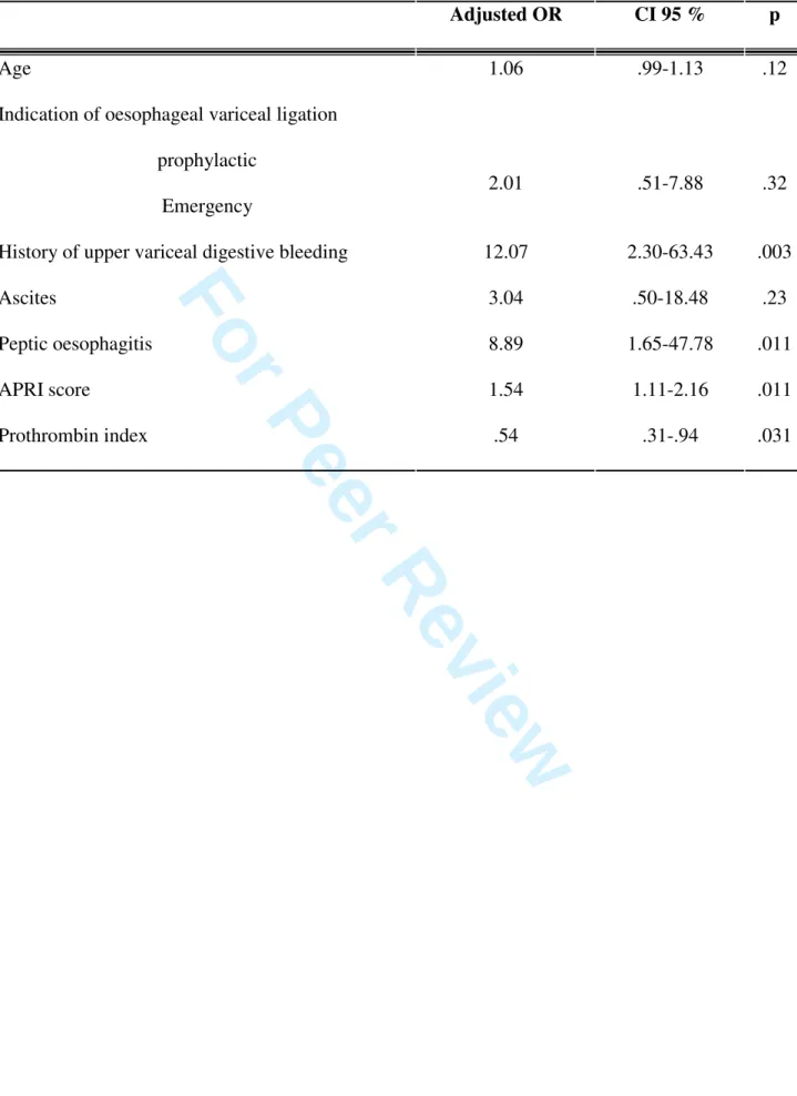
Table 6 – Multivariate analysis
Adjusted OR CI 95 % p
Age 1.06 .99-1.13 .12
Indication of oesophageal variceal ligation
prophylactic
Emergency
2.01 .51-7.88 .32
History of upper variceal digestive bleeding 12.07 2.30-63.43 .003
Ascites 3.04 .50-18.48 .23 Peptic oesophagitis 8.89 1.65-47.78 .011 APRI score 1.54 1.11-2.16 .011 Prothrombin index .54 .31-.94 .031 3 4 5 6 7 8 9 10 11 12 13 14 15 16 17 18 19 20 21 22 23 24 25 26 27 28 29 30 31 32 33 34 35 36 37 38 39 40 41 42 43 44 45 46 47 48 49 50 51 52 53 54 55 56 57 58 59 60
For Peer Review
FIGURE
Figure 1 0 10 20 30 40 50 60 70 80 90 100 0 10 20 30 40 50 60 70 80 90 100 1 - Sp S eBest Cut Off PI = 45% Se = 47% ; Sp = 81% 3 4 5 6 7 8 9 10 11 12 13 14 15 16 17 18 19 20 21 22 23 24 25 26 27 28 29 30 31 32 33 34 35 36 37 38 39 40 41 42 43 44 45 46 47 48 49 50 51 52 53 54 55 56 57 58 59 60