HAL Id: hal-01928736
https://hal.archives-ouvertes.fr/hal-01928736
Submitted on 20 Nov 2018
HAL is a multi-disciplinary open access
archive for the deposit and dissemination of
sci-entific research documents, whether they are
pub-lished or not. The documents may come from
teaching and research institutions in France or
abroad, or from public or private research centers.
L’archive ouverte pluridisciplinaire HAL, est
destinée au dépôt et à la diffusion de documents
scientifiques de niveau recherche, publiés ou non,
émanant des établissements d’enseignement et de
recherche français ou étrangers, des laboratoires
publics ou privés.
Anti-Acne, Antioxidant and Cytotoxic Properties of
Ludwigia peploides Leaf Extract
Imen Smida, Alaa Sweidan, Yasmine Souissi, Isabelle Rouaud, Aurélie
Sauvager, Franck Torre, Virgile Calvert, Jean Le Petit, Sophie Tomasi
To cite this version:
Imen Smida, Alaa Sweidan, Yasmine Souissi, Isabelle Rouaud, Aurélie Sauvager, et al.. Anti-Acne,
Antioxidant and Cytotoxic Properties of Ludwigia peploides Leaf Extract. International Journal of
Pharmacognosy and Phytochemical Research, Dr. Yashwant Research Labs Pvt. Ltd., 2018, 10 (7),
pp.271-278. �hal-01928736�
ISSN: 0975-4873
Research Article
Anti-Acne, Antioxidant and Cytotoxic Properties of Ludwigia
peploides Leaf Extract
Smida I
1,4*, Sweidan A
1, Souissi Y
2, Rouaud I
3, Sauvager A
3, Torre F
4, Calvert V
4, Le Petit
J
4, Tomasi S
31INSERM U 1241, INRA, Université de Rennes 1, Université Bretagne Loire, Nutrition Metabolism and Cancer, 2
avenue du Professeur Léon Bernard-35043 Rennes, France
2Laboratoire Biotechnologie & Valorisation des Bio-Géo Ressources (LR11-ES31); Institut Supérieur de Biotechnologie;
Biotechpole Sidi Thabet, 2020, Sidi Thabet, Ariana, Tunisie.
3Univ Rennes, CNRS ISCR-UMR 6226, F-35000 Rennes, France.
4Institut Méditerranéen de Biodiversité et d’Ecologie marine et continentale (IMBE), Aix- Marseille Université,
UMRCNRS-IRD, Avignon Université, Faculté de Saint-Jérôme, Case 422, F-13397 Marseille cedex20, France.
Available Online :25th July, 2018
ABSTRACT
Objective: This work is the first to investigate the in vitro anti-acne and cytotoxic activities of the leaves of Ludwigia
peploides. With its important seasonal biomass production, this plant has great potential in several therapeutic and cosmetic
applications. Methods: The antibacterial activity of the extract was screened against a consortium of skin microorganisms that cause acne vulgaris disease, using disc diffusion and broth microdilution methods. The antioxidant activity of methanolic leaf extract of L. peploides was evaluated by DPPH and NBT assays to assess the free radical scavenging activity of L. peploides, which in turn has a great importance related to its role in minimizing the oxidative stress linked to the pathophysiology of diseases like acne vulgaris. Its putative cytotoxicity was examined against human macrophage-like monocytic leukemia (THP-1) and human keratinocytes (HaCaT) cell lines. In addition, antiproliferative activity was screened against B16 cancer cell lines. Results: The extract displayed antioxidant effect by DPPH (IC50= 58 ± 6.0 µg mL -1) and NBT (IC
50= 30 ± 2.8 µg mL-1) assays, and it was not toxic on HaCaT cells (IC50 > 200 µg mL-1). A strong inhibitory
activity against Propionibacterium acnes (MIC = 1.9 µg mL-1) was registered for the extract, which had a bactericidal
effect against Staphylococcus aureus, Staphylococcus epidermidis, and Salmonella enterica. Finally, the extract was shown to have an antiproliferative activity (IC50=5.5 ± 2.3 µgmL-1). Conclusion: The extract displays antioxidant and anti-acne
effects as well as inhibition potential of B16 melanoma cells proliferation.
Keywords: Ludwigia peploides, antioxidant, anti-acne, antiproliferative properties. INTRODUCTION
Aquatic macrophytes belonging to Ludwigia genus (Onagraceae family) are invasive plants that perturb the functioning of aquatic habitats1,2. Several species of this
genus are known for their antioxidant activity3 and for their
ability to synthesize allelopathic molecules4, which confer
medicinal properties such as antimicrobial5,6,7 and
antiproliferative8 activities. In this context, we examined
the putative medicinal properties of Ludwigia peploides, which can produce, from June to September, large amounts of biomass, reaching 2-3 kg dry weight m-2 2 on a surface
of several hectares in the south of France. Smida et al. (2015)7 have demonstrated the antibacterial properties of
this plant that were superior in leaves and flowers. Among the large number of studies on the antimicrobial properties of plants, several ones have reported their therapeutic effects in the treatment of the acne vulgaris9,10.
The latter has an important impact on the social, emotional, and psychological comportment of 650 million people around the world9. Acne vulgaris appears as inflammation
in skin areas. This disease is due to an oxidative stress in the pilosebaceous unit that creates an anaerobic environment. That is the best suited condition for the development of a Gram-positive obligate anaerobic bacterium, Propionibacterium acnes, which inhabits under the human skin and populates the androgen stimulated sebaceous follicles11. It is often associated to Staphylococcus aureus and other microorganisms such as Staphylococcus epidermidis, Salmonella enterica, Escherichia coli, and Candida albicans, which can also be
responsible for skin infection diseases. The antibiotics used for its treatment are not specific and can generate resistant bacteria12. P. acnes seems to be responsible for
the production of reactive oxygen species, which provoke an inflammation in the pilosebaceous skin structure. The capability of plants to inhibit this phenomenon is, to some extent, due to the fact that they contain non-enzymatic antioxidants such as polyphenols, which are able to reduce reactive oxygen species13,14. Saric et al. (2017)15 reported
Smida et al. / Anti-Acne, Antioxidant…
IJPPR, Volume 10, Issue 7: July 2018 Page 272 secretion and are effective for the treatment of acne. These
antioxidant properties are complementary to antibacterial activity16, in particular in acne disease. Polyphenols have
also been described, particularly those found in green tea, as putative anti-tumor agents through their antioxidant activities, which inhibit the growth of tumor cells and favor apoptosis17,18, the effect of active oxygen radicals being
also known to intervene in the carcinogenesis phenomenon19,20. Because polyphenols are found in
several Onagraceae species21 and in extracts of L. peploides22, we investigated the antioxidant properties in
this plant. Thus, in our study, we have measured the total polyphenolic (TP), flavonoid (FLA), tannin (TAN) and anthocyanin (ANT) compounds from a methanol leaf extract of L. peploides and have assessed its antioxidant property. Furthermore, its anti-acne and antiproliferative activities were studied and compared to that of classical antibiotic and anti-cancer compounds. The cytotoxicity tests were conducted to ensure the safety of this extract.
MATERIAL AND METHODS
Treatment of the Samples and Preparation of Extracts
Plants were collected alongside the bank of a dead arm of the Durance river (43◦ 40 17 N, 5◦ 28 07 E), near Pertuis town (Vaucluse, France). The characteristics of this sampling station were described by Smida et al. (2015)7.
Leaves were immediately separated from plants before being air-dried and then ground into a coarse powder with the aid of a mechanical grinder (IKA Labortechnik type A10). Then, the ultrasound-assisted extraction commenced by soaking one gram of the obtained leaves powder in 50 mL of a methanol: water mixture (80:20 v/v) for 10 min (Bioblock Scientific 88160), then mixed under a vigorous magnetic stirring at 130 tr.min-1 and centrifuged at 3354 g
at 4°C for 30 min. The supernatant was filtered through a Whatman filter paper (0.22 µm) and concentrated under vacuum and reduced pressure at 40°C using a rotary flask evaporator (Heidolph Laborta 4000). The dry crude hydro-methanolic extract was labeled with Dry Plant Extract (DPE) and stored at 4°C in a sealed sterile glass flask until further use. The extract was dissolved in DMSO for all the biological tests performed.
Chemical Analysis Methods
Determination of Total Phenolic (TP) Content
TP content in the methanol extract was determined using Folin-Ciocalteu reagent according to Ksouri et al. (2009)23
and expressed as mg of gallic acid (GA) equivalent per gram of dry plant extract (DPE) or, taking into account the yield of extraction, per gram of dry plant material (DPM).
Determination of Flavonoid (FLAV) Content
Flavonoid content was estimated according to Ksouri et al. (2009)23 and expressed as mg of catechin (CAT)
equivalent per gram of DPE or DPM.
Determination of Total Tannin (TAN) Content
The method to determine TAN content was based on the reaction of tannins with hydrochloric vanillin. Thus, 0.5 mL of diluted (1/100) methanol extract was mixed with 3 mL of a 4 % vanillin solution in methanol. This mixture was vigorously stirred, then let to rest for 15 min before adding 1.5 mL of concentrated HCl (38 %). The
absorbance was measured at 500 nm. Tannin content was expressed as mg CAT equivalent per gram of DPE or DPM.
Determination of Anthocyanin (ANT) Content
The differential pH method described by Giusti and Wrolstad (2001)24 was used. Anthocyanin content was
expressed as mg of cyanidin-3-glycosylated (C3G) equivalent per gram of DPE or DPM.
Antioxidant Assays
Antioxidant activity was performed on the leaf extract using the 1, 1′-Diphenyl-2-Picrylhydrazyl (DPPH) free radical scavenging activity and the superoxide (O2.-)
radical scavenging nitro blue tetrazolium (NBT) assays.
DPPH Radical Scavenging Activity
The capacity of the extract to scavenge DPPH radical by its reduction was measured according to a modified method of Tuberoso et al. (2007)25. In a 96-well plate, a
reaction mixture containing 100 μL of DPPH methanolic solution (0.5 mmol.L-1) and 10 μL of the extract was added
into each well to give a final concentration range (9.37-600 μg.mL-1). Absorbance was measured at 470 nm after
15 min of incubation in the dark. Gallic acid and quercetin were used as references. The percentage of inhibition at the steady state of each concentration was used to graphically determine the inhibitory concentration 50% (IC50), defined
as the extract concentration (µg.mL-1) required to obtain
50 % of the DPPH reduction.
The radical scavenging activity was calculated using the following formula:
% inhibition = (absorbance control – absorbance sample) /
(absorbance control) * 100
Superoxide Radical Scavenging Activity
The leaf extract was tested for its free radical scavenging activity using the non-enzymatic phenazine methosulfate-nicotinamide adenine dinucleotide (PMS/NADH) system, which generates superoxide radicals that reduce NBT to a purple-colored formazan. The measurement was performed in 96-well microplates. The reaction mixture consisted of NADH (78 μmol.L-1), NBT (50 μmol.L-1),
PMS (10 μmol.L-1), and 10 µL of the extract sample was
added into each well to give a final concentration range (9.37‒600 μg.mL-1). The reagents were dissolved in a 16
mmol.L-1 Tris–hydrochloride buffer at pH 8. After
incubation for 5 min at room temperature, the measurement was performed at 560 nm against an appropriate blank to determine the quantity of formazan generated. The percentage of inhibition at the steady state for each concentration was used to calculate the IC50
values, i.e. the extract concentration corresponding to 50 % of the NBT reduction. Ascorbic acid was used as reference.
Biological Analysis
Antimicrobial Activity Assay Microorganisms
Bacterial strains included in this study were the hospital strains, Propionibacterium acnes and Staphylococcus
aureus, Staphylococcus epidermidis (CIP 68.21),
Salmonella enterica serovar “Heidelberg" (S. Heidelberg
B182), Escherichia coli (CIP 53126), and a yeast strain,
were procured at the Department of Microbiology, University Hospital Center of Rennes (France). All strains were maintained on Tryptone soya agar at 4°C.
Disc Diffusion Method
Antibacterial activities were determined according to Smida et al. (2015)7. The dry extract was deposited at a
concentration of 7.5 µg dissolved in 0.15 µL of DMSO per disc.
Minimum Inhibitory Concentration (MIC) and Minimum Bactericidal Concentration (MBC)
Broth microdilution test was done to determine the minimum inhibitory concentration (MIC) as described by the CLSI guidelines26. Serial dilutions of the leaf extract
and the positive control, doxycycline, an antibiotic of the tetracycline family, were prepared with concentrations ranging from 0.0078 to 500 mg mL-1 in Broth Heart
infusion (BHI) in a 96-well microtiter plate (Sterile, Flat bottom, with lid, Greiner Bio-one, Germany). Each well was inoculated with a 107 CFU mL-1 bacterial culture.
Then, the plate was incubated at 37 °C for 24 h under anaerobic conditions. After that, 5 µL of the clear wells were spread on Muller-Hinton Petri dishes and incubated for another 24 h. The clear well with the lowest concentration represented the MIC inhibiting a visible bacterial growth. The Petri dish with the lowest concentration showing no colonial growth corresponds to the minimum bactericidal concentration (MBC) defined as the concentration that killed 99.9 % of the initial inoculum.
Cytotoxicity Tests
Human Macrophage-like Monocytic Leukemia Cells Cell Culture
Human macrophage-like monocytic leukemia cell line, THP-127, was grown in RPMI 1640 medium with sodium
pyruvate (1 mmol.L-1) and Hepes buffer (1000 mmol.L-1)
supplemented with L-Glutamine (2 mmol.L-1), 10 %
heat-inactivated fetal bovine serum (FBS, Lonza, France), and antibiotics (penicillin 100 mg.mL-1 and streptomycin 50
mg.mL-1) in a humidified atmosphere of 5% CO
2 and 95%
air at 37°C. For THP-1 differentiation into macrophages, phorbol 12-myristate 13-acetate (PMA) was used at 10 ng.mL-1 for 72 h. 96-well plates (Sterile, Flat bottom, with
lid, Greiner Bio-one, Germany) were seeded with 7.104
THP-1 cells/well. The plates were incubated with PMA for 72 h and then exposed to decreasing concentrations obtained from an initial plant extract solution dosed at 300 μg.mL-1. Wells without extract constituted the negative
controls. Triton X-100 (1 %), a nonionic detergent, was used as a positive control. The plates were incubated for 24 h and then treated according to the MTT [3-(4, 5-dimethylthiazol-2-phenyl)-2, 5-diphenyltetrazolium bromide] assay28.
Determination of Cell Viability
Ten microliters of 5 mg.mL-1 MTT solution, prepared in
PBS and filter-sterilized through a 0.22 µm filter (Whatman), were added to wells containing 100 µL of culture medium. The 96-well plates were incubated for 4 h at 37 °C under a 5% CO2 atmosphere. Then, 100 µL of 0.04
mol.L-1 HCl in isopropanol were added and mixed to
dissolve the formazan crystals. The intensity of formazan
was read after 5 min at 595 nm. The percent of cell viability was calculated by the following formula:
Cell viability (%) = Mean OD / control OD × 100
All chemical products used in this work were obtained from Sigma Aldrich unless otherwise specified.
Normal Human Keratinocytes and Mouse Melanoma B16 Cells
Cell Culture
The normal human keratinocytes (HaCaT) cell line was a generous gift from the Institute of Genetic and Development (Rennes, France) and the mouse melanoma B16 cell line (ATCC-CRL 6475) was purchased from the Chemical Engineering Laboratory (LGC Toulouse, France). The two cell lines were cultivated in RPMI 1640 medium (Dutscher) with 2 mmol.L-1 L- glutamine
(Dutscher) supplemented with 5% fetal bovine serum (FBS), 1% 10000 U.mL-1 penicillin, and 10 000 U.mL -1streptomycin solution (Lonza). They were often
maintained at 37°C in a humidified atmosphere with 5 % CO2 and 95 % air.
For the experiments, cells were prepared using standard trypsinization procedure with trypsin/EDTA (170 mg L-1
trypsine, 200 mg.mL-1 EDTA, LONZA) and seeded in
96-well-bottom plates for viability assays. The cells were incubated for 24 h and then treated with the appropriate extract dissolved in DMSO and diluted in culture medium. The final DMSO concentration that did not exceed 1 % had no influence on cell viability.
Antiproliferative Activity
Antiproliferative activity of the leaf extract was evaluated according to the OECD guideline 425 with some modifications29. In brief, 100 μL of HaCaT or B16 cells
suspension at 2.104 cells mL-1 were added in each well to
be incubated at 37°C the RPMI culture medium in 5 % CO2
atmosphere for 24 h to form monolayers. Then, the 96-well plates were incubated for 48 h with five different concentrations of the tested compound (in the range 1–200 μg.mL-1). Doxorubicin, an anti-cancer drug, was used as a
positive control. Cell viability is measured using the MTT assay as previously described. The absorbance of the developed yellow color, which was directly proportional to the cells viability, was measured by an automated microplate reader (IMARK, BioRad) at 570 nm. The results are presented in percentage where the untreated cells were considered having 100 % of cell viability.
Statistical Methods
Data are expressed as means ± standard deviations of three replicates.
Cell viability at each concentration of the tested compound was compared to that of the untreated control using Student t-test for adequacy. Computations were performed using Microsoft® Excel 2010 (function T.INV.2T to obtain Student two-sided critical values at 5 %, 1 %, and 0.1 % with parameter n=3-1=2).
RESULTS
Chemical Analysis
The yield of the leaves methanol extraction was 14%. The contents of the different types of polyphenols are reported in Table 1. The values of TP, FLAV, TAN, and ANT were
Smida et al. / Anti-Acne, Antioxidant…
IJPPR, Volume 10, Issue 7: July 2018 Page 274 Table 2: DPPH radical and NBT superoxide radical
scavenging activities, expressed as IC50, for L. peploides leaf extract (DPE), gallic acid, quercetin and
ascorbic acid being used as references. DPE Gallic acid Quer cetin Ascorbic acid DPPH IC50 μg.mL-1 58.0 ± 6.0 6.2 ±0.5 9.0±1 .0 ND NBT IC50 μg. mL-1 30.0 ± 2.8 ND ND 7.5± 1.0 ND = not determined
only indicative of a state of development of L. peploides which, in this study, corresponds to the maximum biological activity of the plant7. Indeed, their relative
proportions vary as in the cycle of any plant.
Table 2 shows the potential of the methanol leaf extract to scavenge DPPH or NBT free radicals. The IC50 values
obtained with the extract were higher than those of the reference products, gallic acid, quercetin and ascorbic acid.
Biological Analysis Antimicrobial Capacities
Table 3 shows the antimicrobial activities of both, the crude extract and doxycycline. The latter was used as reference as it is an antibiotic which is generally prescribed to treat acne vulgaris. The leaf extract was found to be active against all skin microorganisms tested, P. acnes, S
aureus, S. epidermidis, S. enterica, E. coli, and C. albicans,
regardless of the strain Gram type. Its highest activity was obtained against P. acnes and S. epidermidis, with respectively an average inhibition zone of 37 and 28.3 mm and an MIC of 1.9 and 3.9 µg.mL-1. For these two strains,
the MBC was equal to 12.5 µg.mL-1. However, a higher
MIC of 5 µg.mL-1 was registered against S. aureus which
was the most sensitive to the extract lethal effect with a MBC = 7.8 µg.mL-1. A slightly higher MIC was obtained
in case of S. enterica in comparison to the first three strains. But, the MBC was the same as for P. acnes and S.
epidermidis. Finally, E. coli and C. albicans were the least
sensible to the extract among all the strains tested which were more sensitive to doxycycline than to the extract.
Cytotoxicity Test
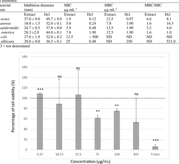
In order to estimate the safety of the methanol extract, its cytotoxicity was evaluated by the MTT assay measuring the viability of macrophage-like cells, THP-1, treated with the extract at various concentrations (Fig.1). Statistically, the extract had a slight significant promotion on the cellular development for the lowest concentration (9.37µg.mL-1), corresponding likely to a hormetic effect.
For the two following values (18.75 µg.mL-1and
37.5µg.mL-1), the extract did not have a significant effect
on the viability of THP-1 cells in contrast to the Triton X-100, which caused a 95.2 % of cell death. For the three
highest concentrations tested, a partial inhibition of cell growth was observed (Fig.1).
Antiproliferative Activity
The antitumor reference, doxorubicin, was toxic against the HaCaT cell line (normal human keratinocytes) with an IC50 of 0.04 ± 0.01 µg.mL-1, whereas the L. peploides
extract was not active against this cell line with an IC50>
200 µg.mL-1 (Table 4). On the other hand, the crude extract
showed a stronger cytotoxic activity against B16 cancer cell lines with an IC50 of 5.5 ± 2.3 µg.mL-1 in comparison
to doxorubicin which displayed an IC50 of 16 ± 1.2 µg.mL -1.
DISCUSSION
The antimicrobial activity of L. peploides7 and other Ludwigia species have been previously reported30,5,31,32,33,34; however, the antioxidant, anti-acne,
and anti-proliferative properties of L. peploides have never been studied.
Measures of the antioxidant activity of L. peploides leaf extract suggest that the latter had effective scavenging activities against free radical (IC50 = 58.0 ± 6.0 µg.mL
-1
) and superoxide anion (IC50= 30 ± 2.8 µg.mL
-1
). Khoudali et al. (2014)35 indicated similar values for methanolic
extracts of Chamaerops humilis leaves (24.5 μg.mL
-1
) and for α-tocopherol (26 μg.mL
-1
). However, the L. peploides extract had a lower antioxidant activity than extracts of known medicinal plants which have registered stronger scavenging activities such as Cerasus avium L.36, with an
IC50 of 17.4 μg mL-1, and extracts of young fruit rinds of G. mangostana L., with an IC50 of 5.6 μg mL-1 37.
Because of their scavenging ability due to their hydroxyl groups, phenolic compounds are often responsible for the antioxidant activity. The principal types of polyphenols, TP, FLAV, TAN, and ANT, were detected in L. peploides leaf extract. Our results were similar to those obtained by Yakob et al. (2012)34 for the methanolic leaf extract of Ludwigia octovalvis. However, the phenolic content of 112
traditional Chinese medicinal plants reported by Cai et al. (2004)38 ranged from 2.2 to 503 mg eq. GAE g-1 of DPM
with average value of 38.7 mg eq. GAE g-1 of DPM, indicates that L. peploides (122.8 ± 0.1 mg eq. GAE g-1
DPM) cannot be classified among plants rich in polyphenol content.
Our study revealed that L. peoploides displayed a potent activity against P. acnes (Table 3), better than those mentioned by Pothitirat et al. (2009)37 for Garcinia mangostana young fruit extract, used in Thai traditional
medicines, (MIC= 15.63 µg.mL-1 and MBC = 31.25
µg.mL-1). Among a great number of plant extracts, Sinha et al. (2014)39 reported the lowest MIC values against P.
Table 1: Total phenolic (TP), flavonoid (FLAV), tannin (TAN) and anthocyanin (ANT) contents in dry plant material (DPM) and dry plant methanolic extract (DPE) of L. peploides leaves.
Polyphenol compounds TP as mg eq. GAE FLAV as mg eq. CATE TAN as mg eq. CATE ANT as mg eq. C3GE g-1 DPM 122.8 ± 0.1 13.2 ± 0.2 9.2 ± 0.0 32.7 ± 0.2 g-1 DPE 17.2 ± 0.2 1.8 ± 0.0 1.3 ± 0.0 ± 0.1
acnes between 4 and 50 µg.mL-1 for Punica granatum, Morus alba, Amanoa anomala and Mahonia aquifolium.
However, Nand et al. (2012)40 reported that the MIC
values of the methanolic extract from Camellia sinensis plant, belonging to Theaceae family, were 1250 µg.mL-1
against P. acnes and S. aureus and 650 µg.mL-1 against S. epidermidis. The MICs were significantly higher than
those obtained with L. peploides (Table 3).
According to Aloni-Grinstein et al. (2015)41, a compound
can be considered bactericidal when its MBC/MIC ratio is lower than four and only bacteriostatic when this ratio is higher than four. In this context, the L. peoploides leaf extract can be considered as bacteriostatic against P. acnes and bactericidal against S. aureus, S. epidermidis, and S.
enterica, while doxycycline is bacteriostatic against P. acnes and S. aureus, and bactericidal against S. epidermidis and S. enterica. The E. coli and C. albicans
strains used can be considered as tolerant. Since the MBC was too high to be determined (E. coli) or 32 times higher than the MIC (C. albicans).
All the MIC and MBC values recorded for P. acnes, S.
aureus, S. epidermidis, and S. enteric were lower than 37.5
µg mL-1 (Table 1), the value above that an inhibition of
THP-1 cell growth (Figure 1) could render the extract unusable. For E. coli and C. albicans, only the MIC values were lower than this limit.
L. peploides leaf extract provided also interesting
antiproliferative activity against B16 cancer cell lines (Table 4). In fact, our results (IC50 = 5.5 µg.mL-1) obtained
with an unpurified extract were better than those of Yang
et al.(2000)42. These authors reported IC
50 values ranging
between 9.7 and 57.5 µg.mL-1 on several human cancer
cell lines with (‒)-epigallocatechin-3-gallate, a purified polyphenol present in green and black tea, which inhibited Table 3: Antimicrobial activities of the L. peploides leaf extract against skin microbial strains. Results are expressed according to the disc diffusion method and measures of the MIC and the MBC, where doxycycline (Dcl) was used as the reference. Bacterial strain Inhibition diameter (mm) MIC µg mL-1 MBC µg mL-1 MBC/MIC
Extract Dcl Extract Dcl Extract Dcl Extract Dcl
P. acnes 37.0 ± 0.0 49.7 ± 0.0 1.9 0.12 12.5 0.97 6.6 8.1 S. aureus 18.0 ± 1.5 32.0 ± 0.1 5.0 0.24 7.8 3.90 1.6 16.3 S. epidermidis 24.7 ± 0.5 37.8 ± 0.0 3.9 0.48 12.5 1.90 3.2 4.0 S. enterica 28.3 ±2.0 44.0 ± 0.1 7.8 1.90 12.5 1.90 1.6 1.0 E. coli 27.0 ± 1.9 32.0 ± 0.2 12.5 > 500 ND ND ND ND C. albicans 28.0 ± 0.0 36.5 ± 0.1 25 0.48 ND 250 ND 521.0 ND = not determined
Figure 1: Evaluation of L. peploides leaf extract cytotoxicity on THP-1 cells by MTT assay. ns P>0.05; **P <0.01; ***P <0.001 for comparison with 100 % viability threshold (dashed line). Error bars are defined according to 95 %
Smida et al. / Anti-Acne, Antioxidant…
IJPPR, Volume 10, Issue 7: July 2018 Page 276 many cell lines. The same molecule has displayed an IC50
ranging from 17.6 to 35.2 µg.mL-1 against prostate cancer
cells43.
The anti-acne and anti-carcinogenic activities of plant extracts, notably of green and black teas42,44,15, are often
associated to their antioxidant potential since reactive oxygen species are in part responsible for these diseases. This correlation does not seem so obvious for L. peploides extracts. Only a statistical study conducted from May to October, period covering the life cycle of L. peploides from its post-winter development to its senescence stage, could determine eventual relationships between polyphenol contents, anti-oxidant, anti-acne, and antiproliferative properties of leaf extracts.
The cytotoxicity test showed that the extract concentration (37.5 μg mL-1), beyond which the viability of
macrophage-like cells THP-1 was inferior to 100 %, was clearly compatible with the MIC and MBC values against the principal microbial strains responsible for the acne disease and with the IC50 for the B16 cancer cell lines. However,
according to Saric et al.15, the bioactivity of plant
compounds depends mainly on their chemical structure and their bioavailability that sometimes leads to the combination of plant extracts with conventional antibiotics or anti-tumor drugs16.
CONCLUSION
Although plants have been used therapeutically for millennia, in a more or less empirical way, biomedical research is becoming more and more interested in them by scientifically highlighting their different capacities, determining the active molecules responsible and using them. This renewed interest comes from the fact that medicinal plants represent an inexhaustible source of natural bioactive compounds, with structural diversity and reduced side effects.
It is in this context that our work has been developed on the study of L. peploides. Leaves of this species have showed encouraging anti-acne and antiproliferative properties that should be studied more thoroughly to be able to consider L. peploides as a new source of compounds which are better than the current drugs that have led to the emergence of resistance among many microbial strains and cancerous cell lines. The rapid propagation of L. peploides and its high growth rate could be easily compatible with an annual exploitation of the biomass of L. peploides for therapeutic or cosmetic purposes.
CONFLICT OF INTEREST
The authors declare that the research was conducted in the absence of any commercial or financial relationships that
could be construed as a potential conflict of interest.This article does not contain any studies with human participants or animals performed by any of the authors.
REFERENCES
1. Dandelot S, Matheron R, Le Petit J, Verlaque R, Cazaubon A. Variations temporelles des paramètres physico-chimiques et microbiologiques de trois écosystèmes aquatiques (Sud-Est de la France) envahis par des Ludwigia. Comptes Rendues de l’Académie des Sciences Paris 2005; 328: 991-999.
2. Dandelot S, Robles C, Pech N, Cazaubon A, Verlaque R. Allelopathic potential of two invasive alien
Ludwigia spp. Aquatic Botany 2008; 88: 311–316.
3. Fodouop SPC, Gatsing D, Tangue BT, Tagne RS, Tala SD, Tchouboué J, Kuiate JR. Effect of Salmonella
typhimurium infection on rat’s cell oxidation and in vivo antioxidant activity of Vitellaria paradoxa and Ludwigia abyssinica aqueous extract. Asian Pacific
Journal of Tropical Disease 2015; 5: 38-46.
4. Sakpere AM, Oziegbe M, Bilesanmi IA. Allelopathic effects of Ludwigia decurrens and L. adscendens subsp. diffusa on germination, seedling growth and yield of Corchorusolitorius L. Notulae Scientia Biologicae 2010; 2: 75-80.
5. Firoj A, Selin MST, Shilpbi JA. Antibacterial activity of Ludwigia adscendens. Fitoterapia 2005; 76: 473-475.
6. Aliyu AB, Musa AM, Abdullahi MS, Oyewale AO. Phytochemical and antibacterial properties of Ludwigia
suffruticosa (Willd) Oliv.ex.O.Ktze (Onagraceae).
International Journal Pure Applications Sciences 2008; 2: 1-5.
7. Smida I, Charpy-Roubaud C, Cherif SY, Torre F, Audran G, Smiti S, Le Petit J. Antibacterial properties of extracts of Ludwigia peploides subsp. montevidensis and Ludwigia grandiflora subsp. hexapetala during their cycle of development. Aquatic Botany 2015; 121: 39–45.
8. Chang CI, Kuo CC, Chang JY, Kuo YH. Three new oleanan-type triterpenes from Ludwigia octovalvis with cytotoxic activity against two human cancer cell lines. Journal natural products 2004; 67: 91-93.
9. Nasri H, Bahmani M, Shahintard N, Natchi AM, Saberianpour S, Kopaei, MR. Medicinal plants for the treatment of Acne Vulgaris: A review of recent evidences. Jundishapur Journal of Microbioliolgy 2015; 8: 1-9.
10. Nelson K, Lyles JT, Li T, Saitta A, Addie-Noye E, Tyler P, Quave CL. Anti-acne activity of Italian medicinal plants used for Skin Infection. Frontiers in Pharmacology 2016; 7: 425.
11. Chaudhary SS, Tariq M, Zaman R, Imtiyaz S. The in
vitro anti-acne activity of two unani drugs. Ancient
Science of Life 2013; 33: 35–38.
12. Wang Y, Kuo S, Shu M, Yu J, Huang S, Dai A, Two A, Gallo RL, Huang CM. Staphylococcus epidermidis in the human skin microbiome mediates fermentation to inhibit the growth of Propionibacterium acnes: implications of probiotics in Acne vulgaris. Applied Table 4: Antitproliferative activity of L. peploides leaf
extract expressed as IC50 (µg.mL-1) on HaCaT and B16
cell lines using MTT assay. Doxorubicin was used as the reference.
Cell lines HaCaT B16
L. peploides IC50 µg.mL-1 >200 5.5 ± 2.3
Microbiology and Biotechnology 2014; 98: 411–424. 13. Scalbert A, Johnson IT, Saltmarsh M. Polyphenols:
antioxidants and beyond. The American Journal of Clinical Nutrition 2005; 81: 215-217.
14. Kasote DM, Katyare SS, Hegde MV, Bae H. Significance of antioxidant potential of plants and its relevance to therapeutic applications. International Journal Biology Science 2015; 11: 982–991.
15. Saric S, Notay M, Sivamani, RK. Green tea and other tea polyphenols: Effects on sebum production and Acne vulgaris. Antioxidants 2017; 6: 2-16.
16. Działo M, Mierziak J, Korzun U, Preisner M, Szopa J, Kulma A. The potential of plant phenolics in prevention and therapy of skin disorders. International Journal of Molecular Science 2016; 17: 1-41.
17. Lambert JD, Yang CS. Mechanisms of cancer prevention by tea constituents. American Society for Nutritional Sciences 2003; 3262S-3267S.
18. Lambert JD, Elias RJ. The antioxidant and pro-oxidant activities of green tea polyphenols: A role in cancer prevention. Archives of Biochemistry and Biophysics 2010; 501:65-72.
19. Okuda, T. Natural polyphenols as antioxidants and their potential use in cancer prevention. In Polyphenol phenomena. Scalbert A. Ed, INRA Publ. Paris, France, 1993, 221-235.
20. Lorenz, M. Cellular target for the beneficial actions of tea polyphenols. American Journal for Clinical Nutrition 2013; 98: 1642S-1650S.
21. Averett JE, Zardini EM, Hoch PC. Flavonoid systematics of ten sections of Ludwigia (Onagraceae). Biochemical Systematics and Ecology 1990; 18: 529– 532.
22. Dandelot S. Les Ludwigia spp., invasives du sud de la France: historique, biosystématique, biologie et écologie. Thèse de Doctorat. Université Aix-Marseille III, France, 2004, 207.
23. Ksouri R, Falleh H, Megdiche W, Trabelsi N, Mhamdi B, Chaieb K, Bakrouf A, Magné C, Abdelly C. Antioxidant and antimicrobial activities of the edible medicinal halophyte Tamarix gallica L. and related polyphenolic constituents. Food and Chemical Toxicology 2009; 47: 2083–2091.
24. Giusti MM, Wrolstad RE. Anthocyanins: characterization and measurement with UV visible spectroscopy. In Current protocols in food analytical chemistry, New York: John Wiley and Sons: Unit. F1 Ed2, 2001, 1-13.
25. Tuberoso, C.I.G., Kowalczyk, A., Sarritzu, E. and Cabras, P. Determination of antioxidant compounds and antioxidant activity in commercial oilseeds for food use. Food Chemistry 2007; 103: 1494–1501. 26. Patel JB, Cokerill FR, Bradford PA, Eliopoulos GM,
Hindler JA, Jenkins SG, Lewis JS, Limbago B, Miller LA, Nicola DP, Powell M, Swenson JM, Traczewski MM, Turnidge, JD, Weinstein MP, Zimmer BL. Performance standards for antimicrobial susceptibility testing; Twenty-Fifth informational supplement. CLSI standards for antimicrobial susceptibility testing 2015; 35: 1-240.
27. Bosshart H, Heinzelmann M. THP-1 cells as a model for human monocytes. Annals of. Translational. Medicine 2016; 4: 438.
28. Mosmann, T. Rapid colorimetric assay for cellular growth and survival application to proliferation and cytotoxicity assays. Journal of Immunological Methods 1983; 65: 55-63.
29. OECD, Organization of Economic Co-Operation and Development. Guideline for the Testing of Chemicals/Section 4: Health Effects Test No. 425 Acute Oral Toxicity: Up-and-Down Procedure. Paris, 2008
30. De Feo V. Ethnomedical field study in northern Peruvian Andes with particular reference to divination practices. Journal of Ethnopharmacology 2003; 85: 243–256.
31. Oyedeji O, Oziegbe M, Taiwo FO. Antibacterial, antifungal and phytochemical analysis of crude extracts from the leaves of Ludwigia abyssinica A. Rich. and
Ludwigia decurrens Walter. Journal of Medicinal
Plants Research 2011; 5: 1192–1199.
32. Dike IP, Obembe OO, Adebiy FE. Ethnobotanical survey for potential antimalarialplants in south-western Nigeria. Journal of Ethnopharmacology 2012; 144: 618–626.
33. Molander M, Saslis-Lagoudakis CH, Jäger AK, Ronsted N. Cross-cultural comparison of medicinal floras used against snakebites. Journal of Ethnopharmacology 2012; 139: 863–872.
34. Yakob HK, Sulaiman SF, Uyub AM. Antioxidant and antibacterial activity of Ludwigia octovalvis on
Escherichia coli O157: H7 and some pathogenic
bacteria. World Applied Sciences Journal 2012; 16: 22–29.
35. Khoudali SD, Benmessaoudleft D, Essaquii A, Zertoubi M, Azzi M, Benaissa M. Étude de l’activité antioxydante et de l’action anticorrosion de l’extrait méthanolique des feuilles du palmier nain (Chamaerops humilis L.) du Maroc. Journal of Materials and Environmental Science 2014; 5: 887-898.
36. Bursal E, Köksal E, Gülçin I, Bilsel G, Gören A. Antioxidant activity and polyphenol content of cherry stem (Cerasus avium L.) determined by LC-MS/MS. Food Research International 2013; 51: 66-74.
37. Pothitirat W, Chomnawang MT, Supabphol R, Gritsanapan W. Comparison of bioactive compounds content, free radical scavenging and anti-acne inducing bacteria activities of extracts from the mangosteen fruit rind at two stages of maturity. Fitoterapia 2009; 80: 442-447
38. Cai Y, Luo Q, Sun M, Corke H. Antioxidant activity and phenolic compounds of 112 traditional Chinese medicinal plants associated with anti-cancer. Life Sciences 2004; 74: 2157-2184.
39. Sinha P, Srivastava S, Mishra N, Yadav NP. New Perspectives on Antiacne Plant Drugs: Contribution to Modern Therapeutics. BioMed Research International 2014; 1–19.
Smida et al. / Anti-Acne, Antioxidant…
IJPPR, Volume 10, Issue 7: July 2018 Page 278 antimicrobial screening of medicinal plants for the
treatment of acne. Indian Journal of Natural Products
and Resources 2012; 3: 28–32.
41. Aloni-Grinstein R, Shifman O, Lazar S, Steinberger-Levy I, Maoz S, Ber R. A rapid real-time quantitative PCR assay to determine the minimal inhibitory extracellular concentration of antibiotics against an intracellular Francisella tularensis Live Vaccine Strain. Frontiers in Microbiology 2015; 6: 12-13. 42. Yang CS, Chung JY, Yang GY, Chhabra SK, Lee MJ.
Tea and tea polyphenols in cancer prevention. The Journal of Nutrition 2000; 130: 472S-478S.
43. Johnson JJ, Bailey HH, Mukhtar H. Green tea polyphenols for prostate cancer chemoprevention: A translational perspective. Phytomedicine 2010; 17: 3-13.
44. Mills OH, Criscito MC, Schlesinger TE, Verdicchio R, Szoke. Addressing free radical oxidation in Acne Vulgaris. Journal of Clinical and Aesthetic Dermatology 2016; 9: 25-30.