Understanding Dopaminergic Cell Death Pathways in Parkinson Disease: Cell Death in Parkinson Disease
Texte intégral
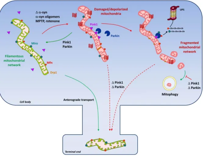
Figure


Documents relatifs
4 6 24–29 Results from these studies indicate that older age, 24 25 28 older age at onset, 4 16 25 more severe disease, 16 28 longer duration of Parkinson’s disease, 4 poorer
Increased activated brain regions during the experience of shame, embarrassment and guilt in fMRI studies comparing these emotions with one another in healthy controls.. The
Tout d’abord, Hardacre (page 850) nous livre un témoignage touchant et révélateur, puis Frank et collègues (page 862) passe en revue le dia- gnostic et le traitement,
Deep brain stimulation is effective only in patients who have exhibited some response to dopaminergic therapy, and will not reverse the condition of all parkinsonian
Gait. Although postural instability is a core feature,
Figure 6.1: Altered expression of TBPH and the expression of alpha-synuclein directed through the ddc-Gal4 HL4.36 transgene does not affects longevity and climbing ability.. 114
The severity of neuronal and oligodendroglial aggrega- tion pathology in representative tissue sections through the deep cerebellar nuclei, the surrounding white matter, and
doi:10.1371/journal.pone.0116641.g004.. CathD activity was not significantly modified by the presence of GAGs extracted from stressed and unstressed cells at doses up to 1 μg/mL in
