HAL Id: inserm-01348875
https://www.hal.inserm.fr/inserm-01348875
Submitted on 26 Jul 2016HAL is a multi-disciplinary open access archive for the deposit and dissemination of sci-entific research documents, whether they are pub-lished or not. The documents may come from teaching and research institutions in France or abroad, or from public or private research centers.
L’archive ouverte pluridisciplinaire HAL, est destinée au dépôt et à la diffusion de documents scientifiques de niveau recherche, publiés ou non, émanant des établissements d’enseignement et de recherche français ou étrangers, des laboratoires publics ou privés.
Parkinson Disease
Patrick P. Michel, Etienne Hirsch, Stéphane Hunot
To cite this version:
Patrick P. Michel, Etienne Hirsch, Stéphane Hunot. Understanding Dopaminergic Cell Death Path-ways in Parkinson Disease: Cell Death in Parkinson Disease. Neuron, Elsevier, 2016, 90 ((4)), pp.675-91. �10.1016/j.neuron.2016.03.038�. �inserm-01348875�
Parkinson Disease
St´
ephane Hunot
To cite this version:
St´ephane Hunot. Understanding Dopaminergic Cell Death Pathways in Parkinson Disease. Neuron, Elsevier, 2016, <10.1016/j.neuron.2016.03.038>. <inserm-01348875>
HAL Id: inserm-01348875
http://www.hal.inserm.fr/inserm-01348875
Submitted on 26 Jul 2016HAL is a multi-disciplinary open access archive for the deposit and dissemination of sci-entific research documents, whether they are pub-lished or not. The documents may come from teaching and research institutions in France or abroad, or from public or private research centers.
L’archive ouverte pluridisciplinaire HAL, est destin´ee au d´epˆot et `a la diffusion de documents scientifiques de niveau recherche, publi´es ou non, ´emanant des ´etablissements d’enseignement et de recherche fran¸cais ou ´etrangers, des laboratoires publics ou priv´es.
Understanding Dopaminergic Cell Death Pathways in Parkinson
Disease
Patrick P. Michel,1,2,3,4,5 Etienne C. Hirsch,1,2,3,4,5,* and Stéphane Hunot1,2,3,4,5
1Inserm, U 1127, F-75013, Paris, France 2CNRS, UMR 7225, F-75013, Paris, France
3Sorbonne Universités, UPMC Univ Paris 06, UMR S 1127, F-75013, Paris, France 4Institut du Cerveau et de la Moelle Epinière, ICM, F-75013, Paris, France
5Equal contributions
Running Title: Cell Death in Parkinson Disease
*Correspondence: etienne.hirsch@upmc.fr
INSERM UMR 1127 - CNRS UMR 7225 - Université Pierre et Marie Curie Institut du cerveau et de la moelle épinière - ICM
Thérapeutique Expérimentale de la Maladie de Parkinson Hôpital de la Salpêtrière
47 boulevard de l'Hôpital 75651 Paris CEDEX 13 France
SUMMARY
Parkinson’s disease (PD) is a multifactorial neurodegenerative disorder, the etiology of which remains largely unknown. Progressive impairment of voluntary motor control, which represents
the primary clinical feature of the disease, is caused by a loss of midbrain substantia nigra
dopamine (DA) neurons. We present, here, a synthetic overview of cell autonomous
mechanisms that are likely to participate in DA cell deathin both sporadic and inherited forms
of the disease. In particular, we describe how damage to vulnerable DA neurons may arise from
cellular disturbances produced by protein misfolding and aggregation, disruption of autophagic
catabolism, endoplasmic reticulum (ER) stress, mitochondrial dysfunction or loss of calcium
homeostasis. Where pertinent, we show how these mechanisms may mutually cooperate to
INTRODUCTION
Parkinson disease (PD) is the second most frequent neurodegenerative disorder of aging. It is
mostly sporadic, less than 10% of PD cases being inherited. The disease is diagnosed clinically
based on the presence of typical motor symptoms that include bradykinesia, rigidity, abnormal
posture, and resting tremor. Although pathological stigmata are detectable in several areas of
the brain, motor symptoms result primarily from the death of substantia nigra (SN) dopamine
(DA) neurons (Damier et al., 1999). This paper is a comprehensive review of how
cell-autonomous mechanisms may participate in DA cell death in both sporadic and hereditary
forms of the disease. Readers are referred to specific reviews for the description of
non-cell-autonomous mechanisms involving activated glial cells and peripheral immune cells in PD
progression (Hirsch and Hunot, 2009; Perry, 2012).
More specifically, we present evidence showing that the progressive deterioration of
vulnerable SN DA neurons may arise from cellular disturbances produced by misfolding and
aggregation of the synaptic protein alpha synuclein (-syn), disruption of the
autophagy-lysosome system, mitochondrial dysfunction, endoplasmic reticulum (ER) stress, or
dysregulation of calcium homeostasis. Where relevant, we will indicate how some of these
mechanisms may act in concert to promote the degeneration of DA neurons.
Synucleinopathy and dysproteostasis.
Pathogenic role of syn aggregation. The discovery in 1997 that point mutation in the
-syn gene was responsible for the inherited form of PD, together with the finding that Lewy
bodies (LBs), a key neuropathological feature of sporadic PD, are primarily composed of misfolded/aggregated α-syn, has dramatically changed our view of PD pathogenesis, which has become in many respects “synuclein-centric” (Polymeropoulos et al., 1997; Spillantini et al., 1997). Genetic evidence for a role of -syn in sporadic PD has emerged from genome-wide
association studies (GWAS) that reported an association between the SNCA locus and the risk
of developing PD (Simon-Sanchez et al., 2009). These studies failed to identify protein-coding
variants at this locus, suggesting that the risk alleles are likely to affect expression levels of
-syn rather than its function. In line with this, the identification of inherited forms of PD with
duplication or triplication of the SNCA gene suggests that an increased expression level of the
normal protein is sufficient to trigger the pathogenic mechanism (Chartier-Harlin et al., 2004;
Singleton et al., 2003).
Alpha-syn is a small 140aa protein normally enriched in the presynaptic compartment (Iwai
et al., 1995), where it is thought to promote the formation of the SNARE complex, thereby
regulating vesicle dynamics and trafficking and, hence, neurotransmitter release (Burré et al.,
2010). Whether this physiological function is lost and plays any role in PD pathogenesis is still
uncertain. While -syn-deficient mice display impaired evoked DA release in the striatum,
these alterations are not associated with obvious features of neuronal distress and degeneration,
suggesting that the pathogenic role of -syn in sporadic PD is unlikely to be linked to a loss of
function but rather to a gain of pathological function (Abeliovich et al., 2000). In fact,
accumulating evidence shows that aggregation of -syn is necessary for its pathogenicity. In
model systems with -syn overexpression, aggregation and deposition of -syn precede
neuronal cell death and strategies to reduce the aggregative process were shown to reduce
neurodegeneration and improve motor deficits in many species, including invertebrates
(nematodes and flies), rodents and non-human primates (Lashuel et al., 2013). It is also worth
noting that familial PD-associated SNCA mutations share a common property of accelerated
aggregation of -syn leading to early-onset and rapidly progressive forms of PD (Kim and Lee,
2008). Finally, aggregate forms of -syn generated from recombinant protein were shown to
be pathogenic when administrated directly into the brain of mice, further supporting the toxic
There is much debate about the toxic aggregate species of -syn involved in
neurodegeneration. While it is now well accepted that under physiological conditions -syn
may exist in various conformational and oligomeric states, the full spectrum of toxic -syn
species and their precise role in the mechanism of neuronal cell death has not been completely
identified. Nonetheless, soluble oligomers and protofibrils, which are formed relatively early
during the fibrillation process of -syn, seem to be particularly toxic compared to the insoluble
amyloid-like fibrils accumulating into LBs (Danzer et al., 2007; Winner et al., 2011) (Figure
1).
Interconnection between synucleinopathy and dysfunction of the autophagy lysosome pathway. What causes accumulation and aggregation of misfolded -syn are important
questions that will lead to major breakthroughs in our understanding of disease development if
solved. Similarly, why some neurons show α-syn accumulation earlier than others during the
course of the disease remains to be understood. Among mechanisms possibly responsible for -syn misfolding and aggregation, dysfunction of degradation pathways, notably the autophagy-lysosome system, has attracted much attention recently (Figure 1).
Macroautophagy (referred hereafter as autophagy), is a self-destructive process whereby
double-membrane-bound structures called autophagosomes engulf cytosolic cargo (damaged
organelles, aggregates, lipid droplets, …) before delivery to the hydrolytic milieu of lysosomes
for degradation (Figure 2). The resulting metabolites are transported into the cytoplasm and
used either for the synthesis of new macromolecules or as a source of energy (Boya et al, 2013).
In SN DA neurons from PD patients, autophagosomes and lysosomal marker proteins are
increased and decreased, respectively, suggesting that the autophagic flux is profoundly
disrupted in these patients (Anglade et al., 1997; Chu et al., 2009). Interestingly, several
PD-related genes have been linked to dysfunctional autophagy. Indeed, the loss of DJ-1 that triggers
leads to the accumulation of microtubule-associated protein 1 light chain 3 (LC3), a reliable
marker for autophagosomes (Thomas et al., 2011) suggesting that oxidative stress represents a
potential trigger for autophagy (Filomeni et al., 2015) (Figure 2). Furthermore, overexpression
of the PD-related protein LRRK2, another PD-related protein shown to exacerbate α-syn
pathology (Lin et al., 2009,) increases autophagosome numbers and lysosomal pH thereby
reducing lysosomal hydrolytic enzyme activities (Gómez-Suaga et al, 2012).
Involvement of dysfunctional autophagy in -syn accumulation is supported by in vivo
gene-targeting experiments in mouse showing that whole-brain specific loss of Atg7 (an enzyme required for autophagosome formation) leads to presynaptic neuronal accumulation of α-syn (Friedman et al., 2012). Note that the presence of aggregates containing K48-linked
polyubiquitin and the ubiquitin binding protein p62 were detected after conditional Atg7
deletion in DA neurons (Ahmed et al., 2012), suggesting that the autophagy-lysosome pathway
and the proteasomal pathway cooperate to reduce misfolded protein burden. These observations
also indicate that selective impairment of one pathway can lead to a compensatory upregulation
of the other as already noted by Ebrahimi-Fakhari and colleagues (2011). Interestingly, mouse
brains deficient in Atg7 also showed increased levels of LRKK2 (Friedman et al., 2012). Also
coherent with these observations, conditional deletion of Atg7 in mouse midbrain DA neurons
resulted in early dendritic and axonal dystrophy associated with delayed neurodegeneration and
late-onset locomotor impairments (Friedman et al., 2012).
Besides macroautophagy, chaperone-mediated autophagy (CMA) has also been implicated
in PD (Cuervo et al., 2014) (Figure 1). CMA is a selective type of autophagy responsible for
lysosomal degradation of a highly specific subset of soluble cytosolic proteins containing a
KFERQ-like motif recognized by the cytosolic chaperone heat shock cognate protein 70
(HSC70) (Figure 2). Of interest, wild-type α-syn is a substrate for CMA (Cuervo et al., 2004)
mutant forms of PD genes SNCA and LRRK2 (Martinez-Vicente et al., 2008; Orenstein et al.,
2013) or post-transcriptionally modified -syn (Martinez-Vicente et al., 2008)exert inhibitory
effects on CMA (Figure 2). Despite being recognized by cytosolic HSC70 and normally
delivered to the lysosomal membrane, mutant forms of -syn and LKKR2 proteins are unable
to reach the lysosomal lumen for CMA-mediated degradation (Orenstein et al., 2013). This is
due to aberrant interactions of these proteins with the CMA receptor LAMP-2A. Providing
additional evidence for decreased CMA activity in PD, both LAMP2A and HSC70 levels are
reduced in PD patients (Alvarez-Erviti et al, 2010). Unexpectedly, however, LAMP-2A is
augmented in patients carrying pathogenic mutations in LRRK2, which is indicative of a
possible compensatory response to CMA dysfunction in these patients (Orenstein et al, 2013).
The implication of the autophagy-lysosome pathway in PD is also supported by the
discovery that mutations in genes encoding proteins of the endosomal/lysosomal system,
vacuolar protein sorting-35 (VPS35), type 5 P-type ATPase and glucocerebrosidase (GBA),
result in parkinsonian syndromes (Figure 2).
VPS35 is part of the retromer, a protein complex that associates with the cytosolic face of
early endosomes to regulateretrograde transport of cargo proteins (plasma membrane receptors,
transporters) to the trans-Golgi network, to rescue them from lysosome-mediated turnover
(Tsika et al, 2014). Mutations in VPS35 have been identified in patients with autosomal
dominant PD (Vilariño-Güell et al. 2011; Zimprich et al. 2011). Confirming that VPS35
dysfunction has a profound impact on the autophagic process, VPS35 deficient DA neurons in
culture contained enlarged late endosomes/lysosomes and exhibited impaired
endosome-to-Golgi retrieval of the CMA receptor LAMP-2A (Tang et al, 2015). Aged VPS35+/- mice
developed PD-related deficits including loss of DA and accumulation of -syn in SN DA
neurons (Tang et al, 2015). Besides, AAV mediated gene transfer of the VPS35 D620N
2014). Of note, another PD gene eukaryotic translation initiation factor 4 gamma, 1 (eIF4G)
involved in autosomal-dominant forms of parkinsonism was reported to genetically and
functionally interact with VPS35 (Dhungel et al, 2015).
ATP13A2, which is localized in acidic membrane compartments of lysosomes is a P-type
ATPase that is expected to function as a cation metal transporter (Gitler et al., 2009) (Figure 2).
ATP13A2 mutations cause familial Kufor-Rakeb syndrome, characterized by early onset
parkinsonism associated with pyramidal degeneration and dementia (Ramirez et al., 2006).
Experiments carried out in cultured fibroblasts from patients with Kufor-Rakeb syndrome not
only confirmed that ATP13A2 loss of function impairs lysosomal catabolism but also revealed
that it favors the accumulation of -syn (Usenovic and Krainc, 2012). Conversely, the loss of
DA neurons induced by -syn overexpression in animal and cellular models of PD is rescued
by coexpression of ATP13A2 (Gitler et al., 2009). The presence of ATP13A2 is also
dramatically reduced within the bulk cytosol of DA SN neurons from patients with sporadic PD
(Dehay et al., 2012), and partially redistributed towards LBs (Dehay et al., 2012; Murphy et al.,
2013), confirming indirectly that the function of the autophagy-lysosome system is impaired in
sporadic forms of PD. Note that cells expressing mutated ATP13A2 were also reported to
accumulate the mutant protein in the ER, making them predisposed to ER-stress induced
degeneration (Ugolino et al, 2011).
The lysosomal enzyme GBA cleaves glucocerebroside (also called glucosylceramide, GC)
into a sugar, glucose and a simpler fat molecule, ceramide. Pathogenetic mutations in both
alleles of GBA result in Gaucher disease, a disorder in which a severe loss of GBA activity
leads to unprocessed GC that accumulates in the bone marrow, lungs, spleen, liver and
sometimes the brain(Platt et al., 2014). Some patients developing a non-neuronopathic form of
Gaucher disease present parkinsonian symptoms (Neudorfer et al., 1996) but, quite
disease, have a 5-fold increase in PD risk (Sidransky et al., 2009), pointing to a possible link
between GBA dysfunction and sporadic PD. Consistent with this view, the analysis of
cerebrospinal fluid and brain tissue samples from PD patients revealed that GBA activity is
reduced regardless of whether or not these patients harbor mutations in GBA (Balducci et al.,
2007; Parnetti et al., 2014). Of note, GBA protein levels and activity are specifically decreased in brain areas where α-syn levels are elevated in early stage PD, suggesting that GBA dysfunction may be a consequence of the accumulation and aggregation of -syn (Murphy et
al, 2014).In keeping with this idea, Mazulli and colleagues demonstrated that α-syn oligomeric
species interrupt ER-Golgi trafficking of GBA, resulting ultimately in defective storage of the
enzyme within lysosomes (Mazzulli et al, 2011). In addition, α-syn can operate as a direct
inhibitor of GBA, at the internal membrane surface of lysosomal vesicles (Yap et al, 2013).
Inversely, experimental evidence suggests that a reduction in GBA lysosomal activity may
promote α-syn misprocessing as AAV-mediated expression of GBA decreases α-syn
aggregation in a presymptomatic mouse model of Gaucher-related synucleinopathy (Sardi et
al., 2013). Overall, these observations suggest the existence of a positive feedback loop in which
reduced GBA lysosomal activity leads to accumulation of α-syn and vice versa, leading
ultimately to neurodegeneration (Mazzulli et al, 2011) (Figure 2). The stabilization of α-syn
soluble oligomers by unprocessed GC may be a possible reason why GBA dysfunction
facilitates α-syn-mediated aggregation (Mazzulli et al, 2011). Overall, a valuable therapeutic
strategy to halt PD progression may be to augment brain GBA activity as recently evidenced in
PD fibroblasts from a GBA carrier using Ambroxol, a secretolytic agent that works as a GBA
activity enhancer (McNeill et al., 2014).
As developed above, if accumulation and subsequent aggregation of -syn can originate
from dysfunctional cellular degradation pathways, -syn aggregates can also profoundly affect
autophagy-lysosomal pathway (Winslow et al., 2010). This pathological feedback loop may therefore
reinforce the synucleinopathic mechanisms and their consequences (Figure 1). Yet, the
pathogenic mechanisms associated with -syn aggregates are likely to be even more diverse
since aggregate species were reported to cause membrane permeabilization (van Rooijen et al.,
2010), golgi fragmentation (Gosavi et al., 2002), mitochondrial dysfunction (Parihar et al.,
2008) and activation of the unfolded protein response associated with endoplasmic reticulum
stress (Colla et al., 2012a) (Figure 1).
Synucleinopathy-associated endoplasmic reticulum stress response. The endoplasmic
reticulum (ER) is central to protein folding in eukaryote cells and any perturbations altering ER
homeostasis can result in the disruption of the folding process and accumulation of misfolded
or unfolded proteins. This cellular condition, also called ER stress, usually induces a
physiological protective response termed unfolded protein response (UPR) which is aimed at
restoring ER homeostasis and normal cell function if it is induced transiently (for review, see
Hetz, 2012) (Figure 3). Yet, under chronic ER stress, the accumulation of unfolded proteins and
sustained UPR activity triggers activation of pro-apoptotic pathways and cell death, thereby
eliminating damaged cells. Among the factors possibly involved in triggering the deleterious
chronic ER stress response in PD, accumulation and aggregation of misfolded -syn is an
obvious candidate. In support of this view, markers of UPR activation have been consistently
observed in mammalian and invertebrate models of synucleinopathy (for review, see Matus et
al., 2011). Some of these findings revealed that -syn conformers, and in particular toxic
oligomers, accumulate at the ER early during the disease process concomitantly with
upregulation of ER chaperones, including Bip and PDIp (Colla et al., 2012b) (Figure 3).
Interestingly, a similar ER accumulation of -syn has also been observed in PD brain tissue
stress and engagement of the UPR machinery occur in nigral dopaminergic neurons from
patients with PD (Hoozemans et al., 2007; Selvaraj et al., 2012; Conn et al., 2004). Apart from
synucleinopathy-associated chronic ER stress response, a number of other reports have also
suggested a link between PD-related genes and ER function/alteration. For instance,
dopaminergic neuronal cell death induced by human -syn in the nematode C. elegans was
shown to be attenuated by LRRK2 through a mechanism involving upregulated expression of
the ER chaperone Bip (Yuan et al., 2011), suggesting that deficiency in LRRK2 may sensitize
neurons to ER stress-induced neurodegeneration associated with -syn misfolding and
accumulation (Figure 3).
As suggested by the above data, if chronic ER stress plays a central role in neuronal cell
death in PD, targeting key elements of the UPR pathway would likely be neuroprotective.
Surprisingly, however, increasing rather than suppressing UPR components was shown to
ameliorate disease phenotypes in both toxic- and genetic-based models of PD (Sado et al., 2009;
Colla et al., 2012a; Gorbatyuk et al., 2012). This apparently counterintuitive result has led to
the concept that a mild ER stress response causing a sustained, yet moderate UPR, may improve
the resistance of dopaminergic neurons during PD progression. This type of response called ER
hormesis was observed with the ER stress inducer, tunicamycin which conferred
neuroprotection in mouse and Drosophila PD models (Fouillet et al, 2012). Thus, fine-tuning
of the UPR, may be a possible therapeutic strategy for PD.
A Disturbance in Mitochondrial Function
Mitochondria, once simply seen as the ‘powerhouses’ of the cell, are now considered to work as initiators and transducers of signaling pathways associated with a number of cell functions,
including apoptosis (Tait and Green, 2013). The crucial implication of mitochondria in PD
The most direct evidence for the implication of mitochondria in PD relies on seminal
observations made in autopsy brain tissue and other tissue samples from PD patients, as well as
on experiments carried out in non-human primates and rodents. In particular, a deficit in
mitochondrial complex I activity is found in autopsy brains from PD patients (Schapira et al.,
1990) and in cytoplasmic hybrid (cybrid) cell lines that contain mitochondrial DNA (mtDNA) from patients’ platelets (Swerdlow et al., 1996). Besides, the mitochondrial complex I inhibitor, 1-methyl-4-phenylpyridinium (MPP+) induces a selective destruction of SN DA neurons in
mice (reviewed by Przedborski et al., 2004) and non-human primates (Langston et al., 1984)
receiving systemic injections of its prodrug MPTP. This explains why accidental intravenous
self-administration of MPTP in humans led to an irreversible neurodegenerative condition that
was clinically and histologically very similar to sporadic PD (Langston et al., 1984). In line
with these findings, other complex I inhibitors, with a chemical structure that is, however,
unrelated to MPP+, such as the alkaloid rotenone (Sherer et al., 2003) and the acetogenin
annonacin (Champy et al., 2004) can also induce irreversible lesions of the dopaminergic
nigro-striatal pathway when systemically administered to rodents. Remarkably, annonacin is present
in soursop, a tropical plant whose consumption is linked to an atypical form of parkinsonism in
the Caribbean island of Guadeloupe (Lannuzel et al., 2008).
Apart from postmortem evidence for complex I deficiency in PD and the parallel drawn with
parkinsonism-induced mitochondrial poisons targeting the electron transport chain (ETC),
major breakthroughs in the field of mitochondrial dysfunction in PD have been made thanks to
the discovery that some of the PD-related genes are involved in key biological processes that
govern mitochondrial homeostasis and stress-response. In particular, it is now well established
that the recessive PD-linked genes PINK1 and Parkin likely play a central role in mitochondrial
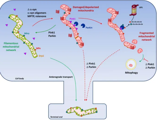
quality control and dynamics (Narendra et al., 2012) (Figure 4). Mitochondrial quality control
network, which is critical for cell physiology and survival. It involves several functional aspects
including damage prevention and repair mechanisms, autophagic elimination of
dysfunctional/damaged organelles (known as mitophagy) and neosynthesis of mtDNA- and
nuclear DNA-encoded mitochondrial proteins for mitochondria biogenesis. To properly
perform these tasks, the mitochondrial network evolves dynamically from elongated to
fragmented states (and vice versa) thanks to a highly specialized molecular machinery that
tightly controls fusion and fission events, which in turn facilitate repair and autophagic
degradation of defective organelles, respectively (Ono et al., 2001). Finally, mitochondrial
dynamics and transport are also essential for suitable distribution of organelles, especially at
neuronal terminal ends, where high energy demand is required to sustain synaptic activity
(Verstreken et al., 2005) (Figure 4).
PINK1 and parkin were shown not only to participate in the functional and morphological
maintenance of the mitochondrial network (Palacino et al., 2004; Gautier et al., 2008) but also,
to critically regulate the removal of dysfunctional mitochondria through mitophagy (Deas et
al., 2011). Remarkably, parkin and PINK1 fulfill these functions collectively, with parkin being
recruited to damaged (i.e., depolarized) mitochondria by PINK1 and acting downstream to
promote their degradation by autophagy (Clark et al., 2006; Park et al., 2006; Narendra et al.,
2010; Vives-Bauza et al., 2010; Bertolin et al., 2013). Although the functional interplay of
parkin and PINK1 on altered mitochondria is still not fully elucidated, it has been shown that
PINK1 accumulation at the outer mitochondrial membrane (OMM) of dysfunctional
mitochondria is required and sufficient to recruit and activate parkin. Parkin in turn drives the
ubiquitination and degradation of mitochondrial substrates, including, mitofusin (Mfn) 1 and 2,
which regulate OMM fusion, and the OMM-associated Miro protein, a Ca2+-binding GTPase
that controls the anterograde transport of mitochondria along the axons (Narendra et al., 2010;
Overall, the parkin/PINK1 pathway-mediated degradation of Mfn1/2 and Miro is believed to
facilitate the elimination of bioenergetically compromised mitochondria by promoting both
their isolation from the filamentous and healthy mitochondrial network and their
immobilization at the somatic compartment away from high-energy-consuming end terminals.
Ubiquitinated OMM proteins then bind to the ubiquitin- and LC3-binding adaptor protein p62
for engulfment by autophagosomes (Geisler et al., 2010; Rakovic et al., 2013). On the basis of
these data, it has been proposed that impairment of mitophagy by mutations of PINK1 or Parkin
may become toxic by accumulation of dysfunctional mitochondria (Narendra et al., 2010;
Vives-Bauza et al., 2010) (Figure 2 and 4). However, chemical uncouplers that are used at
relatively high concentrations to activate mitophagy through mitochondrial depolarization in
immortalized cell lines failedto -activate this process in rat primary neuronal cell cultures (Van
Laar et al., 2011) or in human induced pluripotent stem cell-derived neurons (Rakovic et al.,
2013). This has led to the speculation that mitophagy may be controlled by distinct mechanisms
in neuronal cells, possibly because of the specific bioenergetic requirements of these cells. Yet,
a more recent study suggests that, unlike chemical uncouplers, the short mitochondrial isoform
of ARF (smARF), previously identified as an alternate translation product of the tumor
suppressor p19ARF, has the capacity to depolarize mitochondria and to promote mitophagy in
neuronal cells through the canonical Parkin/PINK1-dependent pathway (Grenier et al., 2014).
It still remains intriguing that, in contrast to the mechanism in invertebrates such as flies, the
control of mitochondrial motility and fusion machinery by the parkin/PINK1 pathway in
mammalian cells appears to be minimal under normal physiological conditions and only
unmasked under stress conditions (Wang et al., 2011; Tanaka et al., 2010). This difference may
underscore evolutionary changes in mitochondrial quality control mechanisms which may be
more tightly regulated in higher eukaryotes that live longer and in which mitochondrial ageing
and there is evidence to suggest that mitophagy is far from being the only process involved in
mitochondrial quality control. Other lines of defense such as activation of mitochondrial matrix
proteases –that degrade unfolded and oxidized soluble proteins, as well as removal of
mitochondrial bits containing damaged cargos through the generation of mitochondrial-derived
vesicles (MDVs) transiting to lysosomes, are rapidly induced mechanisms that could prevent
irreversible mitochondrial damage and elimination by mitophagy (for review see Sugiura et al.,
2014). Interestingly, PINK1, Parkin and Vps35 have been involved in the biogenesis of MDVs
but the functional contribution of MDVs in PD pathophysiology has not been demonstrated so
far.
Enlarged mitochondria with disrupted cristae have been observed in nigral neurons from PD
patients, which confirm the view that mitochondrial quality control is defective in sporadic PD
(Anglade et al., 1997). Similar changes in mitochondrial morphology can be recapitulated in
experimental setups using the mitochondrial complex I inhibitors MPTP/MPP+ and rotenone,
implying that ETC dysfunction and as a corollary to that, oxidative stress and bioenergetic
deficits might primarily impair mitochondrial dynamics in PD (for review, see van Laar and
Berman, 2013) (Figure 4). Yet, other candidates such as -syn accumulation and aggregation
might be involved as well. In support of this statement, many reports have shown that -syn
can not only interact with and accumulate in mitochondria but also trigger morphological
alterations, including swelling and cristae disruption (Martin et al., 2006). More recently, Kamp
and colleagues showed that -syn overexpression in cultured cells and in the nematode C.
elegans results in mitochondrial fusion inhibition and fragmentation, a phenotype that is
rescued by parkin and PINK1 (Kamp et al., 2010). Interestingly, this effect may not rely on
inhibition of the fusion/fission machinery but rather could involve the direct interaction of
-syn oligomers with mitochondrial membranes resulting in their fragmentation (Nakamura et al.,
and defective mitochondrial quality control, is the finding that neuronal expression of the A53T
synuclein mutant is associated with increased mitophagy, mitochondrial loss and cell death.
Intriguingly, stimulating mitochondrial fusion (i.e., inhibiting Drp1 activity or overexpressing
Mfn2) or silencing autophagy-related genes could reverse the mitochondrial loss and protect neurons against A53T α-syn-induced cell death (Choubey et al., 2011). This result contrasts therefore with other reports showing that stimulation instead of inhibition of macroautophagy
and of the PINK/Parkin-associated mitophagy pathway is actually neuroprotective in different
synucleinopathy models including mice overexpressing wild-type -Syn (Lo Bianco et al.,
2004; Spencer et al., 2009; Winslow et al., 2010). The discrepancy may be explained by the
fact that, unlike wild-type -syn, mutated A53T -syn inhibits the CMA pathway resulting in
compensatory over activation of macroautophagy which in turn would lead to excessive
elimination of mitochondria including those that are not depolarized (Xilouri et al., 2009).
These data underline that excessive macroautophagy activity may have negative consequences
and that - fine tuning of mitophagy is required for proper neuronal function and for therapeutic
purposes. These studies underscore also the potential of strategies aimed at stimulating
mitochondrial elongation in distressed neurons. Indeed, filamentous mitochondrial networks
ensure a better response to oxidative stress and other insults through dilution of stress molecules
across the network and induction of compensatory mechanisms (Youle and van der Bliek,
2012). Such an approach has recently been illustrated in toxic-based models of PD where
neuroprotection afforded by the X protein, a virus-derived protein with mitochondrial targeting
and anti-apoptotic properties, was associated with mitochondrial filamentation, i.e.,
mitochondria elongation forming a reticular network (Szelechowski et al., 2014).
Dysregulation of calcium homeostasis may also intervene crucially in the preferential loss
of DA neurons in PD. Defects in calcium-handling may be due principally to changes in the
discharge activity of DA neurons but may also occur as indirect consequence of multiple
PD-related events such as -syn aggregation, mitochondrial deficits or ER dysfunction. DA neuron
survival may be compromised in situations where levels of calcium exceed the upper (Blandini
et al., 2004; Chan et al., 2007) or fall below the lower limit (Salthun-Lassalle et al., 2004) of
the physiological rangein the cytosol and subcellular organelles.
Cytosolic calcium (Ca2+
cyt) overload in DA neurons may result from sustained engagement
of N-methyl-D-aspartate (NMDA) glutamate receptors as a consequence of the overactivity of
subthalamic nucleus glutamatergic inputs (Blandini, et al., 2004). This hypothesis which
presupposes that glutamate is increased extracellularly in the vicinity of SN DA neurons
(Assous et al., 2014) is challenged, however, by the concept of a slowly occurring excitotoxic
process, in which calcium overload and ensuing neurodegenerative events including nitrosative
and oxidative stress (Blandini et al., 2004), may occur in spite of normal extracellular
concentrations of glutamate. Indeed, mitochondrial bioenergetic deficits occurring in PD may
reduce the Mg2+ block of the NMDA channel pore and as a consequence may increase the
sensitivity of DA neurons to glutamate-mediated excitotoxic stress(Schapira and Gegg, 2011).
SN DA neurons are also characterized by Ca2+-dependent pacemaking, an autonomous mode
of discharge that elevates Ca2+cyt through L-type voltage-dependent calcium channels, most
likely a subset of them having a Cav1.3 pore (Chan et al., 2007). This type of activity which
serves to maintain a basal DA tone in the striatum may confer a specific vulnerability to SN
DA neurons, which have low intrinsic calcium buffering capacity (Hirsch et al., 1992). Indeed,
calcium influx through autonomous pacemaking results in elevation of basal mitochondrial
oxidative stress in SN DA neurons, presumably as a direct consequence of an increased
oxidative stress is to reduce the bioenergetic reserve capacity of DA neurons which in turn
makes them particularly vulnerable in conditions of increased metabolic demand (Rivero-Rios
et al., 2014). This probably explains why Isradipine an L-type Cav channel blocker with some
specificity for Cav1.3 over Cav1.2 and Cav1.1 channels not only reduced basal mitochondrial
oxidant stress in SN DA neurons (Guzman et al., 2010) but also protected these neurons from
mitochondrial toxins such as MPP+ and rotenone that impair mitochondrial respiration and
energy production (Chan et al., 2007). Isradipine did not stop pacemaking, however, most
probably becausecalcium supports, but is not necessary to preserve this mode of activity in DA
neurons.
Mitochondrial oxidative stress generated by calcium currents during pacemaking was
reported to be enhanced in SN DA neurons that lack the PD gene DJ-1 (Guzman et al., 2010)
in agreement with the putative role of this gene in regulating oxidant defenses. This type of
stress was also augmented in perinuclear and dendritic compartments of DA neurons exhibiting
intracellular α-syn LB-like inclusions formed in vitro by recruitment of preformed fibrils of -syn (Dryanovski et al, 2013), providing a new link between dysregulated proteostasis and
mitochondrial dysfunction in PD pathogenesis. In line with this last observation, Subramaniam
and colleagues (2014) also reported a progressive and sustained increase in spike rate in mouse nigral DA neurons overexpressing A53T mutant α-syn. Overall, these findings suggest that L-type calcium channel blockers may be helpful in PD. Epidemiological studies of
dihydropyridine class L-type calcium channel blockers for association with PD have so far
yielded conflicting results, however (Rees et al., 2011).
There are alternative scenarios by which high Ca2+ levels may become deleterious for SN
DA neurons. The calcium elevation may increase DA synthesis through the activation of
tyrosine hydroxylase (TH) (Rittenhouse and Zigmond, 1999), thus causing intracellular damage
modifications of the PD gene -syn (Martinez-Vicente et al., 2008). Calcium overload may also exert adverse effects for DA neurons through activation of calpains, a family of
calcium-dependent cysteine proteases. Dufty and colleagues (2007) showed the presence of
C-terminally calpain-cleaved α-syn in LB in brains of PD patients. Most interestingly,
overexpression of the endogenous and specific inhibitor of calpain calpastatin in human A30P α-syn transgenic mice caused a reduction of α-syn-positive aggregates (Diepenbroek et al, 2014). In keeping with these findings, calpains are known to induce cleavage of cdk5, a protein
that has a facilitating role in DA cell death (Smith et al, 2003). Thus, calpains and possibly cdk5
inhibitors may be attractive candidate targets for therapeutic intervention in PD.
Regardless of the nature of the mechanism actually causing Ca2+cyt overload, the capacity of
mitochondria to handle calcium through uptake or efflux mechanisms remains probably crucial
to prevent PD-related neurodegenerative changes (Celardo, 2014). Illustrating this point is the
demonstration that deficiency in the PD protein PINK1 leads to mitochondrial calcium (Ca2+mit)
overload due to a reduction of calcium efflux via the mitochondrial Na+/Ca2+ exchanger
(Gandhi et al., 2009).Mitochondrial Ca2+ overload and ensuing PD-neurodegenerative events
may result from direct accumulation of Ca2+ from the cytosol to mitochondria through the
mitochondrial Ca2+ uniporter (MCU) (Guzman et al, 2010; Qiu et al, 2014). Mitochondria, often
in close apposition to the ER, can also accumulate Ca2+ into the matrix through the coordinated
activation of ER inositol-1,4,5-triphosphate (IP3) receptors and MCU (Celardo et al, 2014).
Therefore, Ca2+mit overload and mitochondrial oxidative stress in DA neurons may also be due
to the fact that too much Ca2+ is pumped out of the cytosol into the ER through the high affinity
sarco-endoplasmic reticulum Ca2+-ATPase (Guzman et al., 2010; Rivero Rios et al., 2014), a
process energetically costly which may as such further amplify mitochondrial dysfunction in a
Despite large evidence implicating stressful pacemaking and calcium overload in PD
progression, it appears that the survival of these neurons may be similarly compromised when
pacemaking is reduced and intracellular calcium drops below threshold levels (Michel et al.,
2013). This notion is actually supported by the following observations; (i) The dopaminergic
toxins MPP+ and rotenone cause an early reduction of DA neuron firing in rodent midbrain
slices (Liss et al, 2005; Yee et al., 2014) and preserving this activity by genetic inactivation of
Kir6.2, the pore forming subunit of KATP channels favors the survival of these neurons (Liss et
al, 2005); (ii) Toxic effects of MPP+’s prodrug MPTP are reduced in vivo by the brain-penetrant blocker of small conductance calcium-activated potassium channels apamin (Alvarez-Fischer
et al, 2013) and by the alkaloid nicotine (NIC) (for review see Quik et al, 2012), two compounds
that raise Ca2+
cyt levels (Toulorge et al, 2011) in DA neurons and stimulate them electrically
(Wolfart et al, 2001; Teo et al, 2004); (iii) Tobacco smoking reduces PD risk, presumably
through an effect of its major component NIC (Quik et al, 2012). Still in line with this
hypothesis, Hirsch and colleagues (1988) found that the rate-limiting enzyme in DA synthesis,
TH whose expression is activity- and calcium-dependent (Brosenitsch and Katz, 2001),
disappears entirely from a large population of neuromelanized SN DA neurons in PD.
Interestingly, SN DA neurons demonstrated decreased discharge frequencies in aged mice that
express disease relevant levels of wild-type α-syn from the complete human SNCA locus
(Janezic et al., 2013). Because DA neurons in these mice die in the absence of overt aggregation
pathology, one may assume that pre-aggregates of -syn were the cause of reduced spiking and
that this deficit may represent an early biomarker of neurodegeneration. One way to reconcile
this set of results with data implicating activity-dependent calcium entry in DA cell death would
be to assume that DA neurons go successively during degeneration through hypo- and
hyperactive phases during which they endure calcium deficiency and calcium overload,
a genetic mitochondrial model of PD where silent DA neurons appear physiologically less
compromised than hyperactive ones (Good et al, 2011).
The possible contribution of activity and calcium deficits to the death of DA neurons may
imply that these neurons have a fundamental need for proteins regulated by calcium. PI3K may
represent one of the cytosolic proteins required for survival (Ries et al., 2009; Toulorge et al.,
2011). The concept that calcium needs to remain above a threshold level to enable survival of
DA neurons is perhaps best illustrated by the mechanism whereby the depolarizing alkaloid
NIC provides protection to spontaneously dying DA neurons in midbrain cultures. Indeed, NIC
was protective only if Ca2+cyt was raised above threshold levels by concurrent depolarizing
stimuli, presumably to keep 7nAChRs, which intervene in the protective action of NIC both
in vitro (Toulorge et al., 2011) and in vivo (Bordia et al., 2014), in an active conformational
state (Changeux, and Edelstein, 1998). Moreover, under adequate depolarizing conditions
NIC-mediated neuroprotection in itself required calcium influx.
Calcium stored in the ER may also enable DA neurons to regulate their own survival. In
particular, elevating basal Ca2+cyt through ER ryanodine receptor channel (RyR) activation
provided protection to midbrain DA neurons in several culture paradigms where
neurodegeneration is either spontaneous or induced by trophic support deprivation or MPP+
intoxication (Guerreiro et al., 2008). The beneficial action of ER calcium mobilization was
attributed to the action of Ca2+
cyt on a putative protein target required for DA cell survival but
it was also proposed that the resultant decrease in ER Ca2+ load may be beneficial by limiting
mitochondrial Ca2+ accumulation occurring through ER calcium mobilization (Rivero-Rios et
al., 2014). Reducing calcium shuttling from ER to mitochondria through blockade of ER IP3R
or inhibition of the mitochondrial calcium uniporter (MCU) was, however, detrimental for DA
neurons (Rousseau et al., 2013), indicating that calcium transfer between these two
bioenergetic machinery functional in these neurons and ultimately preserve their survival (Cali
et al, 2013). Consistent with this interpretation, an early and concomitant depletion in Ca2+mit
(Rousseau et al., 2013) and ATP (Höglinger et al., 2003)preceded DA neuronal loss in midbrain
cultures exposed to the mitochondrial toxin MPP+. These findings also need to be placed in
perspective with data showing that the PD protein -syn facilitates Ca2+
mit transients elicited by
IP3R activation whereas α-syn loss of function in addition to reducing Ca2+ fluxes into
mitochondria also results in increased autophagy (Cali et al., 2012). Coherent with these
observations, α-syn was reported to associate with mitochondria-associated ER membranes
(Guardia-Laguarta et al., 2014), a structurally and functionally distinct subdomain of the ER.
Two other PD-associated proteins, DJ-1 (Ottolini et al., 2013) and parkin, were also reported
to facilitate ER-mitochondria tethering upon stimulation of IP3Rs (Cali et al., 2013), whereas
mutated forms of DJ-1 and parkin siRNA impaired this process. Figure 5 summarizes how
calcium-related events could participate in DA cell loss in PD.
Concluding Remarks
Within a period of just 20 years, we have witnessed tremendous progress in our
understanding of PD pathogenesis. Before the synuclein era, research efforts on the
pathomechanisms of PD were mostly focused on mitochondrial bioenergetic deficits, oxidative
stress and death pathways involved in apoptosis (Jenner and Olanow, 1996; Mizuno et al.,
1995). They have now expanded to embrace a myriad of molecular and biochemical defects
affecting key cellular functions such as autophagic pathways, mitochondrial quality control and
dynamics and the unfolded protein response. The identification of genes linked to inherited
forms of PD has unquestionably contributed massively to this explosion of new knowledge
(Table 1), which in turn should foster the discovery of more efficient therapeutic tools. Yet, the
involved in neurodegeneration which are interconnected and influence each other in a still
poorly defined dynamic cascade. Adding to this complexity is the fact that non-cell autonomous
mechanisms are likely to be important in participating and/or modulating disease mechanisms
as well. For instance, neuroinflammatory processes orchestrated by both innate and adaptive
immune cells have emerged as important contributors of pathogenesis in PD (Hirsch and Hunot,
2009). Non-cell autonomous mechanisms are likely also involved in the spreading of -syn
pathology. Indeed, mounting evidence suggests that α-syn can undergo a toxic templated
conformational change and spread from cell to cell to seed and initiate the formation of
aggregates in the newly “contaminated” neurons (for review see Tyson et al., 2015). According
to such a scenario, the seeding-prone exogenous -syn aggregates can be viewed as non-cell
autonomous culprits that could drive disease progression. Yet, despite strong experimental
evidence for -syn spreading, the possibility that such spreading actually happens in a
pathophysiological context and at disease relevant level of -syn is still lacking. Such
knowledge would however have far reaching therapeutic consequences since trapping and
subsequent degradation of exogenous neuron-released aggregates by antibodies through active
or passive immunization has emerged as a powerful neuroprotective approach in preclinical
settings (for review see Dehay et al., 2015).
To better capture disease complexity, systems biology, which relies on the computational
and mathematical modeling of complex biological systems, has emerged as a powerful
approach to understand in a comprehensive way the intricate interactions between the
dysregulated pathways involved in disease mechanisms (Funke et al., 2013; Lausted et al.,
2014; Fujita et al., 2014). Such approaches have the potential not only to improve our
understanding of disease initiation and progression but also to help the discovery and
implementation of more effective therapeutic strategies. While well advanced in the cancer
few studies that have started to develop and use bioinformatic tools for therapeutic target and
biomarker identification (Büchel et al., 2013; Chandrasekaran and Bonchev, 2013;
Ouzounoglou et al., 2014; Dusonchet et al., 2014). However, with the rapid growth of
PD-related knowledge and technological advances enabling massive databases to be generated from
high-throughput multi-omics profiling, it is predicted that such global and integrated
approaches will gradually be needed and will prove useful in identifying new therapeutic
targets, predicting disease outcome of treatments, and guiding treatment strategies. In line with
the latter point, it is increasingly recognized that there is not a single PD but several PD forms
caused by different factors, all leading to a common pathological denominator that is the
massive and preferential loss of nigral dopaminergic neurons and, to a lesser extent,
synucleinopathy. Genetic and environmental factors are the two major classes of etiological
culprits involved in PD, with pure genetic or environmental forms representing ~10% of all
cases (Lesage and Brice, 2012). For most PD forms, however, it is generally accepted that
disease results from a complex combination of genetic and environmental factors. Yet, their
respective influence on disease initiation could likely vary from one individual to another. For
instance, occupational exposure to pesticides may outweigh genetic burden in PD development
in farmers (Elbaz et al., 2009). On the other hand, carriers of loss-of-function mutations in
glucocerebrocidase (GBA) that have been reliably identified as having a significant risk factor
for sporadic PD (Lesage and Brice, 2012), may develop the disease mostly because of this
genetic defect affecting key cellular functions in neuronal homeostasis. Thus, notwithstanding
the convergence mode of death mechanisms leading to nigral dopaminergic degeneration in
PD, it is conceivable that the number and respective importance of the death pathways described
here may vary from one patient to another.Consequently, therapeutic strategies for preventing
or slowing disease progression might be more efficient if personalized to each PD case. Such
involved in the development of the disease in a given subpopulation of patients and will greatly
benefit from the characterization and validation of biomarkers than can be used to establish a
molecular diagnosis (Gotovac et al., 2014). Associated with a state-of-the-art clinical evaluation
personalized molecular diagnostic tests have the potential to identify PD patients more likely
to respond to a given treatment.
Acknowledgments
The authors wish to acknowledge the support of the funding programs “Investissements d’avenir” ANR-10-IAIHU-06 and “Investissements d’avenir” ANR-11-INBS-0011-NeurATRIS: Translational Research Infrastructure for Biotherapies in Neurosciences. S.H. is
funded by ANR-2010-BLAN-1418-01 (ParKemoS), ANR-12-EMMA-0016-01 (X-Protect),
ANR-13-ISV4-0003-03 (Ire1-PD), ANR-13-BSV1-0013-01 (ParkSTRIM),
ANR-14-JPCD-0005-02 (JPND CrossSeeds) and ANR-15-JPWG-0012-04 (JPND SYNaction). P.P.M. was
Table 1. Cell Autonomous Mechanisms By Which PD-Related Proteins Cause Degeneration of DA Neurons
Gene Protein normal activity Cellular localization Inherited pathology Pathomechanisms linked to PD protein dysfunction Referencesd
- synuclein/SNCA Vesicle dynamics and trafficking Synaptic vesicles, Mitochondria-associated ER membranes AD PD LB formation Mitochondria dysfunction UPS activity macroautophagy CMA Golgi fragmentation ER stress and UPR
ER-mitochondria Ca2+ transfer Pacemakingb; Pacemakingc
Gosavi et al., 2002; Lindersson et al., 2004; Martin et al., 2006; Parihar et al., 2008; Devi et al., 2008; Martinez-Vicente et al., 2008; Kamp et al., 2010; Winslow et al., 2010; Nakamura et al., 2011; Zhu et al., 2011; Colla et al., 2012a; Cali et al., 2012; Janezic et al., 2013; Dryanovski et al., 2013 ; Guardia-Laguarta et al., 2014 ; Subramaniam et al., 2014
Parkin/PARK2 E3 ubiquitin protein ligase
Cytosol AR PD mitophagy
proteasomal activity
ER-mitochondria Ca2+ transfer
Kitao et al., 2007; Geisler et al., 2010; Narendra et al., 2010; Vives-Bauza et al., 2010 ; Boumann et al., 2011; Imai et al., 2011; Cali et al., 2013.
PINK1 Serine/threonine protein kinase Mitochondria outer membrane AR PD mitophagy
Ca2+mit (possibly by reduced extrusion)
Gandhi et al., 2009; Vives-Bauza et al., 2010;
DJ-1/PARK7 Oncogene Mitochondria AR PD mitochondrial oxidant stress autophagy
ER-mitochondria tethering Ca2+ uptake by mitochondria
Aron et al., 2010 ; Guzman et al., 2010; Thomas et al., 2011; Kim et al., 2005; Ottolini et al., 2013. LRRK2 GTPase/kinase Cytosol, intracellular membranes and organelles AD PD CMA -syn pathology ER stress (?) lysosomal pH
Lin et al., 2009 ; Tong et al., 2009; Yuan et al., 2011; Gómez-Suaga et al., 2012 ; Orenstein et al., 2013.
ATP13A2 5 P-type ATPase cation metal transporter Lysosomes AR PD-related disorder with dementia -syn pathology lysosomal function
Gitler et al., 2009; Usenovic and Krainc, 2012; Dehay et al., 2012; Murphy et al., 2013.
GBA Glucocerebrosidase Lysosomes Gaucher disease +
Parkinsonisma (AR) -syn pathology lysosomal function
PD risk in single mutant GBA allele carriers
Balducci et al., 2007; Sidransky et a.l, 2009 ; Sardi et al., 2013; Murphy et al., 2014 ; Parnetti et al., 2014. VPS35 Part of the retromer complex Endosomes AD PD -syn size of endosomes/lysosomes retromer function
LAMP-2A retrieval by trans-Golgi
Vilariño-Güell et al. 2011; Zimprich et al. 2011; Tsika et al., 2014; Tang et al., 2015.
eIF4G Translation initiation
factor Cytosol AD PD Genetically and functionally interacts with VPS35
Dhungel et al, 2015
a In non-neuronopathic forms of Gaucher disease; b In DA neurons expressing the human protein under the control of its native promoter and regulatory elements; c In DA neurons overexpressing A53T mutant -syn under the control of an heterologous promoter; dReferences related to pathomechanisms.
REFERENCES
1. Abeliovich, A., Schmitz, Y., Fariñas, I., Choi-Lundberg, D., Ho, W.H., Castillo, P.E., Shinsky, N., Verdugo, J.M., Armanini, M., Ryan, A., Hynes, M. et al. (2000). Mice lacking alpha-synuclein display functional deficits in the nigrostriatal dopamine system. Neuron 25, 239–252.
2. Alvarez-Erviti, L., Rodriguez-Oroz, M.C., Cooper, J.M., Caballero, C., Ferrer, I., Obeso, J.A., and Schapira, A.H. (2010). Chaperone-mediated autophagy markers in Parkinson disease brains. Arch Neurol. 67, 1464–1472.
3. Alvarez-Fischer, D., Noelker, C., Vulinovi´c, F., Grünewald, A., Chevarin, C., Klein, C., Oertel, W.H., Hirsch, E.C., Michel, P.P., and Hartmann, A. (2013). Bee venom and its component apamin as neuroprotective agents in a Parkinson disease mouse model. PloS One 8, e61700.
4. Ahmed, I., Liang, Y., Schools, S., Dawson, V.L., Dawson, T.M., and Savitt, J.M. (2012). Development and characterization of a new Parkinson's disease model resulting from impaired autophagy. J Neurosci. 32, 16503–16509.
5. Anglade, P., Vyas, S., Javoy-Agid, F., Herrero, M.T., Michel, P.P., Marquez, J., Mouatt-Prigent, A., Ruberg, M., Hirsch, E.C., and Agid, Y. (1997). Apoptosis and autophagy in nigral neurons of patients with Parkinson's disease. Histol. Histopathol. 12, 25–31.
6. Assous, M., Had-Aissouni, L., Gubellini, P., Melon, C., Nafia, I., Salin, P., Kerkerian-Le-Goff, L., Kachidian, P. (2014). Progressive Parkinsonism by acute dysfunction of excitatory amino acid transporters in the rat substantia nigra. Neurobiol Dis. 65, 69–81.
7. Balducci, C., Pierguidi, L., Persichetti, E., Parnetti., L., Sbaragli., M., Tassi., C., Orlacchio, A., Calabresi, P., Beccari, T., and Rossi, A. (2007). Lysosomal hydrolases in cerebrospinal fluid from subjects with Parkinson's disease. Mov. Disord. 22, 1481–1484.
8. Bertolin, G., Ferrando-Miguel, R., Jacoupy, M., Traver, S., Grenier, K., Greene, A.W., Dauphin, A., Waharte, F., Bayot, A., Salamero, J., Lombès, A. et al. (2013). The TOMM machinery is a molecular switch in PINK1 and PARK2/PARKIN-dependent mitochondrial clearance. Autophagy 9, 1801–1817.
9. Blandini, F., Braunewell, K.H., Manahan-Vaughan, D., Orzi, F., and Sarti, P. (2004). Neurodegeneration and energy metabolism: from chemistry to clinics. Cell Death Differ. 11, 479–484.
10. Bordia, T., McGregor, M., Papke, R.L., Decker, M.W., Michael, McIntosh, J., and Quik, M. (2014). The α7 nicotinic receptor agonist ABT-107 protects against nigrostriatal damage in rats with unilateral 6-hydroxydopamine lesions. Exp. Neurol. 263,277–284.
11. Bouman, L., Schlierf, A., Lutz, A.K., Shan, J., Deinlein, A., Kast. J., Galehdar, Z., Palmisano , V., Patenge, N., Berg, D., Gasser, T., et al. (2011) Parkin is transcriptionally regulated by ATF4: evidence for an interconnection between mitochondrial stress and ER stress. Cell Death Differ. 11, 769-792.
12. Boya, P., Reggiori, F. and Codogno, P. (2013). Emerging regulation and functions of autophagy. Nat Cell Biol. 15, 713–720.
13. Brosenitsch, T.A., and Katz D.M. (2001). Physiological patterns of electrical stimulation can induce neuronal gene expression by activating N-type calcium channels. J. Neurosci. 21, 2571–2579.
14. Büchel F., Saliger S., Dräger A., Hoffmann S., Wrzodek C., Zell A., and Kahle P.J. (2013) Parkinson's disease: dopaminergic nerve cell model is consistent with experimental finding of increased extracellular transport of α-synuclein. BMC Neurosci. 14, 136.
15. Burré, J., Sharma, M., Tsetsenis, T., Buchman, V., Etherton, M.R., and Südhof, T.C. (2010). Alpha-synuclein promotes SNARE complex assembly in vivo and in vitro. Science 329, 1663–1667.
16. Calì, T., Ottolini, D., Negro, A., and Brini, M. (2012). -Synuclein controls mitochondrial calcium homeostasis by enhancing endoplasmic reticulum-mitochondria interactions. J. Biol. Chem. 287, 17914–1729.
17. Calì, T., Ottolini, D., Negro, A., and Brini, M. (2013). Enhanced parkin levels favor ER-mitochondria crosstalk and guarantee Ca2+ transfer to sustain cell bioenergetics. Biochim Biophys Acta. 1832, 495– 508.
18. Celardo, I., Martins, L.M., Gandhi, S. (2014). Unravelling mitochondrial pathways to Parkinson's disease. Br. J. Pharmacol. 171, 1943–1957.
19. Champy, P., Höglinger, G.U., Féger J., Gleye, C., Hocquemiller, R., Laurens, A., Guérineau, V., Laprévote, O., Medja, F., Lombès, A. et al. (2004). Annonacin, a lipophilic inhibitor of mitochondrial
complex I, induces nigral and striatal neurodegeneration in rats: possible relevance for atypical parkinsonism in Guadeloupe. J. Neurochem. 88, 63–69.
20. Chan, C. S., Guzman, J. N., Ilijic, E., Mercer, J. N., Rick, C., Tkatch, T., Meredith, G. E., and Surmeier, D.J. (2007). ‘Rejuvenation’ protects neurons in mouse models of Parkinson’s disease. Nature 447, 1081–1086.
21. Chandrasekaran S., and Bonchev D. (2013). A network view on Parkinson's disease. Comput Struct Biotechnol J.7:e201304004.
22. Changeux, J. P., and Edelstein, S. J. (1998). Allosteric receptors after 30 years. Neuron 21, 959–980.
23. Chartier-Harlin, M., Kachergus, J., Roumier, C., Mouroux, V., Douay, X., Lincoln, S., Levecque, C., Larvor, L., Andrieux, J., Hulihan, M., et al. (2004). Alpha-synuclein locus duplication as a cause of familial Parkinson’s disease. Lancet 364, 1167–1169.
24. Choubey, V., Safiulina, D., Vaarmann, A., Cagalinec, M., Wareski, P., Kuum, M., Zharkovsky, A., and Kaasik A. (2011). Mutant A53T alpha-synuclein induces neuronal death by increasing mitochondrial autophagy. J Biol Chem. 286, 10814–10824.
25. Chu, Y., Dodiya, H., Aebischer, P., Olanow, C.W., and Kordower, J.H. (2009). Alterations in lysosomal and proteasomal markers in Parkinson's disease: relationship to alpha-synuclein inclusions. Neurobiol Dis. 35, 385–398.
26. Clark, I.E., Dodson, M.W., Jiang, C., Cao, J.H., Huh, J.R., Seol, J.H., Yoo, S.J., Hay, B.A., and Guo, M. (2006). Drosophila pink1 is required for mitochondrial function and interacts genetically with parkin. Nature 441, 1162–1166.
27. Colla, E., Coune, P., Liu, Y., Pletnikova, O, Troncoso, J.C., Iwatsubo, T., Schneider, B.L., and Lee, M.K. (2012a). Endoplasmic reticulum stress is important for the manifestations of α-synucleinopathy in vivo. J Neurosci. 32, 3306–3320.
28. Colla, E., Jensen, P.H., Pletnikova, O., Troncoso, J.C., Glabe, C., and Lee, M.K. (2012b) Accumulation of toxic synuclein oligomer within endoplasmic reticulum occurs in α-synucleinopathy in vivo. J. Neurosci. 32, 3301–3305.
29. Conn, K.J., Gao, W., McKee, A., Lan, M.S., Ullman, M.D., Eisenhauer, P.B., Fine, R.E., and Wells, J.M. (2004). Identification of the protein disulfide isomerase family member PDIp in experimental Parkinson's disease and Lewy body pathology. Brain Res. 1022, 164–172.
30. Cuervo, A.M., Stefanis, L., Fredenburg, R., Lansbury, P.T. and Sulzer, D. (2004). Impaired degradation of mutant -synuclein by chaperone-mediated autophagy. Science 305, 1292–1295.
31. Cuervo, A.M., and Wong, E. Cell Res. (2014). Chaperone-mediated autophagy: roles in disease and aging. 1, 92–104.
32. Damier, P., Hirsch, E.C., Agid, Y., and Graybiel, A.M. (1999). The substantia nigra of the human brain. II. Patterns of loss of dopamine-containing neurons in Parkinson's disease. Brain. 122, 1437– 1448.
33. Danzer, K.M., Haasen, D., Karow, A.R., Moussaud, S., Habeck, M., Giese, A., Kretzschmar, H., Hengerer, and B., Kostka, M. (2007). Different species of alpha-synuclein oligomers induce calcium influx and seeding. J. Neurosci. 27, 9220–9232.
34. Deas, E., Wood, N.W., and Plun-Favreau, H. (2011). Mitophagy and Parkinson’s disease: The PINK1-parkin link. Biochim. Biophys. Acta. 1813, 623–633.
35. Dehay, B., Ramirez, A., Martinez-Vicente, M., Perier, C., Canron, M.H., Doudnikoff, E., Vita, A., Vila, M., Klein, C., and Bezard, E. (2012). Loss of P-type ATPase ATP13A2/PARK9 function induces general lysosomal deficiency and leads to Parkinson disease neurodegeneration. Proc. Natl. Acad. Sci. U S A. 109, 9611–9616.
36. Dehay, B., Bourdenx, M., Gorry, P., Przedborski, S., Vila, M., Hunot, S., Singleton, A., Olanow, C.W., Merchant, K.M., Bezard, E., et al., (2015). Targeting α-synuclein for treatment of Parkinson's disease: mechanistic and therapeutic considerations. Lancet Neurol, 14, 855–866.
37. Devi, L., Raghavendran, V., Prabhu, B.M., Avadhani, N.G., and Anandatheerthavarada, H.K. (2008). Mitochondrial import and accumulation of alpha-synuclein impair complex I in human dopaminergic neuronal cultures and Parkinson disease brain. J. Biol. Chem. 283, 9089–9100.
38. Diepenbroek, M., Casadei, N., Esmer, H., Saido, T.C., Takano, J., Kahle, P.J., Nixon, R.A., Rao, M.V., Melki, R., and Pieri, L. (2014). Overexpression of the calpain-specific inhibitor calpastatin reduces human alpha-Synuclein processing, aggregation and synaptic impairment in [A30P]αSyn transgenic mice. Hum Mol Genet. 23, 3975–3989.
39. Dryanovski, D.I., Guzman, J.N., Xie, Z., Galteri, D.J., Volpicelli-Daley, L.A., Lee, V.M., Miller, R.J., Schumacker, P.T., and Surmeier, D.J. (2013). Calcium entry and α-synuclein inclusions elevate dendritic mitochondrial oxidant stress in dopaminergic neurons. J. Neurosci. 33, 10154–10164.
40. Du, W. and Elemento, O. (2014) Cancer systems biology: embracing complexity to develop better anticancer therapeutic strategies. Oncogene 1–11.
41. Dhungel, N., Eleuteri, S., Li, L.B., Kramer, N.J., Chartron, J.W., Spencer, B., Kosberg, K., Fields, J.A., Stafa, K., Adame, A., et al. (2015) Parkinson's disease genes VPS35 and EIF4G1 interact genetically and converge on α-synuclein. Neuron. 85, 76–87.
42. Dufty, B.M., Warner, L.R., Hou, S.T., Jiang, S.X., Gomez-Isla, T., Leenhouts, K.M., Oxford, J.T., Fean,y M.B., Masliah, E., and Rohn, T.T. (2007). Calpain-cleavage of alpha-synuclein: connecting proteolytic processing to disease-linked aggregation. Am J Pathol. 170, 1725–38.
43. Dusonchet, J., Li, H., Guillily, M., Liu, M., Stafa, K., Derada Troletti, C., Boon, J.Y., Saha, S., Glauser, L. Mamais, A., et al. (2014). A Parkinson's disease gene regulatory network identifies the signaling protein RGS2 as a modulator of LRRK2 activity and neuronal toxicity. Hum Mol Genet. 23, 4887– 4905.
44. Ebrahimi-Fakhari, D., Cantuti-Castelvetri, I., Fan Z., Rockenstein, E., Masliah, E., Hyman, B.T., McLean, P.J., and Unni, V.K. (2011). Distinct roles in vivo for the ubiquitin-proteasome system and the autophagy-lysosomal pathway in the degradation of α-synuclein. J. Neurosci. 31, 14508–14520.
45. Elbaz, A., Clavel, J., Rathouz, P.J., Moisan, F., Galanaud, J.P., Delemotte, B., Alpérovitch, A., and Tzourio, C. (2009). Professional exposure to pesticides and Parkinson disease. Ann Neurol. 66, 494– 504.
46. Filomeni, G., De Zio, D., and Cecconi, F. (2015). Oxidative stress and autophagy: the clash between damage and metabolic needs. 22, 377–88.
47. Fouillet, A., Levet, C., Virgone, A., Robin, M., Dourlen, P., Rieusset, J., Belaidi, E., Ovize, M., Touret, M., Nataf, S., et al. (2012). ER stress inhibits neuronal death by promoting autophagy. Autophagy. 8, 915–926.
48. Friedman, L.G., Lachenmayer, M.L., Wang, J., He L., Poulose, S.M., Komatsu, M., Holstein, G.R., and Yue, Z. (2012). Disrupted autophagy leads to dopaminergic axon and dendrite degeneration and promotes presynaptic accumulation of α-synuclein and LRRK2 in the brain. J. Neurosci. 32, 7585– 7593.
49. Fujita, K.A., Ostaszewski, M., Matsuoka, Y., Ghosh, S., Glaab, E., Trefois, C., Crespo, I., Perumal, T.M., Jurkowski, W., Antony, P.M., Diederich, N, et al. (2014). Integrating pathways of Parkinson's disease in a molecular interaction map. Mol Neurobiol. 49, 88–102.
50. Funke, C., Schneider, S.A., Berg, D., and Kell, D.B. (2013). Genetics and iron in the systems biology of Parkinson's disease and some related disorders. Neurochem Int. 62, 637–652.
51. Gandhi, S., Wood-Kaczmar. A., Yao, Z., Plun-Favreau, H., Deas, E., Klupsch, K., Downward, J., Latchman, D.S., Tabrizi, S.J., Wood, N.W., Duchen, M.R. et al. (2009). PINK1-associated Parkinson's disease is caused by neuronal vulnerability to calcium-induced cell death. Mol Cell. 33, 627–638.
52. Gautier, C.A., Kitada, T., and Shen, J. (2008). Loss of PINK1 causes mitochondrial functional defects and increased sensitivity to oxidative stress. Proc Natl Acad Sci USA 105, 11364–11369.
53. Gegg, M.E., Cooper, J.M., Chau, K.Y., Rojo, M., Schapira, A.H., and Taanman, J.W. (2010). Mitofusin 1 and mitofusin 2 are ubiquitinated in a PINK1/parkin-dependent manner upon induction of mitophagy. Hum Mol Genet. 19, 4861–4870.
54. Geisler, S., Holmström, K.M., Skujat, D., Fiesel, F. C., Rothfuss, O. C., Kahle, P. J., and Springer, W. (2010). PINK1/Parkin-mediated mitophagy is dependent on VDAC1 and p62/SQSTM1. Nat. Cell Biol. 12, 119–131.