HAL Id: inserm-01241687
https://www.hal.inserm.fr/inserm-01241687
Submitted on 11 Dec 2015HAL is a multi-disciplinary open access archive for the deposit and dissemination of sci-entific research documents, whether they are pub-lished or not. The documents may come from teaching and research institutions in France or abroad, or from public or private research centers.
L’archive ouverte pluridisciplinaire HAL, est destinée au dépôt et à la diffusion de documents scientifiques de niveau recherche, publiés ou non, émanant des établissements d’enseignement et de recherche français ou étrangers, des laboratoires publics ou privés.
Impact of sleep disturbances on kidney function decline
in the elderly
Isabelle Jaussent, Jean-Paul Cristol, Benedicte Stengel, Marie-Laure Ancelin,
Anne-Marie Dupuy, Alain Besset, Catherine Helmer, Karen Ritchie, Claudine
Berr, Yves Dauvilliers
To cite this version:
Isabelle Jaussent, Jean-Paul Cristol, Benedicte Stengel, Marie-Laure Ancelin, Anne-Marie Dupuy, et al.. Impact of sleep disturbances on kidney function decline in the elderly. European Respiratory Journal, European Respiratory Society, 2015, �10.1183/13993003.01147-2015�. �inserm-01241687�
Impact of sleep disturbances on kidney function decline in the elderly
Isabelle Jaussent, PhD1,2*; Jean-Paul Cristol MD, PhD 2,3,4; Benedicte Stengel, MD, PhD5,6,7; Marie-Laure Ancelin, PhD1,2;Anne-Marie Dupuy, MD, PhD 1,2,3, Alain Besset, PhD1,2; Catherine Helmer, MD, PhD8,9; Karen Ritchie, PhD1,2,10;Claudine Berr, MD, PhD 1,2; Yves Dauvilliers, MD, PhD1, 2, 11
1 Inserm, U1061, Montpellier, France
2 Université Montpellier, Montpellier, France
3 Laboratoire de Biochimie, CHRU de Montpellier, Montpellier, France ;
4 Inserm, U1046, CNRS UMR 9214, Montpellier, France
5 Inserm U1018, Team 5, Villejuif, France 6 Université Paris-Sud, Villejuif, France 7 Université Versailles-Saint-Quentin, France 8 Inserm, U897, Bordeaux, France
9 Université de Bordeaux, ISPED, Bordeaux, France
10 Faculty of Medicine, Imperial College, London, UK
11 CHU Montpellier, Service de Neurologie, Unité des Troubles du Sommeil, Hôpital
Gui-de-Chauliac, Montpellier, France
*Corresponding author
Isabelle Jaussent Inserm-U1061
Hôpital La Colombiere
39, avenue Charles Flahault, BP 34493 34093 Montpellier cedex 5
FRANCE
Phone: (33) 4 99 61 45 60 Fax: (33) 4 99 61 45 79
Email: isabelle.jaussent@inserm.fr
Paper : 3236 Abstract: 200 words Title character count: 11 References : 30 Tables: 2 Supplementary table 1 Figures :1
ABSTRACT
While sleep disturbances are frequent in renal disease patients, no studies have examined prospectively the associations between sleep disturbances and kidney function decline in community dwelling elderly subjects.
Glomerular filtration rates (eGFRs) were estimated at baseline and at 11-year follow-up. A glomerular filtration decline over the follow-up was defined as a percentage decline greater than or equal to the cut-off value of the highest tertile of kidney function decline (22%) in 1105
subjects. Excessive daytime sleepiness (EDS), insomnia complaints were self-rated at baseline. Restless legs syndrome (RLS) and its age at onset were assessed at study endpoint. Ambulatory polysomnography recording was performed during the follow-up in 277 subjects. Apnea-hypopnea index (AHI), periodic leg movements during sleep (PLMS) and total sleep time (TST) were analyzed.
An increased risk of eGFR decline was associated with EDS (OR=1.67 95%CI=1.18-2.34) and RLS (OR=1.98 95%CI=1.18-3.30) independently of potential confounders including cardiovascular risk factors. Among insomnia complaints, a borderline association with eGFR decline was found for early morning awakening only. High AHI (≥30/hour), short TST (<6 hours) but not PLMS were linked to eGFR decline in crude associations, but only AHI remained significantly associated after multi-adjustments.
EDS, RLS and AHI constitute independent risk factors for kidney glomerular function decline.
INTRODUCTION
Sleep disturbances and chronic kidney disease (CKD) are often comorbid and are both common conditions in the elderly. Their prevalence increases with age [1, 2]. Sleep disturbances such as insomnia, sleep apnea syndrome (SAS), restless legs syndrome (RLS) and excessive daytime sleepiness (EDS) are very common in patients with early or late end-stage renal disease [3, 4]. However, it remains unclear as to whether the associations between sleep disorders and CKD are unidirectional or bidirectional.
Few longitudinal studies on sleep disorders and renal function have been conducted in clinical populations only. One study reported that patients with various degrees of chronic renal failure had progressively worse sleep quality over a three-year follow-up [5]. Conversely, another study reported that patients newly diagnosed with CKD improved their sleep quality over a four-year period [6]. These inconstancies could be attributed to differences in design, sample size, clinical setting, and heterogeneity in CKD stages. The impact of sleep disorders on the development and progression of cardiovascular disease, hypertension, diabetes, and obesity has been previously observed [7, 8], all factors being associated with decline in glomerular filtration rate [9]. It has been suggested that sleep fragmentation due to periodic limb movements (PLMS), respiratory events, and pain may induce a renin-angiotensin-aldosterone system hyperactivation, an autonomic nervous system dysregulation, and consequently increases in blood pressure in turn increasing risk for CKD progression. High rates of insomnia and EDS complaints in the CKD population may also contribute to the burden of cardiovascular disease in this at-risk population, as already reported in the general population [10, 11].
In view of these conflicting results, further prospective studies are required to better understand the directionality of these associations, notably to determine whether sleep disturbances aggravate CKD or if CKD increases the severity of sleep problems per se. So far, no studies have assessed prospectively whether sleep disturbances alter kidney function in the general elderly population. We thus proposed to assess, over an 11-year follow-up, the relationships between insomnia complaints, EDS, SAS, RLS and glomerular filtration decline in community dwelling elderly people, taking into account confounding factors comorbid with renal alteration.
METHODS Study population
Participants were recruited as part of the Three-City Study, an ongoing multi-site longitudinal study involving three French cities: Bordeaux, Dijon and Montpellier. Briefly, non-institutionalized participants aged ≥65 years were randomly selected from electoral rolls between 1999 and 2001. The study protocol was approved by the ethical committee of the University Hospital of Kremlin-Bicêtre and CPP Sud Méditérannée III, and written informed consent was obtained from each participant. The participants were administered standardized questionnaires and underwent clinical examinations at baseline and at two, four, eight and 11-year follow-up. Serum creatinine measurement was performed at baseline and at 11-11-year
follow-up in both Montpellier and Bordeaux centers only.
Assessment of change in glomerular filtration rate (GFR)
Serum creatinine was measured in a single laboratory using the colometric Jaffé method at both times. To standardize creatinine values, 1720 frozen serum baseline samples from all three centers [12] and 301 from the Montpellier center at endpoint were remeasured using an Isotope Dilution Mass Spectrometry (IDMS) traceable enzymatic assay (Roche assay) [13]. Equations were developed at both times to standardize the results obtained from the Jaffé
assay to enzymatic measurements. GFR expressed as mL/min/1.73m2 was estimated (eGFR)
using the CKD-EPI formula without correction for ethnicity (not available in the 3C study and not recommended in France by Haute Autorité de Santé) [14]. At baseline, eGFR categories were defined according to the National Kidney Foundation guidelines [15] as 1) stages 1-2: normal to mild decrease, eGFR≥60 2) stage 3: moderate alteration, eGFR between 30 and 59 and 3) stage 4 or higher: severe alteration, eGFR<30mL/min/1.73m2. Percentage change in eGFR over time was calculated as: (baseline eGFR-11-year eGFR)/(baseline eGFR)x100. A significant eGFR decline ≥22% (highest tertile of eGFR change) was used to study kidney function decline in our cohort.
Sleep assessment
Sleep complaints were assessed at baseline as part of a clinical interview carried out by a psychologist or nurse, followed by the self-completion of a sleep questionnaire. The participants were invited to answer “never, rarely, frequently, or often” to the following questions: “Do you feel very sleepy during the day?” (EDS), “Do you have any difficulty in falling asleep?” (difficulty in initiating sleep: DIS), “Do you wake up during the night?” (difficulty in maintaining sleep: DMS), “Do you often wake up early in the morning without being able to go back to sleep?” (early morning awakening: EMA), “Do you snore loudly?” In the analyses, EDS was defined as reporting frequently/often being excessively sleepy. Insomnia complaints based on DIS, DMS, and EMA were also dichotomized as frequently/often versus never/rarely and summed to obtain the number of insomnia complaints (range 0-3). The risk of a “potential” SAS was defined as being obese (body mass index (BMI)>30 kg/m2) with frequent/often loud snoring and EDS.
RLS was assessed at study endpoint using the four questions designed to address the minimal diagnostic criteria of the International RLS Group [16]. Diagnosis of RLS was based on the four positive answers, with a further question asked on age at RLS onset. We excluded for this analysis 29 participants with Parkinson’s disease or treated with dopaminergic therapy. Among the participants with creatinine measured at both evaluations, 277 (all from the Montpellier center) underwent, on a voluntary basis, an ambulatory polysomnography (PSG) recording during the follow-up [median at 9.70 years (range, 4.80-12.20)]. No selection was done on sleep complaints and renal function. PSG recordings took place in the subjects' home environment using the Deltamed (Natus) coherence system that include five electroencephalography leads, right and left electro-oculograms, electromyography of chin
and tibialis anterior muscles, electrocardiogram, nasal cannula/pressure transducer, mouth thermistor, chest and abdominal bands, body position and pulse oximeter. Sleep stage, micro-arousals, periodic limb movements (PLM) and respiratory events were scored manually according to standard criteria [17]. Obstructive apnea was defined as a complete airflow cessation for more than 10 seconds associated with thoracoabdominal movements, and hypopnea as airflow reduction of more than 50% associated with a drop in SpO2 (blood oxygen saturation measured using a pulse oximeter) of more than 3% or a micro-arousal. The average number of apneas/hypopneas per hour of sleep (apnea-hypopnea index (AHI)) was calculated. In the present study, only AHI and its related O2 saturation parameters (i.e. mean
and minimum O2 saturation, and percentage of time<90%), PLMS and total sleep time (TST)
were taken into account with TST<6 hours, AHI≥30/hour and PLMS≥15/hour being considered as pathological conditions.
Assessment of covariates
The standardized interview at baseline included questions on socio-demographic characteristics, alcohol consumption, caffeine intake and smoking status, health status and medication use. Case-level depressive symptoms were defined as a score≥16 on the Center for Epidemiological Studies-Depression Scale [18], or taking current antidepressant treatment. Cognitive impairment was defined as a score<26 on the Mini-Mental State Examination [19]. Disability was assessed by the Instrumental Activities of Daily Living scale (IADL) [20]. Anthropometric measurements including height and weight were performed during the clinical examination to calculate BMI defined as weight (kg) divided by height squared (m2). Hypertension was defined by measured systolic blood pressure≥160 mmHg or diastolic blood pressure≥95 mmHg or current antihypertensive treatment. Diabetes was defined as fasting glucose level≥7.0 mmol/L or treatment for diabetes, and hypercholesterolemia as total cholesterol level≥6.2 mmol/L or treatment with lipid-lowering agents. History of respiratory and cardio-cerebrovascular diseases (angina pectoris, myocardial infarction, cardiovascular surgery, arteritis, and stroke) was investigated. Drugs were coded according to the World Health Organization’s Anatomical Therapeutic Chemical Classification [21]. Hypnotics were classified as benzodiazepine (BZD), BZD-like compounds (zolpidem, zopiclone), and miscellaneous medications (including barbiturates, antihistamines, and other pharmacological categories such as neuroleptics) [22].
Statistical analyses
Logistic regression models were used to compare the characteristics of participants according to the eGFR categories (<60 vs ≥60 mL/min/1.73/m2) at baseline after adjustment for study center, age and gender. To analyze the associations of eGFR decline (≥22% vs <22%) with sleep disturbances, logistic regression models were used to estimate odds ratios (OR) and their 95% confidence intervals (CI). Multivariate models included study center and covariates associated with eGFR decline at p<0.15. Where appropriate, the interaction terms were tested using the Wald-2 test. Secondary analyses were implemented in a subsample of subjects free
of CKD (i.e. eGFR≥60 mL/min/1.73m2) at baseline, and in those with ambulatory PSG, to
p<0.05. Analyses were performed using SAS-version 9.4 (SAS Inc, Cary, NC, USA).
RESULTS
Subject characteristics
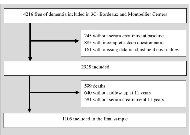
The final sample included 1105 participants from Bordeaux (n=438) and Montpellier (n=667) with a median baseline age of 70.9 years (range, 65.0-87.8) of whom 57.9% were women. As detailed in the flow-chart diagram (Figure 1), these subjects were free of dementia, had fully completed the sleep questionnaire, with eGFRs evaluated at baseline and 11-year follow-up, and without missing data in baseline adjustment covariates. Participants excluded from the study had significantly a lower education level, were older, living alone with activity limitations, more cardio-vascular risk factors, chronic disease and sleep disturbances, taking more hypnotics and with a lower baseline eGFR.
At baseline, 19.2% of the participants had frequent/often EDS, and 74.8% insomnia complaints of whom 30.2% had three insomnia complaints. Furthermore, 17.1% of the participants used regularly sleep medication (67.4% BZD, 32.1% BZD-like compounds, 11.6% miscellaneous medication). Among those subjects, 45.8% had three insomnia complaints and 13.7% none. RLS was reported by 22.2% (n=196) of subjects (62.9% with DMS symptoms, 41.4% with DIS and 35.7% with EMA, and 27.1% without any insomnia complaints), of whom 35.7% declared having RLS before study inclusion.
Median baseline eGFR was 81 mL/min/1.73m2 (interquartile range, 72-88). Only 9.0%
(n=74) had a baseline eGFR<60 including one participant<30. The participants witheGFR<60
differed significantly from those with an eGFR≥60 mL/min/1.73m2 in being frequently with
hypercholesterolemia and hypertension after adjustment for center, age and gender. No significant associations were found between eGFR levels and sleep disturbances (Supplementary table).
Association between sleep disturbances and eGFR decline over 11-year follow-up
The median delay between the collections of both biological samples was 11 years (range, 10.0-12.5). Over this period, 32.1% (n=355) had a moderate to severe eGFR decline including 18 participants with a severe eGFR. The median percentage of eGFR decline was 14% (interquartile range, 7-26) which corresponds to 1.32% per year (interquartile range, 0.59-2.32).
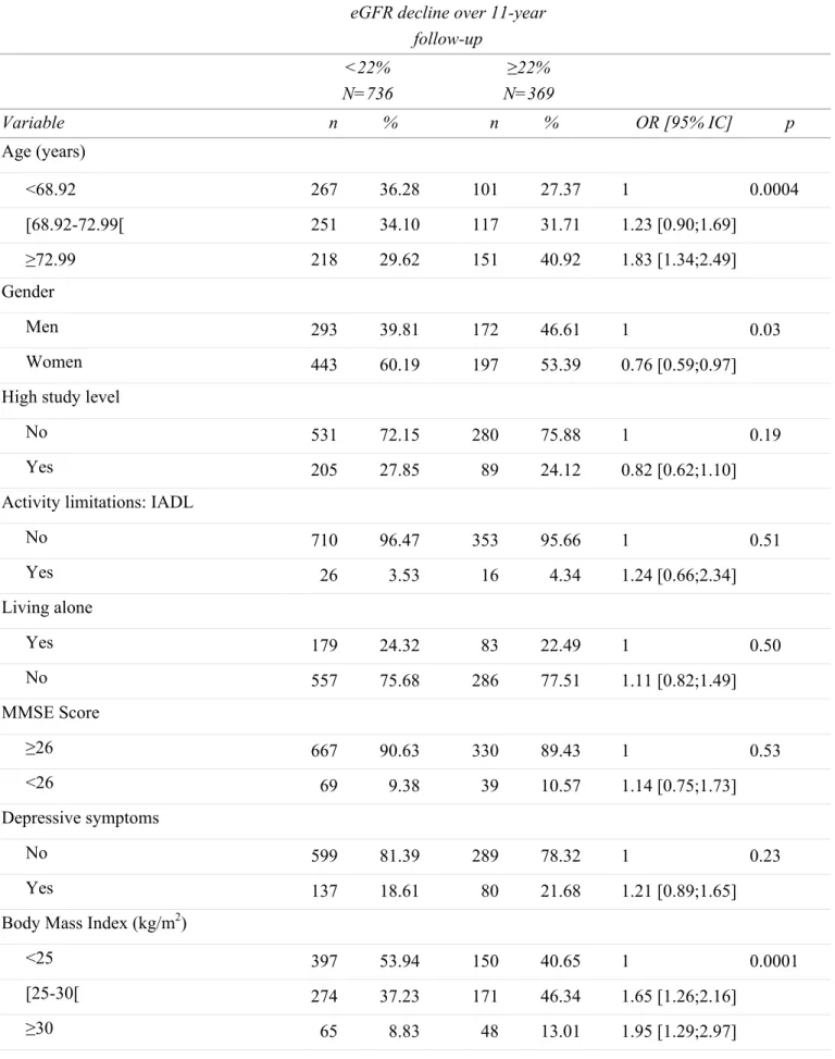
Baseline sociodemographic and clinical characteristics of participants according to kidney function decline are given in Table 1. Participants with a decline ≥ 22% (i.e. highest tertile of kidney function decline) were more frequently men, older, overweight or obese, had diabetes mellitus and hypertension. They also tended to be past or current smoker and had a history of cardiovascular disease (p<0.10). Subsequent analyses were thus adjusted for these factors. Table 2 shows the associations of baseline sleep disturbances with eGFR decline during the follow-up. Whereas DIS, DMS and the number of insomnia complaints were not significantly associated with eGFR decline, a borderline association was observed for EMA after
adjustment for potential confounders (model 2, OR=1.32 95%CI=0.99-1.75). The risk of eGFR decline increased with loud snoring (model 2, OR=1.37 95%CI=1.04-1.81), and clinically-defined apnea (model 2, OR=2.59 95%CI=1.16-5.78). A positive association was also observed between EDS and eGFR decline even after multiple adjustments (model 2, OR=1.67 95%CI=1.18-2.34) and persists even after further adjustment for loud snoring and EMA (OR=1.58 95%CI=1.12-2.23 p=0.009). No significant interactions were found for eGFR decline between EDS and gender or age.
RLS also increased the risk of eGFR decline when its age at onset preceded study inclusion (model 2, OR=1.98 95%CI=1.18-3.30) even after adjustment for EDS (OR=1.90 95%CI=1.13-3.18 p=0.02). In contrast, no significant increased risk of eGFR decline was found when RLS was reported at endpoint only.
Sensitivity analyses
Supplementary analyses were performed 1) excluding the 74 participants with moderate to
severe eGFR impairment (<60 mL/min/1.73m2) at baseline 2) taking into account the new
cerebro-cardiovascular events reported over the follow-up (n=71) and the results remained unchanged.
In the 277 participants having ambulatory PSG recording, only 9.3% had an index of AHI≥30/h and this was associated with loud snoring (p=0.007) but not with EDS (p=0.90) or BMI (p=0.22). A high AHI was also associated with an eGFR decline (OR=2.80 95%CI=1.21-6.44 p=0.02) and the results remained significant after adjustments for age, gender, BMI, smoking status, diabetes mellitus, hypertension, history of cardiovascular disease and EDS (OR=2.50 95%CI=1.01-6.20 p=0.04) (data not shown). No significant interaction was found between AHI and EDS for eGFR decline (p=0.99). Furthermore, no
significant associations were found either for mean (p=0.34) and minimum O2 saturation
(p=0.16) or for percentage of time<90% (p=0.28). A short TST (<6 hours) was found in 50.4%, without any association with insomnia complaints. A short TST was associated with an eGFR decline (OR=1.75 95%CI=1.05-2.93 p=0.03), however the results became non-significant after multi-adjustments (p=0.08). An index of PLMS≥15/h found in 62.4% was associated with RLS at endpoint only (p=0.04) but not when already reported at study inclusion (p=0.10). Finally, no significant associations were found between PLMS and eGFR decline neither in crude (p=0.20) nor in adjusted association (p=0.40).
DISCUSSION
We examined associations between a large range of sleep disturbances and eGFR decline at 11-year follow-up in a general elderly population. Subjects with EDS had a 1.7-fold greater risk of having eGFR decline after adjustment for numerous potential confounders including baseline cardiovascular risk factors. RLS was also found associated with a twice higher risk of eGFR decline independently of EDS. At the exception of EMA which showed a borderline association, insomnia complaints were not related to eGFR decline. In a subsample, high AHI and shorter TST but not PLMS were linked to eGFR decline in crude associations but only AHI remained significant after adjustments.
Although associations between renal functioning and sleep have been previously observed [4], to our knowledge no study has investigated the impact over time of sleep disturbances on eGFR decline in a general population sample. The few clinical studies which have examined the relationship between sleep quality and renal failure in CKD patients with early or end-stage have produced conflicting results [5, 6]. The association between EDS and eGFR decline reported in our study was independent of cardiovascular diseases which failed to support the hypothesis mediating effect of cardiovascular diseases in the relationship between EDS and eGFR in the elderly. Several studies have suggested that cardiovascular and kidney diseases are closely linked through multiple interact effects including neuroendocrine perturbations, fluid dysregulation, inflammation and immune disturbances resulting in amplification loops defined as cardio-renal syndrome. In addition to cardiovascular diseases, diabetes, hypertension and obesity can participate also in the progression of CKD. We report here that EDS is associated with eGFR decline independently of vascular and metabolic risk factors. On the other hand, in previous analyses, we reported that EDS increased the risk of cardiovascular events both fatal and nonfatal, whether first events or recurrent over 6-year follow-up [11]. The underlying pathophysiological mechanism by which EDS interacts with eGFR decline thus appears to be complex and multifactorial.
Sleep disturbances were often associated with activations of hypothalamic–pituitary–adrenal axis and sympathetic nervous system, as well as chronic inflammation that may promote a non-dipping pattern, hypertension and subsequently alter renal function [8]. In our sample, the relationship between EDS and eGFR decline was neither modified by BMI nor by the presence of snoring; however the latter parameters were weaker predictors of SAS in the elderly than in middle aged adults [23]. In the subsample, no associations were found between AHI and EDS or BMI; however we reported an association between AHI and eGFR decline independently of EDS and cardiovascular risk factors without any impact of nocturnal
hypoxia (i.e. no between-group difference for mean and minimum O2 saturations). This
notably differs from the direct nocturnal hypoxia effect already reported on the progression of CKD through a renin-angiotensin-aldosterone system hyperactivation [24, 25]. The link between EDS and eGFR decline may involve a shorter nocturnal sleep duration, a condition frequently associated with increased systemic low-grade inflammation that may trigger cardiovascular disease and subsequent CKD [26]. However the association loses its significant in multivariate model.
The proportion of subjects with RLS (8.3% at baseline) was in the range of previous reported studies in the elderly [27], and it is known to increase with end-stage renal disease [28]. We found that eGFR decline was predicted by RLS at inclusion but not when present at study endpoint only, independently of cardio-vascular risk factors and EDS. These findings suggested that RLS could be an early risk factor for eGFR decline through the bias of an association with sleep fragmentation and periodic-related nighttime increased blood pressure [29], conditions at risk for renal dysfunctioning. However, no associations were found between eGFR decline and PLMS. Except for a borderline association with EMA, other subtypes of insomnia complaints, and depressive symptoms were not significantly associated eGFR decline. Altogether these results supported our previous findings that insomnia
complaints were not risk factors for cardiovascular diseases [11], insomnia being more likely a consequence of these chronic diseases.
The present study benefits from several strengths including standardized creatinine measurements which reduced the bias in the estimate eGFR decline, a large sample size, an 11-year follow-up period of elderly subjects not pre-selected for kidney disease, and a large range of subjective and objective sleep measures.
The present study has some limitations. Selection bias concerned the recruitment of volunteers from electoral rolls and the exclusion of institutionalized elderly people, which limits the extent to which these findings can be generalized to all older adults. Selection bias concerned also the exclusion of subjects due to missing data, loss to follow-up and death. The excluded subjects were older, had more often chronic disorders, a lower baseline eGFR and were more likely to report sleep complaints and to take hypnotics. This progressive selection of the cohort may cause biases which lead to 1) an underreporting of sleep disorders prevalence and kidney failure incidence rate and 2) a modification (to a lesser extent) of the associations between sleeps complaints and eGFR decline.
Sleep complaints were self-reported using a short questionnaire already used [10, 11, 22], which while lacking external confirmation, remains the most common method for initial diagnosis of sleep pathologies in the primary healthcare setting. Unfortunately, validated sleep measures such as the Epworth Sleepiness Scale which provides a measurement of the subject’s general level of daytime sleepiness were not available for this study.
Due to the study design and population (large multicentric elderly cohort), it was not possible to perform PSG in laboratory for the whole sample and this was only possible in the center of Montpellier (one quarter of the sample) in an ambulatory way based on a voluntary basis without selection made on sleep complaints and renal function. PSGs were performed under naturalistic conditions, carried out by skilled sleep specialized technologists with only 2% of recordings being unusable. PSGs were recorded during the follow-up precluding examination of the relationships with baseline sleep complaints. However, the natural course of sleep disturbances is slow in the elderly [30] thus our small sub-sample with ambulatory PSG recording helped to further understand the underlying mechanisms involved in the relationships between sleep disturbances and eGFR decline.
Finally, despite extensive adjustments, the possibility remains that unmeasured factors such as inflammatory biomarkers may also be involved and confound associations.
CONCLUSION
The results of this large 11-year prospective study in the elderly showed that EDS, RLS and AHI constitute risk factors of renal function alteration at a very early stage of decline.
The 3C Study is conducted under a partnership agreement between Inserm, the Victor Segalen – Bordeaux II University and Sanofi-Synthélabo. The Fondation pour la Recherche Médicale funded the preparation and first phase of the study. The 3C-Study is also supported by the Caisse Nationale Maladie des Travailleurs Salariés, Direction Générale de la Santé, MGEN, Institut de la Longévité, Agence Française de Sécurité Sanitaire des Produits de Santé, the Regional Governments of Aquitaine, Bourgogne and Languedoc-Roussillon and, the Fondation de France, the Ministry of Research-Inserm Programme “Cohorts and collection of biological material”.The Lille Génopôle received an unconditional grant from Eisai. The Fondation Plan Alzheimer funded the follow-up of the cohort. Part of this project is financed by grants from the Agence Nationale de la Recherche (projects ANR 2007-LVIE-004 and 06-PNRA-005).
REFERENCES
1. Coresh J, Selvin E, Stevens LA et al. Prevalence of chronic kidney disease in the
United States. JAMA 2007; 298: 2038-2047.
2. Ohayon MM, Carskadon MA, Guilleminault C et al. Meta-analysis of quantitative
sleep parameters from childhood to old age in healthy individuals: developing normative sleep values across the human lifespan. Sleep 2004; 27: 1255-1273.
3. Plantinga L, Lee K, Inker LA et al. Association of sleep-related problems with CKD
in the United States, 2005-2008. Am J Kidney Dis 2011; 58: 554-564.
4. Turek NF, Ricardo AC, Lash JP Sleep disturbances as nontraditional risk factors for
development and progression of CKD: review of the evidence. Am J Kidney Dis 2012; 60: 823-833.
5. Sabbatini M, Pisani A, Crispo A et al. Sleep quality in patients with chronic renal
failure: a 3-year longitudinal study. Sleep Med 2008; 9: 240-246.
6. De Santo RM, Bilancio G, Santoro D et al. A longitudinal study of sleep disorders in
early-stage chronic kidney disease. J Ren Nutr 2010; 20: S59-63.
7. Wolk R, Gami AS, Garcia-Touchard A et al. Sleep and cardiovascular disease. Curr
Probl Cardiol 2005; 30: 625-662.
8. Pepin JL, Borel AL, Tamisier R et al. Hypertension and sleep: overview of a tight
relationship. Sleep Med Rev 2014; 18: 509-519.
9. Yu HT Progression of chronic renal failure. Arch Intern Med 2003; 163: 1417-1429.
10. Blachier M, Dauvilliers Y, Jaussent I et al. Excessive daytime sleepiness and vascular events: the Three City Study. Ann Neurol 2012; 71: 661-667.
11. Jaussent I, Empana JP, Ancelin ML et al. Insomnia, daytime sleepiness and
cardio-cerebrovascular diseases in the elderly: a 6-year prospective study. PLoS One 2013; 8: e56048.
12. Stengel B, Metzger M, Froissart M et al. Epidemiology and prognostic significance of
chronic kidney disease in the elderly--the Three-City prospective cohort study.
Nephrol Dial Transplant 2011; 26: 3286-3295.
13. Pieroni L, Delanaye P, Boutten A et al. A multicentric evaluation of IDMS-traceable
14. Levey AS, Stevens LA, Schmid CH et al. A new equation to estimate glomerular filtration rate. Ann Intern Med 2009; 150: 604-612.
15. National Kidney Foundation DIGO 2012 Clinical Practice Guideline for the Evaluation and Management of Chronic Kidney Diseasee. Kidney International
Supplements 2013; 3: 136-150.
16. Allen RP, Picchietti D, Hening WA et al. Restless legs syndrome: diagnostic criteria,
special considerations, and epidemiology. A report from the restless legs syndrome diagnosis and epidemiology workshop at the National Institutes of Health. Sleep Med 2003; 4: 101-119.
17. Iber C, Ancoli-Israel S, Chesson A et al. The AASM manual for the scoring of sleep
and associated events: rules, terminology, and technical specifications., American Academy of Sleep Medicine, Westchester, IL, USA; 2007.
18. Radloff LS The CES-D Scale: a self-report depression scale for research in the general population. Appl Psychol Meas 1977; 1: 385-401.
19. Folstein MF, Folstein SE, McHugh PR "Mini-mental state". A practical method for grading the cognitive state of patients for the clinician. J Psychiatr Res 1975; 12: 189-198.
20. Lawton MP, Brody EM Assessment of older people: self-maintaining and instrumental
activities of daily living. Gerontologist 1969; 9: 179-186.
21. World Health Organisation World Health Organization Collaborating Centre for Drug
Statistics Methodology. Guidelines for ATC Classification and DDD Assignment.
Oslo, Norway: World Health Organization 2000.
22. Jaussent I, Ancelin ML, Berr C et al. Hypnotics and mortality in an elderly general
population: a 12-year prospective study. BMC Med 2013; 11: 212.
23. Young T, Shahar E, Nieto FJ et al. Predictors of sleep-disordered breathing in
community-dwelling adults: the Sleep Heart Health Study. Arch Intern Med 2002; 162: 893-900.
24. Ahmed SB, Ronksley PE, Hemmelgarn BR et al. Nocturnal hypoxia and loss of
kidney function. PLoS One 2011; 6: e19029.
25. Nangaku M Chronic hypoxia and tubulointerstitial injury: a final common pathway to
end-stage renal failure. J Am Soc Nephrol 2006; 17: 17-25.
26. Cappuccio FP, Cooper D, D'Elia L et al. Sleep duration predicts cardiovascular
outcomes: a systematic review and meta-analysis of prospective studies. Eur Heart J 2011; 32: 1484-1492.
27. Allen RP, Walters AS, Montplaisir J et al. Restless legs syndrome prevalence and
impact: REST general population study. Arch Intern Med 2005; 165: 1286-1292.
28. Unruh ML, Levey AS, D'Ambrosio C et al. Restless legs symptoms among incident
dialysis patients: association with lower quality of life and shorter survival. Am J
Kidney Dis 2004; 43: 900-909.
29. Pennestri MH, Montplaisir J, Colombo R et al. Nocturnal blood pressure changes in
patients with restless legs syndrome. Neurology 2007; 68: 1213-1218.
30. Sforza E, Gauthier M, Crawford-Achour E et al. A 3-year longitudinal study of sleep
Figure 1. Flow chart of participant inclusion
4216 free of dementia included in 3C- Bordeaux and Montpellier Centers
599 deaths
640 without follow-up at 11 years 581 without serum creatinine at 11 years 245 without serum creatinine at baseline 885 with incomplete sleep questionnaire
161 with missing data in adjustment covariables
2925 included
Table 1: Baseline sociodemographic and clinical characteristics of participants according to the estimated GFR (eGFR) decline over 11-year follow-up.
eGFR decline over 11-year follow-up <22% N=736 ≥22% N=369 Variable n % n % OR [95% IC] p Age (years) <68.92 267 36.28 101 27.37 1 0.0004 [68.92-72.99[ 251 34.10 117 31.71 1.23 [0.90;1.69] ≥72.99 218 29.62 151 40.92 1.83 [1.34;2.49] Gender Men 293 39.81 172 46.61 1 0.03 Women 443 60.19 197 53.39 0.76 [0.59;0.97]
High study level
No 531 72.15 280 75.88 1 0.19
Yes 205 27.85 89 24.12 0.82 [0.62;1.10]
Activity limitations: IADL
No 710 96.47 353 95.66 1 0.51 Yes 26 3.53 16 4.34 1.24 [0.66;2.34] Living alone Yes 179 24.32 83 22.49 1 0.50 No 557 75.68 286 77.51 1.11 [0.82;1.49] MMSE Score ≥26 667 90.63 330 89.43 1 0.53 <26 69 9.38 39 10.57 1.14 [0.75;1.73] Depressive symptoms No 599 81.39 289 78.32 1 0.23 Yes 137 18.61 80 21.68 1.21 [0.89;1.65]
Body Mass Index (kg/m2)
<25 397 53.94 150 40.65 1 0.0001
[25-30[ 274 37.23 171 46.34 1.65 [1.26;2.16]
eGFR decline over 11-year follow-up <22% N=736 ≥22% N=369 Variable n % n % OR [95% IC] p
Caffeine intake (mg/day)
≤125 167 22.69 98 26.56 1 0.27 ]125-375] 409 55.57 202 54.74 0.84 [0.62;1.14] >375 160 21.74 69 18.70 0.73 [0.50;1.07] Alcohol (g/day) 0 99 13.45 53 14.36 1 0.15 [1-36[ 568 77.17 268 72.63 0.88 [0.61;1.27] ≥36 69 9.38 48 13.01 1.30 [0.79;2.14] Smoking status Never 454 61.68 202 54.74 1 0.08 Past 245 33.29 144 39.02 1.32 [1.01;1.72] Current 37 5.03 23 6.23 1.40 [0.81;2.41] Diabetes mellitus No 696 94.57 332 89.97 1 0.005 Yes 40 5.43 37 10.03 1.94 [1.22;3.09] Hypercholesterolemia No 474 64.40 246 66.67 1 0.46 Yes 262 35.60 123 33.33 0.90 [0.69;1.18] Hypertension No 409 55.57 153 41.46 1 <0.0001 Yes 327 44.43 216 58.54 1.77 [1.37;2.27] Respiratory disease No 702 95.38 352 95.39 1 0.99 Yes 34 4.62 17 4.61 1.00 [0.55;1.81]
History of cardiovascular disease
No 593 80.57 279 75.61 1 0.06
Yes 143 19.43 90 24.39 1.34 [0.99;1.81]
eGFR decline over 11-year follow-up <22% N=736 ≥22% N=369 Variable n % n % OR [95% IC] p No 613 83.29 302 81.84 1 0.55 Yes 123 16.71 67 18.16 1.11 [0.80;1.53]
Table 2: Baseline sleep complaints of participants according to the percentage decline in estimated GFR decline over 11-year follow-up.
Decline over 11-year follow-up <22 % N=736
≥22%
N=369 Model 0 (1) Model 1 (2) Model 2 (3)
Variable n % N % OR [95% CI] p OR [95% CI] p OR [95% CI] p
Difficulties in initiating sleep
Never/Rarely 442 60.05 217 58.81 1 0.69 1 0.32 1 0.25
Frequently/Often 294 39.95 152 41.19 1.05 [0.82;1.36] 1.15 [0.87;1.52] 1.18 [0.89;1.57] Difficulties in maintaining sleep
Never/Rarely 268 36.41 141 38.21 1 0.56 1 0.48 1 0.45
Frequently/Often 468 63.59 228 61.79 0.93 [0.72;1.20] 0.91 [0.70;1.19] 0.90 [0.69;1.18] Early morning awakening
Never/Rarely 469 63.72 217 58.81 1 0.11 1 0.05 1 0.05
Frequently/Often 267 36.28 152 41.19 1.23 [0.95;1.59] 1.32 [1.00;1.74] 1.32 [0.99;1.75] Number of insomnia complaints
0 187 25.41 91 24.66 1 0.87 1 0.61 1 0.49
1 230 31.25 113 30.62 1.01 [0.72;1.41] 0.97 [0.69;1.37] 0.94 [0.66;1.33] 2 158 21.47 76 20.60 0.99 [0.68;1.43] 1.01 [0.69;1.48] 0.98 [0.66;1.44] 3 161 21.88 89 24.12 1.14 [0.79;1.63] 1.24 [0.83;1.84] 1.25 [0.84;1.87] Excessive daytime sleepiness
Never/Rarely 615 83.56 278 75.34 1 0.001 1 0.0007 1 0.003
Frequently/Often 121 16.44 91 24.66 1.66 [1.22;2.26] 1.78 [1.27;2.49] 1.67 [1.18;2.34] Snoring loudly
Never/Rarely 432 58.70 184 49.86 1 0.005 1 0.002 1 0.02
Frequently/Often 304 41.30 185 50.14 1.43 [1.11;1.84] 1.53 [1.17;2.00] 1.37 [1.04;1.81] RLS retrospectively defined at baseline
No 532 93.66 244 87.77 1 0.004 1 0.004 1 0.009
Yes 36 6.34 34 12.23 2.06 [1.26;3.37] 2.07 [1.26;3.42] 1.98 [1.18;3.30] Clinically-defined apnea
No 724 98.37 354 95.93 1 0.02 1 0.01 1 0.02 (4)
Decline over 11-year follow-up <22 % N=736
≥22%
N=369 Model 0 (1) Model 1 (2) Model 2 (3)
Variable n % N % OR [95% CI] p OR [95% CI] p OR [95% CI] p
(1) Model 0: Crude associations
(2) Model 1: Adjusted for center, age, gender
(3) Model 2: adjusted for all the covariates on model 1 plus BMI, smoking status, diabetes
mellitus, hypertension and history of cardiovascular disease.
(4) For apnea, the adjustment for BMI was not applied because of the colinearity.