HAL Id: hal-03165555
https://hal.univ-lorraine.fr/hal-03165555
Submitted on 10 Mar 2021
HAL is a multi-disciplinary open access
archive for the deposit and dissemination of
sci-entific research documents, whether they are
pub-lished or not. The documents may come from
teaching and research institutions in France or
abroad, or from public or private research centers.
L’archive ouverte pluridisciplinaire HAL, est
destinée au dépôt et à la diffusion de documents
scientifiques de niveau recherche, publiés ou non,
émanant des établissements d’enseignement et de
recherche français ou étrangers, des laboratoires
publics ou privés.
Risk stratification with echocardiographic biomarkers in
heart failure with preserved ejection fraction: the media
echo score
Olivier Huttin, Alan Fraser, Lars Lund, Erwan Donal, Cecilia Linde,
Masatake Kobayashi, Tamas Erdei, Jean-Loup Machu, Kévin Duarte, Patrick
Rossignol, et al.
To cite this version:
Olivier Huttin, Alan Fraser, Lars Lund, Erwan Donal, Cecilia Linde, et al.. Risk stratification with
echocardiographic biomarkers in heart failure with preserved ejection fraction: the media echo score.
ESC Heart Failure, Wiley, 2021, �10.1002/ehf2.13251�. �hal-03165555�
Risk strati
fication with echocardiographic biomarkers
in heart failure with preserved ejection fraction: the
media echo score
Olivier Huttin
1,2, Alan G. Fraser
3, Lars H. Lund
4,5, Erwan Donal
6, Cecilia Linde
4,5, Masatake Kobayashi
1, Tamas
Erdei
3, Jean-Loup Machu
1, Kevin Duarte
1, Patrick Rossignol
1, Walter Paulus
7, Faiez Zannad
1, Nicolas Girerd
1,2*
and MEDIA and KaRen investigators
1Inserm, Centre d’Investigations Cliniques-Plurithématique 1433, Inserm U1116, CHRU Nancy, Université de Lorraine, and F-CRIN INI-CRCT (Cardiovascular and Renal Clinical Trialists), Nancy, France;2Service de Cardiologie, Institut Lorrain du Cœur et des Vaisseaux Louis Mathieu, Centre Hospitalier Universitaire de Nancy, 4 Rue du Morvan, Nancy,54500, France;3School of Medicine, Cardiff University, Cardiff, UK;4Department of Medicine, Karolinska Institutet, Solna, Sweden;5Department of Cardiology, Karolinska University Hospital, Solna, Sweden;6CHU Rennes, Inserm, LTSI-UMR1099, University of Rennes, Rennes, France;7Amsterdam Cardiovascular Sciences, Amsterdam University Medical Centers, Amsterdam, The Netherlands
Abstract
Aims Echocardiographic predictors of outcomes in heart failure with preserved ejection fraction (HFpEF) have not been systematically or independently validated. We aimed at identifying echocardiographic predictors of cardiovascular events in a large cohort of patients with HFpEF and to validate these in an independent large cohort.
Methods and results We assessed the association between echocardiographic parameters and cardiovascular outcomes in 515 patients with heart failure with preserved left ventricular (LV) ejection fraction (>50%) in the MEtabolic Road to DIAstolic Heart Failure (MEDIA) multicentre study. We validated out findings in 286 patients from the Karolinska-Rennes Prospective Study of HFpEF (KaRen). After multiple adjustments including N-terminal pro-brain natriuretic peptide (NT-proBNP), the signif-icant predictors of death or cardiovascular hospitalization were pulmonary arterial systolic pressure> 40 mmHg, respiratory variation in inferior vena cava diameter> 0.5, E/e’ > 9, and lateral mitral annular s’ < 7 cm/s. The combination of these four variables differentiated patients with <10% vs. >35% 1 year risk. Adding these four echocardiographic variables on top of clinical variables and NT-proBNP yielded significant net reclassification improvement (33.8%, P < 0.0001) and increase in C-index (5.3%, a change from 72.2% to 77.5%, P = 0.015) of similar magnitude as the addition of NT-proBNP on top of clinical variables alone. In the KaRen cohort, these four variables yielded a similar improvement in net reclassification improvement (22.3%, P = 0.014) and C-index (4.0%, P = 0.029).
Conclusions Use of four simple echocardiographic parameters (within the MEDIA echo score), indicative of pulmonary hypertension, elevated central venous pressure, LV diastolic dysfunction, and LV long-axis systolic dysfunction, independently predicted prognosis and improved risk stratification additionally to clinical variables and NT-proBNP in HFpEF. This finding was validated in an independent cohort.
Keywords Heart failure, diastolic; Preserved ejection fraction; Echocardiography; Cardiac oedema; Diastolic function; Risk predic-tion; Cardiovascular diseases
Received:18 September 2020; Revised: 14 January 2021; Accepted: 26 January 2021
*Correspondence to: Nicolas Girerd, Service de Cardiologie, Institut Lorrain du Cœur et des Vaisseaux Louis Mathieu, Centre Hospitalier Universitaire de Nancy, 4 Rue du Morvan,54500 Nancy, France. Tel: +33 3 83 15 74 96; Fax: +33 3 83 15 73 24. Email: nicolas_girerd@yahoo.com
Introduction
Heart failure (HF) with preserved ejection fraction (HFpEF) is a heterogeneous syndrome resulting from multiple aetiol-ogies characterized by acutely or chronically increased
cardiac filling pressures.1 Diagnostic algorithms were pro-posed by Paulus et al. and later criteria for diastolic dysfunc-tion by Nagueh et al.2,3Recently, the HFA-PEFF4and H2FPEF5
scores have emerged as important integrative diagnostic tools for HFpEF. Considerable efforts have been made to
identify patients with HFpEF at greatest risk, in whom closer monitoring and/or more intense treatment might improve outcomes, and to identify potential mechanisms that might be targets for therapy.6 Among a number of stratification tools, cardiac imaging, usually currently used for diagnostic purposes, could be pivotal for improving the correlation be-tween diagnostic information and prognosis and potentially also treatment response in HFpEF.7
Importantly, no single echocardiographic parameter is suf-ficiently accurate and reproducible to be used in isolation for stratifying risk in an individual patient with HFpEF. Recent secondary analyses of large trials in HFpEF identified many echocardiographic variables that might provide additional prognostic insights and predict poor outcomes, including indi-cators of ventricular and atrial remodelling and left ventricular (LV)filling.8–12Congestion and reduced diastolic compliance, rather than reduced LV contractility, appear important for diagnosis and for prediction of cardiac events.13Other studies suggest that right ventricular function and load are also important.14–17 If a number of isolated echocardiographic parameters associated with outcome have been reported, the set of echocardiographic variables most associated with outcome in patients with HFpEF is yet to be better deter-mined. Important progress has been made in HFpEF using multiple variable algorithms, such as the HFA-PEFF4 and H2FPEF scores,
5
but this approach primarily intends to improve diagnosis rather than prognosis assessment.
We speculated that detailed echocardiographic phenotyp-ing of well-characterized patients with HFpEF could yield parameters that correlate with natriuretic peptides and be associated with hospitalization and death from cardiovascular causes. This hypothesis was tested in the prospective cohort of the European MEtabolic Road to DIAstolic Heart Failure (MEDIA) project.18 We validated out findings in the Swed-ish–French KaRen HFpEF cohort.19
Methods
MEDIA project study population
A total of 515 patients with HFpEF were enrolled prospec-tively from 2011 to 2013 into the MEDIA multicentre study, in 13 participating university hospitals in Europe.
Inclusion criteria according to the then current consensus statement of the European Society of Cardiology2 were (i) age > 18 years; (ii) signs and symptoms of HF with a pre-served LV ejection fraction (LVEF) (>50%) and an LV end-diastolic volume index (LVEDVi) < 97 mL/m2; (iii) ele-vated serum concentration of brain natriuretic peptide [BNP concentration > 100 pg/mL or N-terminal pro-brain natri-uretic peptide (NT-proBNP) > 300 pg/mL]; and (iv) patients being able and willing to provide written informed consent.
We excluded all patients with acute myocardial infarction, haemodynamically significant valvular disease, chronic dialysis, chronic liver disease, or any concomitant malignant disease during the previous 5 years.
The primary objective was to evaluate the value of imaging and biological markers to predict cardiovascular prognosis in patients with HFpEF. One of the secondary objectives was to evaluate cardiovascular outcomes including a combined end-point of death and all cardiovascular hospitalization (whether for HF or another cardiovascular cause).
The study conforms to the principles of the Declaration of Helsinki20 and was approved by relevant ethics bodies. All subjects provided written informed consent. Patients pro-vided a detailed clinical history, and blood tests (including haematology, biochemistry profile, and NT-proBNP), electro-cardiograms, and echocardiograms were obtained on the same day in most patients.
Echocardiography
All patients underwent echocardiography according to a common protocol, and images were stored in a digital cine-loop format for offline analysis according to the recommendations.21
Left ventricular structure and function were evaluated from standard apical four-chamber, apical two-chamber, and parasternal long-axis and short-axis views. Ventricular dimen-sions, wall thickness, mass, and geometry were determined from 2D parasternal short-axis and long-axis views. LV volumes, stroke volume, and ejection fraction were calcu-lated using the biplane method of disks (modified Simpson’s rule). All cardiac chamber volumes and mass measures were indexed to body surface area.
Additional information regarding echocardiographic methods can be found in Supporting Information.
Echocardiographic data were complete in >75% of patients, apart from the following variables: medial s’, Ard-Ad, S/D, isovolumic relaxation time (IVRT), and E/Vp (which were available in 59%, 63%, 50%, 65%, and 51% of the population, respectively).
Natriuretic peptide analysis
Peripheral venous blood samples for natriuretic peptide analysis were obtained on the same day as echocardio-graphy. Raised serum BNP (>100 pg/mL) and NT-proBNP (>300 pg/mL) were used as inclusion criteria, but some sub-jects were recruited who had normal natriuretic peptide levels as long as they had sufficient other diagnostic criteria to fulfil the requirements for HFpEF.2As biomarker endpoint,
we used NT-proBNP, both as a continuous variable and a cat-egorical variable using age-specific cut-off values [<50 years
(450 pg/mL), between 50 and 75 years (900 pg/mL), and >75 years (1800 pg/mL)22
]. NT-proBNP measurements were performed on automated analysers after the completion of the study.
Cardiovascular events
The primary outcome was a composite of admission for worsening HF or cardiovascular causes and cardiac death. Admission for HF was defined as an admission for worsening of relevant symptoms resulting in substantial intensification of treatment for HF. Follow-up was 100% complete with vital status (in September 2015). Primary outcome events were adjudicated by a dedicated committee.
Replication in the KaRen cohort
It is increasingly recognized that given the heterogeneity of HFpEF, prognostic variables from single studies have been inconsistent and variable and add limited information. Therefore, we performed a validation in an independent cohort, the Swedish–French prospective KaRen study. KaRen included patients with acute signs and symptoms of HF, according to the Framingham criteria, EF ≥ 45% and BNP > 100 pg/mL or NT-proBNP >300 pg/mL, between 1 May 2007 and 1 December 2011 in 10 French and 3 Swedish university hospitals.19These patients were subsequently seen 4–8 weeks after the acute HF episode for detailed echocardi-ography and clinical assessment.
Statistical analyses
Continuous variables are shown as means ± standard devia-tions as specified, and categorical variables are presented as counts and percentages. Logistic regression models were performed to assess the associations between echocardio-graphic parameters and high level of natriuretic peptides.
Cox proportional hazards models were used to identify factors associated with an increased risk of death and hospi-talization (cardiovascular and HF). The follow-up time for each patient was calculated from the date of theirfirst eval-uation to the date of reaching death or hospitalization for cardiovascular cause or to the date of their most recent eval-uation. Odds ratios and hazard ratios (HRs) are presented with their 95% confidence intervals. In order to account for potential confounding, models were adjusted firstly for age, NT-proBNP (either as a log-transformed continuous var-iable or as a categorical varvar-iable using the thresholds detailed earlier22), and glomerular filtration rate and
secondly for the same variables and also for body mass index, atrialfibrillation rather than sinus rhythm, and clinical presentation (acute rather than non-acute). We also
computed a multivariable model using a backward selection procedure to determine a subset of variables that were in-dependently associated with high levels of natriuretic peptides (logistic model) or with the primary composite out-come (Cox model). We included in the multivariable model echocardiographic parameters and potential confounding variables that were associated with the dependent variable in univariable analysis (for which P was <0.10). We used a missing value indicator approach due to some missing values for echocardiographic variables. Multicollinearity was assessed using variance inflation factor in logistic and Cox regression models.
The increased discriminative value associated with the addition of NT-proBNP and echocardiographic variables on top of the aforementioned covariates was evaluated using increase in C-index and continuous net reclassification improvement (cNRI). Changes from a baseline clinical model (including age, estimated glomerular filtration rate, atrial fibrillation, and HF status) were assessed. We also compared the performance of our MEDIA echo score with the HFA-PEFF4and H2FPEF
5
scores.
All statistical analyses were carried out using SAS software Version 9.4 (SAS Institute Inc., Cary, NC, USA) and R software (the R Foundation for Statistical Computing). The two-tailed significance level was set at P < 0.05.
Results
Baseline clinical characteristics in the MEDIA
project (Table 1)
Participants were 74 ± 10 years old, 38% were male, and 71% were in New York Heart Association Class 2. Co-morbidities included hypertension (88%), coronary artery disease (32%), atrial fibrillation (30%), diabetes (39%), and chronic obstructive pulmonary disease (16%). Fifty-one (10%) patients were included in the acute phase during hospitalization for HF, and 71 (14%) in the first month following a hospitalization for HF (<30 days). The remain-der (391, 76%) were ambulatory patients from the outpatient clinic.
Echocardiographic characteristics (Table 2)
Mean LVEF was 60.9 ± 7.2%, and mean indexed LVEDVi was 44.8 ± 14.9 mL/m2. Longitudinal diastolic e’ velocities
at the septum were <8 cm/s in 81.2% and at the lateral wall <10 cm/s in 73.3%. Approximately half (42.5%) of the patients had a mean E/e’ > 13. S/D was <1 in 36.3% of the patients, and A reverse–A duration (Ard-Ad) was >30 ms in 24.9%.
Associations between echocardiographic
variables and N-terminal pro-brain natriuretic
peptide (Figure 1 and Tables S1 and S2)
Left ventricular and right ventricular structure and function Left ventricular longitudinal function (s’) was significantly associated with elevated NT-proBNP in multivariable analysis (Table S1 and Figure 1).
Indexes of relaxation
A longer IVRT (>100 ms) was associated with elevated NT-proBNP, both in univariable analysis and after adjustment
(Table S1 and Figure 1). In contrast, e’ velocities were not as-sociated with elevated NT-proBNP in multivariable analysis (as in Model 2).
Congestion parameters (including estimated left ventricular elevatedfilling pressure and left atrial volume)
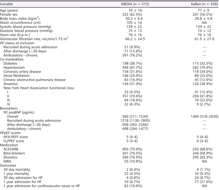
In multivariable analysis (as in Model 2), E/e’ > 15, S/D, short DT, and dilated left atrial (LA) (which can also be an indicator of structural remodelling) were all significantly associated with elevated NT-proBNP. Markers of pulmonary hyperten-sion and right atrial pressure including pulmonary artery systolic pressure (PASP) > 40 mmHg and increased inferior Table 1 Clinical and demographic characteristics in the MEDIA project and KaRen cohort
Variable MEDIA (n = 515) KaRen (n = 356)
Age (years) 74 ± 10 77 ± 9
Female sex 322 (62.5%) 201 (56.5%)
Body mass index (kg/m2) 30.3 ± 6.0 28.8 ± 5.8
Waist circumference (cm) 105 ± 14 NA
Systolic blood pressure (mmHg) 139 ± 23 139 ± 22 Diastolic blood pressure (mmHg) 75 ± 13 74 ± 12
Heart rate (b.p.m.) 70 ± 16 70 ± 16
Glomerularfiltration rate, mL/min/1.73 m2 66.2 ± 24.5 61.9 ± 27.0 HF status at inclusion
Recruited during acute admission 51 (9.9%) — After discharge (<30 days) 71 (13.8%) —
Ambulatory—chronic 391 (76.2%) —
Co-morbidities
Diabetes 198 (38.7%) 115 (32.3%)
Hypertension 448 (87.7%) 282 (79.4%)
Coronary artery disease 158 (31.6%) 118 (34.0%)
Atrialfibrillation 146 (29.9%) 89 (25.0%)
Chronic obstructive pulmonary disease 83 (16.4%) 45 (12.6%)
Anaemia 144 (31.9%) 126 (38.9%)
New York Heart Association functional class
I 23 (4.5%) 41 (12.4%) II 357 (70.6%) 204 (61.8%) III 94 (18.6%) 76 (23.0%) IV 32 (6.3%) 9 (2.7%) Biomarkers NT-proBNP (pg/mL) Overall 560 (211–1530) 1409 (518–2630)
Recruited during acute admission 1218 (1130–7895) — After discharge (<30 days) 958 (302–2266) —
Ambulatory—chronic 498 (204–1477) — HFpEF scores HFA-PEFF score 5 (4–6) 5 (4–6) H2FPEF score 5 (3–6) 4 (3–6) Medication ACEI/ARB 405 (79.4%) 243 (68.6%) Beta-blockers 361 (70.5%) 244 (68.9%) Diuretics 390 (76.9%) 295 (83.3%) MRA 55 (10.8%) NA Outcomes 30 day mortality 2 (0.4%) 4 (1.1%) 1 year mortality 22 (4.5%) 33 (9.3%)
30 day admission for HF 4 (0.8%) 24 (6.7%)
1 year admission for HF 33 (6.7%) 77 (21.6%)
1 year admission for cardiovascular cause or HF 82 (16.6%) NA ACEI/ARB, angiotensin-converting enzyme inhibitor/angiotensin receptor blocker; HF, heart failure; HFpEF, heart failure with preserved ejection fraction; MRA, mineralocorticoid receptor antagonist; NA, not available; NT-proBNP, N-terminal pro-brain natriuretic peptide. Values are mean ± standard deviation or median (Q1–Q3) for continuous variables and frequency (%) for categorical variables.
vena cava (IVC) diameter at rest were also significantly associated with elevated NT-proBNP in multivariable analysis. Integrative assessment (Table S2)
Five echocardiographic variables were found to be significantly associated with NT-proBNP in a multi-echocardiographic marker model after backward selection. These were two functional variables (stroke volume and s’ medial) and three congestion variables (IVC, PASP, and IVRT).
Associations between echocardiographic
variables and primary endpoint (Figure 2 and
Tables S3 and S4)
During a median follow-up of 361 days, hospitalization for cardiovascular causes occurred in 82 patients (16.6%), among which 33 (6.7%) were admissions for HF. One hundred one patients (20.9%) reached a primary study endpoint.
Table 2 Echocardiographic measurements in the MEDIA project and KaRen cohort
Variable MEDIA (n = 515) KaRen (n = 356)
LV structure
Septal wall thickness (mm) 12.2 ± 2.5 11.6 ± 2.2 Posterior wall thickness (mm) 11.1 ± 2.2 11.0 ± 1.9 LV end-diastolic diameter (mm) 48.5 ± 6.3 47.3 ± 6.2 LV end-systolic diameter (mm) 30.8 ± 5.9 32.1 ± 6.5 LV mass index (g/m2) Overall 119 ± 40 126 ± 36 Male 129 ± 39 137 ± 39 Female 113 ± 39 117 ± 31 RWT 0.47 ± 0.12 0.47 ± 0.12 LV function LVEF (%) 60.9 ± 7.2 62.4 ± 6.9
Global longitudinal strain (%) NA 14.6 ± 4.0
LVEDVi (mL/m2) 44.8 ± 14.9 50.3 ± 14.7
LVEDVi≥ 74 (M)/61 (F) mL/m2
39 (8.2%) 40 (12.6%)
LVESVi (mL/m2) 18.2 ± 8.2 19.3 ± 7.6
SV index (SV indexed for BSA) 36.4 ± 10.1 NA SV index (LVEDVi–LVESVi) 26.6 ± 9.7 31.1 ± 8.8 Cardiac output (TVI PW) indexed 2.4 ± 0.6 2.5 ± 0.6 Systolic velocity of mitral annulus
s’ lateral (cm/s) 7.3 ± 2.0 7.3 ± 2.0
s’ medial (cm/s) 6.3 ± 1.8 5.9 ± 1.7
Mitral annular plane systolic excursion (mm) 13.6 ± 3.6 NA Atrial variables
Left atrial area (cm2) 25.7 ± 13.0 NA
LAVi (mL/m2) 43.3 ± 15.2 49.0 ± 16.0
Left ventricular diastolic function
Early wave mitral valveflow velocity (E) (m/s) 91.8 ± 27.4 92.8 ± 28.6 Late wave mitral valveflow velocity (A) (m/s) 82.2 ± 28.0 68.5 ± 31.2
E/A ratio 1.18 ± 0.69 1.79 ± 1.29 DT (ms) 205 ± 65 194 ± 76 e’ lateral (cm/s) 8.3 ± 2.7 9.6 ± 3.4 e’ medial (cm/s) 6.1 ± 1.9 6.4 ± 2.3 Mean e’ 7.3 ± 2.1 8.0 ± 2.5 E/e’ ratio 13.4 ± 5.1 12.7 ± 5.6
E/e’ ratio lateral 12.0 ± 5.0 10.8 ± 5.1
E/e’ ratio medial 16.0 ± 6.6 16.0 ± 7.5
Velocity of systolic pulmonary venousflow—S 0.56 ± 0.23 NA Velocity of diastolic pulmonary venousflow—D 0.56 ± 0.40 NA
S/D 1.23 ± 0.58 NA
A reverse–A duration (Ard-Ad; ms) 0.2 ± 39.9 NA
E/Vp 1.83 ± 0.84 NA
IVRT (ms) 89.4 ± 26.1 92.1 ± 30.6
RV function
Tricuspid regurgitation velocity (m/s) 2.63 ± 0.63 2.87 ± 0.64 Estimated pulmonary arterial pressure (mmHg) 34.8 ± 12.2 44.1 ± 17.3 IVC diameter—IVC rest (mm) 16.7 ± 5.0 18.4 ± 5.4 IVC during respiration/sniff—IVC insp/sniff 9.2 ± 5.8 11.5 ± 5.8 Ratio IVC insp/IVC rest 0.55 ± 0.58 0.59 ± 0.19
TAPSE (mm) 20.5 ± 4.9 17.2 ± 4.7
BSA, body surface area; DT, deceleration time; IVC, inferior vena cava; IVRT, isovolumic relaxation time; LAVi, left atrial volume index; LVEDVi, left ventricular end-diastolic volume index; LVESVi, left ventricular end-systolic volume index; NA, not available; RV, right ventric-ular; RWT, relative wall thickness; TAPSE, tricuspid annular plane systolic excursion.
Left ventricular and right ventricular structure and function In the multivariable analysis, after adjusting for relevant confounders, lateral s’ velocity remained significantly associ-ated with the primary endpoint [HR = 2.26 (1.11–4.61), P = 0.025].
Indexes of relaxation
Increased IVRT duration (>100 ms) was significantly associ-ated with the primary endpoint in both univariable and adjusted analyses, whereas e’ velocity was not.
Congestion parameters (including elevated estimated left ventricularfilling pressure)
In multivariable analysis (Model 2), E/A> 2 [2.55 (HR = 1.13– 5.76)], PASP > 40 [HR = 2.30 (1.30–4.06)], IVC inspiration/ rest > 0.5 (i.e. decreased collapsibility) [HR = 2.73 (1.29– 5.77)], S/D ratio [HR = 3.66 (1.53–8.79)], and LA dilatation [>40 mL/m2, 1.81 (1.02–3.20)] were significantly associated
with higher risk of the primary endpoint. E/e’ > 9 tended to be associated with the primary endpoint [HR = 2.42 (0.95–621), P = 0.065].
We did not identify significant interactions (all P > 0.10) between HF hospitalization status at baseline or presence of atrial fibrillation and echocardiographic variables with regard to rates of the composite outcome, suggesting a homogeneous effect of these echocardiographic variables across both HF and atrialfibrillation status.
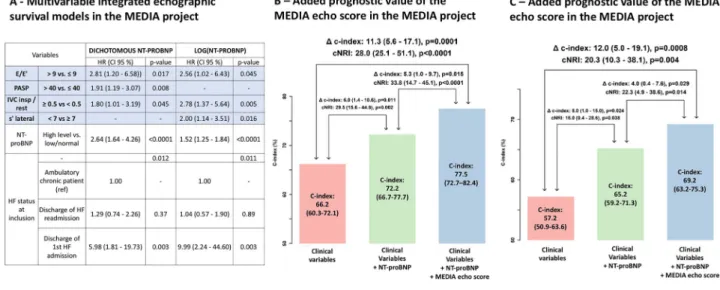
Integrative echocardiographic approach to risk of
cardiovascular hospitalization and mortality
(Figures 3 and 4)
After adjusting for categories of NT-proBNP, the three echocardiographic variables that were retained by the vselection procedure were PAPS [HR = 1.91 (1.19–3.07)], Figure 1 Associations between echocardiographic measurements and high levels of N-terminal pro-brain natriuretic peptide (> 450 pg/mL in patients
below 50 years,>900 in patients aged 50–75 years, and >1800 in patients over 75 years). *Adjusted on age, estimated glomerular filtration rate, body mass index, atrialfibrillation, and clinical presentation.
E/E’ > 9 [HR = 2.81 (1.20–6.58)], and decreased IVC collapsibility [HR = 1.80 (1.01–3.19)]. After adjusting for log-transformed continuous values of NT-proBNP, s’ lateral < 7 was retained in the model [HR = 2.00 (1.14–3.51)] along with E/e’ > 9 and reduced variation in IVC diameter (Figure 3, Panel A).
We estimated the risk for the composite event according to the number of parameters reaching the cut-off among the four echocardiographic variables retained in the multivar-iable survival models (centralfigure). When considering only echocardiographic variables, at 1 year, 37.8% (27.9–46.3%) of the patients with an echo score of 3 or higher (i.e. at least three markers among PAPS> 40, decreased IVC collapsibility, E/e’ > 9, and lateral s’ < 7 cm/s) had an event, whereas patients with a score 0/1 had less than 10% risk [7.9% (2.4–13.1%)] (central figure).
Predictive accuracy of echo parameters in
predicting cardiovascular events at follow-up
(Figure 3)
The addition of NT-proBNP to a clinical model (including age, estimated glomerularfiltration rate, atrial fibrillation, and HF status) improved the net reclassification improvement (NRI = 29.5%, P = 0.002) and C-index (delta C-index 6.0%, P = 0.011) for cardiovascular events. When further adding the four echocardiographic parameters (PAPS > 40, decreased IVC collapsibility, E/e’ > 9, and lateral s’ < 7 cm/s), prediction continued to improve significantly (NRI = 33.8%, P < 0.0001; delta C-index 0.053, P = 0.015) (Figure 3, Panel B); this rise was of similar magnitude to the one observed with the addition of NT-proBNP to the clinical variables.
Figure 2 Associations between echocardiographic parameters and time to cardiovascular/heart failure hospitalization or all-cause death. *Adjusted
for dichotomous N-terminal pro-brain natriuretic peptide, age, estimated glomerularfiltration rate, gender, left ventricular ejection fraction, atrial fibrillation, and clinical presentation.
Added predictive accuracy of MEDIA echo score
on top of the HFA-PEFF and H
2FPEF algorithms
(Figure 3)
The C-index of the MEDIA echo score derived from the four echocardiographic parameters (PAPS > 40, decreased IVC collapsibility, E/e’ > 9, and lateral s’ < 7 cm/s—1 point for each variable) was 0.703 (0.675–0.730). The predictive accuracy of the model including this MEDIA echo score was significantly higher to the ones including the HFA-PEFF score [C-index 0.611 (0.582–0.640); delta C-index 0.092 (0.009–0.175), P = 0.03] or the H2FPEF score [C-index 0.583
(0.550–0.617); delta C-index 0.120 (0.049–0.190), P< 0.001] for cardiovascular events (central figure).
Association of echo variables with outcome and
predictive accuracy of the MEDIA echo score
according to natriuretic peptide levels
To determine whether the pattern of association of echo var-iables with outcome was dependent on natriuretic peptide levels, we constructed a Cox model adjusted of HF status at inclusion including the four echo parameters separately in patients with high levels of NT-proBNP (age-dependent threshold detailed in the Methods section) and patients with lower levels of NT-proBNP. E/E’ > 9, s’ lateral < 7 cm/s, and decreased IVC collapsibility were significantly associated with outcome in patients with lower levels of NT-proBNP [HR = 7.54 (1.01–56.47), P = 0.049; HR = 3.52 (1.57–7.87), P = 0.002; and HR = 3.73 (1.35–10.33), P = 0.011,
respectively]. In patients with high levels of NT-proBNP, PASP> 40 was the only significant factor [HR = 2.68 (1.27– 5.68), P = 0.01], whereas decreased IVC collapsibility tended to be associated with outcome [HR = 2.84 (0.94–8.62), P = 0.07].
The prognostic value of the MEDIA echo score was more important in patients with lower levels of NT-proBNP [C-index 0.729 (0.648–0.810)] than in patients with high levels of NT-proBNP [C-index 0.602 (0.513–0.690)]. The sur-vival curves in patients with high levels or lower levels of NT-proBNP are presented in Figure S1.
Replication in the KaRen cohort
The characteristics of the KaRen cohort have been previously reported.23Briefly, the 539 included patients had very similar characteristics than the MEDIA cohort (mean age 77 ± 9; 56% women, hypertension 78%; Table1).
In the KaRen cohort, we identified a similar significant improvement in prediction when adding NT-proBNP on top of the clinical model. In addition, the added value of the four echocardiographic variables appeared of similar magnitude with significant C-index (4.0%, P = 0.029) and NRI (22.3%, P = 0.014) increases (Figure3, Panel C).
Discussion
We have demonstrated that in a typical population of older patients with HFpEF, the main echocardiographic predictors Figure 3 Multivariable integrated echocardiographic models in the MEDIA project (Panel A) and its added prognostic value in the MEDIA project
(Panel B) and the KaRen cohort (Panel C). Panel A: Cox regression model using subset of variables retained after backward selection (using missing-indicator method) with N-terminal pro-brain natriuretic peptide (NT-proBNP) as a dichotomous or linear variable; Panels B and C: Improve-ment in prognostic value for the primary endpoint on top of clinical model (including age, estimated glomerularfiltration rate, atrial fibrillation, and heart failure status), assessed by net reclassification improvement (NRI) and C-index.
of cardiovascular hospitalization and/or death were related to pulmonary hypertension, high right atrial pressure, and raised E/e’. Importantly, longitudinal LV systolic function as assessed by tissue doppler imaging (TDI) s’ was also associ-ated with clinical outcomes. Importantly, we found that a score of these four variables distinguished patients with HFpEF at low risk (<10% at 1 year) from those at high risk (>35% at 1 year), and it improved risk stratification on top of NT-proBNP (as assessed with C-index and NRI). We found no evidence of interaction with atrial fibrillation or clinical settings (acute/chronic), which suggests that this simple echocardiographic approach could be widely applicable in routine practice. Finally, and in contrast to prior studies, our findings were confirmed in independent validation in a separate large and well-characterized HFpEF cohort.
Echocardiographic markers associated with
N-terminal pro-brain natriuretic peptide
E/e’ ratio, which is widely used as a surrogate measure for mean pulmonary capillary wedge pressure, was not retained as a key echocardiographic predictor of elevated NT-proBNP, whereas PASP and IVC measurements were eventually signif-icantly associated in thefinal multivariable model. Our find-ings are robust because they were very consistent after controlling for the numerous possible confounding factors available in our study, such as cardiac rhythm and whether patients had presented with pulmonary oedema or with dyspnoea on exertion.
Prognostic value of echocardiographic estimates
of left ventricular elevated
filling pressure and
venous pressure
The American College of Cardiology Foundation/American Heart Association Guideline for the Management of Heart Failure suggested an NT-proBNP-guided treatment strategy for optimization of cardioprotective therapy but disfavoured routine repeated echocardiography in stable patients. The 2016 Heart Failure Association guidelines mention natriuretic peptides and echocardiography only as diagnostic tools.24
Consensus recommendations for the diagnosis of diastolic function rely heavily on the E/e’ ratio,2,4,24,25 which is
reported as a validated prognostic marker.24,25Although used
in trials to identify the effects of treatment,26changes only in E/e’ (or any other single echocardiographic measurement) are not accepted surrogates for clinical benefit and, there-fore, not sufficient evidence for approval of a new drug in HFpEF.27Actually, the diagnostic accuracy of E/e’ to predict
LV elevated filling pressure is still debated,28 and a recent
meta-analysis concluded that it cannot reliably estimate fill-ing pressure in patients with preserved LVEF.29Despite these
limitations, we confirm herein the additional prognostic value of E/e’, with however a rather unusual threshold (>9) in pa-tients with known HF (used instead of the usual threshold of eight in light of our data distribution). In addition to E/e’, PASP, another (indirect) marker of filling pressure, was retained as an important and significant risk-stratifying vari-able. Importantly, in our analysis, PASP was significantly asso-ciated with outcome only in patients with the most abnormal NT-proBNP values, which suggests that this marker is most important in the sickest patients. In contrast, E/e’ was most associated with outcome in patients with lower NT-proBNP values. In a way, these two echocardiographic markers appear to complement one another in different settings. Yet the prognostic value of the MEDIA echo score was more pronounced in patients with lower NT-proBNP values.
Two out of four echo variables of the prognostic model we are reporting in this paper are actually indicators of elevated LVfilling pressures, which fits the major issue of pulmonary congestion, resulting in dyspnoea in patients with HFpEF, and is also a key driver of both symptoms and outcomes in HFpEF.30 In addition, IVC was retained in the multivariable model, which further reinforces the value of congestion echo-based variables to stratify the risk of patients with HFpEF.
As emphasized previously, chronic LA remodelling is the final step of chronic intra-cavitary pressure overload31
; in our analysis, LA dilatation was significantly associated with outcome when adjusting on confounding factors. However, it was not retained in ourfinal model by the variable selec-tion procedure, possibly because it is an integrative factor that may overlap with other diastolic function variables.
Prognostic value of left ventricular remodelling
and systolic dysfunction in heart failure with
preserved ejection fraction
In data collected from trials, in hypertension with LV hypertrophy32or in HFpEF,8,10LV mass and other measures
of remodelling had important prognostic implication. In our study, neither the univariable nor the multivariable tests dem-onstrated any significant relationship between LV mass and relative wall thickness with subsequent hospitalization or death at 1 year.
In patients with HFpEF, a varying degree of impaired longi-tudinal systolic function has been demonstrated.33,34LVEF is
an imperfect marker of systolic function: it has high inter-observer variability, it is load dependent, and, in isola-tion, it does not reflect the LV remodelling pattern. Although longitudinal strain generally correlates with LVEF, this corre-lation is relatively modest in patients with HFpEF. Strain imaging detects impaired systolic function despite preserved global LVEF in HFpEF that may contribute to the pathophysi-ology of the HFpEF syndrome.35In a sub-study of the TOPCAT
trial (Treatment of Preserved Cardiac Function Heart Failure with an Aldosterone Antagonist), global longitudinal strain (GLS) was the strongest echocardiographic predictor of the composite outcome of cardiovascular death, aborted cardiac arrest, or HF hospitalization.11Consistently, in our study, s’ was an important prognostic marker. This simple variable does provide information on longitudinal function and could be a pragmatic routine approach to systolic function in patients with HFpEF. Of note, longitudinal systolic function has already been reported to be associated with the transi-tion from hypertensive heart disease to HFpEF.35
Clinical perspectives
After adjusting for a number of important prognostic markers (including NT-proBNP), we demonstrated that a limited set of four echocardiographic variables (E/e’, PASP, IVC, and s’) had predictive value in HFpEF. Of note, the prognostic value of our MEDIA echo score appeared superior (as assessed with C-index) than the one of the HFA-PEFF and H2FPEF (which
are primarily diagnostic algorithms). The major question is whether these variables could also guide the management of patients with HFpEF. Current treatments are empirical9,26 or given for specific indications such as using diuretics to treatfluid retention, giving agents to control systemic blood pressure, and treating underlying ischaemia. There is evidence that residual congestion at discharge from HF hos-pitalization is associated with poor outcome.36 We found that echocardiographic variables assessing congestion pre-dicted outcomes in patients with chronic symptoms as well as those included during a hospitalization for acute symptoms. Therefore, preventing congestion should be beneficial in patients with HFpEF regardless of the context. Indeed, tailoring diuretic and nitrate therapy to changes in pulmonary pressure monitored by an implantable device improves clinical outcome, including in patients with HFpEF.26,37Our study suggests that tailoring anti-congestion therapy using simple echocardiography measures of E/e’, PASP, and IVC is a strategy worth investigating in an appro-priate clinical trial.
Translational outlook
As highlighted earlier, we found that most of the key echocar-diographic variables identified in our study are related to con-gestion. However, congestion animal models are scarce. Our results could suggest that better understanding congestion resolution in controlled settings (such as animal models) would be of interest given the dominance of congestion in HFpEF prognosis.
Strengths and limitations of the study
A particular strength of our study is that it reflects real-life clinical HFpEF practice. On average, patients were older than in many studies (with a mean age of 74 years), and two-thirds were female. There were high proportions of subjects with co-morbidities (which can also be HFpEF aetiologies), includ-ing hypertension in nearly 90% of subjects and diabetes mellitus in more than one-third. Thirty per cent of patients were in atrial fibrillation. In addition, the validation in an external large and well-characterized cohort (the KaRen cohort) strengthens the validity and generalizability of our results. Importantly, the clinical setting and inclusion criteria of the two cohorts were partly different, which further strengthen the generalizability of our results.
Echocardiography was performed in centres with a high level of expertise and with a common protocol, but some studies included in the analysis were not complete with all the measurements.
Our study only focused on rest echocardiography. Exercise echocardiography and lung ultrasound evaluation have emerged as useful diagnostic and prognostic tools in patients with HFpEF.38,39Further studies should determine how these imaging tools should be integrated to best evaluate patients with HFpEF.
Patients could be included in the MEDIA cohort if LVEDVi was <97 mL/m2 and could consequently have mildly or moderately dilated LV upon current standards. However, approximately 10% of the patients we considered in our analysis had LVEDVi≥ 4 (M)/61 (F) mL/m2.
Conclusions
In the MEDIA cohort, we identified four echocardiographic variables (PASP, E/e’, s’, and IVC), three of which are mostly associated with congestion, that independently pre-dicted clinical outcome, regardless of the clinical setting (ambulatory or at acute HF discharge). This important find-ing was validated in the independent KaRen cohort. These results suggest that haemodynamic evaluation of patients using echocardiography (using the MEDIA echo score) could pave the way to future echo-based therapeutic intervention trials.
Acknowledgements
We thank all the participants and investigators of the MEDIA project and KaRen cohort.
Con
flict of interest
L.H.L. related to present manuscript: none; unrelated: research grants to author’s institution and speaker’s and/or consulting fees: AstraZeneca, Boehringer Ingelheim, Novartis, Bayer, Vifor Pharma, Boston Scientific, Sanofi, Myokardia, Pharmacosmos, Mundipharma, Orion Pharma, Merck/MSD, and Medscape. N.G. reports consulting fees, unrelated to this manuscript, from AstraZeneca, Boehringer Ingelheim, and Novartis. P.R. reports personal fees from Relypsa, Inc., a Vifor Pharma Group Company; AstraZeneca; Bayer; CVRx; Fresenius; Novartis; Grunenthal; Servier; Stealth Peptides; Vifor Fresenius Medical Care Renal Pharma; Idorsia; and Novo Nordisk, outside the submitted work; and cofounder: CardioRenal. F.Z. reports personal fees from AstraZeneca, Janssen, Bayer, Novartis, Boston Scientific, Resmed, Amgen, CVRx, General Electric, Boehringer, AstraZeneca, and Vifor Fresenius, outside the submitted work, and cofounder: CardioRenal.
Funding
This work was supported by the EU FP 7 MEDIA project (The MEtabolic Road to DIAstolic Heart Failure) (project number: 261409). T.E. received a research fellowship from the Heart Failure Association of the European Society of Cardiology. L.H.L. was supported by the Swedish Research Council
(grants 2013-23897-104604-23 and 523-2014-2336) and the Swedish Heart Lung Foundation (grants 20120321 and 20150557).
Supporting information
Additional supporting information may be found online in the Supporting Information section at the end of the article.
Table S1: Associations between echocardiographic
measure-ments and high level of NT-proBNP (NT-proBNP > 450 pg/ mL in patients below 50 years, >900 in patients aged 50– 75 years,>1800 in patients over 75 years).
Table S2: Logistic regression model to predict high level of
NT-proBNP on subset of variables retained after backward selection
Table S3: Associations between echocardiographic
parame-ters and time to cardiovascular/HF hospitalization or all-cause death.
Table S4: Hazard ratio for primary outcome based on
echocardiographic parameters analyzed as continuous variables
Figure S1: Kaplan–Meier estimates of time to the combined
end-point of all-cause death and cardiovascular/HF hospitali-zation according to the number of echocardiographic criteria met (E/e’, PASP, IVC insp/IVC rest ratio, s’ lateral) in the ME-DIA project according to NT-proBNP levels.
References
1. Bhatia RS, Tu JV, Lee DS, Austin PC, Fang J, Haouzi A, Gong Y, Liu PP. Outcome of heart failure with preserved ejection fraction in a population-based study. N
Engl J Med 2006;355: 260–269.
2. Paulus WJ, Tschope C, Sanderson JE, Rusconi C, Flachskampf FA, Rademakers FE, Marino P, Smiseth OA, De Keulenaer G, Leite-Moreira AF, Borbély A. How to diagnose diastolic heart failure: a con-sensus statement on the diagnosis of heart failure with normal left ventricular ejection fraction by the Heart Failure and Echocardiography Associations of the European Society of Cardiology. Eur
Heart J 2007;28: 2539–2550.
3. Nagueh SF, Appleton CP, Gillebert TC, Marino PN, Oh JK, Smiseth OA, Waggoner AD, Flachskampf FA, Pellikka PA, Evangelisa A. Recommendations for the evaluation of left ventricular dia-stolic function by echocardiography.
Eur J Echocardiogr 2009;10: 165–193.
4. Pieske B, Tschope C, de Boer RA, Fraser AG, Anker SD, Donal E, Edelmann F, Fu M, Guazzi M, Lam CS, Lancellotti P. How to diagnose heart failure with pre-served ejection fraction: the HFA-PEFF diagnostic algorithm: a consensus recommendation from the Heart Failure Association (HFA) of the European Soci-ety of Cardiology (ESC). Eur J Heart Fail 2020;22: 391–412.
5. Reddy YNV, Carter RE, Obokata M, Redfield MM, Borlaug BA. A simple, evidence-based approach to help guide diagnosis of heart failure with preserved ejection fraction. Circulation 2018;138: 861–870.
6. Burke MA, Katz DH, Beussink L, Selvaraj S, Gupta DK, Fox J, Chakrabarti S, Sauer AJ, Rich JD, Freed BH, Shah SJ. Prog-nostic importance of pathophysiologic markers in patients with heart failure and preserved ejection fraction. Circ
Heart Fail 2014;7: 288–299.
7. Flachskampf FA, Biering-Sorensen T, Solomon SD, Duvernoy O, Bjerner T, Smiseth OA. Cardiac imaging to evaluate left ventricular diastolic function. J Am
Coll Cardiol Img 2015;8: 1071–1093.
8. Zile MR, Gottdiener JS, Hetzel SJ, McMurray J, Komajda M, McKelvie R, Baicu CF, Massie BM, Carson PE, I-PRE-SERVE Investigators. Prevalence and significance of alterations in cardiac structure and function in patients with heart failure and a preserved ejection fraction. Circulation 2011; 124: 2491–2501.
9. Solomon SD, Verma A, Desai A, Hassanein A, Izzo J, Oparil S, Lacourciere Y, Lee J, Seifu Y, Hilkert RJ, Rocha R, Pitt B, Exforge Intensive Control of Hypertension to Evaluate Ef fi-cacy in Diastolic Dysfunction Investiga-tors. Effect of intensive versus standard blood pressure lowering on diastolic function in patients with uncontrolled
hypertension and diastolic dysfunction.
Hypertension 2010;55: 241–248.
10. Shah AM, Claggett B, Sweitzer NK, Shah SJ, Anand IS, O’Meara E, Desai AS, Heitner JF, Li G, Fang J, Rouleau J, Zile MR, Markov V, Ryabov V, Reis G, Assmann SF, McKinlay SM, Pitt B, Pfeffer MA, Solomon SD. Cardiac struc-ture and function and prognosis in heart failure with preserved ejection fraction: findings from the echocardiographic study of the Treatment of Preserved Cardiac Function Heart Failure with an Aldosterone Antagonist (TOPCAT) Trial.
Circ Heart Fail 2014;7: 740–751.
11. Shah AM, Claggett B, Sweitzer NK, Shah SJ, Deswal A, Anand IS, Fleg JL, Pitt B, Pfeffer MA, Solomon SD. Prognostic im-portance of changes in cardiac structure and function in heart failure with pre-served ejection fraction and the impact of spironolactone. Circ Heart Fail 2015;
8: 1052–1058.
12. Shah AM, Cikes M, Prasad N, Li G, Getchevski S, Claggett B, Rizkala A, Lukashevich I, O’Meara E, Ryan JJ, Shah SJ, Mullens W, Zile MR, Lam CSP, McMurray J, Solomon SD, PARAGON-HF Investigators. Echocardiographic fea-tures of patients with heart failure and preserved left ventricular ejection fraction. J Am Coll Cardiol 2019; 74: 2858–2873.
13. Cikes M, Solomon SD. Beyond ejection fraction: an integrative approach for as-sessment of cardiac structure and func-tion in heart failure. Eur Heart J 2016;
37: 1642–1650.
14. Mohammed SF, Hussain I, AbouEzzeddine OF, Takahama H, Kwon SH, Forfia P, Roger VL, Redfield MM. Right ventricular function in heart failure with preserved ejection fraction: a community-based study. Circulation 2014;130: 2310–2320.
15. Melenovsky V, Hwang SJ, Lin G, Redfield MM, Borlaug BA. Right heart dysfunction in heart failure with pre-served ejection fraction. Eur Heart J 2014;35: 3452–3462.
16. Lam CS, Roger VL, Rodeheffer RJ, Borlaug BA, Enders FT, Redfield MM. Pulmonary hypertension in heart failure with preserved ejection fraction: a community-based study. J Am Coll
Cardiol 2009;53: 1119–1126.
17. Abraham WT, Adamson PB, Bourge RC, Aaron MF, Costanzo MR, Stevenson LW, Strickland W, Neelagaru S, Raval N, Krueger S, Weiner S. Wireless pulmo-nary artery haemodynamic monitoring in chronic heart failure: a randomised controlled trial. Lancet (London,
En-gland) 2011;377: 658–666.
18. Stienen S, Ferreira JP, Kobayashi M, Preud’homme G, Dobre D, Machu JL, Duarte K, Bresso E, Devignes MD, López N, Girerd N, Aakhus S, Ambrosio G, Brunner-la Rocca HP, Fontes-Carvalho R, Fraser AG, van Heerebeek L, Heymans S, de Keulenaer G, Marino P, McDonald K, Mebazaa A, Papp Z,
Raddino R, Tschöpe C, Paulus WJ, Zannad F, Rossignol P. Enhanced clinical phenotyping by mechanistic bioprofiling in heart failure with preserved ejection fraction: insights from the MEDIA-DHF study (The Metabolic Road to Diastolic Heart Failure). Biomarkers 2020; 25: 201–211.
19. Donal E, Lund LH, Linde C, Edner M, Lafitte S, Persson H, Bauer F, Öhrvik J, Ennezat PV, Hage C, Löfman I, Juilliere Y, Logeart D, Derumeaux G, Gueret P, Daubert JC. Rationale and design of the Karolinska-Rennes (KaRen) prospec-tive study of dyssynchrony in heart fail-ure with preserved ejection fraction.
Eur J Heart Fail 2009;11: 198–204.
20. World Medical A. World Medical Associ-ation DeclarAssoci-ation of Helsinki: ethical principles for medical research involving human subjects. JAMA 2013; 310: 2191–2194.
21. Lang RM, Badano LP, Mor-Avi V, Afilalo J, Armstrong A, Ernande L, Flachskampf FA, Foster E, Goldstein SA, Kuznetsova T, Lancellotti P, Muraru D, Picard MH, Rietzschel ER, Rudski L, Spencer KT, Tsang W, Voigt JU. Recommendations for cardiac chamber quantification by echocardiography in adults: an update from the American Society of Echocardi-ography and the European Association of Cardiovascular Imaging. Eur Heart J
Cardiovasc Imaging 2015;16: 233–270.
22. Kim HN, Januzzi JL Jr. Natriuretic pep-tide testing in heart failure. Circulation 2011;123: 2015–2019.
23. Donal E, Lund LH, Oger E, Hage C, Persson H, Reynaud A, Ennezat PV, Bauer F, Sportouch-Dukhan C, Drouet E, Daubert JC, Linde C, KaRen Investiga-tors. Baseline characteristics of patients with heart failure and preserved ejec-tion fracejec-tion included in the Karolinska Rennes (KaRen) study. Arch Cardiovasc
Dis 2014;107: 112–121.
24. Ponikowski P, Voors AA, Anker SD, Bueno H, Cleland JG, Coats AJ, Falk V, González-Juanatey JR, Harjola VP, Jankowska EA, Jessup M, Linde C, Nihoyannopoulos P, Parissis JT, Pieske B, Riley JP, Rosano GM, Ruilope LM, Ruschitzka F, Rutten FH, van der Meer P, Authors/Task Force Members, Document Reviewers. 2016 ESC guide-lines for the diagnosis and treatment of acute and chronic heart failure: the Task Force for the diagnosis and treatment of acute and chronic heart failure of the European Society of Cardiology (ESC). Developed with the special contribution of the Heart Failure Association (HFA) of the ESC. Eur J Heart Fail 2016;18: 891–975.
25. Nagueh SF, Appleton CP, Gillebert TC, Marino PN, Oh JK, Smiseth OA, Waggoner AD, Flachskampf FA, Pellikka PA, Evangelista A. Recommendations for the evaluation of left ventricular diastolic function by echocardiography.
J Am Soc Echocardiogr 2009; 22:
107–133.
26. Edelmann F, Wachter R, Schmidt AG, Kraigher-Krainer E, Colantonio C, Kamke W, Duvinage A, Stahrenberg R, Durstewitz K, Löffler M, Düngen HD, Tschöpe C, Herrmann-Lingen C, Halle M, Hasenfuss G, Gelbrich G, Pieske B, Aldo-DHF Investigators. Effect of spironolactone on diastolic function and exercise capacity in patients with heart failure with preserved ejection fraction: the Aldo-DHF randomized con-trolled trial. JAMA 2013;309: 781–791. 27. Edelmann F, Gelbrich G, Duvinage A, Stahrenberg R, Behrens A, Prettin C, Kraigher-Krainer E, Schmidt AG, Düngen HD, Kamke W, Tschöpe C, Herrmann-Lingen C, Halle M, Hasenfuss G, Wachter R, Pieske B. Differential interac-tion of clinical characteristics with key functional parameters in heart failure with preserved ejection fraction—results of the Aldo-DHF trial. Int J Cardiol 2013;
169: 408–417.
28. Santos M, Rivero J, McCullough SD, West E, Opotowsky AR, Waxman AB, Systrom DM, Shah AM. E/e’ ratio in pa-tients with unexplained dyspnea: lack of accuracy in estimating left ventricular filling pressure. Circ Heart Fail 2015; 8: 749–756.
29. Sharifov OF, Schiros CG, Aban I, Denney TS, Gupta H. Diagnostic accuracy of tis-sue Doppler index E/e’ for evaluating left ventricularfilling pressure and dia-stolic dysfunction/heart failure with preserved ejection fraction: a systematic review and meta-analysis. J Am Heart
Assoc 2016;5: e002530.
30. Girerd N, Seronde MF, Coiro S, Chouihed T, Bilbault P, Braun F, Kenizou D, Maillier B, Nazeyrollas P, Roul G, Fillieux L, Abraham WT, Januzzi J Jr, Sebbag L, Zannad F, Mebazaa A, Rossignol P, INI-CRCT, Great Network, and the EF-HF Group. Integrative as-sessment of congestion in heart failure throughout the patient journey. JACC
Heart Fail 2018;6: 273–285.
31. Beltrami M, Palazzuoli A, Padeletti L, Cerbai E, Coiro S, Emdin M, Marcucci R, Morrone D, Cameli M, Savino K, Pedrinelli R, Ambrosio G, Società Italiana di Cardiologia, Sezione Regionale Tosco-Umbra. The importance of integrated left atrial evaluation: from hypertension to heart failure with pre-served ejection fraction. Int J Clin Pract 2018;72.
32. Wachtell K, Bella JN, Rokkedal J, Palmieri V, Papademetriou V, Dahlöf B¨, Aalto T, Gerdts E, Devereux RB. Change in diastolic left ventricular filling after one year of antihypertensive treatment: the Losartan Intervention For Endpoint Reduction in Hypertension (LIFE) study.
Circulation 2002;105: 1071–1076.
33. Donal E, Lund LH, Oger E, Hage C, Persson H, Reynaud A, Ennezat PV, Bauer F, Drouet E, Linde C, Daubert C, KaRen investigators. New echocardio-graphic predictors of clinical outcome in patients presenting with heart failure
and a preserved left ventricular ejection fraction: a subanalysis of the Ka (Karolinska) Ren (Rennes) study. Eur J
Heart Fail 2015;17: 680–688.
34. Vinereanu D, Nicolaides E, Tweddel AC, Fraser AG.“Pure” diastolic dysfunc-tion is associated with long-axis systolic dysfunction. Implications for the diagnosis and classification of heart failure. Eur J Heart Fail 2005; 7: 820–828.
35. Kraigher-Krainer E, Shah AM, Gupta DK, Santos A, Claggett B, Pieske B, Zile MR, Voors AA, Lefkowitz MP, Packer M, McMurray J, Solomon SD, PARA-MOUNT Investigators. Impaired systolic function by strain imaging in heart failure with preserved ejection fraction.
J Am Coll Cardiol 2014;63: 447–456.
36. Coiro S, Porot G, Rossignol P, Ambrosio G, Carluccio E, Tritto I, Huttin O, Lemoine S, Sadoul N, Donal E, Zannad F, Girerd N. Prognostic value of pulmo-nary congestion assessed by lung ultrasound imaging during heart failure hospitalisation: a two-centre cohort study. Sci Rep 2016;6: 39426. 37. Yancy CW, Jessup M, Bozkurt B, Butler J,
Casey DE Jr, Drazner MH, Fonarow GC, Geraci SA, Horwich T, Januzzi JL, John-son MR, Kasper EK, Levy WC, Masoudi FA, McBride PE, McMurray JJV, Mitchell JE, Peterson PN, Riegel B, Sam F, Stevenson LW, Tang WHW, Tsai EJ, Wilkoff BL. 2013 ACCF/AHA guideline for the management of heart failure: executive summary: a report of the American College of Cardiology
Foundation/American Heart Association Task Force on practice guidelines.
Circu-lation 2013;128: 1810–1852.
38. Simonovic D, Coiro S, Carluccio E, Girerd N, Deljanin-Ilic M, Cattadori G, Ambrosio G. Exercise elicits dynamic changes in extravascular lung water and haemodynamic congestion in heart failure patients with preserved ejection fraction. Eur J Heart Fail 2018; 20: 1366–1369.
39. Coiro S, Simonovic D, Deljanin-Ilic M, Duarte K, Carluccio E, Cattadori G, Girerd N, Ambrosio G. Prognostic value of dynamic changes in pulmonary con-gestion during exercise stress echocardi-ography in heart failure with preserved ejection fraction. Circ Heart Fail 2020;