HAL Id: hal-00636551
https://hal.archives-ouvertes.fr/hal-00636551
Submitted on 29 May 2020
HAL is a multi-disciplinary open access
archive for the deposit and dissemination of
sci-entific research documents, whether they are
pub-lished or not. The documents may come from
teaching and research institutions in France or
abroad, or from public or private research centers.
L’archive ouverte pluridisciplinaire HAL, est
destinée au dépôt et à la diffusion de documents
scientifiques de niveau recherche, publiés ou non,
émanant des établissements d’enseignement et de
recherche français ou étrangers, des laboratoires
publics ou privés.
Copyright
Biochemical Characterization of AtHMA6/PAA1, a
Chloroplast Envelope Cu(I)-ATPase.
Patrice Catty, Sylvain Boutigny, Roger Miras, Jacques Joyard, Norbert
Rolland, Daphné Seigneurin-Berny
To cite this version:
Patrice Catty, Sylvain Boutigny, Roger Miras, Jacques Joyard, Norbert Rolland, et al.. Biochemical
Characterization of AtHMA6/PAA1, a Chloroplast Envelope Cu(I)-ATPase.. Journal of Biological
Chemistry, American Society for Biochemistry and Molecular Biology, 2011, 286 (42), pp.36188-97.
�10.1074/jbc.M111.241034�. �hal-00636551�
Biochemical Characterization of AtHMA6/PAA1, a Chloroplast
Envelope Cu(I)-ATPase
*
Received for publication, March 29, 2011, and in revised form, August 30, 2011Published, JBC Papers in Press, August 30, 2011, DOI 10.1074/jbc.M111.241034
Patrice Catty‡§¶, Sylvain Boutigny储**‡‡§§, Roger Miras‡§¶, Jacques Joyard储**‡‡§§, Norbert Rolland储**‡‡§§, and Daphné Seigneurin-Berny储**‡‡§§1
From the‡Laboratoire de Chimie et Biologie des Métaux, UMR5249,CNRS, F-38054 Grenoble, the§Laboratoire Chimie et Biologie des Métaux and **Laboratoire de Physiologie Cellulaire et Végétale, Institut de Recherches en Technologies et Sciences pour le Vivant (iRTSV), Direction des Sciences du Vivant, Commissariat à l’Energie Atomique et aux Energies Alternatives (CEA),
F-38054 Grenoble, the¶Laboratoire de Chimie et Biologie des Métaux, Université Joseph Fourier Grenoble I, F-38054 Grenoble, the
储Laboratoire de Physiologie Cellulaire et Végétale, UMR5168, CNRS, F-38054 Grenoble, the‡‡Laboratoire de Physiologie Cellulaire
et Végétale, UMR1200, Institut National de la Recherche Agronomique (INRA), F-38054 Grenoble, and the§§Laboratoire de Physiologie Cellulaire et Végétale,Université Joseph Fourier Grenoble I, F-38054 Grenoble, France
Background:There is a lack of biochemical characterization of plant PIB-1-type ATPases (copper transporters).
Result:Phosphorylation assays show that the plant P-type ATPase PAA1 is activated by monovalent ions Cu⫹/Ag⫹. Yeast expression validates in vivo the ionic selectivity of the transporter.
Conclusion:PAA1 is a high affinity Cu⫹transporter of the chloroplast envelope.
Significance:This study provides the first biochemical characterization of a plant copper ATPase.
Copper is an essential plant micronutrient playing key roles in cellular processes, among them photosynthesis. In Arabidopsis
thaliana, copper delivery to chloroplasts, mainly studied by
genetic approaches, is thought to involve two PIB-type ATPases:
AtHMA1 and AtHMA6/PAA1. The lack of biochemical charac-terization of AtHMA1 and PAA1, and more generally of plant PIB-type ATPases, is due to the difficulty of getting high
amounts of these membrane proteins in an active form, either from their native environment or after expression in heterolo-gous systems. In this study, we report the first biochemical char-acterization of PAA1, a plant copper-transporting ATPase. PAA1 produced in Lactococcus lactis is active, forming an aspar-tyl phosphate intermediate in the presence of ATP and the ade-quate metal ion. PAA1 can also be phosphorylated using inor-ganic phosphate in the absence of transition metal. Both phosphorylation types allowed us to demonstrate that PAA1 is activated by monovalent copper ions (and to a lower extent by silver ions) with an apparent affinity in the micromolar range. In agreement with these biochemical data, we also demonstrate that when expressed in yeast, PAA1 induces increased sensitiv-ities to copper and silver. These data provide the first enzymatic characterization of a PIB-1-type plant ATPase and clearly iden-tify PAA1 as a high affinity Cu(I) transporter of the chloroplast envelope.
Metal ions are essential for plant growth, having both struc-tural and catalytic roles in proteins involved in a wide variety of cellular processes. However, their concentration must be
tightly regulated as these ions become toxic when present in excess. Therefore, plants have acquired different cellular mech-anisms that ensure safe handlings of metal ions, from assimila-tion to distribuassimila-tion within the plant, and storage. Among the mechanisms that contribute to metal homeostasis, metal trans-port plays a key role. When compared with other organisms, plant genomes encode many different types of metal transport-ers with their own ionic specificity, expression pattern, and sub-cellular localization (for review, see Refs. 1–3).
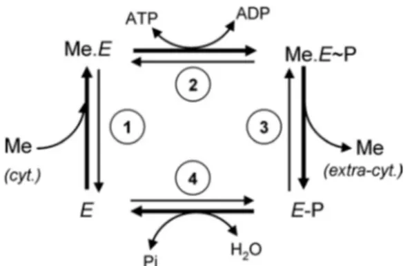
P-type ATPases are multispanning membrane proteins that translocate ions across plasma or organelle membranes at the expense of ATP consumption (4). Their catalytic cycle can be reduced to a four-step process (Fig. 1) accounting for the cou-pling of ion motion to ATP hydrolysis, with the formation of transient phosphorylated states resulting from ATP ␥-phos-phate transfer to the conserved aspartic residue of the DKTGT motif. In its free state E, the P-type ATPase binds cytoplasmic ions at its high affinity membrane site (Fig. 1, step 1). ATP bound to the large cytoplasmic domain of the transporter is then hydrolyzed, leading to the formation of a phosphorylated enzyme (Fig. 1, step 2). The ion binding phosphorylated form of the enzyme (Me.E⬃P) undergoes important conformation changes leading to metal release at the extracytoplasmic side of the membrane (Fig. 1, step 3). In the metal-free phosphorylated enzyme E-P, the aspartyl phosphate bound is hydrolyzed to bring the enzyme back in its free state E (Fig. 1, step 4). For several P-type ATPases such as the Na⫹/K⫹- or H⫹/K⫹ -AT-Pases, a second ion binds from the extracytoplasmic side at step 3 and, on hydrolysis of the phosphorylated Asp (step 4), is released to the cytoplasmic side (4).
On the basis of sequence analysis, P-type ATPases have been classified in several subfamilies; among them, type IB corre-sponds to the transition metal-transporting ATPases (5). On an enzymatic standpoint, PIB-type ATPases display all the major trademarks of P-type ATPases (6). According to the presence of *This work was supported by the CEA (funding of the CEA-PM project), a
Ph. D. fellowship from the CNRS (to S. B.), the INRA, and the University of Grenoble.
1To whom correspondence should be addressed: Laboratoire de Physiologie Cellulaire et Végétale, iRTSV, UMR5168, CNRS/UJF/INRA/CEA-Grenoble, 17 rue des Martyrs, 38054 Grenoble cedex, France. Tel.: 33-4-38-78-23-63; E-mail: daphne.berny@cea.fr.
© 2011 by The American Society for Biochemistry and Molecular Biology, Inc. Printed in the U.S.A.
at INRA Institut National de la Recherche Agronomique on June 13, 2018
http://www.jbc.org/
conserved residues in transmembrane helices 6, 7, and 8, PIB -type ATPases have been divided into five subgroups: (i) sub-group IB-1 of Cu⫹- and Ag⫹-ATPases; (ii) subgroup IB-2 of Cd2⫹-, Zn2⫹-, and Pb2⫹-ATPases; (iii) subgroup IB-3 of Cu⫹/ Cu2⫹-ATPases; (iv) subgroup IB-4 of putative Co2⫹-ATPases; and (v) subgroup IB-5 of PIB-type ATPases with no assigned specificity (7).
The Arabidopsis thaliana genome encodes eight PIB-type ATPases (8, 9). HMAs25, 6, 7, and 8 are members of the sub-group IB-1. In plants, the expression of AtHMA5 is enhanced in the presence of copper and the hma5 knock-out mutant is hypersensitive to copper, suggesting that AtHMA5 could be involved in copper detoxification in A. thaliana roots (10). Originally isolated in the screening of Arabidopsis mutants altered in hormone receptor specificity, AtHMA7 (also called RAN1) was found to functionally replace the yeast Cu⫹ -AT-Pase Ccc2p. It was proposed to be localized in the post-Golgi membrane, providing copper to the ethylene receptor (11–13). AtHMA6/PAA1 and AtHMA8/PAA2 have been localized in the chloroplast envelope and thylakoids, respectively. Because the outer membrane of the chloroplast envelope is not a selec-tive barrier (due to the presence of broad specificity pores), PAA1 is expected to be a specific transporter of the inner mem-brane of the chloroplast envelope. PAA1 and PAA2 mutations were shown to strongly reduce chloroplast copper content (in the stroma for paa1 and in the thylakoid for paa2), and conse-quently, copper-dependent activities (Cu/Zn-superoxide dis-mutase, plastocyanin-dependent photosynthetic electron transport). From in planta analyses, it was proposed that PAA1 provides copper first to a metallochaperone that could interact with the chloroplast Cu/Zn-superoxide dismutase and also to PAA2, which could then provide copper to the plastocyanin in the thylakoid lumen (14, 15).
HMAs 2, 3, and 4 belong to the subgroup IB-2. AtHMA2 and AtHMA4 ensure Zn2⫹translocation from roots to the shoot (16 –18). AtHMA2 is predominantly expressed at the plasma membrane of cells from vascular tissues of roots, stems, and leaves, where it works as an efflux system. When expressed in yeast, AtHMA2 displays Zn2⫹- and Cd2⫹-dependent (and to a lower extent, Pb2⫹-, Ni2⫹-, Cu2⫹-, and Co2⫹-dependent)
ATPase activities and forms an acid-stable phosphorylated intermediate in the presence of metals (16, 18). AtHMA4 is localized at the plasma membrane of cells from tissues sur-rounding the root vascular vessels. A null hma4 mutant exhibits a lower translocation of Zn2⫹and Cd2⫹from roots to the shoot, whereas AtHMA4-overexpressing lines display an increase in Zn2⫹ and Cd2⫹ shoot content, suggesting that AtHMA4 is involved in providing metal to the xylem (17). When expressed in bacteria or yeast, AtHMA4 restores Zn2⫹tolerance to Esch-erichia coli zntAmutant and Cd2⫹ tolerance to yeast ⌬ycf1 strain (19, 20). AtHMA3 is located at the vacuolar membrane, with a predominant expression in guard cells, hydathodes, vas-cular tissues, and the root apex. The hma3 knock-out mutant was found to be more sensitive to Zn2⫹ and Cd2⫹, whereas ectopic overexpression of AtHMA3 improved plant tolerance to Cd2⫹, Co2⫹, and Pb2⫹. It was therefore suggested that AtHMA3 contributes to heavy metal detoxification by partici-pating in their vacuolar sequestration (21). In Saccharomyces
cerevisiae, AtHMA3 expression restores normal metal toler-ance to the Cd2⫹- and Pb2⫹-hypersensitive⌬ycf1 strain but not to the Zn2⫹-hypersensitive⌬zrc1 strain (22).
AtHMA1 was classified in subgroup IB-4 (putative Co2⫹ -ATPase) on the basis of its amino acid sequence (7). In 2006, we localized AtHMA1 in the chloroplast envelope (theoretically in the inner membrane as for PAA1) and showed that its activity was enhanced by copper (23). In addition, hma1 mutants were found to exhibit a reduced plastidial copper content as well as a reduced chloroplast Cu/Zn-superoxide dismutase activity. These data led us to suggest that AtHMA1 functions as a copper transporter (23). Later, Moreno et al. (24) showed that the pre-cursor form of AtHMA1 (i.e. including the chloroplast transit peptide) could complement the lack of yeast Ca2⫹-ATPases as well as restore normal metal resistance of a yeast strain lacking the heavy metal ABC transporter Ycf1p. Additionally, ATPase activity and calcium flux measurements led them to propose that AtHMA1 could act as a Ca2⫹, Cd2⫹, Zn2⫹, Cu2⫹, and Co2⫹ transporter. More recently, in planta analysis and complemen-tation assays in yeast highlighted a possible role of AtHMA1 in Zn2⫹detoxification of A. thaliana chloroplasts (25).
As illustrated above, biochemical characterization of plant PIB-type ATPases is either lacking or at least very partial, with the exception of the Zn2⫹-ATPase AtHMA2 of the PIB-2 sub-group. This is mainly explained by the difficulty to get high amounts of these membrane proteins in an active form either from their native environment or after expression in heterolo-gous systems. Recently, we have shown that Lactococcus lactis, a Gram-positive lactic bacterium, is an attractive system for efficient production of plant membrane proteins and especially for PIB-type ATPases; AtHMA1, AtHMA3, and PAA1, pro-duced at 300 –900g/liter, account for 1–3% of the total mem-brane proteins (26). Thus, this heterologous system provides the unique opportunity to get access to enzymatic parameters of these plant ATPases, parameters that cannot be appre-hended by the characterization of plant mutants alone.
As described above, the chloroplast envelope contains two PIB-type ATPases, AtHMA1 and PAA1, whose specific and rel-ative roles in metal transport are not well understood; either due to the puzzling results concerning the ionic selectivity of
2The abbreviations used are: HMA, heavy metal-associated domain-contain-ing protein; BCA, bicinchoninic acid; BCS, bathocuproine disulfonate. FIGURE 1. Catalytic cycle of P-type ATPases. The bold arrows correspond to the forward cycle of P-ATPases requiring ATP. The four steps are reversible.
cyt., cytoplasmic; extra-cyt., extracytoplasmic.
at INRA Institut National de la Recherche Agronomique on June 13, 2018
http://www.jbc.org/
AtHMA1 or to the lack of biochemical data on PAA1. To address a part of this issue, we have carried out a biochemical study of PAA1, heterologously produced in L. lactis. In this study, we provide the first enzymatic characterization of a PIB-1-type plant ATPase and clearly identify PAA1 as a high affinity Cu(I) transporter of the chloroplast envelope.
EXPERIMENTAL PROCEDURES
PAA1 Expression in L. lactis—The expression of AtHMA6/ PAA1 (At4g33520; Q9SZC9) from the pNZ8148 vector and the preparation of bacterial membranes were performed as described previously (26) with the following modifications. After induction by nisin, bacteria were collected by centrifuga-tion, resuspended in 20 mMHEPES (pH 6.0), 6% (w/v) glycerol and kept at⫺80 °C. After thawing, bacteria were lysed by son-ication followed by two passages through a One Shot (Constant Systems Ltd., Northants, UK) at 35,000 p.s.i. (2.3 kbar). The lysate was centrifuged at 10,000 ⫻ g for 13 min, at 4 °C, to remove unbroken cells and large debris, and the resulting supernatant was centrifuged at 150,000⫻ g for 70 min, at 4 °C. The 150,000⫻ g pellet, containing the membrane proteins, was resuspended in 2 ml of 20 mMHEPES (pH 6.0), 300 mMsucrose, frozen in liquid nitrogen, and stored at ⫺80 °C. The DNA sequence used in the present study to produce PAA1 in L. lactis codes for the mature form of the protein, i.e. the form lacking the 102 first amino acid residues corresponding to the chloro-plast transit sequence (14). Site-directed mutagenesis (QuikChange威, Stratagene) was performed on the pBS-RfA-PAA1 plasmid to produce the D598A mutant (afterward named PAA1-AKT). In this mutant, the aspartate residue of the DKTGT consensus sequence was replaced by an alanine, a sub-stitution known to prevent the transient phosphorylation of any P-type ATPase. Both PAA1 and PAA1-AKT contain a
Strep-Tag II (IBA, Goettingen, Germany) at their C-terminal end (26).
SDS-Polyacrylamide Gel Electrophoresis and Detection— Protein content was estimated using the Bio-Rad protein assay reagent (Bio-Rad). SDS-PAGE analyses were performed as described by Chua (27). Detection of the Strep-tag II was per-formed using the Strep-Tactin HRP conjugate (IBA) at a 1/10,000 dilution followed by a home-made ECL detection.
Phosphorylation Assays—Enzyme phosphorylation from ATP and Piwas performed as described previously (28) with the following modifications. Phosphorylation from Pi was per-formed at 30 °C in a medium containing 1 mg ml⫺1of L. lactis membrane preparations, 20 mMHEPES (pH 6.0), 10 mMMgCl2, 20% (v/v) Me2SO (dimethyl sulfoxide), in the presence of vari-ous concentrations of metals, as indicated in the legends for Figs. 5 and 6, or 1 mMEGTA or a mix of 1 mMbicinchoninic acid (BCA) and 100Mbathocuproine disulfonate (BCS). After a 5-min incubation at 30 °C, the reaction was started by the addi-tion of 100M32Pi(10 –100Ci nmol⫺1) and stopped, 10 min later, by the addition of 1 ml of ice-cold 1 mMKH2PO4in 7% (v/v) trichloroacetic acid. Phosphorylation from ATP was per-formed at room temperature in a medium containing 0.5 mg ml⫺1of L. lactis membrane preparations, 20 mMHEPES (pH 7.0), 100 mMKCl, 5 mMMgCl2, 300 mMsucrose, and metals or EGTA or BCA/BCS as indicated. The reaction was started by
the addition of 0.4M[␥-32P]ATP (50 –500Ci nmol⫺1) and stopped, 30 s later, by the addition of 1 ml of ice-cold 1 mM KH2PO4in 7% (v/v) trichloroacetic acid. After a 30-min incu-bation on ice, precipitated proteins were collected by centrifu-gation (10 min at 10,000⫻ g) and washed twice with 1 ml of 1 mM KH2PO4 in 7% (v/v) trichloroacetic acid. Proteins were then resuspended in 35l of 5 mMTris-PO4, pH 5, 6.7 mMurea, 400 mMDTT, 5% (w/v) SDS, 0.004% (w/v) orange G and loaded on an acidic polyacrylamide gel as described by Weber and Osborn (29). The phosphorylation signal was revealed using a phosphorimaging device (Cyclone, PerkinElmer Life Science) and analyzed using the OptiQuant software (PerkinElmer Life Sciences). The amount of loaded proteins was checked after Coomassie Blue staining.
Cu(I) Determination—Cu⫹ concentration was measured with the Cu⫹chelator BCS and using⑀ ⫽ 12,700M⫺1cm⫺1at 485 nm for the Cu(bcs)23complex (30). This colorimetric assay specifically detects Cu⫹with a sensitivity of 0.5Mand with a precision of 0.1M.
Complementation and Metal Tolerance Assays in S. cerevisiae—For maintenance, the S. cerevisiae YPH499 (31) and YPH499-ccc2⌬ strains were grown at 30 °C in rich YD medium (1% (w/v) KAT yeast extract (Ohly), 2% (w/v) glucose). Cells were transformed as described previously (32). Plasmid selection was performed by growing cells in synthetic minimal Dropout Base medium (2% (w/v) glucose, 0.17% (w/v) yeast nitrogen base without amino acids, and 0.5% (w/v) ammonium sulfate (Bio 101威 Systems)) supplemented with dropout pow-der without uracil and leucine (Bio 101威 Systems) and 2% (w/v) Agar-Y (Bio 101威 Systems). The CCC2 complementation assay was performed as described previously (33). Media containing metals (CuSO4and AgNO3) at the indicated concentrations were buffered to pH 6.1 using 100 mMMES/NaOH. The CCC2 gene and the gene coding for the non-phosphorylating mutant CCC2-AKT (D627A) were expressed from a centromeric vec-tor derived from pRS315 (31), under the control of the consti-tutive and strong PMA1 promoter as described previously (33). The same vector was used for the expression of the cDNA ing for PAA1 modified by the insertion of the Strep-tag II cod-ing sequence at its 3⬘ end as well as the cDNA coding for the non-phosphorylating mutant PAA1-AKT (D597A). The cDNA sequence used for expression of PAA1 codes for the mature form of the protein (i.e. without its chloroplast transit sequence). Vectors were purified by the MidiPrep method (Macherey-Nagel). Sequencing of PAA1 and PAA1-AKT was performed by Beckman Coulter Genomics.
RESULTS
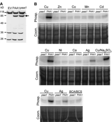
To get the amount of proteins required for biochemical assays, PAA1 and its inactive form PAA1-AKT (in which the aspartate residue of the phosphorylation sequence DKTGT has been mutated to alanine) were produced in L. lactis. In the transformed L. lactis, PAA1 was found to stand for 2–3% of the total membrane proteins (26). As shown in Fig. 2A, PAA1 and PAA1-AKT exhibit the same expression pattern when revealed by detection of the Strep-tag II inserted at their C-terminal end. In addition to PAA1 forms (indicated by an arrow), three other proteins with molecular masses around 26, 36, and 118 kDa and
at INRA Institut National de la Recherche Agronomique on June 13, 2018
http://www.jbc.org/
encoded by the bacterial genome were found to cross-react with the Strep-Tactin HRP conjugate. As these proteins were also detected in the membrane proteins extracted from the strain containing the empty vector (Fig. 2A), these signal were used as internal loading standards.
PAA1 Is Phosphorylated from ATP in the Presence of Monova-lent Metals—In a first set of experiments, we addressed the question of PAA1 activity and selectivity by performing phos-phorylation assays from [␥-32P]ATP in the presence of 5
Mof various metals. As described in the legend for Fig. 1, these experimental conditions should promote forward cycling of PAA1 provided that the supplied metal is transported by the protein. As shown in Fig. 2B, CuCl2and AgNO3are the only metallic salts promoting PAA1 phosphorylation from ATP. In the presence of the other tested metals (supplied as chloride salts), the only visible signal consists of two faint bands. Also observed with membranes containing PAA1-AKT (paa1), a non-phosphorylatable mutant of PAA1, this signal is likely to correspond to endogenous membrane proteins.
To determine which form of copper, Cu⫹or Cu2⫹, triggered PAA1 phosphorylation, we tested the effects of Na2SO3as a reducing agent generating Cu⫹and of a BCA/BCS mixture as a Cu⫹-specific chelator decreasing the amount of available Cu⫹
in the assay. Fig. 2B shows that when Cu⫹chelators are present in the assay, the phosphorylation signal of PAA1 completely disappears, suggesting that PAA1 is activated by monovalent copper. On the other hand, the addition of Na2SO3does not provide any significant enhancement of PAA1 phosphoryla-tion, suggesting that in our experimental conditions and even in the absence of a reducing agent, copper is predominantly under the Cu⫹form.
Using a BCS-based colorimetric assay that specifically detects Cu⫹, we determined that in our assays, copper was actually reduced by the buffer and the L. lactis membranes, even in the absence of Na2SO3. Copper reduction is rapid (less than 5 min) and stable (up to 60 min) at room temperature. We measured that in the experimental conditions used in Fig. 2B, 5 Mof total CuCl2corresponded to 3.5 (⫾ 0.1)MCu⫹in the absence of Na2SO3. In the presence of up to 1 mMNa2SO3 (more than three times the concentration used in Fig. 2B), the measured Cu⫹concentration does not exceed 4.3 (⫾ 0.1)M. In buffer alone (i.e. without L. lactis membranes), 1 mMNa2SO3 completely reduced 5Mof copper. Thus, the partial recovery of Cu⫹(4.3 instead of 5M) in our experimental conditions is likely to be due to the presence of the biological membranes. This was unexpected because the sample is solubilized in deter-gent before analysis and suggests that membranes might tightly trap the metal (0.7MCu⫹), possibly making it not available to PAA1. Together, these measurements show that Cu⫹ concen-trations with and without Na2SO3are close, thereby explaining the similar phosphorylation intensities of PAA1 observed in these two conditions. In conclusion, phosphorylation assays from ATP suggest that PAA1 is only activated by monovalent metal ions, Ag⫹but preferentially Cu⫹.
An interesting point provided by Fig. 2B is that efficiencies of copper and silver in activating PAA1 are different. Indeed, quantifications show that if PAA1 phosphorylation intensity in the presence of 5MCuCl2is taken as 100%, the phosphoryla-tion intensity in the presence of 5MAgNO3is only 40%. To determine whether this difference reflected differences in the whole reaction mechanism of PAA1 or simply in the PAA1 affinity to metals, we performed phosphorylation from ATP within a wide range of copper (with Na2SO3) or silver concen-trations from 0.05 to 50M. As shown in Fig. 3, A and B, PAA1 phosphorylation is biphasic in the two tested conditions. It dis-plays first an increasing phase with a maximum phosphoryla-tion reached at a metal concentraphosphoryla-tion of 3Mfollowed by a steep decreasing phase. The first phase is likely to represent the normal activation process of a P-type ATPase, and using a Hill fitting, we estimated PAA1 apparent affinity around 0.5Mfor copper and 1.1Mfor silver. An interesting point here is that the silver-dependent phosphorylation has a sharp shape, sug-gesting a positive cooperative mechanism of PAA1 activation by this metal. Above 3M, an increase of metal concentration (copper or silver) resulted in an inhibition of PAA1 phospho-rylation almost complete at 30M. As discussed above, PAA1 phosphorylation from ATP displays an unusual dependence to the transported ion (see “Discussion”). This makes the differ-ence in the phosphoenzyme levels observed in Fig. 2B between copper and silver difficult to interpret.
FIGURE 2. Production of PAA1 and PAA1-AKT (paa1) in L. lactis
mem-branes and metal-dependent phosphorylation from ATP. A, expression of
PAA1 and PAA1-AKT in L. lactis. Total membrane proteins (10g) were sepa-rated by SDS-PAGE (10% acrylamide). tag II was detected using a Strep-Tactin HRP conjugate. The arrow indicates PAA1 and PAA1-AKT (paa1). EV, membrane proteins derived from bacteria containing the empty pNZ8148 vector. B, phosphorylation from ATP. Membrane preparations containing PAA1 or PAA1-AKT (paa1) were phosphorylated in the presence of 5Mof various metal ions (with or without Na2SO3for copper) or Cu⫹-specific chela-tors BCA/BCS. Phosphorylation from ATP was performed as described under “Experimental Procedures.” Samples were submitted to an acidic SDS-PAGE, and phosphorylation was detected using a phosphorimaging device. Phosp., phosphorylation signal; Coom., Coomassie Blue-stained gels. paa1 corre-sponds to the membranes containing the mutated protein PAA1-AKT.
at INRA Institut National de la Recherche Agronomique on June 13, 2018
http://www.jbc.org/
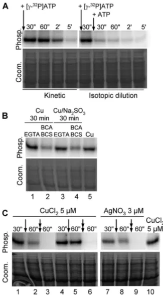
PAA1 Phosphorylation from ATP Is Transient—As shown in Fig. 1, P-type ATPases are transiently phosphorylated when cycling in the forward direction. To demonstrate the transitory character of PAA1 phosphorylation from ATP, and thus to exclude phosphorylation from kinase, two types of experiment were performed. In the “kinetic” experiment, the reaction was carried out in the presence of 5MCuCl2as described previ-ously in the legend for Fig. 2B, except that aliquots were acid-quenched at different times after [␥-32P]ATP addition to stop the reaction. As expected, if the reaction proceeds through the enzymatic cycle shown in Fig. 1, PAA1 phosphorylation inten-sity progressively decreases due to ATP consumption (Fig. 4A,
left). In the “isotopic dilution” experiment, PAA1 phosphoryl-ation intensity decreases almost immediately upon [␥-32P]ATP dilution by 10-fold concentrated cold ATP (Fig. 4A, right). Together, these two experiments demonstrate that PAA1 is active in L. lactis membranes and undergoes a transient phos-phorylation from ATP, emblematic of P-type ATPases.
Confirmation of PAA1 Activation by Monovalent Metal Ions—Previous experiments depicted in Fig. 2B strongly sug-gested that PAA1 was activated by monovalent metal ions, namely Ag⫹and the most physiological one, Cu⫹. To confirm these results for copper, we treated samples before (Fig. 4B) and during (Fig. 4C) the phosphorylation reaction with the divalent chelator EGTA or the monovalent chelators BCA/BCS. In the first experiment, membranes containing PAA1 were
preincu-FIGURE 3. AgNO3and CuCl2dependence of PAA1 phosphorylation from
ATP. A, L. lactis membranes containing PAA1 were phosphorylated as
described under “Experimental Procedures.” Phosp., phosphorylation signal;
Coom., Coomassie Blue-stained gels. B, quantifications were made using the
OptiQuant software (PerkinElmer Life Sciences). 100% corresponds to the amplitude of phosphorylation measured between the BCA/BCS and the 3M metal conditions. Values shown correspond to the mean of three independ-ent experimindepend-ents. Error bars indicate S.E.
FIGURE 4. PAA1 phosphorylation from ATP is transient. A, kinetic and iso-topic dilution experiments on L. lactis membrane expressing PAA1. In the kinetic experiment, phosphorylation from ATP was performed in the pres-ence of 5MCuCl2. The reaction was stopped at different times (30 s to 5 min) after [␥-32P]ATP addition. For isotopic dilution, membranes containing PAA1 were incubated with 5MCuCl2and 0.4M[␥-32P]ATP. After 30 s, an aliquot was taken and acid-quenched. On the remaining sample, ATP concentration was increased to 4Mby the addition of cold ATP, and the reaction was stopped at the indicated times. Phosp., phosphorylation signal; Coom., Coo-massie Blue-stained gels. B, impact of EGTA or BCA/BCS on PAA1 phosphoryl-ation from ATP. L. lactis membranes containing PAA1 were preincubated with 5MCuCl2or with 5MCuCl2⫹ 200MNa2SO3. After 30 min, EGTA or a mix of BCA/BCS was added, and the phosphorylation reaction was started by the addition of [␥-32P]ATP. In lane 5, copper was added just before the addition of [␥-32P]ATP, as described previously in the legend for Fig. 2. C, ADP induced dephosphorylation of PAA1. PAA1 was phosphorylated from ATP in the pres-ence of 5MCuCl2(lanes 1– 6 and 10) or 3MAgNO3(lanes 7–9). Following a 30-s reaction, the sample was acid-quenched (lanes 1, 4, 7, and 10) or incu-bated with chelators (single arrow, lane 5, EGTA; lanes 2 and 8, BCA/BCS) or with chelators plus ADP (double arrow, lane 6, EGTA⫹ ADP; lanes 3 and 9, BCA/BCS⫹ ADP) for 30 s prior to acid-quenching.
at INRA Institut National de la Recherche Agronomique on June 13, 2018
http://www.jbc.org/
bated with CuCl2or (CuCl2⫹ Na2SO3). After 30 min, EGTA or a mixture of BCA/BCS was added just before reaction trigger-ing by ATP. As shown in Fig. 4B, only the BCA/BCS mix was able to significantly reduce PAA1 phosphorylation obtained in the presence of CuCl2(with or without Na2SO3). The presence of EGTA did not affect the phosphorylation levels no matter what copper conditions were used (Fig. 4B, compare lanes 1 and
3with lane 5). In the second experiment, chelators were added on the ongoing reaction. As observed previously in the first experiment, phosphorylation is only impaired by the monova-lent chelators BCA/BCS (Fig. 4C, compare lanes 1 and 2 with
lanes 4and 5). Together, these two experiments demonstrate that PAA1 phosphorylation from ATP strictly depends on the monovalent form of copper.
In the Presence of ATP, PAA1 Mainly Accumulates in the Me.E⬃P Form—As shown in Fig. 1, P-type ATPases can reach
two phosphorylated intermediates; Me.E⬃P still binds the ion to be translocated, whereas E-P occurs after ion release. The accumulation of one of these phosphorylated intermediates
versusthe other depends of the relative velocities of intermedi-ate reactions, a proper feature of the considered P-type ATPase. ADP allows discrimination between these two forms because only Me.E⬃P is able to provide the phosphoryl group back to ADP. Hence, an excess of ADP drives ATP synthesis from Me.E⬃P, and consequently, causes the disappearance of Me.E⬃P.
In the experiment depicted in Fig. 4C, PAA1 was first phos-phorylated from ATP in the presence of copper (lanes 1, 4, and
10). We next compared the effect of ADP on the phosphoryla-tion level remaining after the addiphosphoryla-tion of chelators (see above). The comparison of lanes 2 and 3 with lanes 5 and 6 shows that in any case, ADP totally switches off the phosphorylation signal, suggesting that the PAA1 phosphorylated intermediate from ATP mainly consists of the Me.E⬃P form. The same results were obtained with silver (Fig. 4C, lanes 7–9).
Apparent Affinity of PAA1 to Monovalent Metal Ions Is in the Micromolar Range—P-type ATPase can be phosphorylated from ATP in the forward direction (physiological condition; Fig. 1, steps 1– 4) and from Piin the backward direction (Fig. 1, step 4). Both reactions rely on the presence of the ion to be transported. Although phosphorylation from ATP does require metal ions to occur, metal ions do competitively inhibit phos-phorylation from Pi. Hence, the latter also gives access to the ionic specificity of the enzyme. In addition, involving simple equilibrium reactions (Fig. 1, steps 1 and 4), phosphorylation from Pialso provides a more realistic estimation of the metal-enzyme affinity.
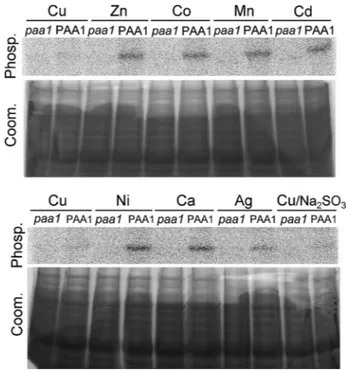
Phosphorylation reactions from Piwere performed on L. lac-tismembranes containing PAA1 or the inactive form PAA1-AKT in experimental conditions previously described for other P-type ATPases, such as the SERCA1a Ca2⫹-ATPase (34) or the CadA Cd2⫹-ATPase (28), i.e. pH 6, no KCl, and 20% Me
2SO. As shown in Fig. 5, 5MCuCl2totally inhibits acylphosphate for-mation in the presence or absence of Na2SO3. A lower inhibi-tory effect is observed with silver, whereas other metals did not prevent PAA1 phosphorylation from Pi(Fig. 5). These results are in good agreement with those obtained from phosphoryla-tion experiments from ATP and confirm the specificity of
PAA1 preferentially to copper, and to a lower extent, to silver. Fig. 6B shows a quantification of copper- and silver-dependent acylphosphate formation from Piat concentrations of metals ranging from 0.05 to 300M. The apparent affinities of PAA1 determined from these data were 0.6Mfor copper and 5M for silver. These values are in the same range as those deter-mined from phosphorylation experiments from ATP (0.5M for copper in the presence of Na2SO3and 1.1Mfor silver, see above) and underline the higher affinity of PAA1 for Cu⫹.
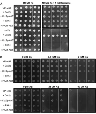
Functional Characterization of PAA1 by Expression in Yeast—To strengthen these biochemical data, we used a yeast complementation assay to validate the ability of PAA1 to transport copper and silver ions in vivo. The yeast CCC2 gene encodes for a Golgi-resident Cu⫹-ATPase required to supply the multicopper oxidase Fet3p with copper. Once loaded with copper, the latter migrates to the plasma mem-brane, where it associates with Ftr1p to create the high affin-ity iron import complex Fet3p-Ftr1p. Thereby, disruption of the CCC2 gene makes yeast unable to survive under limiting iron and copper conditions (35). This complementation assay has been successfully used for the characterization of Cu⫹-ATPases from diverse origins, as illustrated with the human ATP7A (36) and ATP7B (37) or CUA-1 from
Caenorhabditis elegans (38). As shown in Fig. 7A, PAA1 expressed from the constitutive PMA1 promoter is unable to restore the growth of the ⌬ccc2 strain on an iron-limiting medium. PAA1, which does not possess any yeast-targeting sequence, probably stays in the endoplasmic reticulum, thereby explaining its inability to functionally replace Ccc2p,
FIGURE 5. Metal-dependent phosphorylation of PAA1 from inorganic
phosphate. L. lactis membranes containing PAA1 or PAA1-AKT (paa1) were
preincubated for 5 min at 30 °C in 20 mMHEPES (pH 6.0), 10 mMMgCl2, 20% Me2SO, and various metal ions (5M). Phosphorylation was initiated by the addition of 100M32Piand stopped after 10 min (at 30 °C) by the addition of 1 mMKH2PO4, 7% (v/v) TCA. Samples were resolved by SDS-PAGE in an 8% acrylamide acidic gel and visualized using a phosphorimaging device. Phosp., phosphorylation signal; Coom., Coomassie Blue-stained gels. paa1 corre-sponds to the membranes containing the mutated protein PAA1-AKT.
at INRA Institut National de la Recherche Agronomique on June 13, 2018
http://www.jbc.org/
a Golgi-resident protein. As an alternative to strengthen the biochemical data, we then examined metal sensitivity of a wild-type yeast strain expressing PAA1. Our experience with CadA, the Cd2⫹-ATPase from Listeria monocytogenes whose expression in yeast increases sensitivity to cadmium (39), led us to test whether PAA1 could similarly modify yeast toler-ance to copper and silver. As shown in Fig. 7B, PAA1, but not the non-phosphorylating D598A mutant (PAA1-AKT), does actually increase yeast sensitivity to both copper and silver. On the other hand, expression of CCC2 or the non-func-tional D627A CCC2 mutant (CCC2-AKT) does not change yeast sensitivity to copper and silver at the tested concentra-tions. These results obtained with PAA1 can be interpreted along with those obtained with CadA (39, 40). When active in yeast, PAA1 would overload endoplasmic reticulum with copper or silver. The accumulation of these metals would impair endoplasmic reticulum functions and lead to growth defects.
The sensitivity to copper and silver correlated with the pres-ence of a functional PAA1 (no effect of the non-phosphorylat-ing D598A mutant) is comparable with the one previously described for cadmium in yeast strains expressing CadA (39). In good agreement with the biochemical data obtained from phos-phorylation assays, the results obtained using the yeast system show that PAA1 can transport copper and silver in vivo. DISCUSSION
AtHMA6/PAA1 has been previously associated to the chlo-roplast envelope through the in planta expression of a PAA1::GFP fusion and the analysis of its subcellular localiza-tion using confocal microscopy (15). This envelope localizalocaliza-tion was recently confirmed, in our group, using a proteomic approach targeted to the three main compartments of the chlo-roplast, i.e. the envelope, the stroma, and the thylakoids (41; see the AT_CHLORO database). However, the copper-dependent ATPase activity, measured on the purified chloroplast envelope and attributable to PAA1, was very low (23). In purified enve-lope fractions, this activity did not exceed 20 nmol of hydro-lyzed ATP per min and per mg of envelope proteins. Because
FIGURE 6. CuCl2and AgNO3concentration dependence of PAA1
phos-phorylation from Pi. A, membrane fractions (100g) containing PAA1 were incubated with various concentrations of CuCl2or AgNO3. Phosphorylation was initiated by the addition of 100M32Piand stopped after 10 min by the addition of 1 mM KH2PO4, 7% (v/v) TCA. Phosp., phosphorylation signal;
Coom., Coomassie Blue-stained gels. B, phosphorylation intensities were
quantified using the OptiQuant software. 100% represents the phosphoryla-tion level of PAA1 in the absence of metal. Fittings were performed using a Hill equation with a Hill number close to 1. Values correspond to the mean of three independent experiments. Error bars indicate S.E.
FIGURE 7. Phenotypes of yeast strains expressing PAA1. A, CCC2 comple-mentation assay. The yeast strains YPH499 and YPH499-ccc2⌬ expressing the yeast Cu⫹-ATPase Ccc2p, a non-functional Ccc2p mutant (indicated by
Ccc2p-AKT), PAA1 (indicated by PAA1), or a non-functional PAA1 mutant
(indi-cated by PAA1-AKT) were grown on selective media containing either 1 mM ferrozine⫹ 350Miron or 1 mMferrozine⫹ 100Miron (iron-limiting medium). For each expression condition, three independent transformants were grown on the two media. Dilution 1 and 1/10 were spotted as 2-l drops. Dilution 1 corresponds to an optical density of 1.5 at 600 nm. B, metal sensitivity of yeast strains expressing PAA1. The yeast strain YPH499 express-ing the yeast Cu⫹-ATPase Ccc2p, a non-functional Ccc2p mutant (indicated by Ccc2p-AKT), PAA1 (indicated by PAA1), or a non-functional PAA1 mutant (indicated by PAA1-AKT) was grown on selective media supplemented with copper or silver at the indicated concentrations. Drop tests were performed as described above.
at INRA Institut National de la Recherche Agronomique on June 13, 2018
http://www.jbc.org/
this low ATPase activity strongly limited further functional characterization of this transporter in its native environment, we screened alternative expression systems for their ability to produce this membrane protein in amounts compatible with further biochemical studies.
In a recent study, we reported that L. lactis was an efficient host for the production of plant membrane proteins and par-ticularly of P-type ATPases (26). Indeed, AtHMA1, AtHMA3, and PAA1 were found to account for 1–3% of total bacterial membrane proteins, this enrichment being compatible with further biochemical studies. In the present study, we demon-strate that besides producing relatively large amounts of these difficult membrane proteins, L. lactis produces a functional form of PAA1. These data strongly support the choice of L.
lac-tis as a system for the functional characterization of plant ATPases.
PAA1 had been previously associated to the PIB-1 subgroup of heavy metal-transporting P-type ATPases according to the presence of conserved residues in transmembrane segments 6, 7, and 8 (7). This subgroup contains prokaryotic and eukaryotic P-type ATPases. Some of these proteins have been already characterized, such as CopA from Archaeoglobus fulgidus or
E. colior ATP7A and ATP7B from humans (42– 45). However, none of the plant ATPases classified in this PIB-1 subgroup (AtHMA5, AtHMA6/PAA1, AtHMA7/RAN, and AtHMA8/ PAA2) had been biochemically studied so far. This study thus reports the first enzymatic characterization of a plant copper P-type ATPase.
Using phosphorylation assays, we demonstrated that like CopA, ATP7A, and ATP7B, PAA1 is activated in the presence of Cu⫹. Apparent affinity of PAA1 for Cu⫹, estimated using either forward phosphorylation from ATP or reverse phospho-rylation from Pi, is slightly below 1M. This value is close to the ones reported for the previously characterized PIB-1-ATPases: 1 and 2.5Mfor the human ATPases ATP7B and ATP7A and 1.5 and 3.9Mfor CopA from E. coli and from A. fulgidus, respec-tively (Table 1). Estimation of PAA1 apparent affinity was made using Hill equation with a Hill number equal to 1, meaning that Cu⫹ binding to the transporter does not display any cooperativity.
The apparent affinity of PAA1 for Ag⫹is slightly lower than that for Cu⫹, as shown previously for CopA from A. fulgidus (42). In addition, in the presence of ATP, the PAA1 phospho-rylation level reached in the presence of Ag⫹is lower than that reached in the presence of Cu⫹. Ag⫹, which is not a physiolog-ical substrate, is generally used as a mimic of monovalent tran-sition metal, more stable than Cu⫹in solution. The observed
differences between Cu⫹and Ag⫹could be attributed to a dif-ference of ionic radii (0.126 nm for Ag⫹and 0.096 nm for Cu⫹) that could somehow impact on the conformational changes occurring during the enzymatic cycle and thereby affect the phosphorylation rate. Similarly, ZntA from E. coli was found to transport more efficiently Zn2⫹than Cd2⫹, two metals with close chemical properties but with different ionic radii (0.074 nm for Zn2⫹and 0.097 nm for Cd2⫹) (46).
The present results demonstrate that PAA1 behaves as a classic P-type ATPase, forming a transient phosphorylated intermediate in the presence of ATP and the transported ion. The rate of PAA1 dephosphorylation is quite similar to those observed for the yeast Ccc2p and human ATP7B ATPases (44, 47). Indeed, after 2 min, less than 50% of PAA1 is still phospho-rylated, and the enzyme is totally dephosphorylated after 5 min. The transient nature of the phosphorylated intermediate was demonstrated by pulse-chase experiments with cold ATP or ADP. The addition of excess cold ATP results in an almost complete turnover of PAA1 in 60 s, which is comparable with the turnover observed for ATP7A (36). Dephosphorylation assays, in the presence of ADP, indicate a prevalence of the Me.E⬃P conformation of the enzyme when it is phosphoryl-ated in the presence of both copper or silver, suggesting that PAA1 behaves similarly with copper and silver. This is not the case of the CopA from A. fulgidus, which is mainly in the Me.E⬃P form in the presence of copper and in the E-P form in the presence of silver (42).
As discussed previously, acylphosphate formation of PAA1 is copper- and silver-dependent, with a maximum level of phos-phorylation reached at 3M. A further increase in metal con-centration results in the inhibition of phosphorylation. Inhibi-tion of P-type ATPase activity by high concentraInhibi-tions of the transported ion is commonly explained by a reduced dissocia-tion of the ion from the extracytoplasmic binding site (Fig. 1,
step 3), slowing down enzyme turnover rate. Such an explana-tion can be proposed for the inhibitory effect of copper observed on the ATPase activity of the Cu⫹-ATPase CopB from Enterococcus hirae (48) and the Cu2⫹-ATPase CopB from A. fulgidus (49). In this inhibition mechanism, the P-type ATPase, although inactive in terms of ATP hydrolysis, is nev-ertheless fully phosphorylated. The SERCA1a Ca2⫹-ATPase, for example, remains fully phosphorylated even at calcium con-centrations far above the value corresponding to the affinity of the ion to the transporter. This was also reported for the puri-fied Cu⫹-ATPases CopA, from E. coli (43) and A. fulgidus (42), and to a lesser extent for the human Cu⫹-ATPase ATP7B (44). What is observed with PAA1 seems therefore to contradict the
TABLE 1
Apparent affinities for copper and silver determined for PIB-1-ATPases
ND, not determined.
PIB-1ATPases
Apparent affinity for copper Apparent affinity for silver
References Estimated using phosphorylation from ATP Estimated using phosphorylation from Pi Estimated using phosphorylation from ATP Estimated using phosphorylation from Pi CopA A. fulgidus 3.9M ND 23M ND 42 CopA E. coli 1.5M ND ND ND 43 ATP7A human 2.5M ND ND ND 44 ATP7B human 1M ND ND ND 44
PAA1 A. thaliana 0.5M 0.6M 1.1M 5M This work
at INRA Institut National de la Recherche Agronomique on June 13, 2018
http://www.jbc.org/
commonly admitted inhibition scheme and has been reported for another Cu⫹-ATPase, ATP7A, a close paralog of ATP7B that shares with it a similar apparent affinity for copper. Inter-estingly, inhibition of ATP7A phosphorylation has been observed on total membrane preparation above 5Mcopper (36) as well as on the purified protein in detergent above 1M copper (45), somehow excluding an effect of protein environ-ment on the inhibition by copper. For ATP7A, it was suggested that inhibition could be due to “substrate inhibition and/or pro-tein denaturation” (36). The reasons why catalytic phosphoryl-ation of PAA1 is inhibited by high metal concentrphosphoryl-ations remain obscure. However, we can exclude that PAA1 is denatured by cysteine oxidation because PAA1 inhibition is observed in the presence of reduced copper (copper/Na2SO3mixture) and sil-ver. These reducing conditions also exclude an inhibitory effect of the metal by replacement of Mg2⫹in the Mg-ATP complex, the substrate of the reaction, because monovalent ions cannot bind ATP.
To strengthen these biochemical data, we thus performed two independent experiments aiming to validate the specificity of PAA1 in vivo: complementation and metal tolerance assays in the yeast S. cerevisiae. We first found that PAA1 was unable to functionally replace the yeast Cu⫹-ATPase Ccc2p, most probably due to a mistargeting of PAA1 in the endoplasmic reticulum, whereas Ccc2p is a Golgi-resident protein. We then analyzed the metal sensitivity of a wild-type yeast strain expressing PAA1. Similarly to what we previously observed with yeast strains expressing CadA, the Cd2⫹-ATPase from L. monocytogenes(39), PAA1 increases yeast sensitivity to the transported metals. On the one hand, these results show that PAA1 actually transports Cu⫹and Ag⫹in vivoand are thus in good agreement with the in vitro biochemical characterization of PAA1. Furthermore, when considering that Cu⫹is the cop-per form present in yeast, these data also strongly support the biochemical data showing that PAA1 is a Cu⫹-, Ag⫹-ATPase.
The present data show that PAA1 enzymatic properties are similar to those of well known prokaryotic and eukaryotic Cu⫹/ Ag⫹-ATPases such as CopA from E. coli, Ccc2p from S.
cerevi-siae, or ATP7A and ATP7B from humans. Together with the presence of the conserved motifs CPC, NY, and MXXSS in the helices 6, 7, and 8, respectively (7), these properties classify PAA1 as a PIB-1-type ATPase. As extensively illustrated in the literature, PIB-1-ATPases are key actors of copper homeostasis (53, 55). Most of them regulate intracellular copper levels by pumping copper out of the cell (i.e. CopA) or in intracellular compartments where they provide copper to copper-depen-dent enzymes (i.e. ATP7A, ATP7B, Ccc2p). In the cyanobacte-ria Synechococcus PCC7942 and Synechocystis PCC6803, the two PIB-1-type ATPases CtaA have been proposed, from indi-rect measurements, to import copper into the cell (50, 51). This has also been proposed for CopA from E. hirae (52). How sim-ilar proteins can transport copper in two different directions (export in most of the bacteria studied so far versus import in cyanobacteria and E. hirae) is not currently understood. How-ever, the role of PAA1 as chloroplast Cu⫹importer, supported by physiological (14) and biochemical studies (the present work), is consistent with the evolutionary origin of chloroplast (chloroplast originated from cyanobacteria through
endosym-biosis). Abdel-Ghany et al. (15) have suggested that not only PAA1 and CtaA but also PAA2 and PacS, both located in the thylakoids of A. thaliana and cyanobacteria, respectively, might be considered as functional homologs.
The high apparent affinity of PAA1 for copper identified dur-ing this work is in good agreement with previous experiments demonstrating that paa1 mutants are defective in electron transport (plastocyanin) when grow at low copper concentra-tions (14). Recent studies have shown the existence of a tran-scriptional regulatory mechanism that allows cells to down-regulate expression of non-essential copper proteins, and thus, to preserve copper for essential functions under copper defi-ciency (for review, see Ref. 53). In that context, the prioritized delivery of copper to plastocyanin implies the existence of high affinity transporters that could be PAA1 in the chloroplast envelope and PAA2 in the thylakoids. In 2005, Abdel-Ghany et
al. (15) suggested the existence of alternative low-affinity cop-per transporters in the chloroplast envelope because paa1 mutants could be partially rescued by the addition of copper in the plant growth medium. AtHMA1 was proposed to be one of these transporters (23). Thus, PAA1 could be the major copper import system in chloroplasts supplying copper for photosyn-thesis, whereas AtHMA1 could be an alternative pathway, essential under oxidative stress conditions. Another difference between these two transporters could reside in the valence of the transported ion: Cu⫹for PAA1 and Cu2⫹for AtHMA1 (54). Therefore, future characterization of the enzymatic properties of AtHMA1, following the approach described in the present study, should provide key information regarding the relative roles of these two ATPases, PAA1 and AtHMA1, in copper transport across the chloroplast envelope.
Acknowledgment—We thank A. Frelet-Barrand for help in the clon-ing of PAA1-AKT in the pNZ8148 expression vector.
REFERENCES
1. Colangelo, E. P., and Guerinot, M. L. (2006) Curr. Opin. Plant. Biol. 9, 322–330
2. Krämer, U., Talke, I. N., and Hanikenne, M. (2007) FEBS Lett. 581, 2263–2272
3. Palmer, C. M., and Guerinot, M. L. (2009) Nat. Chem. Biol. 5, 333–340 4. Kühlbrandt, W. (2004) Nat. Rev. Mol. Cell Biol. 5, 282–295
5. Palmgren, M. G., and Axelsen, K. B. (1998) Biochim. Biophys. Acta 1365, 37– 45
6. Argüello, J. M., Eren, E., and González-Guerrero, M. (2007) Biometals 20, 233–248
7. Argüello, J. M. (2003) J. Membr. Biol. 195, 93–108
8. Baxter, I., Tchieu, J., Sussman, M. R., Boutry, M., Palmgren, M. G., Grib-skov, M., Harper, J. F., and Axelsen, K. B. (2003) Plant Physiol. 132, 618 – 628
9. Williams, L. E., and Mills, R. F. (2005) Trends Plant Sci. 10, 491–502 10. Andrés-Colás, N., Sancenón, V., Rodríguez-Navarro, S., Mayo, S., Thiele,
D. J., Ecker, J. R., Puig, S., and Peñarrubia, L. (2006) Plant J. 45, 225–236 11. Hirayama, T., Kieber, J. J., Hirayama, N., Kogan, M., Guzman, P.,
Nour-izadeh, S., Alonso, J. M., Dailey, W. P., Dancis, A., and Ecker, J. R. (1999)
Cell 97,383–393
12. Woeste, K. E., and Kieber, J. J. (2000) Plant Cell 12, 443– 455
13. Binder, B. M., Rodríguez, F. I., and Bleecker, A. B. (2010) J. Biol. Chem. 285, 37263–37270
14. Shikanai, T., Müller-Moulé, P., Munekage, Y., Niyogi, K. K., and Pilon, M. (2003) Plant Cell 15, 1333–1346
at INRA Institut National de la Recherche Agronomique on June 13, 2018
http://www.jbc.org/
15. Abdel-Ghany, S. E., Müller-Moulé, P., Niyogi, K. K., Pilon, M., and Shika-nai, T. (2005) Plant Cell 17, 1233–1251
16. Hussain, D., Haydon, M. J., Wang, Y., Wong, E., Sherson, S. M., Young, J., Camakaris, J., Harper, J. F., and Cobbett, C. S. (2004) Plant Cell 16, 1327–1339
17. Verret, F., Gravot, A., Auroy, P., Leonhardt, N., David, P., Nussaume, L., Vavasseur, A., and Richaud, P. (2004) FEBS Lett. 576, 306 –312 18. Eren, E., and Argüello, J. M. (2004) Plant Physiol. 136, 3712–3723 19. Mills, R. F., Krijger, G. C., Baccarini, P. J., Hall, J. L., and Williams, L. E.
(2003) Plant J. 35, 164 –176
20. Mills, R. F., Francini, A., Ferreira da Rocha, P. S., Baccarini, P. J., Aylett, M., Krijger, G. C., and Williams, L. E. (2005) FEBS Lett. 579, 783–791 21. Morel, M., Crouzet, J., Gravot, A., Auroy, P., Leonhardt, N., Vavasseur, A.,
and Richaud, P. (2009) Plant Physiol. 149, 894 –904
22. Gravot, A., Lieutaud, A., Verret, F., Auroy, P., Vavasseur, A., and Richaud, P. (2004) FEBS Lett. 561, 22–28
23. Seigneurin-Berny, D., Gravot, A., Auroy, P., Mazard, C., Kraut, A., Finazzi, G., Grunwald, D., Rappaport, F., Vavasseur, A., Joyard, J., Richaud, P., and Rolland, N. (2006) J. Biol. Chem. 281, 2882–2892
24. Moreno, I., Norambuena, L., Maturana, D., Toro, M., Vergara, C., Orel-lana, A., Zurita-Silva, A., and Ordenes, V. R. (2008) J. Biol. Chem. 283, 9633–9641
25. Kim, Y. Y., Choi, H., Segami, S., Cho, H. T., Martinoia, E., Maeshima, M., and Lee, Y. (2009) Plant J. 58, 737–753
26. Frelet-Barrand, A., Boutigny, S., Moyet, L., Deniaud, A., Seigneurin-Berny, D., Salvi, D., Bernaudat, F., Richaud, P., Pebay-Peyroula, E., Joyard, J., and Rolland, N. (2010) PLoS One 5, e8746
27. Chua, N. H. (1980) Methods Enzymol. 69, 434 – 436
28. Wu, C. C., Gardarin, A., Martel, A., Mintz, E., Guillain, F., and Catty, P. (2006) J. Biol. Chem. 281, 29533–29541
29. Weber, K., and Osborn. M. (1969) J. Biol. Chem. 244, 4406 – 4412 30. Blair, D., and Diehl, H. (1961) Talanta 7, 163–174
31. Sikorski, R. S., and Hieter, P. (1989) Genetics 122, 19 –27 32. Kuo, C. L., and Campbell, J. L. (1983) Mol. Cell Biol. 3, 1730 –1737 33. Morin, I., Gudin, S., Mintz, E., and Cuillel, M. (2009) FEBS J. 276,
4483– 4495
34. Guimarães-Motta, H., and de Meis, L. (1980) Arch. Biochem. Biophys. 203, 395– 403
35. Yuan, D. S., Stearman, R., Dancis, A., Dunn, T., Beeler, T., and Klausner, R. D. (1995) Proc. Natl. Acad. Sci. U.S.A. 92, 2632–2636
36. Voskoboinik, I., Mar, J., Strausak, D., and Camakaris, J. (2001) J. Biol.
Chem. 276,28620 –28627
37. Forbes, J. R., and Cox, D. W. (1998) Am. J. Hum. Genet. 63, 1663–1674 38. Yoshimizu, T., Omote, H., Wakabayashi, T., Sambongi, Y., and Futai, M.
(1998) Biosci. Biotechnol. Biochem. 62, 1258 –1260
39. Wu, C. C., Bal, N., Perard, J., Lowe, J., Boscheron, C., Mintz, E., and Catty, P. (2004) Biochem. Biophys. Res. Commun. 324, 1034 –1040
40. Gardarin, A., Chédin, S., Lagniel, G., Aude, J. C., Godat, E., Catty, P., and Labarre, J. (2010) Mol. Microbiol. 76, 1034 –1048
41. Ferro, M., Brugière, S., Salvi, D., Seigneurin-Berny, D., Court, M., Moyet, L., Ramus, C., Miras, S., Mellal, M., Le Gall, S., Kieffer-Jaquinod, S., Bruley, C., Garin, J., Joyard, J., Masselon, C., and Rolland, N. (2010) Mol. Cell
Proteomics 9,1063–1084
42. Mandal, A. K., Cheung, W. D., and Argüello, J. M. (2002) J. Biol. Chem.
277,7201–7208
43. Fan, B., and Rosen, B. P. (2002) J. Biol. Chem. 277, 46987– 46992 44. Barnes, N., Tsivkovskii, R., Tsivkovskaia, N., and Lutsenko, S. (2005)
J. Biol. Chem. 280,9640 –9645
45. Hung, Y. H., Layton, M. J., Voskoboinik, I., Mercer, J. F., and Camakaris, J. (2007) Biochem. J. 401, 569 –579
46. Rensing, C., Mitra, B., and Rosen, B. P. (1997) Proc. Natl. Acad. Sci. U.S.A.
94,14326 –14331
47. Lowe, J., Vieyra, A., Catty, P., Guillain, F., Mintz, E., and Cuillel, M. (2004)
J. Biol. Chem. 279,25986 –25994
48. Bissig, K. D., Voegelin, T. C., and Solioz, M. (2001) FEBS Lett. 507, 367–370
49. Mana-Capelli, S., Mandal, A. K., and Argüello, J. M. (2003) J. Biol. Chem.
278,40534 – 40541
50. Phung, L. T., Ajlani, G., and Haselkorn, R. (1994) Proc. Natl. Acad. Sci.
U.S.A. 91,9651–9654
51. Tottey, S., Rich, P. R., Rondet, S. A., and Robinson, N. J. (2001) J. Biol.
Chem. 276,19999 –20004
52. Odermatt, A., Suter, H., Krapf, R., and Solioz, M. (1993) J. Biol. Chem. 268, 12775–12779
53. Burkhead, J. L., Reynolds, K. A., Abdel-Ghany, S. E., Cohu, C. M., and Pilon, M. (2009) New Phytol. 182, 799 – 816
54. Pilon, M., Abdel-Ghany, S. E., Cohu, C. M., Gogolin, K. A., and Ye, H. (2006) Curr. Opin. Plant Biol. 9, 256 –263
55. Lutsenko, S., Gupta, A., Burkhead, J. L., and Zuzel, V. (2008) Arch.
Biochem. Biophys. 476,22–32
at INRA Institut National de la Recherche Agronomique on June 13, 2018
http://www.jbc.org/
Daphné Seigneurin-Berny
Patrice Catty, Sylvain Boutigny, Roger Miras, Jacques Joyard, Norbert Rolland and
Cu(I)-ATPase
doi: 10.1074/jbc.M111.241034 originally published online August 30, 2011 2011, 286:36188-36197.
J. Biol. Chem.
10.1074/jbc.M111.241034 Access the most updated version of this article at doi:
Alerts:
When a correction for this article is posted •
When this article is cited •
to choose from all of JBC's e-mail alerts Click here
http://www.jbc.org/content/286/42/36188.full.html#ref-list-1
This article cites 55 references, 26 of which can be accessed free at
at INRA Institut National de la Recherche Agronomique on June 13, 2018
http://www.jbc.org/