HAL Id: inserm-02297922
https://www.hal.inserm.fr/inserm-02297922
Submitted on 26 Sep 2019
HAL is a multi-disciplinary open access
archive for the deposit and dissemination of
sci-entific research documents, whether they are
pub-lished or not. The documents may come from
teaching and research institutions in France or
abroad, or from public or private research centers.
L’archive ouverte pluridisciplinaire HAL, est
destinée au dépôt et à la diffusion de documents
scientifiques de niveau recherche, publiés ou non,
émanant des établissements d’enseignement et de
recherche français ou étrangers, des laboratoires
publics ou privés.
Chronological occurrence of PI3KCA mutations in
breast cancer liver metastases after repeat partial liver
resection
Aldrick Ruiz, Mylène Sebagh, Raphaël Saffroy, Marc-Antoine Allard, Nelly
Bosselut, Giulia Hardoin, Julie Vasseur, Jocelyne Hamelin, René Adam,
Jean-François Morère, et al.
To cite this version:
Aldrick Ruiz, Mylène Sebagh, Raphaël Saffroy, Marc-Antoine Allard, Nelly Bosselut, et al..
Chrono-logical occurrence of PI3KCA mutations in breast cancer liver metastases after repeat partial liver
resection. BMC Cancer, BioMed Central, 2019, 19 (1), pp.169. �10.1186/s12885-019-5365-2�.
�inserm-02297922�
R E S E A R C H A R T I C L E
Open Access
Chronological occurrence of PI3KCA
mutations in breast cancer liver metastases
after repeat partial liver resection
Aldrick Ruiz
1,2, Mylène Sebagh
3,4, Raphaël Saffroy
4,5, Marc-Antoine Allard
2,4,5, Nelly Bosselut
4,5, Giulia Hardoin
4,5,
Julie Vasseur
4,5, Jocelyne Hamelin
4,5, René Adam
2,6, Jean-François Morère
4,7and Antoinette Lemoine
4,5,8*Abstract
Background: Liver metastases of breast cancer are frequent and can recur even after“complete/R0” resection in combination with systemic and hormonal treatments. The aim of this study was to analyze throughout repeat hepatectomies for liver metastases the evolution of PI3KCA gene mutational status.
Methods: All liver metastases nodules (n = 70) from 19 women who underwent at least 2 liver resections were reexamined. DNA extraction from archived tumoral tissue was performed and the major‘hot spot’ mutations in the helical and catalytic domains of PI3KCA have been analyzed using Massarray platform (Agena Bioscience) based on allelic discrimination PCR amplification followed by sensitive mass spectrometry detection.
Results: The two major somatic hot spotPI3KCA mutations were found in 27 (38.6%) nodules corresponding to 8 of the 19 patients (42%). The frequency of women whose breast cancer liver metastases (BCLM) carries PI3KCA mutations increased from the first to the third hepatectomy. Tumors carrying PI3KCA mutations are significantly larger and more frequently observed when resections were R0 compared to patients with no PI3KCA mutation. Conclusion: PI3KCA mutations are frequently observed in BCLM and persist along with the recurrence. Their identification in circulating tumor cells should become a useful biomarker in the routine practice of breast cancer management to prevent tumor recurrence and overcome the problems of intra- and inter-tumoral heterogeneity of the current biomarkers,
Keywords: PI3KCA, Breast, Cancer, Liver, Metastases, Lineage, Evolution Introduction
A significant proportion of breast cancer patients will eventually develop metastases (stage IV) with poor prog-nostic outcome [1–3]. Distant metastatic sites were identi-fied as the brain, liver, lungs, and bones. Once metastatic disease is diagnosed, treatment is generally palliative and usually consists of anthracycline- or taxane-based regimes with or without hormonal or targeted therapeutic agents. This widely implemented palliative approach has yielded median survival ranging between 3 to 16 months [2–7]. For liver metastases, aggressive approaches adopted by ex-perienced hepatobiliary centers, where systemic and
hormonal treatments for metastatic breast cancer are combined with surgical removal, has shown promising results in improving patient survival [8–12]. However, of the patients who undergo removal of breast cancer liver metastases, a portion still develops tumor recur-rence in the liver.
Genomic profiling of tumor tissues has been increas-ingly used to understand and investigate the evolution of metastatic disease and alternative targets for breast can-cer therapies [13, 14]. There are several studies describ-ing genotype profildescrib-ing of breast tumors. Frequently altered genes are ErbB2, PI3K (phosphatidylinositol 3 kinase) pathways, TP53, BRCA1/2, and PTEN [15]. However, following common clinical practice there has few studies examining tumors beyond the usual point of first metastatic presentation or in recurrences after
* Correspondence:antoinette.lemoine@aphp.fr
4Inserm UMR-S 1193, Université Paris-Sud Orsay, France
5AP-HP Hôpital Paul Brousse, Department Oncogénétique, Villejuif, France
Full list of author information is available at the end of the article
© The Author(s). 2019 Open Access This article is distributed under the terms of the Creative Commons Attribution 4.0 International License (http://creativecommons.org/licenses/by/4.0/), which permits unrestricted use, distribution, and reproduction in any medium, provided you give appropriate credit to the original author(s) and the source, provide a link to the Creative Commons license, and indicate if changes were made. The Creative Commons Public Domain Dedication waiver (http://creativecommons.org/publicdomain/zero/1.0/) applies to the data made available in this article, unless otherwise stated.
removal [16–19]. PIK3CA mutations have been reported to be present in over one-third of cases, with enrichment in the luminal and in human HER2-positive subtypes [19]. In human tumors or mammary epithelial cell lines, the two most common mutant alleles (H1047R and E545K) were found to activate PI3K signaling and to be involved in tumorigenesis and resistance to chemother-apy [20–23]. Therefore, the aim of this study was to analyze the chronology of major hotspot mutations in PI3KCA occurrence in a series of patients who under-went at least 2 liver resections for breast cancer liver metastases.
Material and methods
As part of treatment patients with recurrent breast can-cer liver metastases can undergo repeat liver resection. This study is a retrospective case series experiment.
Case selection
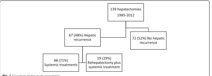
Study size was based on convenient sample of the ex-perience of a hepatobiliary center. All consecutive pa-tients with breast cancer liver metastases (BCLM) who underwent at least two separate liver resections at our center between January 1985 and December 2012 were included in the study (Fig. 1). Patients were selected from our prospectively maintained institutional database, and each medical record was reviewed to update basic clinical and pathological data.
Tissue samples
Representative tumor samples of all nodules within each patient were collected and reviewed to examine genetic ab-normalities. Hematoxylin-eosin-stained slides from tumors were assessed for the ratio percentage of tumor cells/sam-ple area (non-tumor tissue, stroma of the tumor) by pathol-ogists at our department. Three sections of 10μm thickness were obtained from the paraffin-embedded tissue
containing at least 50% of tumor cells. Vascular or lymph-atic invasion features were also analyzed. Immunohisto-chemistry analysis for hormonal receptors was conducted to analyze expression of hormonal receptors and HER2.
DNA extraction
DNA extraction was performed using the QIAmp DNA Mini kit (Qiagen, France), which provides silica-membrane-based nucleic acid purification [20].
Mutations detection
PIK3CA and HER2 mutations was analysed using a Massarray iPlex technology panel and Massarray online design tools (Agena Bioscience). The panel includes main exons (9 and 20) PIK3CA mutations (all mutations from 542 to 545–546-1047 codon), exon 20 HER2 inser-tions (codons 774–776) and exon 2 to 4 KRAS. The Massarray iPlex procedure involves a three-step process consisting of the initial PCR reaction, inactivation of un-incorporated nucleotides by shrimp alkaline phosphatase and a single-base primer extension. Then, the products are nano-dispensed onto a matrix-loaded silicon chip (SpectroChipII, Ageno Bioscience) and finally, the muta-tions are detected by MALDI–TOF (matrix-assisted laser desorption-ionization–time of flight) mass spec-trometry. Data analysis was performed using MassArray-Typer Analyzer software 4.0.4.20 (Agena Bioscience, Hamburg, Germany), which facilitates visualization of data patterns as well as the raw spectra. To allow detec-tion of rare PIK3CA, HER2 or KRAS mutadetec-tions, analysis of the whole exons was completed by High Resolution Melting analysis (HRM). PCR was performed in a 96-well plate with a 20μL volume including 50 ng DNA, 2 mmol/L of each primer, 2.5 mmol/L of MgCl2, 4.7μL of water, and 10μL of LightCycler 480 HRM Master mix (Roche Diagnostics, France). The reaction mix was sub-jected to initial denaturation at 95 °C for 10 min,
Fig. 1 Flowchart of the study population
followed by 45 cycles of amplification consisting of de-naturation at 95 °C for 30 s, annealing at 58 °C for 30 s, and extension at 72 °C for 30 s. Melting was performed with a denaturation step at 95 °C for 1 min, followed by annealing at 40 °C for 1 min and a melt from 70 °C to 97 °C at a ramp rate of 0.03 °C/second with 18 acquisitions per second. LightCycler 480 Resolight Dye (Roche), a fluorescent dye that uniformly binds to the minor groove of double-stranded DNA in a nonsequence-dependent manner for melting analysis was used. In all experi-ments, positive and negative controls were included (Horizon diagnostics).
Clinical data
Hepatectomies were conducted in a tertiary center where data were available, extra information regarding the primary tumor was collected. Chronology of primary tumor, first liver metastasis, first hepatic resection, hep-atic recurrences and repeat hepatectomies for each case were used for interval calculation.
Statistical methods
Categorical variables were compared between groups by the chi-square test or Fisher’s exact test when appropri-ate and continuous variables were compared using the independent-sample t-test. All statistical analyses were performed with SPSS version 21.0 (SPSS Inc., Chicago, IL, USA).
Results
Patient samples
Between 1985 and 2012, 139 female patients underwent partial liver resection for breast cancer liver metastases at our institution (Fig. 1). Sixty-seven women (48%) de-veloped hepatic recurrence of which 19 patients (28%) had subsequent second liver resections with curative in-tent. Four of them had a third hepatectomy. A total of 86 breast cancer liver metastases nodules were removed. Sixteen nodules were not available for molecular analysis due to insufficient tumoral cell content or inability to amplify DNA. High Resolution Melting and allelic dis-crimination PCR amplification using Massarray analyses were performed on samples originating from 70 nodules.
Mutations discovered
Overall, the two major somatic hot spot mutations in the helical and catalytic domains of PI3KCA in the breast cancer liver metastases were found in 27 (38.6%) nodules corresponding to 8 of the 19 patients (42%) (Fig.2). No mutation in HER2 (exon 20) or KRAS (exon 2 to 4) were detected both by the High Resolution Melt-ing analysis and allelic discrimination.
Population characteristics
Table1summarizes clinical characteristics of the patient population stratified by their corresponding hepatec-tomy. Mean maximum size of tumors removed during first hepatectomy was significantly larger in patients with
Table 1 Clinical characteristic according to PI3KCA mutation status
NN First hepatectomy Second hepatectomy Third hepatectomy No Mutation Mutation No Mutation Mutation No Mutation Mutation N = 8 N = 5 N = 11 N = 6 N = 2 N = 2 Hepatic metastases
Mean age at resection + SD Years 42 + 7 44 + 7 51 + 11 50 + 12 63 + 18 42 + 4 Mean interval between primary removal and liver resection
+ SD Months
42 + 20 47 + 37 43 + 21 46 + 33 28 + 9 22 + 9 Mean number of BCLM diagnosed + SD Distribution 2 + 2 2 + 2 2 + 2 2 + 1
Unilateral 5 50% 4 80% 9 82% 4 67% 1 50% 1 100% Bilateral 5 50% 1 20% 2 18% 2 33% 1 50% 0 0% Concomitant extra-hepatic disease
No 10 91% 4 80% 8 73% 4 67% 1 50% 1 100% Yes 1 9% 1 20% 3 27% 2 33% 1 50% 0 0% Chemotherapy before liver resection
No 2 18% 1 20% 0 0% 1 17% 0 0% 0 0% Yes 9 82% 4 80% 11 100% 5 83% 2 100% 2 100% Hormonal Therapy before liver resection
No 8 73% 2 40% 2 18% 2 33% 1 50% 0 0% Yes 3 27% 3 60% 9 82% 4 67% 1 50% 2 100% Targeted Therapy before liver resection **
No 7 64% 4 80% 2 18% 5 83% 1 50% 2 100% Yes 4 36% 1 20% 9 82% 1 17% 1 50% 0 0% Histopathological characteristics
Mean maximum resected tumor size + SD, mm 17 + 15 33 + 15 * 21 + 16 26 + 12 19 + 8 23 + 11 Resection margin***
R0 8 89% 3 60% 8 80% 4 67% 0 0% 2 100% R1 1 11% 2 40% 2 20% 2 33% 1 100% 0 0% Microscopic intrahepatic invasion
No 6 60% 1 20% 7 64% 2 33% – 0 0% Yes 4 40% 4 80% 4 36% 4 67% – 2 Cell surface receptors status
Estrogen Negative 0 0% 2 40% 4 36% 2 33% 0 0% 1 50% Positive 8 100% 3 60% 7 64% 4 67% 2 100% 1 50% Progesterone Negative 5 63% 4 80% 6 55% 2 33% 1 50% 1 50% Positive 3 38% 1 20% 5 45% 4 67% 1 50% 1 50% Her2/Neu Negative 6 75% 4 80% 6 60% 4 67% 0 0% 2 100% Positive 2 25% 1 20% 4 40% 2 33% 1 100% 0 0% Differentiation Well differentiated 0 0% 1 20% 1 14% 2 40% 1 50% 0 0% Moderately differentiated 5 71% 4 80% 3 43% 2 40% 0 0% 1 50% Poorly differentiated 2 29% 0 0% 3 43% 1 20% 1 50% 1 50%
*P < 0.1 ** P < 0.05 *** R0: complete surgical resection with a negative surgical margin at histopathology; R1: invaded surgical margins
PI3KCA mutation compared to patients with no PI3KCA mutation 33 mm vs. 17 mm respectively p = 0.07. At the Second hepatectomy the proportion of pa-tients treated with anti-HER2 targeted therapy is signifi-cantly higher in patients without PI3KCA compared to patients with PI3KCA mutation 82% vs. 17% respectively (p = 0.035). Timeline, anatomical location and extend of metastatic disease are highlighted for the presence of mutation and is presented in Table2.
Chronological occurence of PI3KCA mutation
Four out of 5 patients with “Breast Liver Cancer Metas-tases” with PI3KCA mutations at the first hepatectomy, had the same mutation at the second hepatectomy. Three (16%) women had de novo PI3KCA in their tu-mors at the second or third hepatectomy. Four patients received a third hepatectomy of which 2 had PI3KCA mutations (one possible de novo and one recurrence). At the first, second and third hepatectomy, 38, 47 and 50% respectively of the patients for whom tumors were available hadPI3KCA mutations (Fig.2).
Of the 8 multinodular hepatectomies with PI3KCA mutation, the same mutation was found in all the nod-ules for 5 of them (62.5%). For the 3 remaining
hepatectomies, thePI3KCA mutation was observed in 2 out of 3 nodules.
Discussion
In our case series of patients who underwent repeat hep-atectomies for breast cancer liver metastases, PIK3CA hotspot mutations were frequently observed in most of the nodules for multinodular tumors and persist along with the liver metastases recurrence. Their frequencies also increased along with hepatectomies. When examin-ing the specific mutation H1047R or E545K, mutation remains the same within each patient over time.
The global frequency of PI3KCA mutations found in liver metastases in this series of patients is similar to those reported in sequencing studies in multiple cancers. PIK3CA mutations are present in approximately 30% of all breast cancers, with a higher frequency reported in oestrogene or progesterone positive tumors as well as in HER2–positive breast cancers [13–19]. In our series, al-most all tumors express either HER2 or a hormone re-ceptor, explaining the high frequency of PI3KCA gene mutations.To our knowledge, there are no data reporting neither frequencies of PI3KCA mutations in breast can-cer liver metastases nor a specific gene signature includ-ing PI3KCA mutations. In the molecular classification of
Table 2 Clinical characteristics and presence of PI3KCA mutations
ID Year Primary tumor
First hepatectomy Second hepatectomy Third hepatectomy Survival since primary (months) Margins Interval
(months)a Intrahepatic
Location
xhep Margins Interval
(months)a Intra hepaticLocation xhep Margins Interval(months)a Intra hepaticLocation xhep
1 1989 R0 45 BI No – 53 BI No – 21 UL No 140 2 1998 R1 65 UR Yes R0 40 UR No 142 3 2002 – 22 UR No R0 57 UR No 95 4 1984 – 65 No R0 46 BI No 195 5 1988 R0 27 UL No R1 26 UL No 127 6 1998 R0 49 UR No R0 38 BI Yes 112 7 1992 R0 46 UR Yes R0 28 UL No 108 8 2005 R1 34 UR No R0 34 UR Yes 93 9 1988 R0 30 UR No R1 34 UL No R1 34 BI Yes 205 10 1993 R0 56 UL No R1 113 UL Yes 182 11 2000 R0 34 UL No R1 36 UL No 107 12 1998 R0 27 BI No R0 49 UL No 120 13 1999 – 17 BI No R0 29 BI Yes 72 14 1986 R0 82 UL No R0 40 UL No R0 28 UR Yes 244 15 1999 – 62 No R0 92 UL Yes 157 16 1997 R0 60 UR No R0 44 UR No 145 17 1992 R0 42 BI No R0 8 UR No 75 18 2003 R1 10 BI No R0 32 BI Yes 63 19 1990 R0 36 BI No R0 32 UR No R0 15 UR No 93 a
Hennecke et al., liver metastasis was most frequent in HER-2-enriched variety and PI3KCA mutations have also been enriched in HER-2 breast cancers [4]. We can observe a tendency for more PI3KCA mutated nodules between the first and second hepatectomies for tumors expressing these receptors.
The increase in the mutation rate along with hepatec-tomies can be considered as a marker of clonal evolution in favor of the selection of a dominant clone. In our study, mutations of PI3KCA show such an increase after each hepatectomy and especially a generalization within nodules for multinodular tumors. This increase may be due to malfunctions of the mechanisms for maintaining the integrity of the genome or its exposure to mutagenic agents and thus sign the resistance to treatments. We observe the expansion of mutations within nodules and at each hepatectomy highlighting a potential role of PI3KCA mutations as a marker of treatment resistance and expansion of clones bearing the PI3KCA anomaly. However, since the increase in the frequency of muta-tions is inversely correlated with the HER2 status of pri-mary tumors that guided targeted therapy, PI3KCA mutations do not appear to be involved in the resistance mechanism of anti-HER2 therapies.
Once the oncogenic mutation is acquired, it can be-come a driver excluding generally other oncogenic mu-tations. It can then impact the tumor microenvironment and grow by promoting tumor angiogenesis. This is what we observe in our study. Indeed, tumors with a PI3KCA mutation are larger and trend to be associated with more tumor invasion of microvessels. This probably makes the tumors more aggressive. Similarly, the in-crease rate of PI3KCA mutations in the women who underwent several hepactectomies for tumor relapse even when liver resections were complete is in favor of the emergence of the clones carrying resistance mutations.
The persistence of PI3KCA mutations over time and despite one or two R0 hepatectomies, as well as the delay between 2 hepatectomies, mainly signify the pres-ence of a reservoir of tumor cells in the form of dormant cells or stem cells [22]. These cells would preferentially carry the PI3K mutations conferring on them this prop-erty. They can also circulate in the bloodstream and be identified as prognostic and/or predictive markers to guide therapeutic decisions.
These findings raise more questions than answers. Com-mon knowledge dictates that cancer progresses in steps [23,24] and treatment strategies have been directed at the last step of “evolution”. The basis of systemic palliative treatment has been based on trial and error and our find-ings raise the question whether a better strategy might be to base our strategies on tumor lineage. In other words, tumor cells might have a common origin but might have
taken different evolutionary paths during time. A propos-ition could be to surgically remove and analyze all tumors to identify inter tumor “lineage” variation which in turn would lead to multi-lineage treatment strategies with the ultimate goal to eradicate all malignant cells.
Dan Frumkin et al., developed a way of reconstructing cell lineage trees from genomic variability caused by somatic mutations. This method was applied to cancer and for the first time a lineage tree of neoplastic and ad-jacent normal cells was reconstructed [25]. Other studies have also come to similar findings when examining che-motherapies effect using radiolabeling and imaging stud-ies. Both methods support the idea of multi-lineage and genetic heterogeneity of metastases disease [24,26].
This relatively small series study has some inherit limi-tation. There is no statistical power to make a definitive statement about clinical significance of our findings however these findings have not been presented before and can become the basis for further studies on the tumor heterogeneity and changes overtime. As a special-ized center, the primary tumor tissue is usually not avail-able for genetic studies which is important to complete the picture and lineage link. Furthermore, women with breast cancer liver metastases, usually present years after primary tumor discovery which makes it challenging to obtain primary tumor tissue and perform these genetic mutation detections.
Conclusion
In conclusion, PIK3CA mutations are frequently ob-served in breast cancer liver metastases and persist over time. The frequently observed mutations in PIK3CA that have been associated with resistance to chemotherapy, anti-HER2 or anti-estrogen therapies could be involved in the recurrence of liver metastases and might be a tar-get in the future[27]. The identification of circulating tumor cells carrying PI3KCA should become a useful biomarker in the routine practice of breast cancer to prevent tumor recurrence but also to overcome the problems of intra- and inter-tumoral heterogeneity, as well as temporal, of the current biomarkers, which are hormonal receptor or HER2 expression measurements whose evolutions are not re-evaluated during treatments or in all metastatic sites.
Abbreviations
BCLM:Breast Cancer Liver Metastases; HER2: Human epidermal growth factor receptor; PIK3CA: Phosphatidylinositol-4,5-Bisphosphate 3-Kinase Catalytic Subunit Alpha
Acknowledgments Not applicable
Funding
The Netherlands Organization for Scientific Research (NOW). Grant number: 017.009.053.
Availability of data and materials
The datasets used and/or analysed during the current study are available from the corresponding author on reasonable request.
Authors’ contributions
AR: Lead author, histology preparation. MS: Histology review and manuscript contributor and review. RS: Genetic analysis and manuscript contributor. MA: Tissue analysis and manuscript contributor. NB: Tissue analysis and manuscript contributor. GH: Genetic analysis and manuscript contributor. JV: Genetic analysis and manuscript contributor. JH: Genetic analysis and manuscript contributor. JM: Oncologist and study design. RA: Hepatic surgeon and study design. AL: Department supervisor, study design, genetic analysis interpretation, manuscript contributor. All authors read and approved the final manuscript.
Ethics approval and consent to participate Ethics review is not required under French Law.
Consent for publication Not applicable.
Competing interests
All authors certify that they have NO affiliations with or involvement in any organization or entity with any financial interest (such as honoraria; educational grants; participation in speakers’ bureaus; membership, employment, consultancies, stock ownership, or other equity interest; and expert testimony or patent-licensing arrangements), or non-financial interest (such as personal or professional relationships, affiliations, knowledge or beliefs) in the subject matter or materials discussed in this manuscript.
Publisher’s Note
Springer Nature remains neutral with regard to jurisdictional claims in published maps and institutional affiliations.
Author details
1Department of Surgery, University Medical Center Utrecht, Utrecht, The
Netherlands.2AP-HP Hôpital Paul Brousse, Centre Hépato-Biliaire, Villejuif, France.3Department de Pathologie, AP-HP Hôpital Paul Brousse, Villejuif,
France.4Inserm UMR-S 1193, Université Paris-Sud Orsay, France.5AP-HP
Hôpital Paul Brousse, Department Oncogénétique, Villejuif, France.6Inserm
UMR-S 985, Université Paris-Sud Orsay, France.7Department. Cancérologie, AP-HP Hôpital Paul Brousse, Villejuif, France.8Departement of Oncogenetics,
APHP, GH Paris-Sud, Hôpital Paul Brousse, Inserm UMR-S 1193, Université Paris-Saclay, 14 Avenue Paul Vaillant Couturier, 94800 Villejuif, France.
Received: 15 April 2018 Accepted: 11 February 2019
References
1. Jung SY, Rosenzweig M, Sereika SM, Linkov F, Brufsky A, Weissfeld JL. Factors associated with mortality after breast cancer metastasis. Cancer Causes Control. 2012 Jan;23:103–12.
2. Abuzallouf S, Motawy M, Thotathil Z. Baseline staging of newly diagnosed breast Cancer– Kuwait Cancer control center experience. Med Princ Pract. 2007;16(1):22–4.
3. Schuetz F, Diel IJ, Pueschel M, von Holst T, Solomayer EF, Lange S, et al. Reduced incidence of distant metastases and lower mortality in 1072 patients with breast cancer with a history of hormone replacement therapy. Am J Obstet Gynecol. 2007;196(4):342 e1–9.
4. Kennecke H, Yerushalmi R, Woods R, Cheang MCU, Voduc D, Speers CH, et al. Metastatic behavior of breast Cancer subtypes. J Clin Oncol. 2010 Jul 10; 28(20):3271–7.
5. Pogoda K, Niwińska A, Murawska M, Pieńkowski T. Analysis of pattern, time and risk factors influencing recurrence in triple-negative breast cancer patients. Med Oncol. 2013;30(1):388–96. Available from:https://doi.org/10. 1007/s12032-012-0388-4.
6. Berman AT, Thukral AD, Hwang WT, Solin LJ, Vapiwala N. Incidence and patterns of distant metastases for patients with early-stage breast cancer after breast conservation treatment. Clin Breast Cancer. 2013 Apr;13:88–94. 7. Follana P, Barrière J, Chamorey E, Largillier R, Dadone B, Mari V, et al.
Prognostic factors in 401 elderly women with metastatic breast Cancer. Oncology. 2014;86(3):143–51.
8. Adam R, Aloia T, Krissat J, Bralet MP, Paule B, Giacchetti S, et al. Is liver resection justified for patients with hepatic metastases from breast cancer? Ann Surg. 2006;244:897–907 discussion 907-8.
9. Adam R, Chiche L, Aloia T, Elias D, Salmon R, Rivoire M, et al. Hepatic resection for noncolorectal nonendocrine liver metastases: analysis of 1,452 patients and development of a prognostic model. Ann Surg. 2006;244:524–35. 10. Ruiz A, Castro-Benitez C, Sebagh M, Giacchetti S, Castro-Santa E, Wicherts
DA, et al. Repeat hepatectomy for breast Cancer liver metastases. Ann Surg Oncol. 2015;22(Suppl 3):S1057–66.
11. Ruiz A, Wicherts DA, Sebagh M, Giacchetti S, Castro-Benitez C, van Hillegersberg R, et al. Predictive profile-nomogram for liver resection for breast Cancer metastases: an aggressive approach with promising results. Ann Surg Oncol. 2017 Feb;24:535–45.
12. Ruiz A, Sebagh M, Wicherts DA, Castro-Benitez C, van Hillegersberg R, Paule B, Castaing D, Vibert E, Cunha AS, Cherqui D, Morère JF, Adam R. Long-term survival and cure model following liver resection for breast cancer metastases. Breast Cancer Res Treat. 2018;170(1):89–100. PMID: 29464535.
https://doi.org/10.1007/s10549-018-4714-1. Epub 2018 Feb 20.
13. Cejalvo JM, Perez-Fidalgo JA, Ribas G, Burgues O, Mongort C, Alonso E, et al. Clinical implications of routine genomic mutation sequencing in PIK3CA/ AKT1 and KRAS/NRAS/BRAF in metastatic breast cancer. Breast Cancer Res Treat. 2016 Nov;160:69–77.
14. Tobin NP, Harrell JC, Lovrot J, Egyhazi Brage S, Frostvik Stolt M, Carlsson L, et al. Molecular subtype and tumor characteristics of breast cancer metastases as assessed by gene expression significantly influence patient post-relapse survival. Ann Oncol. 2015;26:81–8.
15. Lee EY, Muller WJ. Oncogenes and tumor suppressor genes. Cold Spring Harb Perspect Biol. 2010;2:a003236.
16. Di Cosimo S, Baselga J. Phosphoinositide 3-kinase mutations in breast cancer: a“good” activating mutation? Clin Cancer Res. 2009 Aug 15;15: 5017–9.
17. Dunlap J, Le C, Shukla A, Patterson J, Presnell A, Heinrich MC, et al. Phosphatidylinositol-3-kinase and AKT1 mutations occur early in breast carcinoma. Breast Cancer Res Treat. 2010 Apr;120:409–18.
18. Kandula M, Chennaboina KK, Ys AR, Raju S. Phosphatidylinositol 3-kinase (PI3KCA) oncogene mutation analysis and gene expression profiling in primary breast cancer patients. Asian Pac J Cancer Prev. 2013;14:5067–72. 19. Yuan TL, Cantley LC. PI3K pathway alterations in cancer: variations on a
theme. Oncogene. 2008 Sep 18;27:5497–510.
20. Allard MA, Saffroy R, de la Maisonneuve PB, Ricca L, Bosselut N, Hamelin J, Lecorche E, Bejarano MA, Innominato P, Sebagh M, Adam R, Morère JF, Lemoine A. Colorectal liver metastases are more often super wild type. Toward treatment based on metastatic site genotyping? Target Oncol. 2015; 10(3):415–21.https://doi.org/10.1007/s11523-014-0346-5.
21. Singh RR, Bains A, Patel KP, Rahimi H, Barkoh BA, Paladugu A, et al. Detection of high-frequency and novel DNMT3A mutations in acute myeloid leukemia by high-resolution melting curve analysis. J Mol Diagn. 2012 Jul;14:336–45.
22. Boros M, Moncea D, Moldovan C, Podoleanu C, Georgescu R, Stolnicu S. Intratumoral heterogeneity for Ki-67 index in invasive breast carcinoma: a study on 131 consecutive cases. Appl Immunohistochem Mol Morphol. 2017;25(5):338–40.
23. Nowell P. The clonal evolution of tumor cell populations. Science. 1976 Oct 1;194(4260):23–8.
24. Morandi L, Pession A, Marucci GL, Foschini MP, Pruneri G, Viale G, et al. Intraepidermal cells of Paget’s carcinoma of the breast can be genetically different from those of the underlying carcinoma. Hum Pathol. 2003 Dec; 34(12):1321–30.
25. Frumkin D, Wasserstrom A, Itzkovitz S, Stern T, Harmelin A, Eilam R, et al. Cell lineage analysis of a mouse tumor. Cancer Res. 2008 Jul 15;68(14):5924–31. 26. Rijpkema M, Boerman OC, Oyen WJG. Tumor targeting using radiolabeled
antibodies for image-guided drug delivery. Curr Drug Targets. 2015;16(6): 625–33.
27. Sobhani N, Roviello G, Corona SP, Scaltriti M, Ianza A, Bortul M, et al. The prognostic value of PI3K mutational status in breast cancer: a meta-analysis. J Cell Biochem. 2018;119(6):4287–92. Available from:https://doi.org/10.1002/ jcb.26687.