Design of a Thermal Diffusion Sensor for Noninvasive Assessment of
Skin Surface Perfusion and Endothelial Dysfunction
by
Vivian V. Li
S.B., Mechanical Engineering hU_ i.. fC hU l1 MASSACHUS T OF TEOHNOLJUN 2
0
2
LIBRARI
1Via•cllUstLL Int111LULe; II 1 •;IIUgIUy•, LVVUSubmitted to the Harvard-MIT Division of Health Sciences and Technology Aii; in Partial Fulfillment of the Requirements for the Degree of
MASTER OF ENGINEERING IN BIOMEDICAL ENGINEERING at the
MASSACHUSETTS INSTITUTE OF TECHNOLOGY
OGY
008
ES
June 2008
02008 Massachusetts Institute of Technology All rights reserved
i
Signature of Author:
Certified by:
INarvard-MIT Dlision of Health Sciences and Technology May 22, 2008
H. Frederick Bowman, Ph.D. Senior Academic Administrator Harvard-MIT Division of Health Sciences and Technology Thesis Supervisor
Accepted by:
Martha L. Gray, Ph.D. Edward Hood Taplin Professor ofMedical and Electrical Engineering Director, Harvard-MIT Division of Health Sciences and Technology
'
Design of a Thermal Diffusion Sensor for Noninvasive Assessment of Skin Surface Perfusion and Endothelial Dysfunction
by
Vivian V. Li
Submitted to the Harvard-MIT Division of Health Sciences and Technology on May 22, 2008 in Partial Fulfillment of the Requirements for the
Degree of Master of Engineering in Biomedical Engineering
Abstract
The skin microcirculation performs a range of vital functions, such as maintaining nutritional perfusion to the tissues and overall thermoregulation. Not only does impairment to the skin blood supply lead to tissue necrosis and other disease complications, increasing evidence shows that dysfunctional vasoreactivity in the skin microcirculation is associated with multiple disease states, including hypertension, diabetes mellitus, hypercholesterolemia, peripheral vascular disease, and coronary artery disease, and it is one of the earliest indicators of systemic endothelial dysfunction, the precursor to atherosclerotic disease.
Endothelial dysfunction is functionally characterized by abnormal vasomotor response to either a
pharmacological or flow-mediated stimulus and can be demonstrated in the skin by measuring reperfusion following a period of ischemia, a phenomenon known as post-occlusive reactive hyperemia (PORH). In my research, I have reviewed the literature regarding endothelial dysfunction and its association with a wide range of cardiovascular risk factors. I have also described the mechanisms thought to link
endothelial function in the central vascular beds (i.e. coronary) to that of peripheral conduit vessels and the microcirculation. The knowledge thus gathered confirmed that the microcirculation of the skin is an appropriate site for endothelial function assessment. The ultimate goal of my thesis is to design a
noninvasive sensor that is capable of obtaining a quantitative measure of skin perfusion, continuously and in real-time, using the principle of thermal diffusion in perfused tissue.
I performed preliminary noninvasive endothelial function testing with a modified Thermal Diffusion Probe (TDP), which has been previously validated for absolute perfusion measurement in an invasive
setting. Based on an initial analysis, I have shown that thermal surface perfusion measurements are feasible and reflect the natural perfusion and temperature fluctuations intrinsic to skin tissue. I also established guidelines for determining quantitative parameters of reactivity from tests of PORH as well as temporal parameters of perfusion variations over time through a spectral analysis of resting blood flow. After establishing the necessary thermal boundary conditions for obtaining surface perfusion
measurements, I embarked on a process of computer-assisted modeling and rapid prototyping of various design iterations on an insulated sensor housing, with subsequent fabrication of first generation
noninvasive sensors. As a result of these initial sensor designs, specifications for the sensor housing were created to ensure that the appropriate thermal field would be established at the skin measurement site - an important step as it permits the most accurate determination of tissue thermal properties. Finally, I propose a candidate design for an ideal sensor capable of improving the reproducibility of noninvasive perfusion measurements on skin.
The development of a noninvasive measure of endothelial dysfunction in the skin is of great value in the early identification of individuals at risk for atherosclerotic complications. Furthermore, the nature of such a technique would provide quantitative information on the presence of a disorder, the extent of dysfunction, and the effectiveness of treatment interventions.
Thesis Supervisor: H. Frederick Bowman, Ph.D.
Acknowledgements
The research I have completed and the knowledge I have gained would not have been possible without the support and guidance of my advisor, Fred Bowman. He has been a true advocate for my education and future aspirations, always inspiring me to overcome any frustrations or obstacles encountered along the way.
I am also grateful to Dr. Robert Lees, who provided me with the clinical perspective I needed to understand the implications of my research. His patient-centered outlook has guided my own path between the fields of engineering and medicine.
I wish to thank the entire engineering team at Thermal Technologies and Hemedex. Their endless patience as well as their willingness to share their time and resources has contributed a great deal to my understanding of the thermal diffusion probe and to the preparation of my thesis.
A special thanks goes to my parents, Jan and Tien-Sheng Li, who have supported my personal and educational ambitions all along the way. I am also fortunate to have my wonderful sisters, Jing and Melissa, who have always been there to make me laugh and enjoy life outside of MIT. I would also like to thank my friends, who inspire me with their enthusiasm and passion every day. In particular, I would like to thank Chewie, whose delight in "making small things look big" aided in the high-magnification photos of certain sensor components; Francisco, for his immense MATLAB expertise; and Charles, whose continual encouragement and editing suggestions have helped me prepare the thesis presented herein.
The funding that made this work possible was provided in part by the Boston Heart Foundation, the Harvard-MIT Division of Health Sciences and Technology, the National Institutes of Health (Grant 5R43DK070408), and Thermal Technologies of Cambridge, MA.
Table of Contents
Abstract ... 3 Acknowledgem ents ... 5 Table of Contents ... 7 List of Figures ... 11 List of Tables ... 13List of Sym bols ... 14
List of Term s and Abbreviations ... 15
Chapter 1. Introduction ... 16
1.1 M otivation ... 16
1.2 Objectives of Research ... 17
Chapter 2. Skin Microvascular Physiology ... ... 20
2.1 Organization of the M icrocirculation ... ... 20
2.1.1 Arteries & Arterioles ... 21
2.1.2 Capillaries... 22
2.1.3 Venules & Veins ... 23
2.2 Levels of Blood Flow Regulation... 23
2.3 Skin Blood Flow: Roles in Tissue Nutrition and Thermoregulation ... . 25
2.4 Reactive Hyperem ia ... 26
2.5 Skin M icrocirculation in Health and Disease... 27
Chapter 3. Endothelial Dysfunction & Methods of Assessment... 28
3.1 Pathophysiology of Endothelial Dysfunction ... 28
3.2 Systemic Impact of Endothelial Dysfunction and Atherosclerosis... ... 31
3.3 Techniques for the Assessm ent of Endothelial Dysfunction ... .... .. 34
3.3.1 Intracoronary Studies... 34
3.3.2 Strain-Gauge Venous Occlusion Plethysmography (VOP) ... ... 35
3.3.3 Brachial Artery Ultrasound Measurement of Endothelium-Dependent Flow-Mediated Dilatation (F M D )... ... .. ... 3 7 3.4 Current Methods for Noninvasive Evaluation of the Microcirculation ... 39
3.4.1 Laser D oppler Flow m etry... ... ... ... ... 39
3.4.2 Orthogonal Polarization Spectral (OPS) Imaging ... ... 42
3.4.3 Peripheral Arterial Tonometry (PAT) ... ... 43
3.5 Current Methods of Noninvasive Assessment: Thermal Methods... ... 45
3.5.1 Skin Surface Temperature Gradients... ... 45
3.5.2 Thermal Clearance ... .... ... 46
3.5.3 Convective Perfusion Bioprobe... ... ... 48
3.5.4 Digital Thermal M onitoring (DTM )... ... ... ... 49
3.6 Clinical Implications of Noninvasive Endothelial Function Assessment... 52
3.6.1 Epidemiology ... .. ... ... 52
3.6.2 R isk A ssessm ent ... ... ... ... ... ... . ... ... .... ... 53
3.6.2 Prim ary Prevention ... .. ... 53
Chapter 4. Heat Transfer Mechanisms and Tissue Perfusion ... . 55
4.1 H eat Transfer Principles ... . ... 55
4.2 Thermal M odel of Bio-Heat Transfer ... .. .. .... ... ... ... 56
4.3 History of Thermal Tissue Perfusion Measurements... 58
Chapter 5. Noninvasive Application of the TDP Probe... ... 60
5.1 The Invasive Perfusion Probe: Design and Operation... 60
5.1.1 D esign... ... 60
5.1.2 Calibration ... ... ... ... . ... ... 63
5.2 Adapting Invasive Probe for Noninvasive Measurement ... ... ... 64
5.2.1 Sensor Placem ent ... .. ... ... 64
5.2.2 Sensor-Tissue Surface Geom etry ... ... 65
5.2.3 Contact Pressure ... ... ... 66
5.2.4 Exposed Thermistor Beads ... ... ... 67
5.3 Measurement Protocol: Post-Ischemic Reactive Hyperemia (PORH) ... 68
5.3.1 Experimental Considerations ... 68
5.3.2 Protocol ... ... ... ... ... ... 68
5.4 M easurem ent Issues ... ... ... ... 71
5.4.1 M otion Artifacts ... .. ... ... ... ... 71
5.4.3 Undetectable Baseline Perfusion... 72
5.4.4 Non-zero Perfusion During Occlusion ... ... 73
5.5 Physiological Issues ... 75
5.5.1 Baseline Temperature Fluctuations ... ... 75
5.5.2 Variability of km ... 76
5.5.3 M ulti-Phasic Perfusion Hyperem ia ... ... 78
5.6 M ethods of Data Analysis ... 80
5.6.1 Filtering ... 80
5.6.2 Perfusion Analysis... 80
5.6.3 Temperature Analysis... 82
5.6.4 Frequency Analysis ... 84
5.7 Exam ples of Subject Data ... 85
5.7.1 24 year old M ale, Healthy ... ... 86
5.7.2 30 year old M ale, Cigarette Sm oker... 87
Chapter 6. Design of the Noninvasive Sensor ... 88
6.1 Design Issues to Address ... 88
6.2 G eom etry ... 89
6.2.1 Disk ... 89
6.2.2 Hem isphere... 89
6.3 Tissue-Sensor Interface ... 90
6.4 Sensor Fixation and Placem ent ... ... 90
6.5 Initial Designs and Rapid Prototyping ... ... 91
6.5.1 M olded Design ... 91
6.5.2 Extruded Design ... 93
6.6 Proposed Sensor Design ... 97
6.6.1 Design and Manufacture of Sensor Housing... 98
6.7 Calibration ... 99
6.8 Sensor Assem bly ... 100
Chapter 7. Conclusion and Recommendations ... 103
List of Figures
Figure 2.1 Layers of skin with microvascular ultrastructure ... ... 21
Figure 2.2 Vessels of the circulatory system ... 24
Figure 3.1 Regulatory functions of the endothelium ... ... 28
Figure 3.2 Endothelial dysfunction as an index of overall cardiovascular risk ... 31
Figure 3.3 Venous Occlusion Plethysmography (VOP) measures the changes in blood volume within a limb during short periods of venous occlusion ... 36
Figure 3.4 Brachial artery ultrasonography and endothelial function testing... 37
Figure 3.5 Basic operating principles of laser Doppler flowmetry (LDF) ... 40
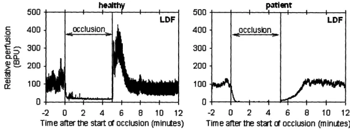
Figure 3.6 Representative LDF measurement during test of Post-Occlusive Reactive Hyperemia (P O R H ) ... 4 1 Figure 3.7 RH-PAT finger plethysmographic device developed by Itamar Medical ... 43
Figure 3.8 Representative RH-PAT signal recorded in subjects with normal and abnormal reactive hyperem ic responses ... ... 44
Figure 3.9 Parameters of Digital Thermal Monitoring (DTM) test of endothelial dysfunction... 50
Figure 3.10 VENDYS® technology ... ... 51
Figure 5.1 The minimally invasive thermal diffusion probe (TDP) produced by Hemedex, Inc.60 Figure 5.2 Temperature profile of the thermistor probe and tissue before and after heating ... 61
Figure 5.3 Noninvasive sensor measurement locations on palmar face of hand ... 65
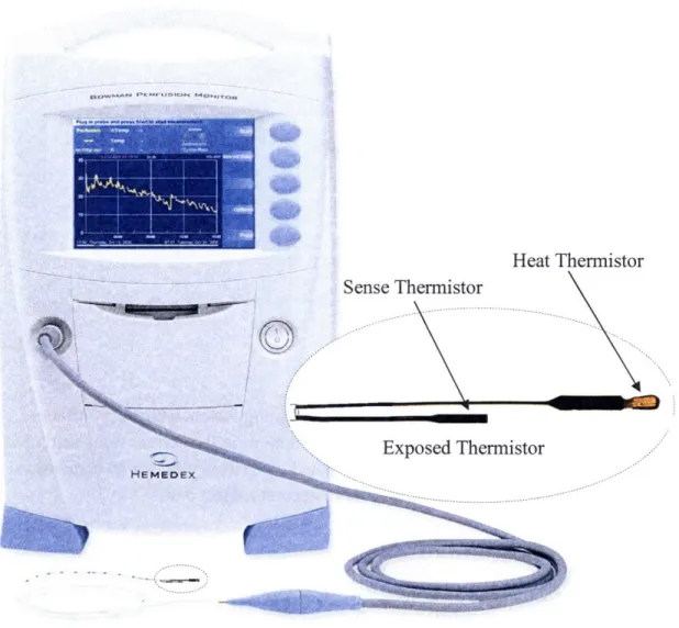
Figure 5.4 Schematic cross-section of exposed thermistors in TDP resting on surface of tissue. 66 Figure 5.5 Bowman Perfusion Monitor and modified thermal diffusion probe (TDP) for noninvasive application ... ... 67
Figure 5.6 Outline of PORH test procedure... 70
Figure 5.7 Perfusion and temperature are measured during an occlusive challenge to the arm.. 70
Figure 5.8 Motion artifact during perfusion measurement ... 71
Figure 5.9 Baseline measurement is undetectable ... ... 72
Figure 5.10 Perfusion during occlusion does not reflect absolute zero flow (no flow) ... 74
Figure 5.11 Temperature fluctuations in resting skin perfusion and temperature ... 76
Figure 5.12 Measured intrinsic thermal conductivity of skin (km) before and after occlusion in test of PO R H ... 78
Figure 5.14 Representative PORH perfusion measurement with suggested parameters of
reactivity ... 82
Figure 5.15 Representative PORH temperature measurement with suggested parameters of reactiv ity ... 83
Figure 5.16 Spectral analysis of perfusion signal measured by thermal sensor ... 85
Figure 5.17 Perfusion and Temperature during test of PORH in 24 year old, healthy male... 86
Figure 5.18 Perfusion and Temperature during test of PORH in 30 year old, male, smoker... 87
Figure 6.1 Two-dimensional noninvasive designs with corresponding one-dimensional models considered by Charles, 2004 ... ... 90
Figure 6.2 Solid model of proposed thermistor bead holder, designed by Savage, 2007... 92
Figure 6.3 Three-dimensional printed acrylic prototype thermistor bead holder ... 92
Figure 6.4 Plastic extruder cut away to reveal operating components ... 94
Figure 6.5 Extruded tubing with channels designed for thermistor beads fixed at specified depths relative to the skin surface ... ... 95
Figure 6.6 Multiple perspectives of solid model and prototype of alternative extruded housing d esig n ... 9 6 Figure 6.7 Dimensioned cross-section of sensor housing for extrusion die manufacture ... 99
Figure 6.8 Three-dimensional model of a small length of sensor housing extrusion ... 99
Figure 6.9 Cross-section of thermistor beads placed in sensor housing ... 100
Figure 6.10 Complete assembly of proposed noninvasive surface perfusion sensor ... 101
Figure 6.11 Schematic cross-section of assembled noninvasive sensor resting on surface of tissue ... ... 102
List of Tables
Table 3.1 Factors associated with endothelial dysfunction in the literature and investigated
therapies that improve endothelial dysfunction ... ... 30 Table 3.2 Endothelial function assessment in standard clinical practice... . 54
List of Symbols
Heat Transfer Symbols
a Thermistor probe radius calibration constant (cm) C1 Zeroth order Steinhart-Hart calibration constant
C2 First order Steinhart-Hart calibration constant C3 Third order Steinhart-Hart calibration constant
Cbl Specific heat of blood (W- s/g- C)
k Thermal conductivity (mW/cm. C) kb Sensor thermal conductivity
km Tissue intrinsic thermal conductivity
P Power (mW)
q Heat flux vector (W/m2)
hbl Volumetric heat exchange between blood and tissue (W/mL) 49 Metabolic volumetric heat exchange (W/ml)
t Time (s)
AT Volume average sensor temperature rise above baseline (oC) Ti Initial baseline tissue temperature (oC)
V Sensor Volume
rmM Tissue intrinsic thermal diffusivity (cm2/s)
Pbl Density of blood (g/mL)
W Perfusion (ml/min- 100g)
Perfusion and Temperature Response Parameters
(Opeak Amplitude of post-occlusion peak perfusion
Maximum increase of perfusion during reactive hyperemia compared to baseline
m(Opeak -mean baseline perfusion)
O% Percent hyperemic response = (xeak-Mean Baseline Perusion X 100
Mean Baseline Perfusion
C0AUC Area under the perfusion curve following release TTP(o Time-to-peak perfusion
Vmean Mean velocity of the post-occlusion hyperemia increase = Wpeak
TTPCo
TNp Nadir-to-peak temperature change (Tpeak - Tnadir)
TR Temperature recovery, defined as Tpeak - Tinitial (initial pre-occlusion temperature) T% Percent temperature rebound = TR X 100
Tinitial -Tnadir TAUC Area under the temperature curve
List of Terms and Abbreviations
ACE Angiotensin Converting Enzyme
Ach Acetylcholine
AVA Arteriovenous Anastomoses
CAD or CHD Coronary Artery Disease or Coronary Heart Disease CVD Cardiovascular Disease
DTM Digital Thermal Monitoring (Endothelix, Inc.) EDHF Endothelium-Derived Hyperpolarizing Factor
EDRF Endothelium-Derived Relaxing Factor (later known as Nitric Oxide, NO)
FBF Forearm Blood Flow
FCD Functional Capillary Density FMD Flow-Mediated Dilatation HDL High-Density Lipoprotein
HF Heart Failure
LDF Laser Doppler Flowmetry
LDI Laser Doppler Imaging
LDL Low-Density Lipoprotein
MRI Magnetic Resonance Imaging
NO Nitric Oxide
NTC Negative Temperature Coefficient
OPS Orthogonal Polarization Spectral Imaging PAD Peripheral Artery Disease
PAT Peripheral Arterial Tonometry PET Positron Emission Tomography PORH Post-Occlusive Reactive Hyperemia
PWA Pulse Wave Amplitude
RBC Red Blood Cell
RH Reactive Hyperemia
RH-PAT Reactive Hyperemia Peripheral Arterial Tonometry (Itamar Medical) RTD Resistance Temperature Detector
SMC Smooth Muscle Cell
TDP Thermal Diffusion Probe (Hemedex, Inc.) VOP Venous Occlusion Plethysmography
Introduction
1.1 Motivation
The microcirculation of tissues and organs is a vital component of the body's cardiovascular network. Blood flow at this level is generally known as perfusion and is comprised of the smallest blood vessels in the body. In addition to regulating the blood flow to individual organs, perfusion is responsible for a number of critical functions including oxygen delivery, carbon dioxide and waste product removal, targeted transport of substances (e.g. blood cells, platelets, hormones), thermoregulation, and blood volume distribution. The efficiency of the circulatory system is demonstrated by the careful balance of blood flow between vital organs (e.g. heart, brain) and peripheral tissues (e.g. skin). This balance is maintained by local as well as global mechanisms which ensure that perfusion matches tissue metabolic and functional needs. An important local regulator of tissue blood flow is the vascular endothelium, which lines the interior walls of blood vessels. Although the endothelium has many functional roles, the term "endothelial function" generally refers to the ability of the endothelium to release compounds that induce relaxation of the smooth muscle cells within the vascular wall. Endothelial
dysfunction is thus defined as an impairment of endothelium-dependent vasodilatation due to an imbalance in the bioavailability of endothelium-derived vasodilators or vasoconstrictors.
Additional elaborations on current scientific understanding of endothelial dysfunction have characterized this state as being prone to atherogenesis through a combination of
proinflammatory, proliferative, and procoagulatory states [1]. Current evidence shows that endothelial dysfunction is an early marker of atherosclerosis and is implicated in an entire
spectrum of cardiovascular diseases.
While most studies have focused on studying endothelial dysfunction in either the coronary or brachial arteries, the microcirculation has also been shown to display functional abnormalities. These abnormalities have typically been regarded as a secondary phenomenon in pathological disease states. However, the development of methods to evaluate the microcirculation have challenged this assumption with evidence that microvascular dysfunction plays a key role in the pathogenesis and pathophysiology of a large range of diseases. A significant association has been shown to exist between microvascular dysfunction and diseases such as hypertension, diabetes mellitus, coronary artery disease (CAD), atherosclerosis, and peripheral arterial disease (PAD). Just as endothelial dysfunction in coronary arteries has been shown to precede the physical presence of atherosclerosis [2], impaired microvascular reactivity may actually precede disorders of the systemic circulation.
Interest in this area has established a vast field of research and development aimed at evaluating microvascular perfusion in vivo. Often, the most reliable, quantitative methods are invasive, involve complicated procedures, do not permit continuous data collection, and are relatively expensive. As a result, there is a strong demand for a routine, noninvasive method for assessing
Chapter 1.
absolute perfusion in appropriate and accessible tissues, particularly in the skin. Not only would such a method hold great potential clinical value in the early identification of patients at risk for cardiovascular complications, but it would also permit noninvasive assessment of wound healing ability, diabetic skin complications, tissue graft perfusion following reconstructive surgery, extent of ischemia and multiple other disorders of the skin perfusion. Other applications include
shock monitoring in critical care settings and the development of a microcirculatory index to assess the impact of drugs, diet, or lifestyle changes. Perfusion in the skin is thus positioned to play a pivotal role in clinical diagnosis, prognosis, and treatment of major systemic disorders as well as in research of the microcirculation.
1.2 Objectives of Research
Early identification of the preclinical signs of atherosclerosis is a necessary target for technical innovation in noninvasive diagnostics as well as patient risk management. The development of a
sensitive screening tool to detect endothelial dysfunction early in a person's life has the potential to identify pre-symptomatic individuals at a high risk of atherosclerotic complications.
Considering the substantial medical resources dedicated to cardiovascular complications (heart disease, diabetes, stroke, etc.), early diagnosis shifts the burden of acute care towards the vital implementation of primary preventive strategies. Ideally, a test of endothelial function should be inexpensive, noninvasive, safe, repeatable, reproducible, and clearly standardized between
different laboratories [3]. Although several methods for evaluating endothelial function exist, no technique meets all the above criteria.
Endothelial dysfunction has been established as a systemic disorder that manifests in the coronary arteries, peripheral conduit arteries, and the peripheral microvasculature. Due to its
superficial location and extensive vascularity, the skin microcirculation is an appropriate
vascular bed for assessing relevant defects in vascular reactivity, functional capillary density, and tissue blood perfusion. Skin perfusion measurement, in particular, is a field of research with a wide range of clinically important applications. A diverse body of literature addresses the
methods of measuring the individual aspects of skin blood flow (capillary morphology, red blood cell velocity, 02 concentration, etc.). However, nearly all have fallen short of developing a noninvasive technique that delivers an absolute measure of the blood flow in the skin. The ideal device for measuring superficial tissue perfusion has been previously defined for reconstructive surgery applications [4, 5]. Generally, such a technique would be able to provide reliable,
sensitive, and continuous data in real-time so that rapid changes in blood flow could be detected. It would be able to track blood flow in a variety of tissues and the monitoring system would be simple to use, safe, inexpensive and portable. The blood flow sensor itself would be noninvasive, not only for patient comfort and safety in routine clinical use, but also to eliminate the effects of tissue trauma on perfusion.
A clinical method of monitoring perfusion has been developed for invasive applications which enables determination of absolute blood flow values based on the physiological heat transfer properties of perfused tissue [6]. This technique has demonstrated significant potential for
obtaining precise, continuous, and real-time assessments of absolute tissue blood flow
invasively, as it must be inserted into (and completely surrounded by) soft tissue. The Thermal Diffusion Probe (TDP) developed by Bowman, et al. has been validated and successfully used for monitoring perfusion following human liver transplantation [7] as well as for monitoring cerebral blood flow in patients with traumatic brain injury [8]. It has also been used to determine the viability of prefabricated skin flaps used for reconstructive surgery [5]. While this device possesses the desired characteristics of continuous measurement of absolute perfusion, the invasive nature of its design limits its applicability in many desired areas. However, the thermal model of the TDP could theoretically be adapted to the geometry of surface perfusion
measurements.
The concepts behind the TDP operation are based on thermal models of the interaction between tissue and blood flow. These two components account for the dominant modes of heat transfer in living tissue such that a tissue's thermal conductivity and thermal diffusivity govern how heat is distributed over time. A mathematical model can simplify the complex thermal interactions between conduction, perfusion, and metabolism in vascularized tissue, despite considerable anisotropy and non-homogeneity. The advantage of such a model lies in the ability to calculate a value for blood flow based on empirically determined thermal properties. This thesis addresses the design of a thermal sensor that can monitor perfusion in skin using heat as an inert diffusible tracer. The noninvasive sensor is based on the invasive TDP, which has proven to be sensitive to
skin blood flow when applied to the tissue surface with an appropriate contact geometry and pressure. Because of its low cost and non-invasiveness, thermal diffusion may be an ideal method for assessing endothelial function by measuring reactive hyperemia responsiveness. Further improvements on such a design would be a significant realization of the need for a safe
and widely applicable method of measuring tissue blood flow.
To begin, I will address the multiple, interconnected elements that must be considered for the development of a truly noninvasive skin surface perfusion sensor, with particular focus on the assessment of endothelial dysfunction. Such an understanding requires a reverence for the physiological structure and regulation of skin blood flow that has made previous attempts in this field so difficult to interpret.
I will devote a great deal of attention to the systemic mechanisms of endothelial dysfunction as it relates to the inherent physiological relationship between various vascular beds in the body. Descriptions of the alternative techniques for assessing endothelial dysfunction are provided to establish a foundation for the methodology as well as a discussion of the challenges that confront peripheral vascular measurements. From the accumulated knowledge of vasoreactivity testing, I hope one can appreciate the breadth of previously documented findings for studies within the peripheral circulation. Additionally, appropriate design parameters which relate to device
operation, cost, complexity, clinical applicability, and functional constraints of user and patient interaction can also be gauged from the alternative technologies available.
I will also discuss the methods that have been developed within the field of noninvasive tissue perfusion measurement, with a focus on thermal modalities. Particular attention is given to thermal diffusion methods and the heat transfer modeling that enables quantification of absolute blood flow values as these form the foundation for my noninvasive thermal sensor design.
I will present the preliminary observations from a noninvasive application of the Thermal Diffusion Probe for functional reactive hyperemia measurements on the digital skin of human subjects. These observations have led to a significant understanding of the challenges inherent to skin perfusion measurement and the design specifications that must be met to ensure optimal sensor reliability. The perfusion measurements obtained from these initial experiments are used to establish suitable parameters to serve as indicators of vascular reactivity and endothelial
function.
Finally, I will present the design and configuration of a potential sensor housing that addresses many of the weaknesses in the preliminary sensor measurements. Key efforts will be made regarding selection of appropriate materials and geometries, analysis of the issues associated with heat transfer between a surface sensor and the human skin, and appropriate specifications to implement this within a clinical setting.
Improvements on the methods for determining peripheral blood flow and vascular reactivity may ultimately contribute to an improved method for assessing cardiovascular risk. Additionally, it is possible to gain a deeper understanding of the control of skin perfusion with respect to
Skin Microvascular Physiology
As the largest organ of the human body, skin takes on a remarkable range of functions and adaptive mechanisms. It serves as a defensive barrier from environmental stresses and is an active participant in the body's immune system. Skin also gathers sensory information from the environment and responds to sensations of touch, pain, heat, and cold. Owing to its position on the surface of the body and its extensive vascularization, the skin is also a site of active
temperature regulation. The structure of skin is uniquely arranged for its multiple roles and is comprised of three distinct layers: the epidermis, dermis, and underlying subcutaneous tissue. The outermost epidermis is avascular stratified epithelium varying in thickness over the body from 75 to 150 jtm, except on the palms and soles where it is between 400 and 600 gm thick. The dermis also varies in thickness depending on location - from 4 mm on the back to 2.5 mm on the thigh to 1.25 mm on the palm [9]. This layer is structurally divided into a superficial region adjacent to the epidermis called the papillary region and a deep thicker area known as the reticular region, and it contains the blood vessels, nerves, and specialized cells and structures such as hair follicles and sweat glands. The subcutaneous tissue is the underlying layer of fat and connective tissue that contains larger blood vessels and nerves. These features contain the
relevant configuration of blood vessels that are often targeted in microvascular assessments of the skin. To understand the skin microcirculation within the larger context of the circulatory system (in health and disease), a description of the major vascular components and regulatory mechanisms is provided.
2.1 Organization of the Microcirculation
Although the skin is the largest and perhaps the most accessible organ for study on the human body, scientific knowledge about the structure and organization of the cutaneous circulation has only been developed in the past three decades. Since then, Irwin Braverman [10] has written
extensively on the organization of the vascular network of human skin. Rather than existing as a randomly anastomosing network lacking stratification, the skin microcirculation is highly
organized, with two horizontal plexuses: the upper, which lies 1-1.5 mm below the epidermis with ascending arterial and descending venous limbs (capillary loops) to supply each papilla, and the lower, which is at the junction between the dermis and the underlying subcutaneous tissue. The lower (reticular) plexus is formed by perforating vessels from the underlying muscles and subcutaneous fat (subcutis) and gives rise to arterioles and venules that directly connect with the upper horizontal (subpapillary) plexus, as well as lateral tributaries that supply the hair bulbs and sweat glands. These two major networks are contained in the papillary dermis 1 to 2 mm below the epidermal surface and make up most of the microvasculature. Figure 2.1 depicts the layers of skin with the corresponding vascular plexuses within each layer.
Epidermis Subpapillary vascular plexus Reticular vascular plexus Dermis Subcutis
Figure 2.1 Layers of skin with microvascular ultrastructure
There are two types of vessels which regulate the terminal blood supply to the skin. The most numerous are arterioles composed of smooth muscle, which supply capillary beds and are innervated with sympathetic constrictor fibers. They provide nutrient flow to the skin. Veins draining these vascular beds comprise large venous networks which provide a large surface area for heat exchange with the environment. This vascular network is described in more detail in Sections 2.1.1 to 2.1.3.
The second type of vessel is found in certain skin regions and is composed almost exclusively of smooth muscle. These vessels, known as arteriovenous anastomoses (AVA), are located in the subcutaneous plexuses and provide a direct connection between arteries and the venous
networks, bypassing the capillary networks. AVA are especially abundant in exposed areas of skin with a high surface area to volume ratio, including the pads and nail beds of toes and fingers, palms, soles of the feet, lips, nose and ears [11] and are important for thermoregulation. They have a relatively large diameter, on average 35 jtm (20-70 gtm) as compared to capillaries
(5-10 gtm) and are richly (and exclusively) supplied with sympathetic nerve fibers [12]. When
they open, large amounts of blood can pass. Activation of sympathetic nerves leads to active vasoconstriction, and decrease in sympathetic activity leads to passive vasodilatation. In a moderately warm environment the AVA are open. In a slightly cold environment, the AVA are almost closed. Since the capillaries provide the nutritional requirements of the skin, with the effective capillary area controlled by pre-capillary sphincters, AVA are capable of controlling capillary blood flow by diverting large volumes of blood through low-resistance pathways. This type of blood flow is termed non-nutritional flow, or shunt flow.
2.1.1 Arteries & Arterioles
The walls of all arteries are made up of an outer layer of connective tissue, the adventitia; a middle layer of smooth muscle, the media; and an inner layer, the intima, made up of the endothelium and underlying connective tissue. The walls of the aorta and other arteries of large
diameter contain a relatively large amount of elastic tissue, primarily located in the inner and external elastic laminas. They are stretched during systole and recoil on the blood during
diastole. The walls of the arterioles contain less elastic tissue but much more smooth muscle. In the skin, arterioles appear with venules in the deep vascular plexus and average 17 to 26 gim in diameter. The arterioles in skin have two layers of smooth muscle cells: the cells of the inner layer are longitudinally oriented while the cells of the outer layer form a spiral [10]. These vessels are capable of augmenting microvascular resistance and blood flow distribution in the skin through dynamic fluctuations in diameter, resulting in blood flow oscillations. The arterioles are the major site of the resistance to blood flow, and small changes in their caliber cause large changes in the total peripheral resistance.
2.1.2 Capillaries
Capillaries are small exchange vessels that are fed by terminal arterioles. The openings of the capillaries are surrounded by precapillary sphincters, which are supplied by sympathetic fibers from the autonomic nervous system. These fibers maintain a certain basal tone in the sphincters. In the skin, capillaries have an inner diameter of 4-6 gm. When the sphincters are dilated, the diameter of the capillaries is just sufficient to permit red blood cells to squeeze through in "single
file." The state of contraction of precapillary sphincters determines the number of perfused capillaries. Compared to capillaries in other parts of the body in which the vascular wall is usually only 0.1 gm thick, capillaries in the skin have relatively thick walls, measuring 2-3 jim
[13].
While capillaries lack smooth muscle cells, their walls contain pericytes outside of the
endothelial cells. Pericytes are thinner than smooth muscle cells, have fewer myofilaments, and lack dense bodies. These cells have long processes that wrap around the vessels. They contain the contractile proteins necessary for cellular contraction and release a wide variety of vasoactive agents. Pericytes also synthesize and release constituents of the basement membrane and
extracellular matrix. One of their physiologic functions appears to be regulation of flow through the junctions between endothelial cells, particularly in the presence of inflammation.
The capillary loop is an important structure in the microcirculation of the skin and supplies the only nutritional perfusion available to the epidermis. In some cases a capillary loop can be up to
100 gm from the epidermal cells which it supplies. It arises from the terminal arteriole in the superficial horizontal plexus and is composed of an ascending limb, a hairpin turn, and a descending limb that returns to the superficial horizontal plexus and connects with the postcapillary venule. Each dermal papilla is supplied by a single capillary loop which is the primary site of nutrient and oxygen exchange. Generally, a single capillary loop supplies 0.04-0.27 mm2 of the skin surface [14]. Compared to most other tissues, capillary density is relatively low in skin, ranging from 10 to 70 capillaries/mm2. At different skin sites, there are no
2.1.3 Venules & Veins
The descending capillary loop feeds into the postcapillary venules, which make up the majority of vessels in the papillary dermis. The internal diameter enlarges from 8 gm (at venous
capillaries) to 26 gtm and the walls of the venules are only slightly thicker than those of the capillaries. Blood then passes through the small venules and into systemic veins of increasing size. Veins have relatively little smooth muscle or elastic tissue and thus are easily distended. In contrast to small arteries and arterioles, which are resistance vessels, venules and veins function as capacitance vessels. Pressures within the venous system are low, ranging from 20 mmHg at the venular ends of capillaries to about 0 mmHg at the entrance to the vena cava. Due to these properties, the venous system normally contains about 2/3 of the total blood volume, and acts as
a blood reservoir for the body.
2.2 Levels of Blood Flow Regulation
The blood vessels are a closed system of vessels that carry blood from the heart to the tissues and back to the heart. Figure 2.2 contains a schematic of the various types of vessels in the
circulatory system and their organization in the circulatory network. The resistance to flow depends mostly on the diameter of the vessels of the microcirculation, principally the arterioles. The blood flow to each tissue is regulated by local chemical and general neural and humoral mechanisms that dilate or constrict the vessels of the tissue. A parallel arrangement permits wide variations in regional blood flow without changing total systemic flow. In humans and other mammals, multiple cardiovascular regulatory mechanisms have evolved. These mechanisms increase the blood supply to active tissues and increase or decrease heat loss from the body by redistributing the blood. In the face of challenges such as hemorrhage, blood flow is maintained at near constant values in the heart and brain. When the challenge is severe, flow to these vital organs is maintained at the expense of the circulation to the rest of the body.
At rest, about 67% of the circulating blood volume is in the systemic veins and venules. Five percent is in the heart cavities, and 12% is in the low-pressure pulmonary circulation. Only 11% is in the aorta, arteries, and arterioles, and 5% in the capillaries [15]. Although only 5% of the circulating blood is in the capillaries, this portion is the most vital component of the blood volume because it is across the systemic capillary walls that 02 and nutrients enter the interstitial fluid and CO2 and waste products enter the bloodstream. The nutrient exchange across the
capillary walls is essential to the survival of the tissues. Thus, the larger vessels of the body can be viewed as distinct conduits to and from the heart and peripheral organs whereas the
microcirculatory vessels are embedded within an organ to enhance communication between the tissue and the vessels.
To te Fromthe
heart heart
er Layer
Capillaries
Figure 2.2 Vessels of the circulatory system. Blood travels from the heart in arteries, which branch into smaller and smaller vessels, eventually becoming arterioles. Arterioles connect with still smaller blood vessels called capillaries. Through the thin walls of the capillaries, oxygen and nutrients pass from blood into tissues, and waste products pass from tissues into blood. From the capillaries, blood passes into venules, and then into veins to return to the heart.
The vascular endothelium modulates smooth muscle tone by releasing several vasoactive
substances. The critical role of the endothelium in regulating vasodilator tone was first described by Furchgott and Zawadzki in 1980 [16]. They demonstrated that the rabbit aorta dilates in response to the application of acetylcholine only in the presence of intact endothelium, confirming the existence of an derived vasodilator. The principal endothelium-derived relaxing factor (EDRF) was later confirmed to be nitric oxide (NO) [17, 18], and numerous subsequent studies have explored the multiple vascular functions of this simple, diatomic molecule, confirming its role as a central signal transducer and essential antiatherogenic molecule. It is released during basal conditions in response to chemical stimuli, such as
acetylcholine (ACh), and in response to mechanical stimuli, such as shear stress.
Endothelial cells also generate several other vasoactive mediators, including the vasodilator molecules prostacyclin and endothelium-derived hyperpolarizing factor (EDHF), and the vasoconstrictor agents endothelin-1, angiotensin II, superoxide anions, and vasoconstrictor prostanoids. The development of an imbalance in the release of vasoconstrictor and vasodilator
agents from the endothelium leads to an impairment of endothelium-dependent vasodilatation, which represents the hallmark of endothelial dysfunction [19]. In parallel with the vasomotor changes, activated dysfunctional endothelial cells also acquire a vascular phenotype that establishes a local environment favoring the initiation and progression of atherosclerosis [20]. This area of blood flow regulation will be addressed more thoroughly with regard to endothelial dysfunction in Section 3.1.
2.3 Skin Blood Flow: Roles in Tissue Nutrition and Thermoregulation
The skin is both a protective barrier and an important thermoregulatory organ. As a result, it has an extensive and well-developed microcirculation. Human skin microcirculation serves three main functions, namely skin tissue nutrition, heat exchange for thermoregulation, and blood flow redistribution during stress. While the oxygen and nutrient requirements of the skin are small and thus little blood flow is necessary to sustain nutritional perfusion, the skin is the primary means of maintaining body temperature. To meet these needs, skin blood flow can vary between less than 1 ml/min per 100 g of tissue to a maximal flow of 150 to 200 ml/min- 100g during times of severe heat stress (up to 60% of total cardiac output) [21]. In a thermoneutral environment, skin blood flow ranges between 10 and 20 ml/min- 100g [22]. This impressive range of possible circulatory states is achieved through a balance of neural and local control mechanisms that precisely modulate the vasomotor tone and vascular resistance of the peripheral microcirculation. In particular, thermoregulatory control is largely mediated through reflex neural control of skin blood flow.
Skin blood flow is controlled by two branches of the sympathetic nervous system: a noradrenergic vasoconstrictor system and an active vasodilator system, which is less well-understood. While both types of nerves innervate all areas of non-acral skin (trunk, leg, forearm, etc.), areas of acral skin (lips, ears, nose, palmar/plantar surface of hands and feet) are innervated only by sympathetic vasoconstrictor nerves [21]. In thermoneutral environments, the
vasoconstrictor system is tonically active such that withdrawal of the impulses to these nerve fibers allows blood vessels to dilate. This allows for a "neutral" or "vasomotor" zone of thermoregulation where the skin responds to subtle changes in the environment by fine-tuning heat loss solely via adjustments in sympathetic tone [23]. In skin areas outside of acral regions, an active sympathetic vasodilation system exists which is inactive in thermoneutral
environments, but is activated during increases in internal temperature. Therefore, as core body temperature begins to rise (such as during exercise or environmental heat exposure), the initial increase in skin blood flow is mediated by release of vasoconstrictor tone. However, upon reaching a specific threshold, sweating and reflex active vasodilatation are initiated in tandem, stimulating the co-release of acetylcholine (ACh) and an associated yet-unknown vasodilator from sympathetic cholinergic nerves [21]. The active vasodilator system is responsible for 80%-90% of the large increases in skin blood flow during heat stress, which can reach levels as high as 6-8 L/min.
As was discussed in Section 2.1, numerous arteriovenous anastomoses (AVA) exist in acral skin. These thick-walled, low-resistance conduits are richly innervated by sympathetic vasoconstrictor nerves and allow high rates of shunt flow between arterioles and venules. Therefore, in acral areas, opening or closing of these AVA can cause substantial changes in the blood volume that passes through the skin. Sympathetic input to AVA is controlled from hypothalamic temperature regulation centers, while cutaneous arterioles are primarily controlled by neural activity, with local metabolic control secondary.
The periodic oscillations recorded in cutaneous blood flow reflect both vasomotion and flow motion and have been quantified by spectral analysis of the laser Doppler perfusion signal [24]. Vasomotion is defined as the rhythmic changes in the diameter of small blood vessels and is produced by contraction and relaxation of the muscular components in their walls. An important component of vasomotion in skin includes the myogenic activity of terminal arterioles as well as endogenous endothelial activity, such that not all skin microvascular units are open and perfused at any moment in time. Flow motion results from the motion of the blood cells and their
interaction with the vessel walls and is dependent on the different vasomotor control
mechanisms. The characteristic oscillation frequencies present in skin flow motion include a cardiac component (-1 Hz in a resting, healthy subject) and respiratory component (-0.3 Hz), which demonstrate the dependence of peripheral blood flow on the pressure difference generated by the heart and lung pumps. There are also contributions from intrinsic myogenic activity of the
vessel wall (0.1 Hz) and neurogenic activity of the local sympathetic nerves (0.03 Hz) [25]. An oscillation with a period of around 1 min (-0.01 Hz) has been suggested as being endothelium-dependent and may reflect either periodic release of NO or the rhythmic response to NO of an oscillator in vascular smooth muscle cells [26].
2.4 Reactive Hyperemia
If arterial inflow to a vascular bed is stopped temporarily, perfusion within that bed decreases to zero. Upon release of the occlusion, the blood flow immediately exceeds the average flow before occlusion and then gradually returns to the baseline level. This increase in flow is called reactive hyperemia. The peak flow and duration of the reactive hyperemia are relatively proportional to the duration of the occlusion within certain limits. If the extremity is exercised during the occlusion period, reactive hyperemia is increased. These suggest a locally mediated mechanism, either metabolic or otherwise, in the regulation of tissue blood flow.
Post-occlusive skin reactive hyperemia has been primarily defined in laser Doppler flowmetry studies of skin perfusion flux and refers to the increase in skin blood flow above baseline levels following the release of a brief arterial occlusion. It is also called post-ischemic hyperemia. It can be characterized by an initial peak in flux (perfusion) that occurs within a few seconds of removal of the occlusion, and a sustained hyperemia. Four major factors have been proposed to be involved in the hyperemic response: metabolic vasodilators, endothelial vasodilators, the myogenic response and sensory nerves. The post-ischemic peak flow reflects the sudden increase
of the blood flow in the skin microvessels after removal of the occlusion and can be considered an indicator of the increased shear stress on the microvascular wall. The more prolonged hyperemic phase reflects the metabolic debt repayment to the tissue [27] and can be considered
as an indicator of successive tissue reperfusion.
Unlike flow-mediated dilatation, reactive hyperemia is a complex response that depends on the local production of non-endothelium-dependent vasodilators which dilate tissue microvessels to accelerate the delivery of oxygen following a period of ischemia [28]. Endothelium-derived
nitric oxide plays a role in reactive hyperemia as well [29, 30]. Despite the similarities in procedure and vascular regions, it is now thought that reactive hyperemia proceeds through different mechanisms compared to conduit artery flow-mediated dilatation (FMD). This topic will be addressed more fully in Section 3.2.
2.5
Skin Microcirculation in Health and Disease
The microcirculation is of interest because it is where fine control of blood supply takes place, and tissue ischemia is dependent on microvascular flow in many circumstances. During circulatory failure (e.g. hypovolemic shock), blood flow is diverted from the less important tissues (skin, subcutaneous fat, muscle, large intestine) to vital organs (heart, brain, kidneys). Thus monitoring perfusion in the less vital tissues such as skin could be an early clinical marker of critical tissue hypoperfusion. Indeed, the assessment of perfusion in peripheral tissues is more easily obtainable using noninvasive monitoring techniques, thus facilitating earlier recognition of abnormalities. The skin can provide a good platform for this, and in diabetes, for example, measurable changes in the skin have been found to pre-date the symptoms of microvascular disease in other organs by many years [31]. In the dermal microcirculation, endothelial function may be assessed noninvasively using several methods, although no one technique is entirely satisfactory. Nevertheless, this vascular bed is highly suitable for assessment of the endothelium, and is particularly convenient.
By posing the dermal microcirculation as a way to interrogate overall vascular health, it is important to recognize that cutaneous blood flow is a dynamically fluctuating biological variable which possesses substantial spatial heterogeneity. Its magnitude and regulation is modulated through many external and internal parameters by virtue of its central role in thermoregulation and tissue perfusion. Microcirculatory flow adjusts as a result of variations in blood gas
concentration, hormones, and physical factors like temperature and pressure. It is also carefully controlled by the autonomous nervous system. Such signals increase or decrease the
microvascular perfusion by stimulating the endothelium to release substances leading to vasoconstriction or vasodilatation of the upstream arterioles. Characterizing these systemic regulators of blood flow also impacts current understanding of perfusion. Several important clinical goals arise from these local and global relationships. Not only is the status of a patient's generalized microvasculature important, but direct associations must be determined to provide information as it relates to underlying pathophysiology.
Chapter 3.
Endothelial Dysfunction & Methods of Assessment
3.1 Pathophysiology of Endothelial Dysfunction
The normal endothelium plays a key role in the local regulation of vascular tone by producing and secreting substances within a region of the vascular bed. To maintain vascular homeostasis, healthy endothelial cells perform a variety of functions [32] (Figure 3.1):
* Promote vasodilation * Antioxidant effects * Anti-inflammatory effects
* Inhibition of leukocyte adhesion and migration
* Inhibition of smooth muscle cell (SMC) proliferation and migration * Inhibition of platelet aggregation and adhesion
* Anticoagulant effects * Profibrinolytic effects
Vasodilation Thrombolysis
Platelet disaggregation Vasoconstriction
Antiproliferation Thrombosis
AntiinflammationAntiinflammation Adhesion molecules
Antioxidant Growth factors
Inflammation
Oxidant activity
Figure 3.1 Regulatory functions of the endothelium. Healthy endothelium protects the vessels from atherosclerosis. Dysfunctional endothelium possesses proatherogenic properties, shifting the balance towards vasoconstriction, thrombosis, inflammation, and ultimately atherogenesis. [33]
Normal endothelial cells promote an antiatherogenic vessel phenotype and are capable of maintaining a delicate balance between local and systemic stimuli. However, when endothelial
cells lose this ability, the resulting imbalance leads to a state of endothelial "activation," which is characterized by abnormal vasomotor function in addition to a proinflammatory, proliferative, and prothrombotic state that favors the initiation and progression of atherosclerosis [1]. Although the precise mechanisms that cause endothelial dysfunction are not fully understood, it is thought to be a multifactorial process involving mechanical or biochemical damage to endothelial cells and the loss of appropriate response to certain stimuli. One possible mechanism relates to the increased production of reactive oxygen species, such as superoxide, which have been shown to reduce endothelial nitric oxide availability via multiple pathways [34]. Furthermore,
wall. In fact, increased vascular superoxide production has been demonstrated in all major conditions predisposing to atherosclerosis [35].
Endothelial dysfunction is most effectively defined by the diminished production or
bioavailability of vasodilators (e..g. nitric oxide, or NO) or an increase in endothelium-derived contracting factors, leading to impaired endothelium-dependent vasodilatation [36]. This can be assessed functionally by a measure of abnormal vasoreactivity (response to pharmacological or physiological stimulation). However, it is the positive feedback loop in which inflammatory
factors promote monocyte and T-cell adhesion, foam cell formation, extracellular matrix digestion, and vascular smooth muscle migration and proliferation that ultimately leads to atherosclerotic plaque formation. This can include multiple mechanisms, such as up-regulation of adhesion molecules, increased chemokine secretion and leukocyte adherence, increased cell permeability, enhanced low-density lipoprotein (LDL) oxidation, platelet activation, cytokine elaboration, and vascular smooth muscle cell proliferation and migration [33]. The host defense response is triggered, allowing the endothelium to be invaded by lipids and leukocytes and inciting an inflammatory response within the vessel wall. Fatty streaks appear and develop into plaques that are susceptible to rupture with subsequent thrombogenesis and vascular occlusion [33].
Given the relationship between endothelial dysfunction and atherosclerosis, it is likely that the status of an individual's endothelial function reflects their susceptibility to develop
atherosclerotic disease and may thus serve as a marker of an unfavorable cardiovascular
prognosis. Both novel and conventional cardiovascular risk factors are associated with abnormal endothelial function and may influence the bioavailability of endothelial NO or lead to a state of endothelial activation. For example, hypertension, chronic heart failure, diabetes,
hypercholesterolemia, smoking, aging, and a family history of premature atherosclerotic disease are all associated with impaired endothelium-mediated vasodilatation. Table 3.1 summarizes many of the established and recent conditions that have been associated with endothelial dysfunction.
Furthermore, many interventions that are known to limit cardiovascular risk have been shown to restore endothelial function. Thus, endothelial dysfunction is a reversible disorder, and strategies aimed at reducing cardiovascular risk factors, such as cholesterol lowering [37], antihypertensive therapy [38], smoking cessation [39], estrogen replacement therapy in postmenopausal women [40], supplementation with folic acid [41], and physical exercise [42] also translate into an improvement in endothelial health, further supporting the association between risk factors and endothelial dysfunction. Moreover, the observation that several pharmacological interventions that improve endothelial function are associated with a decrease in cardiovascular events independent of risk factor modification supports the concept that cardiovascular risk factors share a common pathway that leads to endothelial dysfunction, such as oxidative stress. A Medline search reveals that several hundred articles are published every year on the role of
different therapeutic interventions aimed at influencing endothelial function. It is clear that evaluation of endothelial function in humans is of great clinical relevance.
Table 3.1 Factors associated with endothelial dysfunction in the literature and investigated therapies that improve endothelial dysfunction
Factors related to Factors improving
References References
endothelial dysfunction endothelial function
Angiotensin-converting
Age [43-46] [38, 47, 48]
enzyme inhibitors
Male sex [43] Hormone-replacement [40, 49, 50]
Smoking [3, 39, 51, 52] Smoking cessation [39, 53]
Passive Smoking [54, 55] Exercise [42, 56-58]
Diabetes mellitus: [59-62] L-Arginine
[53, 63] Types 1 and 2
Hyperglycemia [64] Calcium channel blockers [65, 66]
Family History of CAD [67, 68] Statin therapy [69-74]
Hypertension [52, 75-77] LDL apheresis [78]
High cholesterol [79-82] Antioxidants [83-85]
Low HDL [67, 86] Homocysteine lowering [87, 88]
Obesity [89, 90] Weight loss [91]
Heart failure [92-94] Omega-3 Fatty Acids [95]
Hyperhomocysteinemia [96, 97] Aspirin [98]
End-Stage Renal Disease [99, 100] Elevated C-reactive protein [101]
Chronic Systemic
inflammation
[102]
Given the endothelium's unique position as both the mechanical and biological barrier between blood and the vascular wall, endothelial function represents an integrated index of overall cardiovascular risk factors and local factors (e.g. shear stress), genetic predisposition, and yet-unknown factors (protective and harmful) that confer individual susceptibility to developing atherosclerotic disease as well as predisposition to the subsequent acute complications (Figure 3.2). Indeed, some investigators consider endothelial dysfunction to be the "risk of risk factors" [32]. Therefore, the status of endothelial function offers unparalleled potential for improved risk stratification, as well as the possibility of targeted therapies and prognosis.
Genetic Predisposition
Traditional/Non-traditional Risk Factors
Atherogenesis and Complications
(Myocardial Infarction, Stroke, PAD, Heart Failure)
Figure 3.2 Endothelial dysfunction as an index of overall cardiovascular risk
3.2 Systemic Impact of Endothelial Dysfunction and Atherosclerosis
Because atherosclerosis is a generalized macrovascular disease, lesions in one vascular territory predict disease in other arterial regions. This systemic nature was documented by the
Framingham study which showed that patients first presenting with myocardial infarction, cerebral vascular accident, heart failure, or peripheral vascular insufficiency would, in the following 10 years, manifest disease in another vascular territory in proportions varying from
16% to 50%, both for men and women [104]. Similar risk factors are present among patients
with coronary, peripheral, and carotid atherosclerosis. Of particular importance is evidence that disease in noncoronary arteries is a powerful predictor of coronary heart disease (CHD)
mortality. Aortic, peripheral, and carotid artery diseases have been termed "Coronary Heart Disease Equivalents" because the level of CHD risk and CHD event rates associated with these conditions is approximately equivalent to the level of risk seen in stable CHD. That is, the rate of
CHD events in persons with atherosclerotic vascular disease in other territories is similar to event
rates in patients with known CHD. Additionally, macro- and micro-vascular disease are the principle causes of morbidity and mortality in patients with diabetes mellitus, which is, by itself,
Unknown
a powerful predictor for CHD, stroke, and peripheral arterial disease. In fact, diabetes is also known as a CHD risk equivalent.
Endothelial dysfunction thus appears to be a systemic phenomenon, affecting resistance and conduit vessels in the extremities as well as the coronary circulation [105]. Systemic endothelial dysfunction is often found to be reflected in the peripheral circulation such that macrovascular and microvascular abnormalities share common functional characteristics. There has been a demonstrated correlation between brachial artery reactivity (flow-mediated dilatation) and coronary artery response to intracoronary infusion of vasoactive substances [106, 107],
suggesting that the vascular endothelium exhibits systemic characteristics. Furthermore, as with the coronary circulation, peripheral vasomotor function is diminished in subjects at risk for atherosclerosis, and medical interventions and lifestyle changes that reduce atherosclerotic risk are also associated with improved peripheral vascular function. The correlation in endothelial function in both the coronary and the peripheral vasculature suggests that a common pathway contributes to endothelial dysfunction in both vascular beds. Therefore, the peripheral vascular bed may be used as a surrogate for assessing endothelial function in the coronary arteries, minimizing the invasive nature of coronary investigations.
There is growing evidence that microvascular disease plays a prominent role in the pathogenesis of vascular diseases and mortality [108]. There is also increasing evidence that coronary
microvascular dysfunction may be one underlying mechanism in patients with symptoms and signs of myocardial ischemia without angiographically detectable coronary artery disease, and even in asymptomatic patients with cardiovascular risk factors [109, 110]. Microvascular processes, primarily in the coronary microcirculation, have also been implicated in left ventricular dysfunction and subsequent heart failure, particularly in people with diabetes and hypertension, as well as in patients with dilated or hypertrophic cardiomyopathy. However, the majority of these studies have been cross-sectional and have focused on small samples of highly selected symptomatic patients. In addition, since the coronary circulation cannot be visualized in vivo and methods to assess the coronary microcirculation are invasive and applicable only in experimental settings, what is known about microvascular mechanisms in the pathogenesis of congestive heart failure has been derived indirectly from studies of functional parameters such as myocardial blood flow and coronary flow reserve.
Several studies have suggested that microcirculation in the skin resembles the microcirculation in other tissues and could be used to mirror the state of the microcirculation in other vascular beds to gauge overall cardiovascular health. Furthermore, the skin is highly vascular and an easily accessible model of the microcirculation. Therefore, skin microcirculation offers an opportunity to noninvasively explore the relation of systemic microvascular dysfunction to cardiovascular disease and its risk factors [111].
For example, in individuals with hypertension, microvascular defects can be demonstrated in heart, skeletal muscle and skin. Similarly, an association of diabetes and insulin resistance with
microvascular function has been reported in heart, skeletal muscle and skin tissue [112]. Not only have the metabolic and vascular effects of insulin been demonstrated in skin [113, 114], several studies have demonstrated that impaired microvascular reactivity in the skin are associated with elevated blood pressure and insulin resistance [111, 115-118]. Impairment of post-ischemic reactive hyperemia as measured by laser Doppler signal in forearm skin has also been associated with increased heart disease risk according to the Framingham risk score [119]. These studies suggest that microvascular function in the skin resembles microvascular function in other tissues, particularly with regard to functional deficits related to disease. Additional support comes from studies demonstrating that impaired responses of the skin microcirculation may be reversed after cholesterol-lowering therapy [70, 120, 121]. These findings suggest that microvascular function in skin may be a valid model for the study of the relationships between cardiovascular risk factors and vascular function. What's more, individuals at an increased risk for cardiovascular disease can be characterized by impaired microvascular function in skin. It is nevertheless important to note that the relationship between the commonly assessed brachial FMD does not clearly correspond to the magnitude of skin reactive hyperemia [122, 123], and there seems to be uncertainty regarding the relationship between conduit artery function and downstream peripheral perfusion. While some investigators have found a correlation between the two [123, 124], others have shown no correlation between conduit artery reactivity (FMD) and microvascular vasomotion (LDF perfusion) [122, 125]. In one such study showing no
correlation, it was found that laser Doppler-derived indices during reactive hyperemia were actually more specific for diagnosis of coronary artery disease than measurement of brachial FMD [122]. Thus, further research must investigate the possibility of different vasodilatory mechanisms or factors that would account for this lack of correlation between conduit vessels and the cutaneous microcirculation.
A notable finding in the vasodilatory mechanism of skin microvascular reactivity has revealed that different mechanisms of endothelium-dependent responses vary among different vascular beds. It was established that brachial artery reactivity is principally mediated through
endothelium-dependent release of nitric oxide [126]. However, reactivity of skin microvessels has been demonstrated to be largely nitric oxide-independent [127], although it has also been shown to play a role [29, 30]. Instead, skin reactive hyperemia appears to be principally regulated by endothelial release of prostaglandins (vasodilators) [71] as well as endothelium-independent factors induced by ischemia such as adenosine, pH, pO2, and myogenic control [125]. This is an important consideration when interpreting data from different techniques employing tests of reactive hyperemia as skin perfusion reactivity may or may not be an endothelium-dependent response.
Kullo et al. [52] suggest that reactive hyperemia is primarily a microvascular response to transient ischemia and involves the local release of ischemia-induced vasodilator substances from resistance vessels, although a portion of the hyperemic response may be endothelium-dependent. FMD, on the other hand, is mainly dependent on the release of the vasodilator NO


![Figure 3.9 Parameters of Digital Thermal Monitoring (DTM) test of endothelial dysfunction [218].](https://thumb-eu.123doks.com/thumbv2/123doknet/14685504.560151/50.918.169.751.186.535/figure-parameters-digital-thermal-monitoring-dtm-endothelial-dysfunction.webp)