HAL Id: hal-02181342
https://hal.umontpellier.fr/hal-02181342
Submitted on 15 Jul 2019
HAL is a multi-disciplinary open access
archive for the deposit and dissemination of
sci-entific research documents, whether they are
pub-lished or not. The documents may come from
teaching and research institutions in France or
abroad, or from public or private research centers.
L’archive ouverte pluridisciplinaire HAL, est
destinée au dépôt et à la diffusion de documents
scientifiques de niveau recherche, publiés ou non,
émanant des établissements d’enseignement et de
recherche français ou étrangers, des laboratoires
publics ou privés.
Anti-Tumor Response
Virginie Lafont, Françoise Sanchez, Emilie Laprevotte, Henri-Alexandre
Michaud, Laurent Gros, Jean-Francois Eliaou, Nathalie Bonnefoy
To cite this version:
Virginie Lafont, Françoise Sanchez, Emilie Laprevotte, Henri-Alexandre Michaud, Laurent Gros, et
al.. Plasticity of gammadelta T Cells: Impact on the Anti-Tumor Response. Frontiers in Immunology,
Frontiers, 2014, 5, pp.622. �10.3389/fimmu.2014.00622�. �hal-02181342�
Plasticity of
γδT cells: impact on the anti-tumor response
Virginie Lafont1,2*, Françoise Sanchez1,2, Emilie Laprevotte1,2, Henri-Alexandre Michaud1,2, Laurent Gros1,2,
Jean-François Eliaou1,2,3
and Nathalie Bonnefoy1,2
1U896, Institut de Recherche en Cancérologie de Montpellier (IRCM), INSERM, Montpellier, France
2Centre Régional de Lutte Contre le Cancer CRLC Val d’Aurelle – Paul Lamarque, Université Montpellier 1, Montpellier, France 3
Département d’Immunologie, Centre Hospitalier Régional Universitaire de Montpellier et Faculté de Médecine, Université Montpellier 1, Montpellier, France
Edited by:
Julie Dechanet-Merville, Centre National de la Recherche Scientifique, France
Reviewed by:
Julie Marie Jameson, California State University San Marcos, USA Julie Ribot, Instituto de Medicina Molecular, Portugal
*Correspondence:
Virginie Lafont , Institut de Recherche en Cancérologie de Montpellier, Inserm U896 – Université Montpellier 1 – CRLC Val d’Aurelle, Campus Val d’Aurelle, 208 rue des Apothicaires, 34298 Montpellier Cedex, 5, France e-mail: virginie.lafont@inserm.fr
The tumor immune microenvironment contributes to tumor initiation, progression, and response to therapy. Among the immune cell subsets that play a role in the tumor microen-vironment, innate-like T cells that express T cell receptors composed of γ and δ chains (γδ T cells) are of particular interest. γδ T cells can contribute to the immune response against many tumor types (lymphoma, myeloma, melanoma, breast, colon, lung, ovary, and prostate cancer) directly through their cytotoxic activity and indirectly by stimulating or regulating the biological functions of other cell types required for the initiation and estab-lishment of the anti-tumor immune response, such as dendritic cells and cytotoxic CD8+ T cells. However, the notion that tumor-infiltratingγδ T cells are a good prognostic marker in cancer was recently challenged by studies showing that the presence of these cells in the tumor microenvironment was associated with poor prognosis in both breast and colon cancer. These findings suggest thatγδ T cells may also display pro-tumor activities. Indeed, breast tumor-infiltrating γδ T cells could exert an immunosuppressive activity by negatively regulating dendritic cell maturation. Furthermore, recent studies demonstrated that signals from the microenvironment, particularly cytokines, can confer some plasticity toγδ T cells and promote their differentiation into γδ T cells with regulatory functions. This review focuses on the current knowledge on the functional plasticity ofγδ T cells and its effect on their anti-tumor activities. It also discusses the putative mechanisms underlying γδ T cell expansion, differentiation, and recruitment in the tumor microenvironment.
Keywords: plasticity,γδT cells, cytokines, anti-tumor response, pro-tumor response
INTRODUCTION
Cancer initiation, progression, and invasion rely on the active com-munication between cancer cells and the different cell types in the tumor microenvironment, such as fibroblasts, endothelial cells, and immune cells. It is now well established that the immune contexture of the tumor microenvironment can influence cancer progression and outcome (1). All subsets of immune cells can be found within tumors, but their density, functionality, and orga-nization vary according to the tumor type and stage and also from patient to patient. Within the tumor microenvironment, several sub-populations of effector cells participate in controlling and eliminating cancer cells. Among them, innate-like T cells that express T cell receptors (TCR) composed ofγ and δ chains actively contribute to the anti-tumor immune response in many tumors (lymphoma, myeloma, melanoma, breast, colon, lung, ovary, and prostate cancer) (2–12). They can do this directly through their cytotoxic activity against tumor cells, or indirectly by stimulating and regulating the biological functions of other immune cell types, such as dendritic cells (DC) or interferonγ (IFN-γ)-producing CD8+ T cells, required for the initiation and establishment of an efficient anti-tumor immune response.
γδ T cells belong to the non-conventional or innate lympho-cyte family. They differ from conventionalαβ T cells, since most ofγδ T cells do not express the CD4 and CD8 co-receptors and, as a consequence, antigen recognition byγδ TCR is not restricted
to major histo-compatibility complex (MHC) molecules (13,14). Thus, whileαβ TCR interact with peptides bound to MHC class I or class II molecules,γδ TCR recognize a diverse array of self and non-self antigens, such as small peptides, soluble or membrane proteins, phospholipids, prenyl pyrophosphates, and sulfatides. Because of this antigenic diversity, a single mechanism might not explain all observed TCR-dependentγδ T cell responses (15). Moreover asγδ T cell activation does not require antigen processing and presen-tation by antigen-presenting cells (APC),γδ T cells can be rapidly activated and act during the early phase of the immune response. Like natural killer (NK) cells,γδ T cells also respond to stimula-tion by stress- and/or infecstimula-tion-induced ligands, such as the MHC class I-related molecules H60, RAE1, and MULT-1 in mice (16), or MICA/B and ULBP in humans (17). Normally, these ligands are weakly or not expressed, they are up-regulated only in the pres-ence of stress (DNA damage, heat stress) or infection and activate γδ T cells by binding to the activating NKG2D receptor expressed on these cells (18–21) and, in some cases, through direct recog-nition by humanγδ TCR (22,23). Moreover, humanγδ T cells also express pattern recognition receptors (PRR), such as Toll-like receptors (TLR), which modulate their activation (24).
In humans,γδ T cells represent 0.5–16% (on average: 4%) of all CD3+ cells in adult peripheral blood, in organized lymphoid tissues (thymus, tonsil, lymph nodes, and spleen),<5% in tongue and reproductive tract and 10–30% in intestine (25,26). In adult
mice, 1–4% of all T cells in thymus, secondary lymphoid organs and lung areγδ T cells. γδ T cells are more abundant in other mucosal sites. Indeed, they constitute 10–20% of all T cells in female reproductive organs (27), 20–40% of the intestinal intraep-ithelial T cells (28) and 50–70% of skin dermal T cells (29,30). Moreover γδ TCR repertoire is restricted and depends on the tissue type and their localization. Specifically, Vγ9Vδ2 TCR are expressed by 50–95% ofγδ T cells from human peripheral blood (31), whereas, TCR including other Vδ elements are
predomi-nantly found in intestinal (Vδ1 and Vδ3) or skin (Vδ1) γδ T cells (32,33). In mice,γδ T cells with distinct Vγ/Vδ usage are present in spleen (Vγ1 and Vγ4), skin and intestine (Vγ7Vδ4, Vγ7Vδ5, and Vγ7Vδ6), lung (Vγ4 and Vγ6), and reproductive organs (Vγ6Vδ1) (33,34). While bothαβ and γδ T cell subsets are found in human skin (35),γδ T cells expressing the invariant Vγ5Vδ1 are the major population found in mice skin. They form a dense network of dendritic-like cells that are called dendritic epidermal T cells (DETCs) (36).
γδ T cells share many functional characteristics with conven-tional effectorαβ T cells, for instance human Vγ9Vδ2 T cells can display cytotoxic activity against infected or transformed cells and produce pro-inflammatory cytokines, such as tumor necrosis fac-torα (TNF-α), IL-17, and IFN-γ (33,34,37). A unique feature of human Vγ9Vδ2 T cells is the TCR-dependent recognition of non-peptidic phosphorylated antigens, called phosphoantigens. Natural phosphoantigens, such as (E)-4-hydroxy-3-methyl-but-2-enyl pyrophosphate (HMBPP) are produced by many bacteria through the prokaryotic isoprenoid pathway (also called non-mevalonate isoprenoid pathway or Rohmer pathway) and are extremely potent activators of human Vγ9Vδ2 T cells (38, 39). On the other hand, eukaryotic cells use the mevalonate iso-prenoid pathway to produce phosphoantigens, such as isopentenyl pyrophosphate (40), which are much less active than the natural phosphoantigens produced by bacteria. As the mevalonate path-way plays a key role in multiple cellular processes, the increased metabolism of tumor cells stimulates the over-production and secretion of endogenous phosphoantigens that are sensed by humanγδ T cells as tumor-related antigens (40). Through their unique capacity to recognize phosphoantigens, Vγ9Vδ2 T cells play an essential role in anti-infection immunity and also in tumor immune surveillance (41,42).
Vγ9Vδ2 T cells have rapidly emerged as an attractive ther-apeutic target for anti-tumor therapies. Indeed, they display a very efficient, non-MHC restricted lytic activity against a broad panel of tumors, they abundantly produce IFN-γ and can be eas-ily expanded from peripheral blood with agonist molecules. Many clinical trials have been carried out based on the adoptive transfer of in vitro stimulated Vγ9Vδ2 T cells or on the in vivo stimulation of their activity using clinical-grade agonists (43,44). So far, no concluding result has been obtained from clinical trials based on the adoptive transfer of expanded autologous Vγ9Vδ2T cells; how-ever, in vivo stimulation ofγδ T cells showed objective responses in 10–33% of patients (45). Although, the lack of response to therapy could be attributed, in some cases, to deficient expansion of effec-tor Vγ9Vδ2 T cells (5,10,12), many patients who did not respond to the treatment exhibited significant and sustained Vγ9Vδ2 T cell activation and proliferation. These results suggest that the current
γδ T cell-based treatments are feasible and safe, but have some obvious limitations. Thus, a better understanding of effectorγδ T cell regulation is required to improve their efficacy (45). Interest-ingly, recent in vitro and in vivo data highlighted thatγδ T cells show some degree of plasticity driven by environmental signals that can affect and modify their anti-tumor functions and limit their efficacy. Therefore, much research effort is currently focused on precisely understanding the molecular mechanisms that gov-ern the functional plasticity of Vγ9Vδ2 T cells and other γδ T sub-populations and the role of cancer cells and of the tumor microenvironment on the recruitment, polarization, and biologi-cal functions of such cells. This knowledge is required to develop optimal strategies for the expansion ofγδ T cells with anti- rather than pro-tumor activity.
Here, we provide an overview of the current knowledge on γδ T cell functional plasticity and its effect on their tumor activ-ities. We also discuss the putative mechanisms that underlieγδ T cell expansion, differentiation, and recruitment in the tumor microenvironment.
FUNCTIONAL PLASTICITY OFγδ CELLS
The differentiation of conventionalαβ T cells into effector cells is driven by TCR engagement and specific environmental signals. For example, naive αβ CD4 T cells can differentiate into Th1 or Th2 cells following priming by viruses or extracellular para-sites, respectively (46–49). This polarization is stably imprinted by lineage-specific transcription factors to allow the generation of memory T cells with appropriate functions to rapidly eliminate the infectious agents after new exposure. However, recent studies demonstrated considerable flexibility, or plasticity, in T cell fate, unraveling the complex relationships among effector and regula-toryαβ T cell sub-populations. Similarly, γδ T cells also present some plasticity that contributes to their functional specialization.
PLASTICITY OF HUMAN Vγ9Vδ2 T CELLS
Several studies showed that after phosphoantigen activation, peripheral human Vγ9Vδ2 T cells promote a Th1 immune response (50–52) characterized by potent TNF-α and IFN-γ
production and cytotoxic responses (53,54). This Th1 cell-like polarization of Vγ9Vδ2 T cells is probably acquired during their postnatal peripheral expansion upon exposure to environmen-tal microbial antigens. Gibbons and collaborators reported that neonatalγδ T cells can produce IFN-γ and that they acquire the ability to produce TNF-α after 1 month of post-partum envi-ronmental exposure (55). However in vitro, depending on the cytokines and the γδ TCR stimulus provided, adult Vγ9Vδ2 T cells can be polarized into cells with features associated with Th2 cells, Th17 cells, follicular T helper cells (Tfh), or regulatory T cells (Treg) (56–60) (see Table 1).
It has been first demonstrated that,Vγ9Vδ2 T cells can be polar-ized toward IFN-γ-secreting Th1-like γδ T cells upon activation by IPP in the presence of IL-12 and an anti-IL-4 antibody, or toward IL-4-producing Th2-likeγδ T cells upon stimulation by IPP in the presence of IL-4 and an anti-IL-12 antibody (56).
Interestingly, Thedrez et al. demonstrated that expansion of phosphoantigen-activated Vγ9Vδ2 T cells from peripheral blood mononuclear cells (PBMCs) in the presence of IL-21 and IL-2
Table 1 |γδT cell functional plasticity.
γδT cell subsets TCR activation Cytokines Polarization Transcription factors Effector molecules Reference
Adult blood Vγ9Vδ2 T cells + IL-12 or IL-18 Th1-like T-bet, eomesodermin IFN-γ, TNF-α (56)
+ IL-4 Th2-like GATA-3 IL-4 (56)
+ IL-15 +TGF-β Treg-like Foxp3 IL-10, TGF-β (60) + IL-6 + IL-23 + IL-1β +TGF-β
+Ahraagonists
Th17-like RORγt IL-17 (61)
+ IL-23 + IL-1β +TGF-β Th17-like, RORγt Th1/17 like,
RORγt, T-bet Th22, FOX04
IL-17 IFN-γ, IL-17 IL-22
(62)
+ IL-2 APC functions ND MHC I and II (63,64)
Adult blood and tonsillar Vγ9Vδ2 T cells
+ IL-21 Tfh-like Bcl6 IL-4, IL-10, CXCL13 (58,59)
Th1 Vγ9Vδ2 T cells − IFN type I Th1-like ND IFN-γ (65)
Cord blood Vγ9Vδ2 T cells + IL-6 + IL-1β +TGF-β Th17-like, RORγt Th22-like, FOX04 IL-17 IL-22 (62) + IL-6 + IL-1β+TGF-β + IL-23 Th1/17 like RORγt, T-bet IFN-γ, IL-17 (62) Human Vγ1+ and Vγ2+
thymocytes
− IL-2 or IL-15 Th1 like T-bet, eomesodermin IFN-γ, TNF-α (66) Murineγδ T cells − IL-23 + IL-1β Th17 RORγt IL-17, IL-21, IL-22 (67)
aAryl hydrocarbon receptor.
promotes their cytolytic function (Th1 function), with increased expression of CD56 and several lytic molecules and also higher tumor-induced degranulation capacity (68). However, IL-21 can also promote differentiation of Vγ9Vδ2 T cells toward a Tfh-like phenotype. Indeed, activation of purified Vγ9Vδ2 T cells with phosphoantigens in the presence of IL-21 induces Tfh-associated features, as indicated by the expression of the BCL-6 transcrip-tion factor, ICOS, CD40-L, and CXCR5 as well as IL-21R, CD244, CXCL10, and CXCL13 and their trafficking to lymph node germi-nal centers (59). Both soluble and contact-dependent mechanisms seem to be involved in the B cell helper activity of Tfh-like Vγ9Vδ2 T cells. Indeed, Ig production is consistently impaired by inhibition of CD40-L and ICOS interaction with their respective receptor and ligand or by neutralization of IL-4 and IL-10 (58). It would be interesting to determine whether the interaction between Tfh-like Vγ9Vδ2 T cells and B cells in reactive tumor-associated lymphoid tissues might positively affect the production of high affinity anti-bodies against tumor antigens, thus favoring antibody-dependent cell cytotoxicity (ADCC) mechanisms (Figure 1E).
Besides these effects on the cytotoxic activity and B cell helper functions of Vγ9Vδ2 T cells, our preliminary data suggest that IL-21 might also confer some regulatory functions toγδ T cells. Overall these data suggest that IL-21 together with environmen-tal signals can strongly influence Vγ9Vδ2 T cell functions by polarizing them toward Th1-, Tfh-, or Th1/Treg-like T cells.
Other co-signals can induce the polarization of Vγ9Vδ2 T cells into Treg cells. Particularly, when they are activated by IPP in the presence of IL-15 and TGF-β, Vγ9Vδ2 T cells express the FOXP3 transcription factor and display regulatory/immunosuppressive activity as demonstrated by their capacity to suppress the pro-liferation of anti-CD3/anti-CD28-stimulated PBMCs (60). How-ever, they do not simultaneously display regulatory and Th1-like
effector functions, differently from regulatoryγδ T cells developed in the presence of IL-21. Interestingly, treatment with decitabine (a DNA hypomethylating agent) and IL-15/IL-2/transforming growth factor-β (TGF-β) associated with phosphoantigen activa-tion facilitates the inducactiva-tion of the immunosuppressive funcactiva-tions of Vγ9Vδ2 T cells derived from human PBMCs and favors the regulatory activity of Vγ9Vδ2 T cells (69).
First established for murineγδ T cells (67), the production of IL-17 by humanγδ T cells was also recently demonstrated (70). In both mouse and human, IL-7 promotes substantially an expansion of IL-17-producingγδ T cells (71). Moreover, several studies have shown that when cultured in the presence of various combina-tions of cytokines, naive Vγ9Vδ2 T cells acquire an IL-17-secreting Th17-like phenotype or a mixed Th1/Th17 phenotype and pro-duce both IFN-γ and IL-17 (61–63). Human cord blood-derived Vγ9Vδ2 T cells stimulated with HMBPP require IL-6, IL-1β, and TGF-β to differentiate into γδTh17 cells, whereas, differentiation intoγδTh1/Th17 cells needs also IL-23 (62,63). In adults, differ-entiation of naiveγδ T cells into memory γδTh1/Th17 T cells and γδTh17 T cells requires IL-23, IL-1β, and TGF-β, but not IL-6. γδTh17 cells can also produce IL-22 (especially cells in the cord blood) (62,63). Recently, Wu et al. demonstrated that, in a colorec-tal cancer model, activated inflammatory DCs polarize Vγ9Vδ2 cells into γδTh17 cells that secrete high amount of IL-17, but also IL-8, TNF-α,and granulocyte macrophage colony-stimulating factor (GM-CSF) in an IL-23- dependent manner (64).
Besides their T cell effector functions, phosphoantigen-activated Vγ9Vδ2 T cells can express lymph node migration recep-tors (e.g., CXCR5) and display several hallmarks of professional APCs, such as up-regulation of MHC class I and II molecules and of the co-stimulatory molecules CD40 and CD83 and also the abil-ity to phagocytose and process antigens and to activate naiveαβ T
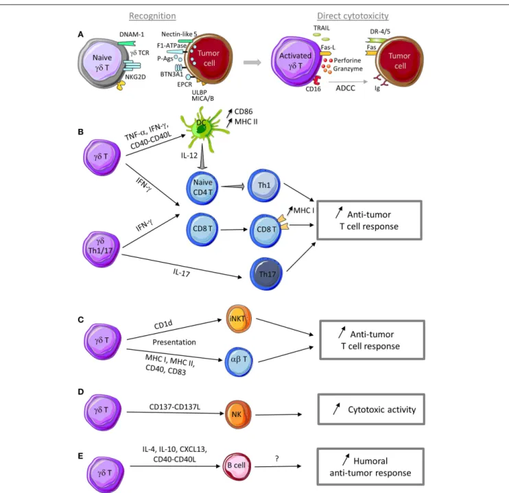
FIGURE 1 | Anti-tumor functions ofγδT cells. (A) γδ T cells can recognize tumor cells through interaction with (i) TCR ligands, such as phosphoantigens (P-Ags), F1-ATPase, BTN3A1, EPCR,. . ., and (ii) innate receptor ligands, such as ULBP, MICA/B, and nectin-like 5. Following sensing of tumor antigens or stress signals,γδ T cells are activated and can kill tumor cells through cytotoxic mechanisms that rely on the perforin/granzyme pathway, the death receptor pathway in response to TRAIL or Fas-L expression, and ADCC in the presence of tumor-specific antibodies.(B)γδ T cell activation leads to TNF-α and IFN-γ production and CD40-L expression that promote DC maturation and T cell differentiation into Th1 cells. IL-17-producingγδ Th17 cells favor Th17 effector cell development. Th1 and Th17 effector T cells display anti-tumor
functions to control tumor development.(C) Through a trogocytosis mechanism, activatedγδ T cells can capture and express CD1d molecules and then promote iNKT cell activation. Activatedγδ T cells can also display APC functions (MHC I and II, CD40, CD83, and CD86 expression) and activate both naive and effector T cells with cytotoxic activity against tumor cells. (D) Activatedγδ T cells can provide a co-stimulatory signal to NK cells through CD137L expression to promote their anti-tumor activity.(E) In the presence of specific signals, activatedγδ T cells can display a Tfh profile (i.e., IL-4, IL-10, and CXCL13 production and CD40-L expression) to help B cell antibody production. Although not yet demonstrated, production of antibodies against specific tumor antigens could be involved in the humoral anti-tumor response.
cells (72–74) (Figure 1E). These observations are based on results obtained in vitro. The APC functions ofγδ T cells in vivo have not been evaluated and remain to be demonstrated.
Moreover, similarly toαβ T cells, the differential induction of specific effector functions may also depend on the innate immu-nity receptor class that is engaged and the nature of the cytokine
stimuli. For example, NKG2D engagement triggers the induction of human Vγ9Vδ2 T cell cytotoxic functions, thereby influencing the fate of target cells (lysis or survival), but has limited effects on cytokine production (21). Similarly, type I IFN-released by stim-ulated myeloid and plasmacytoid DCs induces exclusively IFN-γ, but no TNF-α, production by human Vγ9Vδ2 T cells (65).
In conclusion, Vγ9Vδ2 T cells display a surprisingly broad array of functional activities. One essential question is to deter-mine whether such functional plasticity is an intrinsic feature of the whole Vγ9Vδ2 T cell population or whether it is restricted to specific Vγ9Vδ2 T cell subsets. This is an important issue, because it could directly affectγδ T cell-based therapeutic strate-gies. Indeed, boostingγδ T cell regulatory activity is suitable in some instances (i.e., autoimmune disease), conversely optimiz-ing, for example, their APC or cytotoxic functions could be more important for the treatment of tumors or infections. In terms of cytokine production and cytotoxic activity, Vγ9Vδ2 T cells can be divided in different subsets based on the expression of cell sur-face markers. Upon in vitro activation and extended culture in the presence of IL-2, naive Vγ9Vδ2 T cells (CD27+CD45RA+) can sequentially differentiate into TCM (CD27+CD45RA−), TEM (CD27−CD45RA−), and TEMRA (CD27−CD45RA+) cells. CD45RA−CD27− TEM cells show the highest IFN-γ secre-tion, while CD45RA+CD27− TEMRA cells are characterized by a strong cytotoxic activity. In contrast, naive CD45RA+CD27+ Vγ9Vδ2 T cells display very low, if any, functional activity (75). Studies using cell sorter-purified Vγ9Vδ2 T cell subsets have deter-mined that only naive CD45RA+CD27+ Vγ9Vδ2 T cells can differentiate into IL-17-producing cells when exposed to IL-1β, IL-6, IL-23, and TGF-β (61). IL-17-producing Vγ9Vδ2 T cells
dis-play a TEMRA phenotype, promote neutrophil migration through production of CXCL8 and up-regulateβ-defensin production in epithelial cells (61). Similarly, Vγ9Vδ2 T cell cytotoxic activity can
be assigned to specific subsets, especially to (CD45RA+CD27−) TEMRA and (CD56+CD16+) cells (75–77), but their clonal plasticity remains uncertain.
In addition, whether a given Vγ9Vδ2 T cell phenotype induced by specific environmental stimuli, such as cytokines, is stable or reversible, remains to be investigated. Although the expression of lineage-associated transcription factors in Vγ9Vδ2 T cells has been assessed in some studies, so far no clear correlation between the expression of transcription factors and a specific stable cytokine profile has been reported.
Finally, most of these studies concerned the Vγ9Vδ2 T cell sub-set thus raising the question of whether other human or mouse γδ T cell populations display similar plasticity. Ribot and collab-orators have reported that also human Vγ1 and Vγ2 thymocytes show functional phenotypic plasticity and can differentiate into cytotoxic type 1 effector cells following IL-2 or IL-15 stimulation (66) but no investigation was reported on other humanγδ T cell subsets.
PLASTICITY OF MOUSEγδ T CELLS
In mice, several studies demonstrated that γδ thymocytes are functionally pre-committed and polarized in term of cytokine production (78–80). During fetal development,γδ T cells are gen-erated from two waves of thymocytes that express invariant TCR.
The first group migrates into the skin (Vγ5Vδ1 DETC) and is programed to produce IFN-γ; the second group migrates into the vaginal epithelium and the peritoneal cavity (Vγ6Vδ1 subset) and is programed to produce IL-17 (33,81). Otherγδ T cell subsets appear postnatally in the thymus and express TCR with various Vδ and Vγ combinations. In adult mice, these cells are found in all lymphoid organs and below the epithelium or mucosal surfaces of many tissues, including the small intestine and lung. Most of them display a programed polarization acquired during thymic selection (33,81) through a process regulated by TCR (78–80) and co-receptor signaling (81). Thus,γδ T cell differentiation into
IFN-γ-producing cells require TCR and CD27 signals (78–80). CD27, a member of the tumor necrosis factor receptor family, regulates the balance between IFN-γ and IL-17 producing γδ T cell subsets (82). CD27+γδ T cells are committed to express IFN-γ genes, whereas, CD27−γδ T cells display a permissive chromatin config-uration at loci encoding IFN-γ and IL-17 as well as their regulatory transcription factors. They can thus differentiate into both IFN-γ-and IL-17-producing cells (82). It has also been shown that IL-23 in combination with IL-1β promotes IL-17, IL-21, and IL-22 expression by mouseγδ T cells in the absence of additional signals; however, the authors did not investigate CD27 expression in this setting (67).
Altogether, these results suggest that mouseγδ T cells have a low plasticity compared to humanγδ T cells. Nevertheless further investigation on mouse and humanγδ T cell functional plasticity are required to better characterize the molecular mechanisms and the precise role of eachγδ T cell subset in the immune response and in pathologic conditions in order to improveγδ T cell-based therapies.
IMPACT OFγδ T CELLS ON THE TUMOR IMMUNE RESPONSE
γδ T cells can: (i) detect and sense any type of stress through a MHC-independent mechanism, (ii) produce huge quantities of pro-inflammatory cytokines, and (iii) exert potent cytotoxic activity against a broad panel of tumors. For these reasons,γδ T lymphocytes are key players in the tumor immune response. Like other cytotoxic effectors,γδ T cells directly participate in the elimination of tumor cells, but they also control indirectly the tumor immune response by modulating the activity and functions of other immune cells. In this section, we will summarize both pro- and anti-tumor activities ofγδ T cells by focusing mainly on their tumor recognition mechanisms and the triggered biological responses.
ANTI-TUMOR ACTIVITY OFγδ T CELLS Mechanisms of tumor cell recognition
Similarly to any other T cell population,γδ T cell activation and acquisition of effector functions are triggered by TCR engage-ment (Figure 1A). Specifically,γδ TCR recognize molecules that are over-expressed in stress conditions. In normal cells, the con-centration of metabolites of the isoprenoid pathway, such as IPP, is too low to be sensed as a danger signal by Vγ9Vδ2 T cells. Deregulation of the isoprenoid pathway in some tumors leads to IPP over-production that is detected and considered as a tumor antigen by Vγ9Vδ2 TCR (40,83). Similarly, incubation of tumor cells with bisphosphonates that inhibit the farnesyl pyrophosphate
synthase enzyme in the isoprenoid pathway leads to IPP accumu-lation and makes tumor cells more sensitive to Vγ9Vδ2 T cell cytotoxicity (84–86). Several reports have shown that phospho-antigens need to interact with specific proteins to be recognized by TCR and to activate Vγ9Vδ2 T cells. First, Mookerjee-Basu et al. showed that F1-ATPase, which is expressed on the sur-face of some tumor cells, binds to the adenylated derivative of IPP and is involved in triggering Vγ9Vδ2 T cell activation and anti-tumoral activity (87,88). More recently, it was reported that butyrophilin 3 A1 (BTN3A1) can contribute toγδ T cell activa-tion by sensing changes in phosphoantigen concentraactiva-tion within tumor cells. Specifically, phosphoantigen binding to the intracellu-lar domain of BTN3A1 could initiate a cascade of events that result in extracellular changes or cell surface rearrangements (includ-ing immobilization of BTN3A extracellular domain) and lead to Vγ9Vδ2 T cell activation (89,90). Dechanet-Merville and collab-orators found that a humanδ2 negative T cell subset recognizes both CMV-infected and transformed cells through the interaction between the endothelial protein C receptor (EPCR) and the TCR (91). EPCR is over-expressed in CMV-infected endothelial cells and transformed cells and it is conceivable that it might act as a determinant of stress surveillance during epithelial cell trans-formation to communicate a state of “dysregulated self ” toγδ T cells.
In addition to TCR engagement, stimulation of NKR expressed byγδ T cells and particularly engagement of NKG2D receptor can also efficiently trigger the anti-tumor functions ofγδ T cells. NKG2D is expressed by Vγ9Vδ2 T cells and binds to non-classical MHC molecules of the MIC and ULBP families that are expressed by tumor cells (18,20,21). Ligand binding to NKG2D induces the release of IFN-γ and TNF-α, increases the expression of CD25, theα chain of the IL-2 receptor and promotes γδ T cell cytolytic activity (21). In particular, ULBP molecules have been involved in the recognition by Vγ9Vδ2 T cells of leukemia and lymphoma (92) and also of solid tumors, such as ovarian and colon carcinomas (93,
94). For instance, ULBP1 expression level determines lymphoma susceptibility toγδ T cell-mediated cytolysis upon NKG2D bind-ing (92). ULBP4 also can bind to Vγ9Vδ2 TCR and thus induce
the cytotoxic activity of Vγ9Vδ2T cells toward tumor cells through both TCR and NKG2D engagement (22). More recently, Lamb and collaborators have shown that temozolomide (TMZ), the main chemotherapeutic agent used to treat glioblastoma multiforme (GMB), increases the expression of stress-associated NKG2D lig-ands on TMZ-resistant glioma cells, potentially making them more susceptible toγδ T cell recognition and lysis (95). Furthermore, as described for Vγ9Vδ2 T cells, recognition of MICA, MICB, or ULBP expressed on cancer cells by human Vγ1δ1 T lymphocytes can trigger or increase their cytolytic activity against tumor cells that express NKG2D ligands (23,96). Indeed, ULBP and MICA interact with NKG2D or TCR on Vδ1 γδ T cells and induce their activation. However, MICA binds in mutually exclusive manner to NKG2D and TCR, suggesting that the two receptors might be sequentially engaged following recognition of target tumor cells (97).
DNAM-1 (also called CD226) is another NKR involved in the regulation of the cytotoxic activity ofγδ T cells. It is expressed on the surface of both Vγ9Vδ2 and γ1 T cell populations and its
ligand nectin-like-5 has been detected on certain tumors. DNAM-1 cooperates with TCR and NKG2D signaling in γδ T cells to positively regulate their IFN-γ production and cytotoxic activity against tumor cells (98,99).
Like NK cells, humanγδ T cells also express the CD16 (FcγRIII) receptor that binds to the Fc portion of immunoglobulin G (IgG). CD16 expression on Vγ9Vδ2 T cells can be up-regulated follow-ing stimulation with phosphoantigens (100). Its engagement leads to ADCC (101), a process that can result in lysis of tumor cells bound by specific antibodies. Indeed, several in vitro studies have clearly shown that γδ T cells are activated through CD16 and mediate ADCC of tumor cells in the presence of therapeutic anti-tumor monoclonal antibodies, such as rituximab, trastuzumab, atumumab, and alemtuzumab (102–105). Reinforcing the rele-vance of such in vitro data, it has been shown that stimulatedγδ T cells increase the efficacy of Trastuzumab in Her2+ breast cancer patients (105).
Impact on immune cell activity
In addition to these direct effects against tumor cells,γδ T cells can also control indirectly the anti-tumor immune response by pro-moting the recruitment and modulating the activation of other cell types in the tumor microenvironment, such as DCs, NK cells, and effector T cells (Figures 1B–D).
In the presence of tumor cells, or following stimulation with TCR agonists, NKG2D ligands, cytokines (such as 12 and IL-18), or DNAM-1 engagement, humanγδ T cells produce IFN-γ and TNF-α (21,56,94,106–108). These two cytokines can inhibit tumor growth through several mechanisms, but especially by enhancing CD8 T cell anti-tumor activity (Figure 1B) and by inhibiting tumor angiogenesis (109–111). Mouseγδ T cells also are an important and early source of IFN-γ within the tumor microenvironment where IFN-γ enhances MHC class I expression on tumor cells and CD8+ T cell responses (112–114). Altogether these findings suggest that both human and mouseγδ T cells pos-itively influence the anti-tumor immune response by increasing the adaptive anti-tumor immunity (115) (Figure 1B).
As previously mentioned, both mouse and humanγδ T cells could be an important source of IL-17. This cytokine plays an essential role in the host defense against microbial infections, but also in autoimmune disorders and cancer (116). IL-17 contri-bution to the tumor immune surveillance is still controversial. Indeed, IL-17 has often been described as a cytokine with pro-tumor properties, but several studies highlighted that it can also display anti-tumor functions (117). Therefore, IL-17 heteroge-neous sources and, perhaps, targets in the tumor microenviron-ment may determine whether it will negatively or positively affect tumor growth. In human, the majority of αβ and γδ Th17 cell populations that produce IL-17 also concomitantly produce IFN-γ (63) and the anti-tumor functions of IL-17-producingαβ T cells strongly depend on IFN-γ (118). Moreover, IL-17-producingαβ T cells stimulate the release of several cytokines (such as 6, IL-12, CXCL9, and CXCL10) by immune or cancer cells, leading to DC maturation or effector T cell recruitment to the tumor, and as a consequence, to an increase of the anti-tumor immunity (119,
120) (Figure 1B). It is likely thatγδ Th17 cells might do the same, but this remains to be formally demonstrated.
Importantly, in mice, IL-17-producingγδ T cells (Vγ4+ and Vγ6+) contribute to chemotherapy efficacy because they are required for the priming of IFN-γ-secreting tumor-specific T cells. In this context,γδ T cells are considered as part of the innate immune response that is involved in the subsequent specific anti-tumor T cell response following treatment with chemother-apeutic agents (121,122). Nevertheless, it is not known whether human IL-17γδ T cells also contribute to the efficacy of anti-cancer chemotherapy and whether combination treatments with γδ T cell agonists and anthracyclines could improve the patient outcome.
Dendritic cells are potent inducers ofγδ T cell effector func-tions through their ability to expressγδ TCR ligands and to provide co-stimulation signals (123,124). Inversely, interactions between activatedγδ T cells and DCs were shown to induce DC activa-tion and maturaactiva-tion, thus facilitating the establishment of the T cell response (125, 126). Indeed, activated human Vγ9Vδ2 T
cells enhance IL-12 production by monocyte-derived DCs through an IFN-γ- and IL-12-mediated positive feedback loop that can then promote naiveαβ T cell activation and differentiation into Th1-type cells (127), an effect that may positively influenced the anti-tumor immunity (Figure 1B).
As already mentioned, when activated by phosphoantigens, Vγ9Vδ2 T cells can display APC features and acquire the ability to activate naive and effector T cells (72,73) (Figure 1C). Similarly, Vγ9Vδ2 T cells can also present antigens to invariant NKT cells (iNKT). Schneiders et al. demonstrated that, when co-cultured with CD1d-positive cells, activated Vγ9Vδ2 T cells uptake CD1d on their membrane through trogocytosis and acquire the capac-ity to present glycolipid antigens to iNKT cells and activate them (128) (Figure 1C). iNKT cell activation triggers the production of large amounts of cytokines that play an important role in ini-tiating and orchestrating anti-tumor immune responses, such as Th1-biased pro-inflammatory responses.
Natural killer cells also have a role in anti-tumor responses and their activity can be regulated byγδ T cells. When co-localized within tumors, humanγδ T cells can provide co-stimulatory sig-nals to NK cells and induce NK cell-mediated killing of tumor cells (129). Indeed, CD137L is expressed on activatedγδ T cells and interacts with the cognate receptor CD137 on NK cells, lead-ing to the up-regulation of the activation markers CD25, CD54, CD69, and NKG2D on the surface of NK cells and to the increase of their cytotoxic function, particularly against solid tumors that are usually resistant to NK cytolysis (129) (Figure 1D).
PRO-TUMOR ACTIVITY OFγδ T CELLS
In some conditions,γδ T cells can also promote tumor growth via regulatory functions that impair the anti-tumor immune responses (Figure 2).
Human Vγ9V δ2 T cells
Vγ9Vδ2 T cells with immunosuppressives functions may play an important role in human cancers. Upon activation, human periph-eral Vγ9Vδ2 T cells also can express IL-4, IL-10, and TGF-β and inhibit T cell proliferation, thus developing a regulatory profile that may play a role in the suppression of anti-tumor responses (130). Indeed, depending on the context, Vγ9Vδ2 T cells may display a
Th1-, Th2-, Th17-, or Th1/reg-like profile and synthetize IFN-γ, IL-4, IL-17 or IL-10, and TGF-β, respectively.
While IL-4 is a cytokine involved in Th2 responses (which are not appropriate for anti-tumor immunity), IL-10 and TGF-β are cytokines with immunosuppressive functions and thus could be involved in the pro-tumor activities ofγδ T cells. TGF-β has a cru-cial role in tumor development because it can promote tumor cell invasiveness and metastasis formation mainly by modulating the immune system and the tumor microenvironment (Figure 2A). The most important mechanisms of tumor progression linked to TGF-β activities are the epithelial-to-mesenchymal transition (EMT), immune system evasion, and promotion of cancer cell pro-liferation by modulation of the tumor microenvironment (131). The expression of IL-10 and TGF-β is frequently increased in vari-ous cancer types. IL-10 directly affects APC function by inhibiting the expression of MHC and co-stimulatory molecules, which induces immune suppression or tolerance (Figure 2B). Addition-ally, IL-10 down-regulates the expression of Th1 cytokines and induces T-regulatory responses.
IL-17 plays a dual role by promoting both tumor growth and anti-tumor immunity, depending on the tumor type, stage, and target cells present in tumor microenvironment. The number of IL-17-producing cells is increased in cancer and this is associ-ated with poor prognosis (117,132,133). Several IL-17 activi-ties contribute to tumor progression. In breast cancer, IL-17 can directly promote tumor cell proliferation and dissemination (119) and favor the development of cancer resistance to conventional chemotherapeutic agents, such as docetaxel (133) (Figure 2A). IL-17 can also act on cells in the tumor microenvironment. For instance, IL-17 up-regulates the secretion of pro-angiogenic and pro-tumor factors (e.g., VEGF, IL-6, and IL-8) by stromal cells and fibroblasts, thus promoting angiogenesis and sustained chronic inflammation (119, 120). In colorectal cancer, Vγ9Vδ2 T cells
can differentiate into Th17 cells that secrete 17 and also IL-8, TNF-α, and GM-CSF and thus contribute to the accumula-tion of immunosuppressive polymorphonuclear-myeloid-derived suppressor cells (PMN-MDSCs) within the tumor microenvi-ronment and influence the anti-tumor immune response (64) (Figure 2C).
Human Vδ1 T cells
Besides Vγ9Vδ2 T cells, other human γδ T cell subsets can display immunosuppressive functions. First, Peng et al. demonstrated that Vδ1 γδT cells infiltrating human breast cancer suppress DC mat-uration and T cell effector functions both in vitro and in vivo. When stimulated by tumor cells and an anti-CD3 antibody, Vγ1 T cells express IFN-γ and GM-CSF, but not IL-1β, TNF-α, IL-12, IL-2, IL-4, IL-10, or TGF-β (134). Thus neither IL-10 nor TGF-β
seems to play a role in this immunosuppressive activity. Although, the involved factor(s) remain to be identified, these authors found that the suppressive activity was in the soluble fraction with a mol-ecular mass higher than 100 kDa and could be inactivated by heat, but not by DNAse or RNAse treatments (134) (Figure 2D). These Vδ1 γδ T cells represent a large percentage of tumor-infiltrating lymphocytes in breast and also in prostate cancer, suggesting that they may play an important role in promoting an immunosup-pressive tumor microenvironment. Interestingly, stimulation of
FIGURE 2 | Pro-tumor functions ofγδT cells. (A) Activation of γδ T cells in the presence of specific stimuli can promote their polarization into Th17- or Treg-like cells that produce IL-17 and TGF-β, thus favoring tumor cell proliferation and dissemination. IL-17 produced byγδ T cells confers chemotherapy resistance to tumor cells.(B) Activatedγδ T cells can inhibit DC maturation and their APC functions, thus impairing naive T cell activation and differentiation into effector T cells.(C) IL-17 produced byγδ Th17 cells promotes the development of Th17 cells with pro-tumor
functions.γδ Th17 cells also produce a cocktail of cytokines and chemokines involved in the recruitment of myeloid-derived suppressive cells (MDSC) and small peritoneal macrophages (SPM) with
immunosuppressive and pro-tumor functions.(D)γδ Treg cells produce cytokines (IL-4, IL-10, and TGF-β) and other immunosuppressive factors that impair CD8 T and NK cell cytotoxic activity.γδ Treg cells can also promote senescence of DC orαβ T cells and consequently favor tumor growth.
suppressive Vδ1 γδ T cells in breast cancer by using a TLR8 agonist reversed the anti-tumor response inhibition (134). More recently, the same group demonstrated that regulatoryγδ T cells can induce both T cell and DC senescence. Specifically, regulatoryγδ T cells induce senescence of both naive and effector T cells, as indicated by the impaired expression of the co-stimulatory molecules CD27 and CD28 and the low proliferative capacities of both Th1 and Th17 T cell subsets. Senescent T cells and DCs become suppressive
cells, further amplifying the immunosuppression mediated byγδ Treg cells (135). Furthermore, Ma and collaborators found that highγδ T cell level in breast cancer tissues is correlated with poor survival and high risk of relapse (136). Similarly, in colon ade-nocarcinoma, a significant correlation has been observed between presence ofγTCR cells and disease stage. These two reports sug-gest that γδ T cells may have a key prognostic role in colon adenocarcinoma and breast cancers (137).
Mouseγδ T cells
γδ T cells with immunosuppressive functions have also been observed in mouse tumor models (138, 139). Seo et al. found that murine γδ T cells that infiltrate tumors arising from B16 melanoma cells produce large amounts of IL-4 and IL-10 and inhibit NK and iNKT cell activity (138) (Figure 2D). They demon-strated that supernatants from these γδ T cells did not affect NK and iNKT cell cytotoxicity, but reduced their proliferation, suggesting that soluble IL-4 and IL-10 could contribute to the inhibition of NK and iNKT cell activity by γδ T cells in this model (138). Additional studies from this group showed thatγδ T cells that infiltrate MM2 mammary tumors in mice express IL-10 and TGF-β, but not IFN-γ or IL-4. γδ T cells isolated from these tumors and from the spleen hindered the cytotoxic activity of NK and CD8 T cells. IL-10 and TGF-β neutralization inhibited some of the immunosuppressive effects of theseγδ T cells, sug-gesting the involvement of these cytokines (Figure 2D). Moreover, depletion of IL-10- and TGF-β-secreting γδ T cells by using a spe-cific antibody enhanced the anti-tumor immunity and reduced tumor growth in xenografted mice (139). More recently, Hao et al. using the B16 melanoma model, showed that mouse Vγ1 T cells suppress the anti-tumor functions of the Vγ4 T cell sub-set, thus promoting tumor growth. Specifically, Vγ1 γδ T cells reduced IFN-γ, perforin, and NKG2D expression in Vγ4 γδ T cells through contact-independent mechanisms involving IL-4 (140). Collectively, these data strongly suggest that within the tumor microenvironment, some mouseγδ T cell populations express IL-4, IL-10, and TGF-β and inhibit the anti-tumor immune response. IL-17-secretingγδ T cells show pro-tumor activity also in mouse models. Recently, Rei et al. demonstrated that murine CD27-Vγ6 T cells that produce IL-17 promote ovarian cancer growth via mobilization of small peritoneal macrophages (141) (Figure 2C). Overall, these findings support the idea thatγδ T cells, at least in some cancers, can behave as Tregs or Th17 T cells that impair the anti-tumor immune response and promote tumor growth, through the secretion of different cytokines with regulatory func-tions or the recruitment of immunosuppressive cells within the tumor microenvironment.
CONCLUSION
During the last decade, our knowledge on the role ofγδ T cells in the tumor microenvironment has hugely improved. Plasticity of γδ T cells increases the range of their biological responses as differentγδ T cell sub-populations can regulate different aspects of the tumor immunity. Functional plasticity also can explain the heterogeneous responses and contradictory functions of this unconventional T cell population in the context of cancer immune surveillance. As discussed in this review, due to the TCR-mediated recognition and activation mechanisms and the fine regulation of their activation through innate and cytokine receptors,γδ T lymphocytes are attractive targets for immunotherapeutic proto-cols with the final objective of boosting the anti-tumor immune response. Several clinical trials have already assessedγδ T cell-based immunotherapy in patients with advanced hematological malignancies and solid cancers with encouraging results. However, high density ofγδ T cells in the breast and colon tumor microen-vironment has been associated with poor clinical outcome. We
are convinced that a better characterization of the mechanisms regulating their polarization should allow the development of optimal therapeutic strategies to favor the expansion ofγδ T cell populations with anti-tumor rather than pro-tumor functions.
ACKNOWLEDGMENTS
This work was supported by institutional grants from INSERM and Université de Montpellier 1. Henri-Alexandre Michaud is sup-ported by a post-doctoral fellowship from the Fondation pour la Recherche Médicale (FRM).
REFERENCES
1. Hanahan D, Weinberg RA. Hallmarks of cancer: the next generation. Cell (2011) 144(5):646–74. doi:10.1016/j.cell.2011.02.013
2. Bouet-Toussaint F, Cabillic F, Toutirais O, Le Gallo M, Thomas de la Pin-tiere C, Daniel P, et al. Vgamma9Vdelta2 T cell-mediated recognition of human solid tumors. Potential for immunotherapy of hepatocellular and colorectal carcinomas. Cancer Immunol Immunother (2008) 57(4):531–9. doi:10.1007/s00262-007-0391-3
3. Cordova A, Toia F, La Mendola C, Orlando V, Meraviglia S, Rinaldi G, et al. Characterization of human gammadelta T lymphocytes infiltrating primary malignant melanomas. PLoS One (2012) 7(11):e49878. doi:10.1371/journal. pone.0049878
4. Corvaisier M, Moreau-Aubry A, Diez E, Bennouna J, Mosnier JF, Scotet E, et al. V gamma 9V delta 2 T cell response to colon carcinoma cells. J Immunol (2005)
175(8):5481–8. doi:10.4049/jimmunol.175.8.5481
5. Dieli F, Vermijlen D, Fulfaro F, Caccamo N, Meraviglia S, Cicero G, et al. Tar-geting human {gamma}delta} T cells with zoledronate and interleukin-2 for immunotherapy of hormone-refractory prostate cancer. Cancer Res (2007)
67(15):7450–7. doi:10.1158/0008-5472.CAN-07-0199
6. Dudley ME, Wunderlich JR, Yang JC, Sherry RM, Topalian SL, Restifo NP, et al. Adoptive cell transfer therapy following non-myeloablative but lymphode-pleting chemotherapy for the treatment of patients with refractory metastatic melanoma. J Clin Oncol (2005) 23(10):2346–57. doi:10.1200/JCO.2005.00.240 7. Kang N, Zhou J, Zhang T, Wang L, Lu F, Cui Y, et al. Adoptive immunotherapy of lung cancer with immobilized anti-TCRgammadelta antibody-expanded human gammadelta T-cells in peripheral blood. Cancer Biol Ther (2009)
8(16):1540–9. doi:10.4161/cbt.8.16.8950
8. Kunzmann V, Bauer E, Feurle J, Weissinger F, Tony HP, Wilhelm M. Stim-ulation of gammadelta T cells by aminobisphosphonates and induction of antiplasma cell activity in multiple myeloma. Blood (2000) 96(2):384–92. doi:10.1080/10428190500051893
9. Kunzmann V, Wilhelm M. Anti-lymphoma effect of gammadelta T cells. Leuk
Lymphoma (2005) 46(5):671–80. doi:10.1080/10428190500051893
10. Meraviglia S, Eberl M, Vermijlen D, Todaro M, Buccheri S, Cicero G, et al. In vivo manipulation of Vgamma9Vdelta2 T cells with zoledronate and low-dose interleukin-2 for immunotherapy of advanced breast cancer patients. Clin
Exp Immunol (2010) 161(2):290–7. doi:10.1111/j.1365-2249.2010.04167.x
11. Viey E, Lucas C, Romagne F, Escudier B, Chouaib S, Caignard A. Chemokine receptors expression and migration potential of tumor-infiltrating and peripheral-expanded Vgamma9Vdelta2 T cells from renal cell car-cinoma patients. J Immunother (2008) 31(3):313–23. doi:10.1097/CJI. 0b013e3181609988
12. Wilhelm M, Kunzmann V, Eckstein S, Reimer P, Weissinger F, Ruediger T, et al. Gammadelta T cells for immune therapy of patients with lymphoid malignan-cies. Blood (2003) 102(1):200–6. doi:10.1182/blood-2002-12-3665 13. Morita CT, Beckman EM, Bukowski JF, Tanaka Y, Band H, Bloom BR,
et al. Direct presentation of nonpeptide prenyl pyrophosphate antigens to human gamma delta T cells. Immunity (1995) 3(4):495–507. doi:10.1016/ 1074-7613(95)90178-7
14. Morita CT, Tanaka Y, Bloom BR, Brenner MB. Direct presentation of non-peptide prenyl pyrophosphate antigens to human gamma delta T cells. Res
Immunol (1996) 147(5):347–53. doi:10.1016/0923-2494(96)89649-0
15. Born WK, Kemal Aydintug M, O’Brien RL. Diversity of gammadelta T-cell antigens. Cell Mol Immunol (2013) 10(1):13–20. doi:10.1038/cmi.2012.45 16. Cao W, Xi X, Hao Z, Li W, Kong Y, Cui L, et al. RAET1E2, a soluble
NKG2D-mediated NK cytotoxicity. J Biol Chem (2007) 282(26):18922–8. doi:10.1074/jbc.M702504200
17. Groh V, Steinle A, Bauer S, Spies T. Recognition of stress-induced MHC molecules by intestinal epithelial gammadelta T cells. Science (1998)
279(5357):1737–40. doi:10.1126/science.279.5357.1737
18. Das H, Groh V, Kuijl C, Sugita M, Morita CT, Spies T, et al. MICA engagement by human Vgamma2Vdelta2 T cells enhances their antigen-dependent effector function. Immunity (2001) 15(1):83–93. doi:10.1016/S1074-7613(01)00168-6 19. Groh V, Wu J, Yee C, Spies T. Tumour-derived soluble MIC ligands impair expression of NKG2D and T-cell activation. Nature (2002) 419(6908):734–8. doi:10.1038/nature01112
20. Wrobel P, Shojaei H, Schittek B, Gieseler F, Wollenberg B, Kalthoff H, et al. Lysis of a broad range of epithelial tumour cells by human gamma delta T cells: involvement of NKG2D ligands and T-cell receptor- versus NKG2D-dependent recognition. Scand J Immunol (2007) 66(2–3):320–8. doi:10.1111/j.1365-3083. 2007.01963.x
21. Rincon-Orozco B, Kunzmann V, Wrobel P, Kabelitz D, Steinle A, Herrmann T. Activation of V gamma 9V delta 2 T cells by NKG2D. J Immunol (2005)
175(4):2144–51. doi:10.4049/jimmunol.175.4.2144
22. Kong Y, Cao W, Xi X, Ma C, Cui L, He W. The NKG2D ligand ULBP4 binds to TCRgamma9/delta2 and induces cytotoxicity to tumor cells through both TCRgammadelta and NKG2D. Blood (2009) 114(2):310–7. doi:10.1182/blood-2008-12-196287
23. Wu J, Groh V, Spies T. T cell antigen receptor engagement and speci-ficity in the recognition of stress-inducible MHC class I-related chains by human epithelial gamma delta T cells. J Immunol (2002) 169(3):1236–40. doi:10.4049/jimmunol.169.3.1236
24. Shojaei H, Oberg HH, Juricke M, Marischen L, Kunz M, Mundhenke C, et al. Toll-like receptors 3 and 7 agonists enhance tumor cell lysis by human gam-madelta T cells. Cancer Res (2009) 69(22):8710–7. doi:10.1158/0008-5472. CAN-09-1602
25. Groh V, Porcelli S, Fabbi M, Lanier LL, Picker LJ, Anderson T, et al. Human lymphocytes bearing T cell receptor gamma/delta are phenotypically diverse and evenly distributed throughout the lymphoid system. J Exp Med (1989)
169(4):1277–94. doi:10.1084/jem.169.4.1277
26. Parker CM, Groh V, Band H, Porcelli SA, Morita C, Fabbi M, et al. Evidence for extrathymic changes in the T cell receptor gamma/delta repertoire. J Exp Med (1990) 171(5):1597–612. doi:10.1084/jem.171.5.1597
27. Itohara S, Farr AG, Lafaille JJ, Bonneville M, Takagaki Y, Haas W, et al. Hom-ing of a gamma delta thymocyte subset with homogeneous T-cell receptors to mucosal epithelia. Nature (1990) 343(6260):754–7. doi:10.1038/343754a0 28. Goodman T, Lefrancois L. Intraepithelial lymphocytes. Anatomical site, not
T cell receptor form, dictates phenotype and function. J Exp Med (1989)
170(5):1569–81. doi:10.1084/jem.170.5.1569
29. Cai Y, Shen X, Ding C, Qi C, Li K, Li X, et al. Pivotal role of dermal IL-17-producing gammadelta T cells in skin inflammation. Immunity (2011)
35(4):596–610. doi:10.1016/j.immuni.2011.08.001
30. Gray EE, Suzuki K, Cyster JG. Cutting edge: identification of a motile IL-17-producing gammadelta T cell population in the dermis. J Immunol (2011)
186(11):6091–5. doi:10.4049/jimmunol.1100427
31. Hinz T, Wesch D, Halary F, Marx S, Choudhary A, Arden B, et al. Identification of the complete expressed human TCR V gamma repertoire by flow cytometry.
Int Immunol (1997) 9(8):1065–72. doi:10.1093/intimm/9.8.1065
32. Holtmeier W, Pfander M, Hennemann A, Zollner TM, Kaufmann R, Caspary WF. The TCR-delta repertoire in normal human skin is restricted and distinct from the TCR-delta repertoire in the peripheral blood. J Invest Dermatol (2001)
116(2):275–80. doi:10.1046/j.1523-1747.2001.01250.x
33. Bonneville M, O’Brien RL, Born WK. Gammadelta T cell effector functions: a blend of innate programming and acquired plasticity. Nat Rev Immunol (2010)
10(7):467–78. doi:10.1038/nri2781
34. Hayday AC. [gamma][delta] Cells: a right time and a right place for a con-served third way of protection. Annu Rev Immunol (2000) 18:975–1026. doi:10.1146/annurev.immunol.18.1.975
35. Toulon A, Breton L, Taylor KR, Tenenhaus M, Bhavsar D, Lanigan C, et al. A role for human skin-resident T cells in wound healing. J Exp Med (2009)
206(4):743–50. doi:10.1084/jem.20081787
36. Macleod AS, Havran WL. Functions of skin-resident gammadelta T cells. Cell
Mol Life Sci (2011) 68(14):2399–408. doi:10.1007/s00018-011-0702-x
37. O’Brien RL, Roark CL, Born WK. IL-17-producing gammadelta T cells. Eur J
Immunol (2009) 39(3):662–6. doi:10.1002/eji.200839120
38. Hintz M, Reichenberg A, Altincicek B, Bahr U, Gschwind RM, Kollas AK, et al. Identification of (E)-4-hydroxy-3-methyl-but-2-enyl pyrophosphate as a major activator for human gammadelta T cells in Escherichia coli. FEBS Lett (2001) 509(2):317–22. doi:10.1016/S0014-5793(01)03191-X
39. Altincicek B, Moll J, Campos N, Foerster G, Beck E, Hoeffler JF, et al. Cut-ting edge: human gamma delta T cells are activated by intermediates of the 2-C-methyl-D-erythritol 4-phosphate pathway of isoprenoid biosynthesis. J
Immunol (2001) 166(6):3655–8. doi:10.4049/jimmunol.166.6.3655
40. Gober HJ, Kistowska M, Angman L, Jeno P, Mori L, De Libero G. Human T cell receptor gammadelta cells recognize endogenous mevalonate metabolites in tumor cells. J Exp Med (2003) 197(2):163–8. doi:10.1084/jem.20021500 41. Bonneville M, Scotet E. Human Vgamma9Vdelta2 T cells: promising new leads
for immunotherapy of infections and tumors. Curr Opin Immunol (2006)
18(5):539–46. doi:10.1016/j.coi.2006.07.002
42. Riganti C, Massaia M, Davey MS, Eberl M. Human gammadelta T-cell responses in infection and immunotherapy: common mechanisms, common mediators? Eur J Immunol (2012) 42(7):1668–76. doi:10.1002/eji.201242492 43. Catros V, Toutirais O, Bouet F, Cabillic F, Desille M, Fournie JJ. [Tgammadelta
lymphocytes in oncology: unconventional killer lymphocytes]. Med Sci (Paris) (2010) 26(2):185–91. doi:10.1051/medsci/2010262185
44. Buccheri S, Guggino G, Caccamo N, Li Donni P, Dieli F. Efficacy and safety of gammadeltaT cell-based tumor immunotherapy: a meta-analysis. J Biol Regul
Homeost Agents (2014) 28(1):81–90.
45. Gomes AQ, Martins DS, Silva-Santos B. Targeting gammadelta T lymphocytes for cancer immunotherapy: from novel mechanistic insight to clinical appli-cation. Cancer Res (2010) 70(24):10024–7. doi:10.1158/0008-5472.CAN-10-3236
46. Mosmann TR, Sad S. The expanding universe of T-cell subsets: Th1, Th2 and more. Immunol Today (1996) 17(3):138–46. doi:10.1016/0167-5699(96) 80606-2
47. O’Garra A, Arai N. The molecular basis of T helper 1 and T helper 2 cell differ-entiation. Trends Cell Biol (2000) 10(12):542–50. doi:10.1016/S0962-8924(00) 01856-0
48. Romagnani S. Human TH1 and TH2 subsets: regulation of differentiation and role in protection and immunopathology. Int Arch Allergy Immunol (1992)
98(4):279–85. doi:10.1159/000236199
49. Sher A, Coffman RL. Regulation of immunity to parasites by T cells and T cell-derived cytokines. Annu Rev Immunol (1992) 10:385–409. doi:10.1146/ annurev.iy.10.040192.002125
50. Dunne MR, Mangan BA, Madrigal-Estebas L, Doherty DG. Preferential Th1 cytokine profile of phosphoantigen-stimulated human Vgamma9Vdelta2 T cells. Mediators Inflamm (2010) 2010:704941. doi:10.1155/2010/704941 51. Garcia VE, Sieling PA, Gong J, Barnes PF, Uyemura K, Tanaka Y, et al. Single-cell
cytokine analysis of gamma delta T cell responses to nonpeptide mycobacterial antigens. J Immunol (1997) 159(3):1328–35.
52. Tsukaguchi K, de Lange B, Boom WH. Differential regulation of IFN-gamma, TNF-alpha, and IL-10 production by CD4(+) alphabetaTCR+ T cells and vdelta2(+) gammadelta T cells in response to monocytes infected with Mycobacterium tuberculosis-H37Ra. Cell Immunol (1999) 194(1):12–20. doi:10.1006/cimm.1999.1497
53. Thedrez A, Sabourin C, Gertner J, Devilder MC, Allain-Maillet S, Fournie JJ, et al. Self/non-self discrimination by human gammadelta T cells: simple solu-tions for a complex issue? Immunol Rev (2007) 215:123–35. doi:10.1111/j. 1600-065X.2006.00468.x
54. Morita CT, Verma S, Aparicio P, Martinez C, Spits H, Brenner MB. Function-ally distinct subsets of human gamma/delta T cells. Eur J Immunol (1991)
21(12):2999–3007. doi:10.1002/eji.1830211215
55. Gibbons DL, Haque SF, Silberzahn T, Hamilton K, Langford C, Ellis P, et al. Neonates harbour highly active gammadelta T cells with selective impairments in preterm infants. Eur J Immunol (2009) 39(7):1794–806. doi:10.1002/eji. 200939222
56. Wesch D, Glatzel A, Kabelitz D. Differentiation of resting human peripheral blood gamma delta T cells toward Th1- or Th2-phenotype. Cell Immunol (2001) 212(2):110–7. doi:10.1006/cimm.2001.1850
57. Vermijlen D, Ellis P, Langford C, Klein A, Engel R, Willimann K, et al. Dis-tinct cytokine-driven responses of activated blood gammadelta T cells: insights
into unconventional T cell pleiotropy. J Immunol (2007) 178(7):4304–14. doi:10.4049/jimmunol.178.7.4304
58. Bansal RR, Mackay CR, Moser B, Eberl M. IL-21 enhances the potential of human gammadelta T cells to provide B-cell help. Eur J Immunol (2012)
42(1):110–9. doi:10.1002/eji.201142017
59. Caccamo N, Todaro M, La Manna MP, Sireci G, Stassi G, Dieli F. IL-21 regu-lates the differentiation of a human gammadelta T cell subset equipped with B cell helper activity. PLoS One (2012) 7(7):e41940. doi:10.1371/journal.pone. 0041940
60. Casetti R, Agrati C, Wallace M, Sacchi A, Martini F, Martino A, et al. Cut-ting edge: TGF-beta1 and IL-15 Induce FOXP3+ gammadelta regulatory T cells in the presence of antigen stimulation. J Immunol (2009) 183(6):3574–7. doi:10.4049/jimmunol.0901334
61. Caccamo N, La Mendola C, Orlando V, Meraviglia S, Todaro M, Stassi G, et al. Differentiation, phenotype, and function of interleukin-17-producing human Vgamma9Vdelta2 T cells. Blood (2011) 118(1):129–38. doi:10.1182/ blood-2011-01-331298
62. Ness-Schwickerath KJ, Jin C, Morita CT. Cytokine requirements for the differentiation and expansion of IL-17A- and IL-22-producing human Vgamma2Vdelta2 T cells. J Immunol (2010) 184(12):7268–80. doi:10.4049/ jimmunol.1000600
63. Ness-Schwickerath KJ, Morita CT. Regulation and function of IL-17A- and IL-22-producing gammadelta T cells. Cell Mol Life Sci (2011) 68(14):2371–90. doi:10.1007/s00018-011-0700-z
64. Wu P, Wu D, Ni C, Ye J, Chen W, Hu G, et al. GammadeltaT17 cells promote the accumulation and expansion of myeloid-derived suppressor cells in human col-orectal cancer. Immunity (2014) 40(5):785–800. doi:10.1016/j.immuni.2014. 03.013
65. Devilder MC, Allain S, Dousset C, Bonneville M, Scotet E. Early triggering of exclusive IFN-gamma responses of human Vgamma9Vdelta2 T cells by TLR-activated myeloid and plasmacytoid dendritic cells. J Immunol (2009)
183(6):3625–33. doi:10.4049/jimmunol.0901571
66. Ribot JC, Ribeiro ST, Correia DV, Sousa AE, Silva-Santos B. Human gam-madelta thymocytes are functionally immature and differentiate into cyto-toxic type 1 effector T cells upon IL-2/IL-15 signaling. J Immunol (2014)
192(5):2237–43. doi:10.4049/jimmunol.1303119
67. Sutton CE, Lalor SJ, Sweeney CM, Brereton CF, Lavelle EC, Mills KH. Interleukin-1 and IL-23 induce innate IL-17 production from gammadelta T cells, amplifying Th17 responses and autoimmunity. Immunity (2009)
31(2):331–41. doi:10.1016/j.immuni.2009.08.001
68. Thedrez A, Harly C, Morice A, Salot S, Bonneville M, Scotet E. IL-21-mediated potentiation of antitumor cytolytic and proinflammatory responses of human V gamma 9V delta 2 T cells for adoptive immunotherapy. J Immunol (2009)
182(6):3423–31. doi:10.4049/jimmunol.0803068
69. Hu Y, Cui Q, Gu Y, Sheng L, Wu K, Shi J, et al. Decitabine facilitates the gen-eration and immunosuppressive function of regulatory gammadeltaT cells derived from human peripheral blood mononuclear cells. Leukemia (2013)
27(7):1580–5. doi:10.1038/leu.2012.345
70. Deknuydt F, Scotet E, Bonneville M. Modulation of inflammation through IL-17 production by gammadelta T cells: mandatory in the mouse, dispensable in humans? Immunol Lett (2009) 127(1):8–12. doi:10.1016/j.imlet.2009.08.003 71. Michel ML, Pang DJ, Haque SF, Potocnik AJ, Pennington DJ, Hayday AC.
Interleukin 7 (IL-7) selectively promotes mouse and human IL-17-producing gammadelta cells. Proc Natl Acad Sci U S A (2012) 109(43):17549–54. doi:10.1073/pnas.1204327109
72. Brandes M, Willimann K, Bioley G, Levy N, Eberl M, Luo M, et al. Cross-presenting human gammadelta T cells induce robust CD8+ alphabeta T cell responses. Proc Natl Acad Sci U S A (2009) 106(7):2307–12. doi:10.1073/pnas. 0810059106
73. Brandes M, Willimann K, Moser B. Professional antigen-presentation function by human gammadelta T Cells. Science (2005) 309(5732):264–8. doi:10.1126/ science.1110267
74. Himoudi N, Morgenstern DA, Yan M, Vernay B, Saraiva L, Wu Y, et al. Human gammadelta T lymphocytes are licensed for professional antigen presentation by interaction with opsonized target cells. J Immunol (2012) 188(4):1708–16. doi:10.4049/jimmunol.1102654
75. Dieli F, Poccia F, Lipp M, Sireci G, Caccamo N, Di Sano C, et al. Differen-tiation of effector/memory Vdelta2 T cells and migratory routes in lymph
nodes or inflammatory sites. J Exp Med (2003) 198(3):391–7. doi:10.1084/jem. 20030235
76. Alexander AA, Maniar A, Cummings JS, Hebbeler AM, Schulze DH, Gast-man BR, et al. Isopentenyl pyrophosphate-activated CD56+ {gamma}{delta} T lymphocytes display potent antitumor activity toward human squamous cell carcinoma. Clin Cancer Res (2008) 14(13):4232–40. doi:10.1158/1078-0432. CCR-07-4912
77. Angelini DF, Borsellino G, Poupot M, Diamantini A, Poupot R, Bernardi G, et al. FcgammaRIII discriminates between 2 subsets of Vgamma9Vdelta2 effector cells with different responses and activation pathways. Blood (2004)
104(6):1801–7. doi:10.1182/blood-2004-01-0331
78. Jensen KD, Su X, Shin S, Li L, Youssef S, Yamasaki S, et al. Thymic selec-tion determines gammadelta T cell effector fate: antigen-naive cells make interleukin-17 and antigen-experienced cells make interferon gamma.
Immu-nity (2008) 29(1):90–100. doi:10.1016/j.immuni.2008.04.022
79. Ribot JC, deBarros A, Pang DJ, Neves JF, Peperzak V, Roberts SJ, et al. CD27 is a thymic determinant of the balance between interferon-gamma- and interleukin 17-producing gammadelta T cell subsets. Nat Immunol (2009) 10(4):427–36. doi:10.1038/ni.1717
80. Turchinovich G, Hayday AC. Skint-1 identifies a common molecular mecha-nism for the development of interferon-gamma-secreting versus interleukin-17-secreting gammadelta T cells. Immunity (2011) 35(1):59–68. doi:10.1016/ j.immuni.2011.04.018
81. Turchinovich G, Pennington DJ. T cell receptor signalling in gammadelta cell development: strength isn’t everything. Trends Immunol (2011) 32(12):567–73. doi:10.1016/j.it.2011.09.005
82. Schmolka N, Serre K, Grosso AR, Rei M, Pennington DJ, Gomes AQ, et al. Epigenetic and transcriptional signatures of stable versus plastic differenti-ation of proinflammatory gammadelta T cell subsets. Nat Immunol (2013)
14(10):1093–100. doi:10.1038/ni.2702
83. Uchida R, Ashihara E, Sato K, Kimura S, Kuroda J, Takeuchi M, et al. Gamma delta T cells kill myeloma cells by sensing mevalonate metabolites and ICAM-1 molecules on cell surface. Biochem Biophys Res Commun (2007) 354(2):613–8. doi:10.1016/j.bbrc.2007.01.031
84. Mattarollo SR, Kenna T, Nieda M, Nicol AJ. Chemotherapy and zoledronate sensitize solid tumour cells to Vgamma9Vdelta2 T cell cytotoxicity. Cancer
Immunol Immunother (2007) 56(8):1285–97. doi:10.1007/s00262-007-0279-2
85. Dhar S, Chiplunkar SV. Lysis of aminobisphosphonate-sensitized MCF-7 breast tumor cells by Vgamma9Vdelta2 T cells. Cancer Immun (2010) 10:10. 86. D’Asaro M, La Mendola C, Di Liberto D, Orlando V, Todaro M, Spina
M, et al. V gamma 9V delta 2 T lymphocytes efficiently recognize and kill zoledronate-sensitized, imatinib-sensitive, and imatinib-resistant chronic myelogenous leukemia cells. J Immunol (2010) 184(6):3260–8. doi:10.4049/ jimmunol.0903454
87. Mookerjee-Basu J, Vantourout P, Martinez LO, Perret B, Collet X, Perigaud C, et al. F1-adenosine triphosphatase displays properties characteristic of an antigen presentation molecule for Vgamma9Vdelta2 T cells. J Immunol (2010)
184(12):6920–8. doi:10.4049/jimmunol.0904024
88. Scotet E, Martinez LO, Grant E, Barbaras R, Jeno P, Guiraud M, et al. Tumor recognition following Vgamma9Vdelta2 T cell receptor interactions with a surface F1-ATPase-related structure and apolipoprotein A-I. Immunity (2005)
22(1):71–80. doi:10.1016/j.immuni.2004.11.012
89. Harly C, Guillaume Y, Nedellec S, Peigne CM, Monkkonen H, Monkko-nen J, et al. Key implication of CD277/butyrophilin-3 (BTN3A) in cellular stress sensing by a major human gammadelta T-cell subset. Blood (2012)
120(11):2269–79. doi:10.1182/blood-2012-05-430470
90. Sandstrom A, Peigne CM, Leger A, Crooks JE, Konczak F, Gesnel MC, et al. The intracellular B30.2 domain of butyrophilin 3A1 binds phosphoantigens to mediate activation of human Vgamma9Vdelta2 T cells. Immunity (2014)
40(4):490–500. doi:10.1016/j.immuni.2014.03.003
91. Willcox CR, Pitard V, Netzer S, Couzi L, Salim M, Silberzahn T, et al. Cytomegalovirus and tumor stress surveillance by binding of a human gam-madelta T cell antigen receptor to endothelial protein C receptor. Nat Immunol (2012) 13(9):872–9. doi:10.1038/ni.2394
92. Lanca T, Correia DV, Moita CF, Raquel H, Neves-Costa A, Ferreira C, et al. The MHC class Ib protein ULBP1 is a nonredundant determinant of leukemia/lymphoma susceptibility to gammadelta T-cell cytotoxicity. Blood (2010) 115(12):2407–11. doi:10.1182/blood-2009-08-237123