HAL Id: inserm-00979278
https://www.hal.inserm.fr/inserm-00979278
Submitted on 15 Apr 2014
HAL is a multi-disciplinary open access
archive for the deposit and dissemination of
sci-entific research documents, whether they are
pub-lished or not. The documents may come from
teaching and research institutions in France or
abroad, or from public or private research centers.
L’archive ouverte pluridisciplinaire HAL, est
destinée au dépôt et à la diffusion de documents
scientifiques de niveau recherche, publiés ou non,
émanant des établissements d’enseignement et de
recherche français ou étrangers, des laboratoires
publics ou privés.
Sébastien Gauvrit, Josette Philippe, Matthieu Lesage, Marc Tjwa, Isabelle
Godin, Stéphane Germain
To cite this version:
Sébastien Gauvrit, Josette Philippe, Matthieu Lesage, Marc Tjwa, Isabelle Godin, et al.. The role
of RNA interference in the developmental separation of blood and lymphatic vasculature.. Vascular
Cell, BioMed Central, 2014, 6 (1), pp.9. �10.1186/2045-824X-6-9�. �inserm-00979278�
R E S E A R C H
Open Access
The role of RNA interference in the developmental
separation of blood and lymphatic vasculature
Sébastien Gauvrit
1,2,3,4, Josette Philippe
1,2,3, Matthieu Lesage
1,2,3, Marc Tjwa
5,6, Isabelle Godin
7,8and Stéphane Germain
1,2,3,9,10*Abstract
Background: Dicer is an RNase III enzyme that cleaves double stranded RNA and generates functional interfering RNAs that act as important regulators of gene and protein expression. Dicer plays an essential role during mouse development because the deletion of the dicer gene leads to embryonic death. In addition, dicer-dependent interfering RNAs regulate postnatal angiogenesis. However, the role of dicer is not yet fully elucidated during vascular development.
Methods: In order to explore the functional roles of the RNA interference in vascular biology, we developed a new constitutive Cre/loxP-mediated inactivation of dicer in tie2 expressing cells.
Results: We show that cell-specific inactivation of dicer in Tie2 expressing cells does not perturb early blood vessel development and patterning. Tie2-Cre; dicerfl/flmutant embryos do not show any blood vascular defects until embryonic day (E)12.5, a time at which hemorrhages and edema appear. Then, midgestational lethality occurs at E14.5 in mutant embryos. The developing lymphatic vessels of dicer-mutant embryos are filled with circulating red blood cells, revealing an impaired separation of blood and lymphatic vasculature.
Conclusion: Thus, these results show that RNA interference perturbs neither vasculogenesis and developmental angiogenesis, nor lymphatic specification from venous endothelial cells but actually provides evidence for an epigenetic control of separation of blood and lymphatic vasculature.
Keywords: Dicer, Lymphangiogenesis, Veino-lymphatic separation, Angiogenesis, RNA interference Background
RNA interference (RNAi) is a gene silencing pathway by which specific messenger RNAs (mRNAs) are either de-graded or translationally suppressed [1]. It is mediated by microRNA (miRNA) or short interfering RNA (siRNA), both non coding RNAs of 20–22 nucleotides which are matured by the RNase Dicer and are involved in base pairing with target mRNAs. In mice, dicer is critical for early mouse development because its abrogation prevents the production of functional interfering RNAs resulting in embryonic lethality at E7.5 [2]. A second study reported death at E13.5 which was associated with angiogenesis defects [3] but both studies were unable to decipher the role of Dicer in specific vascular cell types. Conditional
ablation of dicer developed to investigate its function in limb buds [4], in immune cells [5], and heart development [6] have suggested important roles of RNA interference in various biologic processes such as cell survival, prolifera-tion, differentiaprolifera-tion, and maintenance of cell function.
In angiogenesis, the role of Dicer-regulated miRNAs was further suggested in mice expressing a hypomorphic Dicer1 allele, which resulted in female infertility caused by corpus luteum insufficiency and defective ovarian angiogenesis [7]. In addition, Dicer has been shown to have multiple roles in vascular biology. Tamoxifen-inducible and smooth muscle cell (SMC)-specific dele-tion of Dicer achieved by Cre-Lox recombinadele-tion showed that miRNAs are necessary for vascular smooth muscle growth, differentiation, and function [8,9]. Dicer-deficient mice exhibited a dramatic reduction in blood pressure due to significant loss of vascular contractile function and SMC contractile differentiation as well as
* Correspondence:stephane.germain@college-de-france.fr
1Collège de France, Center for Interdisciplinary Research in Biology (CIRB), 11,
place Marcelin Berthelot, Paris F-75005, France
2CNRS UMR 7241, Paris F-75005, France
Full list of author information is available at the end of the article
VASCULAR CELL
© 2014 Gauvrit et al.; licensee BioMed Central Ltd. This is an Open Access article distributed under the terms of the Creative Commons Attribution License (http://creativecommons.org/licenses/by/2.0), which permits unrestricted use, distribution, and reproduction in any medium, provided the original work is properly credited. The Creative Commons Public Domain Dedication waiver (http://creativecommons.org/publicdomain/zero/1.0/) applies to the data made available in this article, unless otherwise stated.
Gauvrit et al. Vascular Cell 2014, 6:9 http://www.vascularcell.com/content/6/1/9
vascular remodeling. This phenotype pointed to miRNAs as important mediators for the modulation of the VSMC phenotype by targeting transcription factors and the cytoskeleton, which acts as molecular switches for VSMC differentiation [10]. In these cells, the Mir143/ 145 gene cluster plays a major role in regulating the contractile phenotype and controling responses to vari-ous types of injury [11-13].
The reduction of endothelial miRNAs by inactivation of Dicer both in vitro [14] and in vivo using Cre-recombinase under the regulation of tie2 promoter/en-hancer or tamoxifen inducible expressed Cre-recombinase (Cre-ERT2) under the regulation of vascular endothelial
cadherin promoter was shown to reduce postnatal
angio-genic response to a variety of stimuli, including exogenous VEGF, tumors, limb ischemia, and wound healing [15]. In
vitro studies demonstrated the presence of miRNAs in
endothelial cells [16,17] and silencing of Dicer using short interfering (si)RNA in human endothelial cells resulted in impaired capillary-like structures and reduced cell growth [18-21]. The angiogenic properties of members of the mir 17–92 cluster have been extensively studied [15,22,23]. Also, miR-92a, miR-15a, miR-126 were identified to target mRNAs corresponding to several proangiogenic proteins, such as FGF2 and VEGF [22,24-28]. In addition, recent studies reported the role of 99b, 181a, and miR-181b in the differentiation of human embryonic stem cells to vascular endothelial cells [29]. In the vascular endothe-lium, recent findings have shown that miRNAs such as mir-210 orchestrate the response to hypoxia [30,31] and that down-regulation of Dicer under chronic hypoxia is an adaptive mechanism that serves to maintain the cellular hypoxic response through HIF-α and miRNA-dependent mechanisms [29]. Functional deficiency of Dicer in chronic hypoxia is relevant to both HIF-α isoforms and hypoxia-responsive/HIF target genes. The regulation of Prox1 by miR-181 further highlighted the contribution of RNA interference in the induction of lymphatic endothe-lium. Indeed, miR-181 is highly expressed in the blood vasculature, but significantly reduced in lymphatic endo-thelial cells, reciprocally to Prox1 expression [32].
However, whether Dicer could regulate angiogenesis, especially during development when hypoxia is a major stimulus remains largely unclear. There is still insuffi-cient evidence for the involvement of RNA interference during the early stages of vascular cell development, and particularly in the control of endothelial arterial-, venous-, and lymphatic- fate specification. Here, we show that conditional inactivation of Dicer in mice ex-pressing Cre recombinase under the control of the tie2 promoter causes no major alterations in EC fates and dif-ferentiation but leads to unexpected functional and morphologic alterations in the separation of blood and lymphatic vasculature.
Methods
Mice
The experiments were performed in accordance with the guidelines of the French Ministry of Agriculture. This study conforms to the standards of INSERM (the French National Institute of Health) in accordance with European Union Council Directives (86/609/EEC). All experiments were performed blindly, meaning that the experimenter was blind to the mouse genotype.
Mice were backcrossed to the C57BL/6 J background for more than 10 generations.
tie2-Cre:dicerfl/+ (dicerΔEC/+
) males were crossed with
dicerfl/flfemales to generate embryos. The day of vaginal plug observation was considered as E0.5. Genotyping was performed on embryonic fragments using the fol-lowing PCR primer pairs: Cre-R 5′-AACAGCATTGCT GTCACTTGGTCG-3′ and Cre-F 5′-ATTACCGGTCG ATGCAACGAGTGA-3′ (product size: 350-bp); DicerF1 5′-CCTGACAGTGACGGTCCAAAG-3′ and DicerR1 5′-CATGACTCTTCAACTCAAACT-3′ (product sizes: 420-bp dicerΔ
allele and 351-bp wild-type dicer allele). ROSA26-R embryos were genotyped by PCR using three oligonucleotides: ROSA-1 5′-AAAGTCGCTCTGAGTT GTTAT-3′, ROSA-2 5′-GCGAAGAGTTTGTCCTCAA CC-3′ and ROSA-3 5′-GGAGCGGGAGAAATGGATA TG-3′. Dicerfl/+ and dicerfl/fl are thereafter designated as wild type (WT) embryos, dicerΔEC/+
and dicerΔEC/ΔEC called heterozygous and mutant embryos respectively.
Efficient Cre recombinase-mediated excision of the floxed dicer allele was detected on PECAM+ endothelial cells from dicerΔEC/+ and dicerΔEC/ΔEC embryos. Briefly, mouse tissues were incubated in 5 mL Dulbecco modified Eagle medium containing 200 U/mL collagenase I (Invi-trogen) for 45 minutes at 37°C with occasional shaking followed by filtering through a 40-μm nylon mesh. The cells were then centrifuged for 5 minutes at 4°C, resus-pended in Buffer 1 (0.1% bovine serum albumin, 2 mM EDTA pH 7.4 in phosphate-buffered saline) and incubated with anti rat immunoglobulin G-coated magnetic beads (Invitrogen) precoupled with rat anti–mouse platelet/ endothelial cell adhesion molecule-1 (PECAM-1; MEC13.3, BD Pharmingen) for 30 minutes at 4°C. Beads were sepa-rated using a magnetic particle concentrator (Dynal MPC-S, Invitrogen). The beads were washed 5× with Buffer 1 and centrifuged for 5 minutes at 3400 g, and the super-natant removed as previously described [33]. PCR analysis was performed using primers DicerF1 and DicerDel 5′-CCTGAGCAAGGCAAGTCATTC-3′. The deletion allele produced a 471-bp PCR product whereas a wild-type al-lele resulted in a 1,300-bp product.
X-Gal staining
Embryos were harvested at different stages and fixed in 4% formaldehyde for 10 min at RT, rinsed twice in 1X
phosphate-buffered saline, and incubated overnight at 37°C in buffer containing PBS 1X, 0.1 M sodium phos-phate (pH 7.3), 2 mM magnesium chloride, 0.02% NP-40, 0.01% sodium deoxycholate, 5 mM potassium ferricyan-ide, 5 mM potassium ferrocyanferricyan-ide, and 1 mg/ml X-gal (5-bromo-4-chloro-3-indoyl β-D-galactopyranoside).
Histological analysis
Embryos were harvested, fixed in 4% paraformaldehyde overnight and embedded in paraffin. Histologic speci-men of mouse tissue was stained with hematoxylin and eosin.
Immunohistochemistry
Paraffin-embedded sections were deparaffinized, perme-abilized, and incubated with goat polyclonal anti-VEGFR-3 (1:100, R&D Systems) or anti VEGFR-2 (1:100, R&D Systems) followed by biotin-streptavidin-HRP amplifica-tion using the Vectastain-ABC kit (Vector Lab), and post-stained with eosin.
For whole-mount staining, tissues were fixed overnight in 4% PFA and blocked overnight in blocking buffer (PBS, 5% goat serum, 0.3% Triton X-100, and 0.2% BSA). Tissues were incubated overnight at 4°C with bio-tinylated anti–mouse LYVE-1 (1:100, R&D Systems) or PECAM-1 (1:100, BD Biosciences) in blocking buffer followed by biotin-streptavidin-HRP amplification using the Vectastain-ABC kit.
Results
To bypass the early embryonic lethality of dicer-null mice [2], we developed a new Cre-loxP-mediated condi-tional deletion of dicer in tie2-expressing cells in order to investigate its role in vascular development. To this end, we crossed dicer-floxed mice (dicerfl/fl) [4] with
tie2-Cre transgenic mice [4,34]. The resulting
heterozy-gous double transgenic mice (dicerΔEC/+) were viable. In-tercrosses of dicerΔEC/+
male with dicerfl/fl females yielded no dicerΔEC/ΔEC
pups out of 293 viable offspring at birth (see Table 1). These data suggested that mice bearing dicer gene deficiency in tie2-expressing cells do not survive embryogenesis. To determine when the
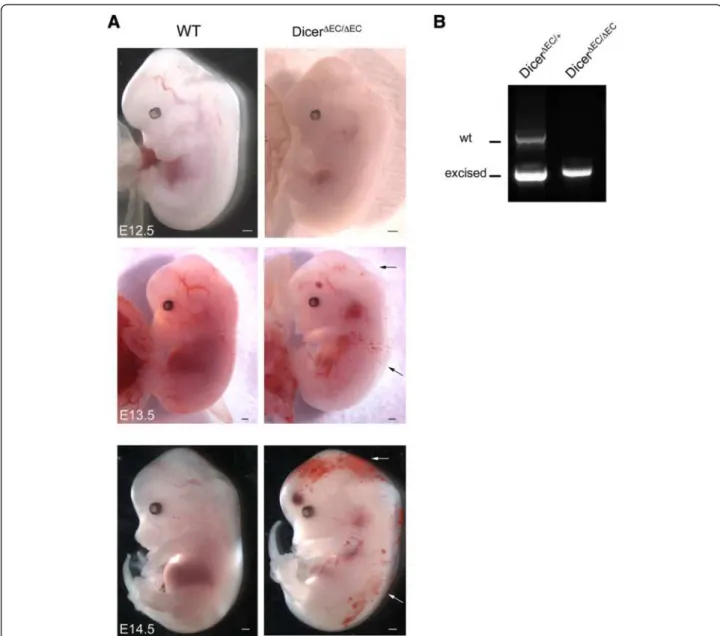
dicerΔEC/ΔEC mice died, embryos were examined from E10.5 to birth. Embryos were removed and embryonic DNA was analyzed for homo- or heterogeneity of the floxed allele. This genotype was then correlated with the viability of the embryo. Mendelian ratios were observed from E10.5 to E13.5 (see Table 1). Mutant embryos from E10.5 to E11.5 were macroscopically indistinguishable from the control littermates. At later stages, E12.5 on-wards, macroscopic examination revealed the presence of hemorrhages and edema in mutant embryos that in-creased in size and number with age (Figure 1A). Geno-typing PECAM+ endothelial cells showed efficient dicer
inactivation in E13.5 dicerΔEC/ΔEC embryos compared to E13.5 dicerΔEC/+
embryos here used as controls (Figure 1B). Recombination was also monitored in tie2-expressing cells using the ROSA26 (R26) reporter line [35]. We then crossed the dicerΔEC/+
males with homozygous
dicerfl/fl:R26/R26 females to generate dicerΔEC/+ :R26/+ (heterozygous, here as a control) and dicerΔEC/ΔEC:R26/+ (mutant) triple transgenic embryos. As indicated by whole-mount X-Gal staining, the recombination was ef-ficient in blood endothelial cells thereby allowing us to compare the pattern of the vascular network in mutant and control embryos using LacZ staining (Figure 2). Be-tween E10.5 and E12.5, dicerΔEC/ΔEC embryos did not display obvious blood vascular defects: avascular regions were not observed in control embryos. X-Gal-stained blood vessels formed properly and vascular density was comparable in both control and mutant embryos (Figure 2). Whole-mount staining using an anti-PECAM antibody confirmed these observations as reported in Figure 3A showing that vascular patterning of blood ves-sels was comparable to controls in E11.5 dicerΔEC/ΔEC
em-bryos. In order to study the development of the blood vessel network in greater details, branchpoints of the cra-nial vascular network (internal carotid artery) were quantified on E11.5 embryos. The number of branch-points in the internal carotid artery was not statistically different in dicerΔEC/ΔEC
embryos compared to WT em-bryos (Figure 3B).
Between E12.5 and E14.5, extensive edema gradually progressed on the back of the embryos and was some-times filled with blood cells in dicerΔEC/ΔEC embryos which were all dead at E14.5 (Figure 1 and Table 1). This also phenocopies the effects observed upon genetic dele-tion of Prospero homeobox 1 (prox-1) [36], Src hom-ology domain-containing leukocyte protein-76 (slp-76) [37] or C-type lectin-like receptor 2 (clec-2) [38]. All show impaired lymphatic vessel development and die in
utero with severe edema and hemorrhages. To establish
whether dicerΔEC/ΔEC
embryos also present defects in
Table 1 Genotype analysis in percentages of live embryos resulting from the cross of a dicerΔ/+male with a dicerfl/fl female
WT dicerΔEC/+ dicerΔEC/ΔEC Expected ratios 50% 25% 25% E10.5 n = 119 47.1% 28.5% 24.4% E11.5 n = 49 49% 30.8% 20.4% E12.5 n = 31 37.5% 38.7% 23.8% E13.5 n = 90 36.6% 26.7% 36.7% E14.5 n = 29 34.5% 23.8% 9.5% E15.5 n = 4 50% 50% 0% P14 n = 293 63.9% 36.1% 0%
Gauvrit et al. Vascular Cell 2014, 6:9 Page 3 of 10 http://www.vascularcell.com/content/6/1/9
lymphatic vessels development, we examined transverse sections of mutant embryos. At E13.5, we never ob-served any disruption of the main blood vessels i.e. the thoracic aorta or the cardinal vein in dicerΔEC/ΔEC
em-bryos (Additional file 1: Figure S1 and Figure 4). The lymph sacs, the first lymphatic structure that emerges from the cardinal vein [39] during development, also ap-peared normal (Additional file 2: Figure S2). At E13.5 however, in contrast to control embryos, these lymph sacs were filled with blood cells in dicerΔEC/ΔEC
embryos (Figure 4A). The lymphatic identity of the blood-filled structures was confirmed by the expression of lymphatic markers VEGFR-3 (Figure 4A) and PROX-1 (Additional file 3: Figure S3). Moreover, LYVE-1 whole-mount
immunostaining evidenced a complete overlap between blood-filled structures and the lymphatic vasculature in E14.5 mutant embryos (Figure 4B), confirming the blood-filled lymphatics phenotype.
Altogether, these data indicate that dicer inactivation in tie2 expressing cells leads to embryonic lethality at E14.5, and to a failure in the separation of lymphatic vessels during embryonic angiogenesis.
Discussion
Here, using Cre/loxP-mediated inactivation of dicer in
tie2-expressing cells, we demonstrate for the first time
that embryonic venous-lymphatic separation is submit-ted to epigenetic control by RNA interference. Previous
Figure 1 Conditional deletion of dicer gene leads to hemorrhage and edema. A) Whole-mount view of WT and dicerΔEC/ΔECembryos from E12.5 to E14.5. Hemorrhagic regions and edema are indicated (arrows). Scale Bar: 500 μm. B) PCR genotyping analysis of PECAM+endothelial cells from E13.5 dicerΔEC/+and dicerΔEC/ΔECembryos. Detection of Cre and dicer fragments (floxed, excised and WT) are presented.
studies using a similar approach of conditional dicer de-ficiency using tie2-Cre and ve-cadherin-CRE-ERT2 have reported reduced postnatal angiogenesis but no develop-mental defects [15]. The likely explanation for this discrepancy probably relies on the use of a different dicer-floxed mouse leading to the presence of residual Dicer protein levels in tie2-Cre:dicerfl/flendothelial cells, reflecting an incomplete excision of the dicer allele [15]. Thus, these mice were hypomorphic for dicer in ECs and tie2-Cre:dicerfl/fl newborn litters were overtly nor-mal and indistinguishable from their littermate controls. In contrast, in the present study, efficient dicer inactiva-tion was evidenced in PECAM+ endothelial cells which showed complete excision of dicer in dicerΔEC/ΔEC em-bryos. The present study thus shows that dicer gene de-letion in Tie2 expressing cells leads to embryonic lethality at E14.5. Mutant embryos, which display hem-orrhages and edema, showed blood-filled lymphatics without evident angiogenesis defects at early stages.
We here used the well-documented tie2-Cre trans-genic mice that express Cre in a pan-endothelial fashion for vascular endothelial targeting [34]. With the Rosa26
reporter line, we showed recombination in lymphatic vessels (Additional file 4: Figure S4). Using the same
tie2-Cre ROSA26 strain, Srinivasan et al. demonstrates
that at E11.5, Prox1+ endothelial cells in the anterior cardinal vein and those budding from it were lacZ+. Similarly, all E13.5 and E14.5 Prox1+endothelial cells in the lymph sacs were lacZ+[40]. Nevertheless, it should be noted that it has also been reported that tie2-Cre transgenic mice express Cre in blood island progenitors [41,42]. Recent studies have highlighted the role of hematopoietic cells during the process of separation between the venous and the lymphatic vasculature. It has been shown that podoplanin, a transmembrane pro-tein expressed on lymphatic endothelial cells, engages the platelet receptor CLEC-2 leading to Syk-Slp-76-dependent platelet activation [43]. Deletion of these genes leads to aberrant vascular connection between blood and lymphatic vessels. Similar lymphovenous con-nections were also observed in mice deficient for the homeodomain transcription factor Meis1 (myeloid eco-tropic viral integration site 1) which completely lack megakaryocyte/platelets and for the transcription factor
Figure 2 Mutant embryos do not display vascular defects. A-C) Whole-mount view of X-Gal staining of dicerΔEC/+:R26/+ embryos and
dicerΔEC/ΔEC:R26/+ embryos from E10.5 to E12.5. Scale Bar: 500 μm. The vascular network is identical in mutant and control embryos (n = 3 for
each condition).
Gauvrit et al. Vascular Cell 2014, 6:9 Page 5 of 10 http://www.vascularcell.com/content/6/1/9
Runx1 which lack hematopoietic stem cells [40,44]. It should also be noted that runx1 mutant embryos, which lack platelets, present hemorrhages in the brain [45], which could also be observed in some dicerΔEC/ΔEC em-bryos. Because platelets also act to maintain vascular in-tegrity and as the brain and lungs are more susceptible to haemorrhage in a mouse model of acute severe thrombocytopenia induced by platelet depletion [46], these hemorrhages most likely occur secondary to the lack of platelets. These data showed that platelets are re-quired during embryonic lymphangiogenesis for the sep-aration of the nascent lymphatic vasculature from blood vessels [47,48]. However, recent studies by Yang et al. [49] and Hägerling et al. [50] have disproved a direct involment of platelets in the emergence of the first jugu-lar lymph sacs. Podoplanin expression only starts after lymphatic endothelial cells leave the cardinal vein sug-gesting that platelets have a role restricted to the region where lymphatics and blood vessels coalesce, in the lym-phovenous valves. Nevertheless, the presence of blood cells in lymphatic vessels may also indicate an incom-plete separation of blood and lymph vessel, but could also result from de novo connections of previously sepa-rated blood and lymph vessels. Recently, Hess et al. proved that platelets interact with lymphatic endothelium
valves specifically at the thoracic duct-subclavian vein junction [51]. Blood-filled lymphatics arise due to backfill-ing of the lymphatic vascular network from this site either due to a lymphovenous valve defect or due to a platelet aggregation defect. We therefore looked at the thoracic duct-subclavian vein junction and we determined that the lymphovenous valves appears normal (Additional file 5: Figure S5) suggesting a defect in platelet aggregation.
We therefore sought to decipher whether perturbing
dicer expression in megakaryocytes could also reproduce
a blood-filled lymphatic phenotype during development by generating pf4-cre:dicerfl/flmice. Pf4-cre express Cre-recombinase in the megakaryocytic lineage as previously shown [52] and are a useful tool to study megakaryopoi-esis, and platelet function. These mice were born at nor-mal mendelian ratio and the separation of the lymphatic vasculature from the blood vessels was not disrupted during development (Additional file 6: Figure S6 and Table 2). Recombination was observed in liver megakar-yocytes before venous-lymphatic separation, as soon as E11.5 (data not shown) and persisted at E16.5 (Additional file 4: Figure S4B). However, the pf4-Cre transgene is also partially expressed in other hematopoietic lineages and the recombination pattern during early embryogenesis is not clear [53]. A megakaryocyte specific promoter that
Figure 3 Blood vessel patterning normally occurs in mutant embryos. A) Whole-mount immunohistochemical staining by anti-PECAM-1 antibody on E11.5 embryos. Scale Bar: 500 μm. B) Branchpoints quantification (means ± SEM) of the internal carotid artery (ica) on E11.5 embryos. The number of branchpoints is similar in WT and dicerΔEC/ΔECembryos (ica; dots represent arterial branchpoints; e, eye; v, veins). (WT n = 10,
could allow earlier deletion might be useful but does not exist.
Also, cells from the myeloid lineage play a critical role in this separation. Abnormal infiltration of a specific monocyte population in syk-deficient mice leads to lymph-atic hyperplasia, vessel dilation and blood-lymphlymph-atic shunts [54]. Tie2 is expressed in the early yolk sac meso-derm suggesting that recombination may occur in hematopoietic cells [55]. The use of a more endothelial specific strains such as ve-cadherin-CRE-ERT2 [56] or
pdgfb-CRE-ERT2 [57] would also be very useful for
under-standing the specific role of Dicer in the endothelium.
Figure 4 Mutant embryos present blood-filled lymphatics. A) Histological analysis of E13.5 WT and dicerΔEC/ΔECembryos. dicerΔEC/ΔECembryos
display blood-filled structures contrary to WT embryos as revealed after hematoxylin/eosin staining (HE) (upper panel). Immunostaining with the lymphatic marker VEGFR-3 confirmed the lymphatic identity of the blood filled structures (lower panel). Cv: cardinal vein, ls: lymph sac. Scale bar: 50 μm. (n = 2 for each condition). B) Whole-mount view of a E14.5 dicerΔEC/ΔECembryo after dissection (Left panel). Hemorrhages are indicated by
Δarrows. Immunohistochemical staining by anti-LYVE-1 antibody on the same mutant embryo (Right panel). Scale Bar: 500 μm. Higher magnification of the dicerΔEC/ΔECembryo after dissection and after LYVE-1 staining respectively (Lower panel). There is a complete overlap between hemorrhages and
LYVE-1 staining indicating blood-filled lymphatics in the dicerΔEC/ΔECembryo. (n = 2).
Table 2 Genotype analysis in percentages of live pups resulting from the cross of a pf4-cre:dicerΔ/+male with a dicerfl/flfemale
WT pf4-cre:dicerΔ/+ pf4-cre: dicerΔ/Δ
Expected ratios 50% 25% 25% P14 n = 40 45% 30% 25%
Gauvrit et al. Vascular Cell 2014, 6:9 Page 7 of 10 http://www.vascularcell.com/content/6/1/9
However, the CRE activation is tamoxifen-dependent making these models more suitable for postnatal angio-genesis as recombination at a precise embryonic time point might be somewhat difficult to achieve in a very re-producible manner.
MicroRNAs are involved in many aspects of physio-logical and malignant hematopoiesis but surprisingly, no existing studies have focused on the role of dicer during hematopoietic development. However, dicer invalidation in adult has been described. Buza-Vidas et al. showed that dicer is required during erythroid lineage differenti-ation [58]. It was also suggested that Dicer is involved in the regulation of the hematopoietic stem cell niche as well as the regulation of hematopoietic stem cell number [59,60]. The blood filled phenotype that we observed could result from either a defect of hematopoiesis or a volume expansion of the blood stream indirectly affect-ing lymphatic development. We therefore believe that further experiments, outside of the scope of the present manuscript, will be needed to determine precisely whether hematopoiesis is modulated in dicerΔEC/ΔEC
em-bryos and to fully decipher the cellular and molecular mechanisms responsible for the blood-filled lymphatic phenotype in these mice.
Conclusion
Taken together, these results show a new role for RNA interference in epigenetic control of embryonic venous-lymphatic separation and provide a knowledge base for further investigations to validate functional roles for microRNAs.
Additional files
Additional file 1: Figure 1. Histological analysis of E13.5 thoracic aorta in WT and dicerΔEC/ΔECembryos. Immunostaining with VEGFR-2 confirmed
a normal patterning of the thoracic aorta of dicerΔEC/ΔECembryos. Scale
Bar: 2 μm. (n = 3).
Additional file 2: Figure 2. Mutant embryos do not present lymph sacs defect. Whole-mount view of E12.5 WT and dicerΔEC/ΔECembryos after
LYVE-1 staining. The mutant embryo do not show a lymph sac defect. (n = 3 for each condition).
Additional file 3: Figure 3. Prox1 expression on transversal sections of E13.5 WT and dicerΔEC/ΔECembryos (n = 2 for each condition). Prox1
expression is maintained in lymphatics vessels in mutant embryos (upper panel), and the number of Prox1 expressing cells is similar in WT and dicerΔEC/ΔECembryos (lower panel).
Additional file 4: Figure 4. Whole-mount view of X-Gal staining of dicerΔEC/ΔEC:R26/+ embryos at E13.5. Mutant embryo present recombination
in lymphatic vessels (indicated by arrows). (n = 5).
Additional file 5: Figure 5. Histological analysis of E13.5 lymphovenous valves in WT and dicerΔEC/ΔECembryos (indicated by arrows).
Immunostaining with VEGFR-3 showed a normal patterning and morph-ology of the lymphovenous valves of dicerΔEC/ΔECembryos. Scale Bar: 2
μm. (n = 2 for each condition).
Additional file 6: Figure 6. Conditional deletion of dicer in megakaryocytes does not lead to embryonic lethality. A) Whole-mount view of WT and pf4-cre:dicerΔ/Δ embryos at E16.5. Mutant embryos do
not present any obvious phenotype. B) Whole-mount view of X-Gal staining of a pf4-cre:dicerΔ/Δliver at E16.5 (Left panel). Histological analysis
of the same E16.5 liver (Right panel). Recombination occurs in typical large megakaryocytes in the liver. (n = 3).
Abbreviations
CLEC-2:C-type lectin-like receptor 2; Cv: Cardinal vein; E: Embryonic day; Ica: Internal carotid artery; Ls: Lymph sac; miRNA: microRNA;
mRNA: Messenger RNA; Pecam-1: Platelet endothelial cell adhesion molecule 1; siRNA: Short interfering RNA; Vegfr-3: Vascular endothelial growth factor receptor 3; WT: Wild type.
Competing interests
The authors declare that they have no competing interests. Authors’ contribution
SGe, SGa designed experiments. SGa, JP and ML performed experiments. SGe, SGa, IG and MT wrote the paper. All authors read and approved the final manuscript.
Acknowledgements
This work has received support under the program « Investissements d’Avenir » launched by the French Government and implemented by the ANR, with the references:
ANR-10-LABX-54 MEMO LIFE.
ANR-11-IDEX-0001-02 PSL* Research University. Sources of funding
This work was supported by a grant from Agence Nationale de la Recherche (R10032JJ - RPV10032JJA) and a grant from La Ligue Contre le Cancer. Author details
1Collège de France, Center for Interdisciplinary Research in Biology (CIRB), 11,
place Marcelin Berthelot, Paris F-75005, France.2CNRS UMR 7241, Paris F-75005, France.3INSERM U 1050, Paris F-75005, France.4ED 394: Physiologie
et Physiopathologie, Université Pierre et Marie Curie, Paris F-75005, France.
5Lab of Vascular Hematology/Angiogenesis, Goethe University Frankfurt,
Frankfurt, Germany.6Institute for Transfusion Medicine, DRK
Blutspendedienst, Goethe University Frankfurt, Frankfurt, Germany.7INSERM
U1009, Villejuif F-94805, France.8Gustave Roussy, Villejuif F-94805, France.
9Equipe labellisée Ligue contre le Cancer, Paris, France.10Department of
Pathology, Saint-Louis Hospital, AP-HP, Paris F-75010, France.
Received: 2 October 2013 Accepted: 25 February 2014 Published: 1 April 2014
References
1. Ketting RF: The many faces of RNAi. Dev Cell 2011, 20:148–161. 2. Bernstein E, Kim SY, Carmell MA, Murchison EP, Alcorn H, Li MZ, Mills AA,
Elledge SJ, Anderson KV, Hannon GJ: Dicer is essential for mouse development. Nat Genet 2003, 35:215–217.
3. Yang WJ, Yang DD, Na S, Sandusky GE, Zhang Q, Zhao G: Dicer is required for embryonic angiogenesis during mouse development. J Biol Chem 2005, 280:9330–9335.
4. Harfe BD, McManus MT, Mansfield JH, Hornstein E, Tabin CJ: The RNaseIII enzyme Dicer is required for morphogenesis but not patterning of the vertebrate limb. Proc Natl Acad Sci U S A 2005, 102:10898–10903. 5. Cobb BS, Nesterova TB, Thompson E, Hertweck A, O’Connor E, Godwin J,
Wilson CB, Brockdorff N, Fisher AG, Smale ST, Merkenschlager M: T cell lineage choice and differentiation in the absence of the RNase III enzyme Dicer. J Exp Med 2005, 201:1367–1373.
6. Chen JF, Murchison EP, Tang R, Callis TE, Tatsuguchi M, Deng Z, Rojas M, Hammond SM, Schneider MD, Selzman CH, Meissner G, Patterson C, Hannon GJ, Wang DZ: Targeted deletion of Dicer in the heart leads to dilated cardiomyopathy and heart failure. Proc Natl Acad Sci USA 2008, 105:2111–2116.
7. Otsuka M, Zheng M, Hayashi M, Lee JD, Yoshino O, Lin S, Han J: Impaired microRNA processing causes corpus luteum insufficiency and infertility in mice. J Clin Invest 1944–1954, 2008:118.
8. Albinsson S, Skoura A, Yu J, DiLorenzo A, Fernandez-Hernando C, Offermanns S, Miano JM, Sessa WC: Smooth muscle miRNAs are critical for post-natal regulation of blood pressure and vascular function. PLoS One 2011, 6:e18869.
9. Albinsson S, Suarez Y, Skoura A, Offermanns S, Miano JM, Sessa WC: MicroRNAs are necessary for vascular smooth muscle growth,
differentiation, and function. Arterioscler Thromb Vasc Biol 2010, 30:1118–1126. 10. Albinsson S, Sessa WC: Can microRNAs control vascular smooth muscle
phenotypic modulation and the response to injury? Physiol Genomics 2011, 43:529–533.
11. Boettger T, Beetz N, Kostin S, Schneider J, Kruger M, Hein L, Braun T: Acquisition of the contractile phenotype by murine arterial smooth muscle cells depends on the Mir143/145 gene cluster. J Clin Invest 2009, 119:2634–2647.
12. Cheng Y, Liu X, Yang J, Lin Y, Xu DZ, Lu Q, Deitch EA, Huo Y, Delphin ES, Zhang C: MicroRNA-145, a novel smooth muscle cell phenotypic marker and modulator, controls vascular neointimal lesion formation. Circ Res 2009, 105:158–166.
13. Xin M, Small EM, Sutherland LB, Qi X, McAnally J, Plato CF, Richardson JA, Bassel-Duby R, Olson EN: MicroRNAs miR-143 and miR-145 modulate cytoskeletal dynamics and responsiveness of smooth muscle cells to injury. Genes Dev 2009, 23:2166–2178.
14. Kuehbacher A, Urbich C, Zeiher AM, Dimmeler S: Role of Dicer and Drosha for endothelial microRNA expression and angiogenesis. Circ Res 2007, 101:59–68.
15. Suarez Y, Fernandez-Hernando C, Yu J, Gerber SA, Harrison KD, Pober JS, Iruela-Arispe ML, Merkenschlager M, Sessa WC: Dicer-dependent endothelial microRNAs are necessary for postnatal angiogenesis. Proc Natl Acad Sci USA 2008, 105:14082–14087.
16. Heusschen R, van Gink M, Griffioen AW, Thijssen VL: MicroRNAs in the tumor endothelium: novel controls on the angioregulatory switchboard. Biochim Biophys Acta 1805, 2010:87–96.
17. Poliseno L, Tuccoli A, Mariani L, Evangelista M, Citti L, Woods K, Mercatanti A, Hammond S, Rainaldi G: MicroRNAs modulate the angiogenic properties of HUVECs. Blood 2006, 108:3068–3071.
18. Kuehbacher A, Urbich C, Dimmeler S: Targeting microRNA expression to regulate angiogenesis. Trends Pharmacol Sci 2008, 29:12–15.
19. Landskroner-Eiger S, Moneke I, Sessa WC: miRNAs as modulators of angiogenesis. Cold Spring Harbor perspectives in medicine 2013, 3:a006643. 20. Suarez Y, Fernandez-Hernando C, Pober JS, Sessa WC: Dicer dependent
microRNAs regulate gene expression and functions in human endothe-lial cells. Circ Res 2007, 100:1164–1173.
21. Suarez Y, Sessa WC: MicroRNAs as novel regulators of angiogenesis. Circ Res 2009, 104:442–454.
22. Bonauer A, Carmona G, Iwasaki M, Mione M, Koyanagi M, Fischer A, Burchfield J, Fox H, Doebele C, Ohtani K, Chavakis E, Potente M, Tjwa M, Urbich C, Zeiher AM, Dimmeler S: MicroRNA-92a controls angiogenesis and functional recovery of ischemic tissues in mice. Science 2009, 324:1710–1713.
23. Doebele C, Bonauer A, Fischer A, Scholz A, Reiss Y, Urbich C, Hofmann WK, Zeiher AM, Dimmeler S: Members of the microRNA-17-92 cluster exhibit a cell-intrinsic antiangiogenic function in endothelial cells. Blood 2010, 115:4944–4950.
24. Fish JE, Santoro MM, Morton SU, Yu S, Yeh RF, Wythe JD, Ivey KN, Bruneau BG, Stainier DY, Srivastava D: miR-126 regulates angiogenic signaling and vascular integrity. Dev Cell 2008, 15:272–284.
25. Hinkel R, Penzkofer D, Zuhlke S, Fischer A, Husada W, Xu QF, Baloch E, van Rooij E, Zeiher AM, Kupatt C, Dimmeler S: Inhibition of MicroRNA-92a Protects Against Ischemia/Reperfusion Injury in a Large-Animal Model. Circulation 2013, 128:1066–1075.
26. Kuhnert F, Mancuso MR, Hampton J, Stankunas K, Asano T, Chen CZ, Kuo CJ: Attribution of vascular phenotypes of the murine Egfl7 locus to the microRNA miR-126. Development 2008, 135:3989–3993.
27. Wang S, Aurora AB, Johnson BA, Qi X, McAnally J, Hill JA, Richardson JA, Bassel-Duby R, Olson EN: The endothelial-specific microRNA miR-126 governs vascular integrity and angiogenesis. Dev Cell 2008, 15:261–271. 28. Yin KJ, Olsen K, Hamblin M, Zhang J, Schwendeman SP, Chen YE: Vascular
endothelial cell-specific microRNA-15a inhibits angiogenesis in hindlimb ischemia. J Biol Chem 2012, 287:27055–27064.
29. Kane NM, Howard L, Descamps B, Meloni M, McClure J, Lu R, McCahill A, Breen C, Mackenzie RM, Delles C, Mountford JC, Milligan G, Emanueli C,
Baker AH: Role of microRNAs 99b, 181a, and 181b in the differentiation of human embryonic stem cells to vascular endothelial cells. Stem Cells 2012, 30:643–654.
30. Fasanaro P, D’Alessandra Y, Di Stefano V, Melchionna R, Romani S, Pompilio G, Capogrossi MC, Martelli F: MicroRNA-210 modulates endothelial cell response to hypoxia and inhibits the receptor tyrosine kinase ligand Ephrin-A3. J Biol Chem 2008, 283:15878–15883.
31. Fasanaro P, Greco S, Lorenzi M, Pescatori M, Brioschi M, Kulshreshtha R, Banfi C, Stubbs A, Calin GA, Ivan M, Capogrossi MC, Martelli F: An integrated approach for experimental target identification of hypoxia-induced miR-210. J Biol Chem 2009, 284:35134–35143.
32. Kazenwadel J, Michael MZ, Harvey NL: Prox1 expression is negatively regulated by miR-181 in endothelial cells. Blood 2010, 116:2395–2401. 33. del Toro R, Prahst C, Mathivet T, Siegfried G, Kaminker JS, Larrivee B, Breant
C, Duarte A, Takakura N, Fukamizu A, Penninger J, Eichmann A: Identification and functional analysis of endothelial tip cell-enriched genes. Blood 2010, 116:4025–4033.
34. Kisanuki YY, Hammer RE, Miyazaki J, Williams SC, Richardson JA, Yanagisawa M: Tie2-Cre transgenic mice: a new model for endothelial cell-lineage analysis in vivo. Dev Biol 2001, 230:230–242.
35. Soriano P: Generalized lacZ expression with the ROSA26 Cre reporter strain. Nat Genet 1999, 21:70–71.
36. Wigle JT, Oliver G: Prox1 function is required for the development of the murine lymphatic system. Cell 1999, 98:769–778.
37. Abtahian F, Guerriero A, Sebzda E, Lu MM, Zhou R, Mocsai A, Myers EE, Huang B, Jackson DG, Ferrari VA, Tybulewicz V, Lowell CA, Lepore JJ, Koretzky GA, Kahn ML: Regulation of blood and lymphatic vascular separation by signaling proteins SLP-76 and Syk. Science 2003, 299:247–251.
38. Suzuki-Inoue K, Inoue O, Ding G, Nishimura S, Hokamura K, Eto K, Kashiwagi H, Tomiyama Y, Yatomi Y, Umemura K, Shin Y, Hirashima M, Ozaki Y: Essential in vivo roles of the C-type lectin receptor CLEC-2: embryonic/ neonatal lethality of CLEC-2-deficient mice by blood/lymphatic misconnections and impaired thrombus formation of CLEC-2-deficient platelets. J Biol Chem 2010, 285:24494–24507.
39. Wang Y, Oliver G: Current views on the function of the lymphatic vasculature in health and disease. Genes Dev 2010, 24:2115–2126. 40. Srinivasan RS, Dillard ME, Lagutin OV, Lin FJ, Tsai S, Tsai MJ, Samokhvalov IM,
Oliver G: Lineage tracing demonstrates the venous origin of the mammalian lymphatic vasculature. Genes Dev 2007, 21:2422–2432. 41. Li W, Ferkowicz MJ, Johnson SA, Shelley WC, Yoder MC: Endothelial cells in
the early murine yolk sac give rise to CD41-expressing hematopoietic cells. Stem Cells Dev 2005, 14:44–54.
42. Lancrin C, Sroczynska P, Stephenson C, Allen T, Kouskoff V, Lacaud G: The haemangioblast generates haematopoietic cells through a haemogenic endothelium stage. Nature 2009, 457:892–895.
43. Bertozzi CC, Hess PR, Kahn ML: Platelets: covert regulators of lymphatic development. Arterioscler Thromb Vasc Biol 2010, 30:2368–2371.
44. Carramolino L, Fuentes J, Garcia-Andres C, Azcoitia V, Riethmacher D, Torres M: Platelets play an essential role in separating the blood and lymphatic vasculatures during embryonic angiogenesis. Circ Res 2010, 106:1197–1201. 45. Okuda T, van Deursen J, Hiebert SW, Grosveld G, Downing JR: AML1, the
target of multiple chromosomal translocations in human leukemia, is essential for normal fetal liver hematopoiesis. Cell 1996, 84:321–330. 46. Goerge T, Ho-Tin-Noe B, Carbo C, Benarafa C, Remold-O’Donnell E, Zhao BQ,
Cifuni SM, Wagner DD: Inflammation induces hemorrhage in thrombocytopenia. Blood 2008, 111:4958–4964.
47. Uhrin P, Zaujec J, Breuss JM, Olcaydu D, Chrenek P, Stockinger H, Fuertbauer E, Moser M, Haiko P, Fassler R, Alitalo K, Binder BR, Kerjaschki D: Novel function for blood platelets and podoplanin in developmental separation of blood and lymphatic circulation. Blood 2010, 115:3997–4005.
48. Bertozzi CC, Schmaier AA, Mericko P, Hess PR, Zou Z, Chen M, Chen CY, Xu B, Lu MM, Zhou D, Sebzda E, Santore MT, Merianos DJ, Stadtfeld M, Flake AW, Graf T, Skoda R, Maltzman JS, Koretzky GA, Kahn ML: Platelets regulate lymphatic vascular development through CLEC-2-SLP-76 signaling. Blood 2010, 116:661–670.
49. Yang Y, Garcia-Verdugo JM, Soriano-Navarro M, Srinivasan RS, Scallan JP, Singh MK, Epstein JA, Oliver G: Lymphatic endothelial progenitors bud from the cardinal vein and intersomitic vessels in mammalian embryos. Blood 2012, 120:2340–2348.
Gauvrit et al. Vascular Cell 2014, 6:9 Page 9 of 10 http://www.vascularcell.com/content/6/1/9
50. Hagerling R, Pollmann C, Andreas M, Schmidt C, Nurmi H, Adams RH, Alitalo K, Andresen V, Schulte-Merker S, Kiefer F: A novel multistep mechanism for initial lymphangiogenesis in mouse embryos based on ultramicroscopy. EMBO J 2013, 32:629–644.
51. Hess PR, Rawnsley DR, Jakus Z, Yang Y, Sweet DT, Fu J, Herzog B, Lu M, Nieswandt B, Oliver G, Makinen T, Xia L, Kahn ML: Platelets mediate lymphovenous hemostasis to maintain blood-lymphatic separation throughout life. J Clin Invest 2014, 124:273–284.
52. Tiedt R, Schomber T, Hao-Shen H, Skoda RC: Pf4-Cre transgenic mice allow the generation of lineage-restricted gene knockouts for studying megakaryocyte and platelet function in vivo. Blood 2007, 109:1503–1506.
53. Calaminus SD, Guitart AV, Sinclair A, Schachtner H, Watson SP, Holyoake TL, Kranc KR, Machesky LM: Lineage tracing of Pf4-Cre marks hematopoietic stem cells and their progeny. PLoS One 2012, 7:e51361.
54. Bohmer R, Neuhaus B, Buhren S, Zhang D, Stehling M, Bock B, Kiefer F: Regulation of developmental lymphangiogenesis by Syk(+) leukocytes. Dev Cell 2010, 18:437–449.
55. Ema M, Yokomizo T, Wakamatsu A, Terunuma T, Yamamoto M, Takahashi S: Primitive erythropoiesis from mesodermal precursors expressing VE-cadherin, PECAM-1, Tie2, endoglin, and CD34 in the mouse embryo. Blood 2006, 108:4018–4024.
56. Pitulescu ME, Schmidt I, Benedito R, Adams RH: Inducible gene targeting in the neonatal vasculature and analysis of retinal angiogenesis in mice. Nat Protoc 2010, 5:1518–1534.
57. Claxton S, Kostourou V, Jadeja S, Chambon P, Hodivala-Dilke K, Fruttiger M: Efficient, inducible Cre-recombinase activation in vascular endothelium. Genesis 2008, 46:74–80.
58. Buza-Vidas N, Cismasiu VB, Moore S, Mead AJ, Woll PS, Lutteropp M, Melchiori L, Luc S, Bouriez-Jones T, Atkinson D, O’Carroll D, Jacobsen SE, Nerlov C: Dicer is selectively important for the earliest stages of erythroid development. Blood 2012, 120:2412–2416.
59. Guo S, Lu J, Schlanger R, Zhang H, Wang JY, Fox MC, Purton LE, Fleming HH, Cobb B, Merkenschlager M, Golub TR, Scadden DT: MicroRNA miR-125a controls hematopoietic stem cell number. Proc Natl Acad Sci USA 2010, 107:14229–14234.
60. Raaijmakers MH, Mukherjee S, Guo S, Zhang S, Kobayashi T, Schoonmaker JA, Ebert BL, Al-Shahrour F, Hasserjian RP, Scadden EO, Aung Z, Matza M, Merkenschlager M, Lin C, Rommens JM, Scadden DT: Bone progenitor dysfunction induces myelodysplasia and secondary leukaemia. Nature 2010, 464:852–857.
doi:10.1186/2045-824X-6-9
Cite this article as: Gauvrit et al.: The role of RNA interference in the developmental separation of blood and lymphatic vasculature. Vascular Cell 2014 6:9.
Submit your next manuscript to BioMed Central and take full advantage of:
• Convenient online submission
• Thorough peer review
• No space constraints or color figure charges
• Immediate publication on acceptance
• Inclusion in PubMed, CAS, Scopus and Google Scholar
• Research which is freely available for redistribution
Submit your manuscript at www.biomedcentral.com/submit