HAL Id: inserm-00168108
https://www.hal.inserm.fr/inserm-00168108
Submitted on 27 Aug 2007HAL is a multi-disciplinary open access
archive for the deposit and dissemination of sci-entific research documents, whether they are pub-lished or not. The documents may come from teaching and research institutions in France or abroad, or from public or private research centers.
L’archive ouverte pluridisciplinaire HAL, est destinée au dépôt et à la diffusion de documents scientifiques de niveau recherche, publiés ou non, émanant des établissements d’enseignement et de recherche français ou étrangers, des laboratoires publics ou privés.
Aurore Brugeaud, Cécile Travo, Danielle Demêmes, Marc Lenoir, Jordi
Llorens, Jean-Luc Puel, Christian Chabbert
To cite this version:
Aurore Brugeaud, Cécile Travo, Danielle Demêmes, Marc Lenoir, Jordi Llorens, et al.. Control of hair cell excitability by vestibular primary sensory neurons.. Journal of Neuroscience, Society for Neuroscience, 2007, 27 (13), pp.3503-11. �10.1523/JNEUROSCI.5185-06.2007�. �inserm-00168108�
Control of hair cell excitability by vestibular primary sensory neurons
Journal: Journal of Neuroscience Manuscript ID: JN-RM-5185-06.R1 Manuscript Type: Regular Manuscript
Manuscript Section: Development Plasticity Repair Date Submitted by the
Author: n/a
Complete List of Authors: Brugeaud, Aurore; INSERM U 583, Neurosciences Travo, Cecile; INSERM U 583, Neurosciences Dememes, Danielle; INSERM U 583, Neurosciences Lenoir, Marc; INSERM U 583, Neurosciences
Llorens, Jordi; Universitat de Barcelona, Departament de Ciencies Fisiologiques II
Puel, Jean-Luc; INSERM U 583, Neurosciences
CHABBERT, CHRISTIAN; INSERM U583, Neurosciences
Keywords: Excitotoxicity, Development, Excitability, Hair Cell, repair, synapse impairement, utricle, voltage-gated sodium channel
Themes & Topics:
a. Hair celss, endorgans, and nerve < 7. Vestibular < Theme C: Sensory and Motor Systems, a. Synapse formation: PNS < 3. Synaptogenesis and Activity-Dependent Development < Theme A: Development
ScholarOne, 375 Greenbrier Drive, Charlottesville, VA, 22901
Title: Control of hair cell excitability by vestibular primary sensory neurons. Abbreviated title: Innervation modulates vestibular hair cells excitability
Authors and author addresses: Aurore Brugeaud, Cécile Travo, Danielle Demèmes, Marc Lenoir, Jordi LLorens*, Jean-Luc Puel and Christian Chabbert
INSERM U583 Montpellier, France. * Departament de Ciencies Fisiologiques II, Universitat de Barcelona, l'Hospitalet de Llobregat, Espa a.
Correspondence should be addressed to Dr Christian Chabbert, INSERM U583, Institut des Neurosciences de Montpellier, Equipe Physiopathologie et Thérapie de l’Oreille Interne, Groupe d’Etude des Désordres Vestibulaires, Hôpital St Eloi, 34091 Montpellier cedex 5 (France). E-mail: chabbert@univ-montp2.fr
Number of figures: 7 Number of tables: 2 Number of pages: 18
Key words: voltage-gated sodium channel, hair cells, utricle, excitability, development, excitotoxicity, nerve impairment, repair.
Acknowledgments: This work was supported by the Centre National d’Etudes Spatiales, the French Ministry of Research and New Technologies and the Spanish Ministry of Education and Science (BFI2003-01606 FEDER). We thank Drs. G. Desmadryl, G. Geleoc and W. Marcotti for valuable comments on that work, and Drs. S. Bartolami and J. Ruel for helpful discussions and the Montpellier Regional Center of Cellular Imaging (CRIC).
ABSTRACT
In the rat utricle, synaptic contacts between hair cells and the nerve fibers arising from the vestibular primary neurons form during the first week after birth. During that period, the sodium-based excitability that characterizes neonate utricle sensory cells is switched off. To investigate whether the establishment of synaptic contacts was responsible for the modulation of the hair cell excitability, we used an organotypic culture of rat utricle in which the setting of synapses was prevented. Under this condition, the voltage-gated sodium current and the underlying action potentials persisted in a large proportion of non-afferented hair cells. We
then studied whether impairment of nerve terminals in utricle of adult rats may also affect hair cell excitability. We induced selective and transient damages of afferent terminals using glutamate excitotoxicity in vivo. The efficiency of the excitotoxic injury was attested by selective swellings of the terminals and underlying altered vestibular behavior. Under this condition, the sodium-based excitability transiently recovered in hair cells. These results indicate that the modulation of hair cells excitability depends on the state of the afferent terminals. In adult utricle hair cells this property may be essential to set the conditions required for restoration of the sensory network after damage. This is achieved via re-expression of a biological process that occurs during synaptogenesis.
INTRODUCTION
The developmental sequence of innervation of vestibular organs has been well described in rodents, based on morphological and functional studies (for review see Eatock and Hurley, 2003). Nerve fibers arising from Scarpa’s ganglion enter the sensory epithelia a few days before birth and first immature synaptic contacts can already be identified at birth. Synapses acquire their mature phenotype by the end of the first postnatal week (Desmadryl and Sans, 1990, Dechesne et al., 1986), giving rise to the first recordable synaptic activities (Curthoys, 1982; Desmadryl et al., 1986). During that period, hair cells gradually acquire the biophysical properties that bring them their mature character (Rush et al., 1998).
We have previously shown that, at birth (P0), rat utricle hair cells transiently express a neuronal-like tetrodotoxin (TTX)-sensitive voltage-gated Na+ current (INa), enabling these cells to generate sodium-driven action potentials (AP) (Chabbert et al., 2003). INa is downregulated during the first postnatal week, decreasing the sodium based excitability in mature hair cells. This developmental excitability, also observed in the mouse utricle (Geleoc et al., 2004), has also been reported in other sensory organs of the higher vertebrates, such as the cochlea (Evans and Fuchs, 1987; Kros et al., 1998) and the retina (Pan and Hu, 2000; Kawai et al., 2001). Although in most cases, the physiological relevance of the INa remains unknown, we first reported that INa was involved in the activity-dependent secretion of brain-derived neurotrophic factor (BDNF) in the neonate rat utricle (Chabbert et al., 2003). Regarding the major role of BDNF in the establishment and the stabilization of synaptic contacts (Ernfors et al., 1995; Shimmang et al., 1995), we proposed that the transient hair cell excitability may contribute to the synaptogenesis process in the vestibular organs. A direct demonstration of this hypothesis remains to be provided.
In this study, we tested the hypothesis that the modulation of hair cell excitability is controlled by the vestibular nerve fibers. We first prevented the establishment of synaptic
contacts in neonate rat utricles cultured in vitro before synaptic stabilization. Under this condition, the membrane expression of INa and the ability to fire AP persisted in the non-afferented hair cells. We then selectively damaged afferent terminals in adult utricles using glutamate excitotoxicity in vivo. In this condition, a large proportion of hair cells transiently expressed INa and recovered the ability to fire AP. These observations demonstrate that the state of the afferent terminals directly affects hair cell excitability. The physiological relevance of this biological process on post-injury repair processes is discussed.
METHODS
Animals: Experiments were performed on newborn and adult (40 days old) female Wistar rats in
accordance with French Ministry of Agriculture regulations and European Community Council Directive no. 86/609/EEC, OJL 358, 18 December 1986. Neonate rats were decapitated, and adult animals were anesthetized with pentobarbital solution injection (0.1 ml/100 g) before decapitation. All efforts were made to minimize the number of animals used and their suffering.
Denervated utricle organotypic culture: Utricle cultures were prepared as previously described
(Gaboyard et al., 2005). Briefly, utricles were explanted aseptically from P0 rats. The otolith membrane was removed and utricles were excised. Nerve fibers were trimmed close to the epithelium. Epithelia were embedded in 10 µl of extracellular matrix extracted from Engelbreth-Holm-Swarm tumors (Harbor Bio-products, Norwood, MA) on laminin-coated coverslips, and covered with 2 ml of feeding medium, a 1:1 mixture of Dulbecco's modified Eagle Medium (DMEM) and Ham's F-12 nutrient (Gibco BRL, Invitrogen, Gaithersburg, MD, USA) medium supplemented with 10% fetal bovine serum, glucose (5 g/L), glutamine (1.5 mM), sodium bicarbonate (1.1 g/l), and HEPES buffer (15 mM, pH 7.4). Cultures were maintained for 10 days in a humidified 5% CO2 atmosphere before electrophysiology and immunocytochemical experiments. The feeding medium was renewed every three days.
Excitotoxic impairment of vestibular nerve terminals: Experiments were performed on 78 adult female
rats (body weight 180-250 g). Pharmacological agents were applied to the inner ear as previously described by Guitton et al. (2003). Surgery was carried out under general anesthesia induced with isoflurane (Baxter S.A., Lessignes, Belgium), using a Minerve apparatus (induction 10 min at 2.5 %, maintenance at 2% under 0.8 L O2/min). Under aseptic condition, the right otic bulla was exposed
through a retro-auricular incision and a hole was created with a micro-drill, revealing the round window. Gelfoam (Gelita Tampon, B. Braun Medical Inc., Melsungen, Germany) filled with 2.5 µl of artificial perilymph solution (APS) (see below), containing 5 mM kainate, a glutamate receptor agonist (Sigma, St Louis, USA), was applied in the round window niche. The tympanic bulla was closed with dental cement (Unifast Trad, GC Corporation, Tokyo, Japan) and the surgical incision was sutured. We
treated 42 rats with kainate alone (17 for electron microscopy, 12 for immunochemmistry and 13 for electrophysical investigations), and 15 rats with kainate plus 200 µM DNQX (6, 7-dinitroquinoxaline-2, 3 (1H, 4H)-dione, Sigma), a competitive kainate/quisqualate (non-N-methyl-D-Aspartate, NMDA) receptor antagonist (6 for electron microscopy, 6 for immunochemistry and 3 for electrophysiology). 21 animals were used as control with gelfoam filled with 2.5 µl APS alone. After surgery, animals were returned to their cages and maintained at 22 ± 2°C.
Thin sections and transmission electron microscopy
Utricles were processed as previously reported (Seoane et al., 2001). Briefly, sensory epithelia were rapidly removed and fixed by incubation for two hours in 1% paraformaldehyde and 2.5% glutaraldehyde in 0.1 M PBS. They were then kept overnight in 0.1 M PBS at 4°C. Classical techniques were used to prepare the samples for transmission electron microscopy: post-fixation for 1 hour in 1% OsO4 in cacodylate buffer (pH 7.2), subsequent dehydration with increasing concentrations of ethanol
(70°, 95°, 100°) and embedding in epoxy resin. Utricles were longitudinally sectioned in 1µm sections with a diamond knife on an ultra-microtome (Reichert OMU3 Vienna, Austria). Sections were stained with toluidine blue (Rhône Poulenc, France). Ultrathin transverse sections were cut, stained with 2% uranyl acetate and lead citrate and examined using a JEOL 6300F electron microscope.
Morphological evaluation of nerve terminal damages. The degree of nerve terminal swelling was
estimated by measuring the calices thickness at half its height (corresponding to the level of the type I hair cell nucleus) and the diameter of the boutons innervating the type II hair cells under each experimental condition using the METAMORPH software (Universal Imaging). Swelling was measured from semithin sections of three utricles for each experimental condition. The proportion of nerve terminals damaged by the kainate treatment in each epithelium was estimated on entire semi-thin sections cut through the longitudinal axis of treated (n=5) and control utricles (n=5). Nerve terminals displaying widths larger than 1µm were considered as swollen. We counted the total number of hair cells present in each semithin section and determined the percent of hair cells that displayed swollen terminals. The evaluation was made on single sections chosen randomly over series of consecutive sections of each utricle to mimic the conditions encountered in the electrophysiological recordings. Student t-Test (p<0.05) was used to characterised the significant relevance.
Evaluation of vestibular dysfunction: Vestibular dysfunction was evaluated by observation of
spontaneous motor behavior as previously described (Boadas-Vaello et al., 2005). Briefly, rats were placed for one minute on a table top and the experimenter rated the animals from 0 to 4 for circling, retropulsion and abnormal head movements. Circling was defined as stereotyped circling movement. Retropulsion consisted of backward displacement of the animal. The head bobbing consisted of intermittent extreme backward extension of the neck. The rats were also rated 0 to 4 for the tail-hang reflex, contact inhibition of the righting reflex and air righting reflex tests. When lifted by the tail, normal rats exhibit a “landing” response consisting of forelimb extension. Rats with impaired vestibular
function bent ventrally, sometimes “crawling” up towards their tails, thus tending to occipital landing. For the contact inhibition of the righting reflex, rats were placed supine on a horizontal surface and a metal bar grid was lightly placed in contact with the soles of the animals’ feet. Healthy rats quickly right themselves, whereas the vestibular-deficient rats lie on their back, with their feet up and “walk” with respect to the ventral surface. For the air righting reflex, the animals were held supine and dropped from a height of 40 cm onto a foam cushion. Normal rats are successful in righting themselves in the air whereas vestibular-deficient rats are not. The results of all tests were combined and expressed as a percentage (versus a maximal score of 24). To provide a positive control condition for the behavioral assessment of vestibular dysfunction, four animals were injected intraperitoneally with 1000 mg/kg of 3,3’-iminodipropionitrile (IDPN) 2 days before behavioral assessment. This treatment causes a complete loss of vestibular hair cells and hence of vestibular function (Llorens et al., 1993).
Electrophysiological recordings: Whole-cell voltage and current clamp recordings were made using an
Axopatch 200B amplifier (Axon Instruments; Molecular Devices Corp. Sunnyvale, CA, USA) in hair cells from cultured or acutely isolated rat utricles as previously described (Chabbert et al., 2003). Epithelia were placed in an experimental chamber containing 2 ml of artificial perilymph solution (in mM): 137 NaCl, 0.7 NaH2PO4, 5.8 KCl, 1.3 CaCl2, 0.9 MgCl2, 5.6 D-glucose, 10 HEPES-NaOH, pH
7.4, adjusted to 305 mOsm/l. Amino acids and vitamins for Eagle's MEM were added from concentrates (Gibco BRL). The pipette solution contained (in mM): 135 KCl, 0.1 CaCl2, 5 EGTA, 3
MgATP, 1 NaGTP, 5 HEPES-NaOH, pH 7.3, adjusted to 300 mOsm/l. After seal formation (>10 G ) onto the basolateral membrane of hair cells and membrane disruption, the membrane capacitance (Cm,
5.4±1.4 pF, n=16) and series resistance (Rs, 4 to 10 M ) were estimated from the decay of the
capacitive transient induced by a ± 10 mV pulse from a holding potential of -100 mV. Rs was
compensated for up to 85% after cancellation of the capacitive transients. Voltage errors resulting from uncompensated Rs did not exceed 5 mV and were not corrected. No linear leakage compensation was
performed and the liquid junction potential was not corrected (-2 mV). Data were collected using pClamp 9.0 software (Axon Instruments) and analyzed with Origin 4.1 software (Microcal Software, Northampton, MA). Data were sampled at 5 or 10 kHz and filtered at half sampling rate (8-pole Bessel filter). The mean chord conductances of INa were calculated from the currents adjusted for driving force,
assuming a Na+ reversal potential of +56 mV. This value was estimated by applying 1 ms voltage steps to -30 mV from holding potential -110 mV to fully activate INa followed by 20 ms depolarizations from
-80 to +70 mV in 10 mV increments. Protocols for voltage activation and inactivation of INa and current
injections are described in the legends of the Figure 1. Relative activation curves for INa were best fitted
with a single Boltzmann function of the form: GNa/G Na,max= 1/(1+exp (V1/2-Vm)/k for activation where
GNa is the voltage-dependent sodium conductance, G Na,max is the maximal sodium conductance, V1/2 is
the potential at which activation is half-maximal, Vm is the membrane potential, and k the slope factor.
Relative inactivation curves of INa were fitted using the equation: INa/INa,max = 1/(1+exp (Vm -V1/2)/k
where INa,max is the peak sodium current elicited after the more hyperpolarized prepulse, Vm is the
preconditioning pulse potential and V1/2 is the half-inactivation potential.
We checked for the presence of gK,L a negative activating delayed rectifier current that distinguishes
between type I and type II hair cells, using a protocol described in detail elsewhere (Gaboyard et al., 2005). Hair cells displaying INa with a half inactivation more hyperpolarized than -85 mV, characteristic
of the tetrodotoxin-insensitive sodium current, carried by the Nav1.5 subunit and mostly found in the
striolar region of the rat utricle (Wooltorton et al., 2005), were excluded from the analysis. The chamber was continuously superfused at a rate of 1 ml/min, using a peristaltic pump. All experiments were conducted at room temperature (22-24°C). Results are presented as mean ± standard deviation (SD). n values represents the total number of hair cells studied in each experimental condition.
Immunocytochemistry: Utricles were fixed by incubation overnight at 4°C with 4% paraformaldehyde
in 0.1 M phosphate-buffered saline (PBS, pH 7.4). They were embedded in 4% agarose (Invitrogen Ltd, Palsley, UK) in PBS and cut into 60 µm sections with a vibrating blade microtome in ice-cold PBS. Free-floating sections were incubated for 1 h at 4°C in PBS supplemented with 10% normal donkey serum and 0.3% Triton X-100. The sections were incubated overnight at 4°C with a rabbit polyclonal antibody recognizing the -subunit of the voltage-gated sodium channel (Nav1.2) (brain type II, 1/100,
Sigma), together with a mouse monoclonal anti-neurofilament 200 antibody (NF) (clone N52, 1/500, Sigma), in PBS supplemented with 5 % normal donkey serum and 0.1% Triton X-100. Sample sections were rinsed with PBS, and incubated for 2 h at room temperature with biotinylated anti-rabbit IgGs (1:300; Jackson ImmunoResearch Labs, West Grove, PA, USA), and then with streptavidin Alexa-Fluor 546 conjugate (1:500; Molecular Probes, Invitrogen) and fluorescein-isothiocyanate (FITC)-conjugated anti-mouse IgGs (1/300, Jackson Labs). Sections were mounted in Fluorsave Reagent (Calbiochem, Meudon, France) and observed with a Bio-Rad MRC 1024 laser scanning confocal microscope equipped with 40x and 63x oil immersion lenses. For all ages of rat studied, negative controls, for which the primary antibody was omitted, showed no specific fluorescence (data not shown).
Statistical tests: Statistical analysis were performed using Khi² test (significant at least p<0.05) for the
percentage of INa expression and Student’s t-Test (p<0.05) to compare INa densities. ANOVA (p<0.05)
followed by Duncan’s test (p<0.05) was used for behavioral scores. Statistically significant differences were indicated by asterisks (*) in figures.
RESULTS
Persistence of electrical excitability in non-afferented utricle hair cells
At birth in the rat utricle, most hair cells (78.9 %; 45/57) transiently express a voltage-gated sodium current (INa) that brings them the ability to fire sodium-based action potentials (Chabbert et al., 2003). During the first postnatal week, INa is dramatically down regulated and
remains in a small population of hair cells from P10 until adulthood (Fig.1A). Since the first postnatal week, is a period of intense synaptogenesis in the rodent vestibular organs (Curthoys, 1982; Desmadryl et al., 1986), we hypothesized that the down regulation of INa in hair cells resulted from the establishment of synaptic contacts with the nerve fibers arising from the vestibular ganglion. To test this hypothesis, we checked whether preventing the setting of synaptic contacts may affect the developmental expression of INa in hair cells. Utricles were acutely excised at P0 and maintained in culture for 10 days (P0+10DIV) without vestibular ganglion. In this condition, whole-cell patch-clamp recordings revealed that a large percentage of P0+10DIV hair cells still expressed INa (64.0 %, 16/25) (Fig.1A), conversely to those after 10 days of normal development, P10 (9.6 %, 3/31). At P0+10DIV, both hair cell types could be distinguished (40% type I, 60% type II; n=20) based on the presence of the low-voltage activating outwardly rectifying conductance (gK,L) (Correia and Lang, 1990, Chen and Eatock, 2000). INa was present in half of the type I hair cells (4/8) and in two third of the type II hair cells (8/12). The density of INaobtained by dividing the maximal sodium conductance (GNa max)
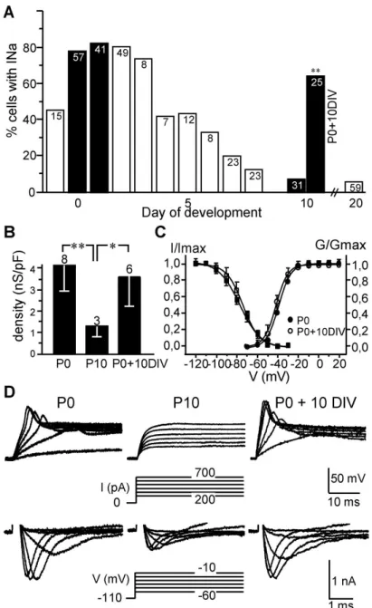
by the hair cell membrane capacitance, did not differ between hair cells after 10 days in culture (3.6 ± 1.4 nS/pF, n = 6) and P0 hair cells (4.1 ± 1.2 nS/pF, n = 8), but was significantly higher (p<0.05) than those of P10 hair cells (1.3 ± 0.5 nS/pF, n = 3) (Fig. 1B). The pharmacological properties (complete block by 100 nM tetrodotoxin-TTX, not shown) and biophysical properties of INa in P0+10DIV hair cells did not differ from those of P0 hair cells (Fig. 1C and Table 1). Figure 1D (bottom) shows representative INa in P0, P10 and P0+10DIV hair cells respectively. Similar to what is observed at P0, TTX-sensitive action potentials (AP) could be evoked in hair cells at P0+10DIV by the injection of depolarizing currents in the range of those that would be carried by mechanosensitive transduction channels (Fig. 1D). No such APs were elicited in acutely excised P10 hair cells. All together these observations reveal that the down regulation of INa in the utricle hair cells does not occur in absence of ongoing innervation.
Recovery of excitability in hair cells following nerve terminals impairment
To examine whether a similar property may explain the lack of excitability in adult hair cells, we investigated whether impairment of afferent nerve terminals in adult utricles (subsequently to synapse stabilization) led to the recovery of hair cell excitability. Initially, we aimed to record INa in utricles explanted from adult rats and maintained for several days in culture. The nerve terminals present in the epithelia degenerated in one day. Unfortunately, we could not achieve satisfactory culture conditions for recording hair cell currents. We overcame this problem by inducing selective damage of nerve terminals in vivo, in adult rats. We adapted to the rat vestibule, a method previously developed in the mammalian cochlea to induce transient excitotoxic injuries to the first auditory synapse (Puel et al., 1994; Guitton et al.,
2003). We applied a gelfoam containing 5 mM of kainate into the round window niche of adult P40 rats. Indeed, kainate has been shown to induce excitotoxic damages to vestibular synapses (Liu, 1999). Figure 2 illustrates the consequences of kainate treatment on the nerve terminals in utricles. Swellings of calyx and bouton nerve terminals were clearly identifiable on the entire utricle 48 hours after kainate application (K+48h; Fig. 2A). These swellings were particularly marked in the striolar region, in which type I hair cells were often expulsed from their calyx anchorage. Swellings were less pronounced in the peristriolar region, but both calyx and bouton nerve terminals were destructured with respect to those present in control ears (Fig. 2B-C and Fig. 3A-D). Quantitative assessment revealed that about 75 % (75.1 ± 5.82 %, over 5 epithelia) of hair cells displayed swollen afferent terminals in treated utricles versus less than 6 % (5,8 ± 2.7 %, over 5 epithelia) in control utricles (Fig. 2D). In our experimental conditions, kainate-induced impairments were transient. No morphological trace of swelling was observed one week after kainate application (n = 3) (Fig. 3E, F). The specificity of the excitotoxic injury was confirmed by the absence of morphological damage when DNQX (n = 3), a competitive non-NMDA receptor antagonist, was applied simultaneously with kainate (Fig. 3G, H). Estimation of the nerve terminals size in each condition is given in Table 2. Morphological examination of the synapses structure at higher magnification did not reveal significant alteration of the active zone organization both in hair cells and in afferent nerve terminals (Fig. 4). Ribbons were always found at the contact with the basal hair cell membrane and surrounded by numerous synaptic vesicles as reported in control preparations. Postsynaptic densifications were also identifiable at the afferent terminals membrane facing the ribbons, although disorganization of their internal ultrastructure was clearly identifiable in K+48h condition. Efferent terminals never displayed any sign of swelling (Fig. 4I-K).
Consequences of the afferent terminals impairment on the vestibular function were evaluated using a battery of tests that included observation of spontaneous motor behavior previously described (Boadas-Vaello et al., 2005). Rats treated with kainate transiently displayed signs of vestibular dysfunction. This was attested by significant increase in the rating scores of vestibular dysfunction at K+48h relative to control rats. Signs of vestibular dysfunction were no longer detected one week after kainate application or under neuroprotection conditions (Fig. 5).
During afferent terminals impairment (K+48h), the proportion of utricle hair cells expressing INa increased dramatically with respect to control hair cells still afferented at P40 (55.2 %, 16/29 versus 5.8 %, 1/17; p<0.01; Fig. 6A). 40 % of the tested cells (6/15) were identified as type I hair cells. INa was present in one third (2/6) of these latter cells. The density of INa in hair cells at K+48h was in the same order to that estimated in P0 cells (3.1 ± 0.1 nS/pF, n = 4,
versus 4.1 ± 1.2 nS/pF, n = 8). The pharmacological (not shown) and biophysical properties of INa in hair cells at K+48h did not differ significantly from those of hair cells in utricles acutely isolated at P0 (Table 1). Hair cells displaying INa density 3 nS/pF displayed TTX-sensitive AP upon injection of depolarizing currents (Fig. 6B). One week after kainate application, hair cells were no longer able to express high INadensity and to generate sodium-based AP (n = 11). These properties were also not observed if DNQX was applied simultaneously with kainate (n = 14).
We previously reported that Nav1.2, one of the commonest voltagegated sodium channel -subunits in rat utricle hair cells, is down regulated during synaptogenesis (Chabbert et al. 2003). To study whether the regulation of Nav1.2 expression during synaptic impairment may support the observed re-expression of INa, we checked its membrane expression using immunochemistry. Nav1.2 was not detected in utricle hair cells from adult control rats (Fig. 7A). However, 48 hours after kainate application, Nav1.2 immunostaining dramatically increased in hair cells. The labeling was prominent at the hair cells basal membrane (Fig. 7B). Recovery of Nav1.2 staining did not persist beyond one week after kainate treatment in most studied utricles (4 of 5) (Fig. 7C). Nav1.2 membrane re-expression was not observed when DNQX was applied with kainate (all 3 rats tested) (Fig. 7D). These observations reveal that impairment of the afferent terminals in adult rat utricle triggers the re-expression of Nav1.2 at the hair cell membrane.
DISCUSSION
This study shows that the modulation of the sodium-based excitability of the sensory cells is dependent on the state of the afferent terminals in the mammal utricle. The voltage-gated sodium current expression and underlying hair cell excitability are down regulated in neonate utricles as the ongoing nerve fibers contact the hair cells. Conversely, when mature afferent terminals are selectively impaired in vivo, hair cells re-express INa and recover the ability to fire sodium-based action potentials. These observations provide new data which help to understand of the cellular interactions involved in synaptogenesis and post-injury repair of the vestibular sensory organs.
Persistence of electrical excitability in non afferented utricle hair cells
Denervated organotypic culture has been previously used to test whether morphological or electrophysiological differentiation depended on ongoing innervation (Rush et al., 1998). Results obtained on mouse utricles demonstrated that neither calyx formation nor sustained postnatal innervation was required for morphological differentiation of the two types of hair
cells, nor for the developmental acquisition of voltage-gated potassium currents. Using similar preparation in the rat, we confirmed that the developmental expression of gK,L is independent on innervation (Gaboyard et al., 2005). Conversely, present study shows that the developmental expression of INadepends on the ongoing innervation, since the down regulation of voltage-gated sodium currents does not occur in the absence of synaptogenesis. This applies to both hair cell types. At P0+10DIV the density of INa in hair cells remained relatively high, allowing hair cells to fire sodium-based action potentials as in neonate period. This could be interpreted as a consequence of a bloc of hair cell differentiation under the culture conditions, since the large density of INa is usually restricted to P0-P3 hair cells in the rat utricle (Chabbert et al., 2003). However, since other voltage-gated ionic currents, such as gK,L that mark utricle hair cell maturity (Rusch et al., 1998) were observed at P0+10DIV (see also Gaboyard et al., 2005), it is unlikely that only intrinsic factors that would affect INa expression in vivo are lost in culture. A study recently published by our group, strongly support the idea of a control of INa expression by the ongoing innervation. We studied the expression INa in utricle hair cells from rats developed under enhanced gravity (Brugeaud et al., 2006). This experimental condition was reported to delay synaptogenesis (specifically the synaptic stabilization) by four days (Gaboyard et al., 2003; Bouet et al., 2004). In this model, INa down regulation was also delayed by four days, whereas its biophysical properties were unaffected. This additional observation confirms the relationships between the innervation and the down regulation of INa, and strengthens the idea of a control of the developmental expression INa by the innervation. In type II hair cells that develop and become innervated earlier than type I, it can be assume that the down regulation of INa would occur earlier. This remains to be demonstrated. We can question whether the influence of the nerve fibers on the hair cell electrical phenotype results from the cell-to-cell contact of the nerve terminals with the hair cell membrane or whether it results from synaptic stabilization. Since at birth, when immature synaptic contacts are identifiable, most hair cells express INa, the latter hypothesis appears more likely.
Recovery of excitability in adult utricle hair cells following nerve terminals impairment
in vivo
The use of the gelfoam method to produce selective excitotoxic impairment of the nerve terminals is to our knowledge unique in the vestibule. Such a technical approach appears appropriate since we observed morphological damage similar to that reported after intra-labyrinthine application of glutamatergic agonists in mammals (Liu 1999; Shimogori and Yamashita, 2004). The transient occurrence of the selective damages and the underlying vestibular disorders confirms the efficiency of the method. The glutamatergic nature of the excitotoxic impairment was attested first by the selective swelling of afferent terminals bearing
glutamate receptors conversely to the efferents, and second, by the neuroprotective effect of DNQX. This result is in aggreement with our recent demonstration that the non-NMDA receptors play a major role at the primary vestibular synapse (Bonsacquet et al., 2006). The fact that the biophysical and pharmacological properties of INa, in the kainate-treated utricles are similar to those in neonate utricles, suggests that the same sodium channel is re-expressed after excitotoxicity. This is supported by the re-expression of the Nav1.2 subunit. The dependency between hair cell excitability and the state of the nerve terminals was confirmed by the down-regulation of INa as the nerve fibers re-contacted the hair cells one week after the kainate application. The lack of reafferentation after kainate treatment in 20% of the tested rats may result from the toxic effect of kainate previously reported on auditory ganglion neurons (Juiz et al., 1989). The persistence of INa in a small population of adult hair cells under control conditions remains unexplained. It may result from hair cells undergoing re-afferentation after hair cell regeneration, which has been reported to occur in adult mammal utricles (Warchol et al., 1993). However, the small degree of turnover would not account for the estimated proportion of persistent INa. Another possibility is that the persistent INa might involve the Nav1.5 subunit, the expression of which is restricted to striolar hair cells and is not developmentally regulated (Wooltorton et al., 2005). Based on the observations reported here, we suggest that the recovery of hair cell excitability results from the release of cellular mechanisms that normally prevent membrane expression of INa after synaptogenesis is complete.
Plasticity in voltage-gated sodium channel expression
The present results strongly suggest that in utricular hair cells, the expression of the sodium current is a dynamic process. Such plasticity has been widely studied in spinal sensory neurons in which the expression of various sodium channels isoforms is regulated during development (Roy and Narahashi 1992) or following nerve injury (Waxman et al., 1994, Black et al., 1999; Dib-Hajj et al., 1999; Kim et al., 2001). Using immunocytochemistry, we demonstrated that Nav1.2 subunit is down-regulated in hair cells during synaptogenesis (Chabbert et al., 2003). This result is consistent with other observations in mammalian retinal ganglion cell axons in which Nav1.2, first clustered at immature nodes of Ranvier, is down-regulated and replaced by Nav1.6 as myelination proceeds. In contrast, Nav1.2 continues to be expressed throughout adult axons of retinal ganglion cells in shiverer mice (Boiko et al., 2001), which lack compact myelin, and also on adult photoreceptors in the human retina (Kawai et al., 2005), which lacks myelin. In the present study, we showed that Nav1.2 membrane expression is up-regulated following nerve terminal impairments. Such a phenomenon has also been reported to occur in rat hippocampal neurons following kainate-induced seizure (Gastaldi et al., 1997). Together,
these observations suggest that Nav1.2 may be specifically involved in a wide variety of developmental processes in sensory neurons. However, since various Nav isoforms are expressed in utricular hair cells (Chabbert et al., 2003; Mechaly et al., 2005) it would be of interest to test whether other Nav isoforms are also regulated after nerve terminals impairment. Several neurotrophic factors have been shown to regulate sodium channel transcription in spinal sensory neurons. For example, exposure to nerve growth factor (NGF) and/or glial cell neurotrophic factor (GDNF) differentially promotes the expression of sodium currents (Leffler et al., 2002; Fjell et al., 1999). In addition, in PC12 cells expressing trkB receptor, BDNF increases type II sodium channel mRNA expression (Fanger et al., 1995). We previously reported that an activity-dependent secretion of BDNF occurs in the rat utricle during synaptogenesis (Chabbert et al., 2003). Therefore, it would certainly be worthwhile studying whether this is reactivated following nerve terminals impairment and to what extent it controls the expression of the voltage-gated sodium current.
Physiological relevance and perspectives
Ischemic damage to the inner ear constitutes a major cause of hearing loss and vertigo in man (Puel, 1995). It has now been clearly established that such damage is mediated by the massive release of glutamate by hair cells, resulting in excitotoxic damage to auditory (Puel et al., 1994; Puel et al., 1995; Hakuba et al., 2003) and vestibular (Liu, 1999; Shimogori and Yamashita, 2004) synapses. The transient nature of such damage, which is well documented in the mammalian cochlea, indicates that local repair processes may occur under specific conditions (Puel et al., 1995). We provide here the first evidence that such processes also exist in the vestibular endorgans and that they occur through the modulation of hair cell sodium excitability. The involvement of the efferents in that repair process remains to be elucidated. Further experiments using selective bloc of the efferent synaptic transmission would allow to asses their involvement in the restoration of the vestibular function following excitotoxicity. The data presented here are thus of particular importance since they help to understand how synaptic contacts between hair cells and their cognate afferents are formed and repaired in mammalian vestibular organs. Studies of this type are a prerequisite for the development of strategies to protect the vestibular synapse against local ischemia and repairing damage caused by this condition.
REFERENCES
Black JA, Cummins TR, Plumpton C, Chen Y, Clare J, Waxman SG (1999) Upregulation of a previously silent sodium channel in axotomized DRG neurons. J Neurophysiol 82:2776-85.
Boadas-Vaello P, Riera J, Llorens J (2005) Behavioral and pathological effects in the rat define two groups of neurotoxic nitriles. Toxicol Sci 88:456-466.
Boiko T, Rasband MN, Levinson SR, Caldwell JH, Mandel KG, Trimmer JS, Matthews G (2001) Compact myelin dictates the targeting of two sodium channel isoforms in the same axon. Neuron 30:91–104.
Bonsacquet J, Brugeaud A, Compan V, Desmadryl G, Chabbert C (2006) AMPA type glutamate receptor mediates neurotransmission at turtle vestibular calyx synapse. J Physiol (Lond) 576:63-71.
Bouet V, Wubbels RJ, Jong HAA, Gramsbergen A (2004) Behavioural consequences of hypergravity in developing rats. Dev Brain Res 153:69–78.
Brugeaud A, Gaboyard-Niay S, Puel JL, Chabbert C (2006) Hypergravity affects the developmental expression of voltage-gated sodium current in utricular hair cells. Neuroreport 17:1697-1701.
Chabbert C, Mechaly I, Sieso V, Giraud P, Brugeaud A, Lehouelleur J, Couraud F, Valmier J, Sans A (2003) Voltage-gated Na+ channels activation regulates both action potential and BDNF release in rat utricular hair cells during a restricted period of development. J Physiol (Lond) 553:113-123.
Chen JW, Eatock RA, (2000) Major potassium conductance in type I hair cells from rat semicircular canals: characterization and modulation by nitric oxide. J Neurophysiol 84:139-151.
Correia MJ, Lang DG (1990) An electrophysiological comparison of solitary type I and type II vestibular hair cells. Neurosci Lett 116:106-111.
Curthoys IS (1982) Postnatal developmental changes in the response of rat primary horizontal semicircular canal neurons to sinusoidal angular accelerations. Exp Brain Res 47:295-300. Dechesne CJ, Mbiene JP, Sans A (1986) Postnatal development of vestibular receptor surfaces
in the rat. Acta Otolaryngol 101:11-18.
Desmadryl G, Raymond J, Sans A (1986) In vitro electrophysiological study of spontaneous activity in neonate mouse vestibular ganglion neurons during development. Brain Res 390:133-136.
Desmadryl G, Sans A (1990) Afferent innervation patterns in crista ampullaris of the mouse during ontogenesis. Brain Res Dev Brain Res 52:183-189.
Dib-Hajj SD, Fjell J, Cummins TR, Zheng Z, Fried K, LaMotte R, Black JA, Waxman SG (1999) Plasticity of sodium channel expression in DRG neurons in the chronic constriction injury model of neuropathic pain. Pain 83:591-600.
Eatock RA, Hurley KM (2003) Functional development of hair cells. Curr Top Dev Biol 57:389-448.
Ernfors P, Van De Water T, Loring J, Jaenisch R (1995) Complementary roles of BDNF and NT-3 in vestibular and auditory development. Neuron 14:1153-1164.
Evans MG, Fuchs PA (1987) Tetrodotoxin-sensitive, voltage-dependent sodium currents in hair cells from the alligator cochlea. Biophys J 52:649-652.
Fanger GR, Jones JR, Maue RA (1995) Differential expression of neuronal sodium expression by endogenous and exogenous tyrosine receptors expressed in pheochromocytoma cells. J Neurosci 15:202- 213
Fjell J, Cummins TR, Dib-Hajj SD, Fried K, Black JA, Waxman SG (1999) Differential role of GDNF and NGF in the maintenance of two TTX-resistant sodium channels in adult DRG neurons. Mol Brain Res 67:267-282.
Gaboyard S, Chabbert C, Travo C, Bancel F, Lehouelleur J, Sans A (2005) Three dimensional culture of newborn rat utricle with extracellular matrix: A new functional in vitro model. Neuroscience 133:253-265.
Gastaldi M, Bartolomei F Massacrier A, Planells R, Robaglia-Schlupp A, Cau P (1997) Increase in mRNAs encoding neonatal II and III sodium channel a-isoforms during kainateinduced seizures in adult rat hippocampus. Mol. Brain Res. 44:179–190.
Geleoc G, Risner JR, Holt JR (2004) Developmental acquisition of voltage-dependent conductances and sensory signaling in hair cells of the embryonic mouse inner ear. J Neurosci 24:11148-11159.
Guitton MJ, Caston J, Ruel J, Johnson RM, Pujol R, Puel JL (2003) Salicylate induces tinnitus through activation of cochlear NMDA receptors. J Neurosci 23:3944-3952.
Hakuba N, Matsubara A, Hyodo J, Taniguchi M, Maetani T, Shimizu Y, Tsujiuchi Y, Shudou M, Gyo K (2003) AMPA/kainate-type glutamate receptor antagonist reduces progressive inner hair cell loss after transient cochlear ischemia. Brain Res 979(1-2):194-202.
Juiz JM, Rueda J, Merchán JA, Sala ML (1989) The effects of kainic acid on the cochlear ganglion of the rat. Hear Res 40:65-74.
Kawai F, Horiguchi M, Susuki H, Miyachi E (2001) Na+ action potentials in human photoreceptors. Neuron 30:451-458.
Kawai F, Horiguchi M, Ichinose H, Ohkuma M, Isobe R, Miyachi E (2005) Suppression by an h current of spontaneous Na+ action potentials in human cone and rode photoreceptors. Invest Ophtalmol Vis Sci 46(1):390-397.
Kim CH, Oh Y, Chung JM, Chung K. (2001) The changes in expression of three subtypes of TTX sensitive sodium channels in sensory neurons after spinal nerve ligation. Mol Brain Res 95:153-161.
Kros CJ, Ruppersberg P, Rusch A (1998) Expression of a potassium current in inner hair cells during development of hearing in mice. Nature 394:281-284.
Leffler A, Cummins TR, Dib-hajj SD, Hormuzdiar WN, Black JA, Waxman SG (2002) GDNF and NGF reverse changes in repriming TTX-sensitive Na+ currents following axotomy of dorsal root ganglion neurons. J Neurophysiol 88:650-658.
Liu TC (1999) The effects of kainic acid on the vestibular ganglion cells. Neurosci Res Com 24:81-88.
Llorens J, Demêmes D, Sans A (1993) The behavioral syndrome caused by 3,3'-iminodipropionitrile and related nitriles in the rat is associated with degeneration of the vestibular sensory hair cells. Toxicol Appl Pharmacol 123:199-210.
Mechaly I, Scamps F, Chabbert C, Couraud F, Sans A, Valmier J (2005) Molecular diversity of voltage-gated sodium channels alpha subunits expressed in neuronal and non-neuronal excitable cells. Neuroscience 130:389-396.
Pan ZH, Hu HJ (2000) Voltage-dependent Na+ currents in mammalian retinal cone bipolar cells. J Neurophysiol 84:2564-2571.
Puel JL (1995) Chemical synaptic transmission in the cochlea. Prog in Neurobiol 47:449-476. Puel JL, Pujol R, Tribillac F, Ladrech S, Eybalin M (1994) Excitatory amino acid antagonists
protect cochlear auditory neurons from excitotoxicity. J Comp Neurol 341:241-256.
Puel JL, Saffiedine S, Gervais d’Aldin, Eybalin M, Pujol R (1995) Synaptic regeneration and functional recovery after excitotoxic injury in the guinea pig cochlea. C R Acad Sci III 318:67-75.
Roy ML, Narahashi T (1992) Differential properties of sensitive and tetrodotoxin-resistant sodium channels in rat dorsal root ganglion neurons. J Neurosci 12:2104-11. Rusch A, Lysakowski A, Eatock RA (1998) Postnatal development of type I and type II hair
cells in the mouse utricle: acquisition of voltage-gated conductances and differentiated morphology. J Neurosci 18:7487-7501.
Schimmang T, Minichiello L, Vazquez E, San Jose I, Giraldez F, Klein R, Represa J (1995) Developing inner ear sensory neurons require TrkB and TrkC receptors for innervation of their peripheral targets. Development 121:3381-3391.
Seoane A, Dememes D, Llorens J (2001) Pathology of the rat vestibular sensory epithelia during subchronic 3,3’-iminodipropionitrile exposure: hair cells may not be the primary target of toxicity. Acta Neuropathol 102:339-348.
Shimogori H, Yamashita H (2004) Peripheral vestibular disorder induced by (±)- -amino-3-hydroxy-5-methyl-isoxazole-4-propionic acid (AMPA). Neurosci Lett 371:69-72.
Warchol ME, Lambert PR, Goldstein BJ, Forge A, Corwin JT (1993) Regenerative proliferation in inner ear sensory epithelia from adult guinea pigs and humans. Science 259:1619-1622.
Waxman SG, Kocsis JD, Black JA (1994) Type III sodium channel mRNA is expressed in embryonic but not in adult spinal sensory neurons, and is reexpressed following axotomy. J Neurophysiol 72:466-470.
Wooltorton J, Hurley K, Eatock RA (2005) Voltage-dependent sodium currents in hair cells of inner ear. In: Topics in Integrative Neuroscience: From Cells to Cognition, Cambridge University Press, Cambridge.
LEGENDS
Figure 1. Persistence of electrical excitability in non afferented utricle hair cells in culture. (A) Diagram showing the percentage of utricle hair cells expressing INa as a function of rat age. Data in white columns were taken from Chabbert et al. (2003). Data in black columns were recorded in hair cells from utricle acutely isolated at P0, P1, and P10 and from cultured utricle (P0+10DIV) (p<0.01). (B) Comparison of INa density (mean ± SD) at indicated stages (*p<0.05, **p<0.01). Mean membrane capacitances (Cm) did not differ significantly between stages: 4.3±1.4 pF at P0, 3.7±0.9 pF at P10 and 4.0±1.1 pF at P0+10DIV. In A and B, the total number of cells studied at each stage of development is reported at the top of each column. (C) Plot of relative activation ( ; between -70 and +20 mV from a Vhold of -110 mV) and inactivation ( ; test potentials: 5 ms at 30 mV; conditioning prepulses 10 ms between -120 and -30 mV from at Vhold of -110 mV) of INa at indicated stages, expressed as G/Gmax and I/Imax as a function of test potentials and conditioning prepulses respectively. (D) Representative voltage responses (upper traces) in the current-clamp mode of the whole-cell patch-clamp technique, following given protocol and at indicated stages of development. Underlying whole-cell currents recorded in voltage-clamp mode are given below. Capacitive transients have been truncated for clarity. Depolarizing current injections (100 pA steps) were applied to cells held under hyperpolarizing holding current. Traces shown in the figure are single traces.
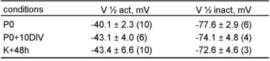
Table 1. Electrophysiological properties of INa. Mean potentials at which half of the cell’s
sodium channels are activated (V1/2 act) or inactivated (V1/2 inact) in given experimental conditions. No significant difference in V1/2 act and V1/2 inact was observed between the studied conditions. Data are given as mean ± SD.
Figure 2. Morphological evaluation of excitotoxic damages. (A) Longitudinal sections
through the entire utricle revealing numerous swollen nerve terminals below hair cells at K+48h. (B, C) High magnification observations in control and K+48h conditions. (D) Diagram
showing the mean percent of hair cells with swollen nerve terminals over the total number of hair cells studied (59/1015 in control (C) and 540/728 in kainate-treated rats) in semithin sections of five epithelia in given conditions (***p<0.001). Scale Bars = 30 µm in A, 10 µm in
B, C.
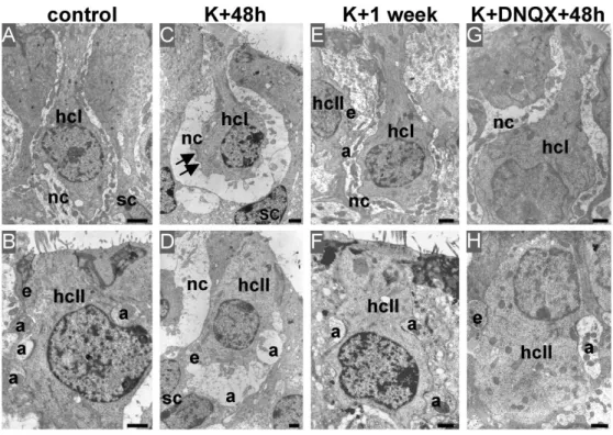
Figure 3. Morphological consequences of transient nerve terminals impairment. Electron
microscopy observations of vestibular nerve terminals at type I (hcI, top) and type II (hcII, bottom) hair cells in the peristriolar region of utricles from adult rats in given conditions. (A, B) Illustration of intact nerve calyx (nc), afferent (a) and efferent (e) bouton terminals in control utricles. (C, D) 48 hours after kainate application, both calyx and bouton terminals displayed characteristic swellings. The calyx membrane often came off from the hair cells membrane (arrow). Bouton swellings resulted in indentations of type II hair cells basal membrane. No damage persisted in the sensory epithelium one week after kainate application (E, F) or if DNQX was applied with kainate (G, H). (sc) supporting cells. Scale bars indicate 1 µm in all panels.
Table 2. Size analyze of nerve terminals. Sizes (in µm) of calyx and bouton terminals
measured in given experimental conditions. Nerve terminals size is significantly larger in K+48h condition relative to other given conditions (***p<0.001). Values are given as mean ±
SDin each condition and total number of cells studied is given between parentheses.
Figure 4. Synapse ultrastructure after kainate treatment. (A-D) Synaptic ribbons in type I
hair cells in given conditions. (A, B) Synaptic ribbon (arrow) in type I hair cell (hcI) surrounded by a swollen nerve calyx (nc) at K+48h. Synaptic vesicles (arrow head) are close to the hair cell basal membrane facing the postsynaptic density (asterisk). (C) Two synaptic ribbons in a single type I hair cell at K+48h. (D) Ribbon structure in control condition. (E-H) Synaptic ribbons in type II hair cells (hcII). (E, G) Postsynaptic densities in swollen afferent boutons contacting type II hair cells at K+48h. Ribbon is closed to basal membrane and surrounded by synaptic vesicles. (H) Ribbon and postsynaptic density in control type II hair cell. (I-K) No damage of efferent terminals (e) was observed at K+48h.
Figure 5. Behavioral consequences of transient nerve terminals impairment. Rating scores
for vestibular dysfunction from a behavioral test battery. Rats were assessed at 2 days (K+48h) or 7 days (K+1week) after kainate application, or 2 days after kainate+DNQX (K+DNQX+48h). The kainate-induced vestibular dysfunctions were transient and prevented by
DNQX. IDPN data form a positive control group of animals treated with a dose of 3,3’-iminodipropionitrile that induces a complete loss of hair cells.
Figure 6. Transient recovery of excitability in adult utricle hair cells following nerve terminals impairment. (A) Diagram showing the percentage of utricle hair cells expressing INa in given conditions. The total number of recorded cells is reported at the top of each column. (**p<0.01) (B) Representative voltage responses (upper traces) for the given protocol, with the underlying whole-cell currents (bottom traces). Capacitive transients have been truncated for clarity. All traces are single traces. A high-density INa supporting a sodium-based AP was observed only 48 hours after kainate application (K+48h). Voltage and current traces shown under neuroprotection (K+DNQX+48h) and recovery (K+1week) conditions are those of the only cells expressing INa in these two sets of conditions (mean Cm: 3.3 and 3.9 pF, respectively).
Figure 7. Transient recovery of Nav1.2 expression in adult utricle hair cells following
nerve terminals impairment. Immunolocalisation of neurofilament-N52 (red) and Nav1.2 subunits (green) in adult rat utricles in given conditions. Nav1.2 labelling is not detected in utricle hair cells or in supporting cells in control (A). 48h after kainate application, a strong Nav1.2 immunoreactivity is observed at the basal hair cells membrane of (arrows) (B). Nav1.2 immunoreactivity do not persist one week after kainate application (C) and is absent in K+DNQX+48h experimental conditions (D). Scale bars indicate 10 µm in all panels.
Table 1
Table 2
Figure 1. Persistence of electrical excitability in non afferented utricle hair cells in culture. (A) Diagram showing the percentage of utricle hair cells expressing INa as a function of rat age. Data in white columns were taken from Chabbert et al. (2003). Data
in black columns were recorded in hair cells from utricle acutely isolated at P0, P1, and P10 and from cultured utricle (P0+10DIV) (p<0.01). (B) Comparison of INa density (mean ± SD) at indicated stages (*p<0.05, **p<0.01). Mean membrane capacitances (Cm) did not differ significantly between stages: 4.3±1.4 pF at P0, 3.7±0.9 pF at P10 and 4.0±1.1 pF at P0+10DIV. In A and B, the total number of cells studied at each stage
of development is reported at the top of each column. (C) Plot of relative activation (6; between -70 and +20 mV from a Vhold of -110 mV) and inactivation ( ; test potentials: 5 ms at 30 mV; conditioning prepulses 10 ms between 120 and 30 mV from at Vhold of
-110 mV) of INa at indicated stages, expressed as G/Gmax and I/Imax as a function of test potentials and conditioning prepulses respectively. (D) Representative voltage
ScholarOne, 375 Greenbrier Drive, Charlottesville, VA, 22901
responses (upper traces) in the current-clamp mode of the whole-cell patch-clamp technique, following given protocol and at indicated stages of development. Underlying
whole-cell currents recorded in voltage-clamp mode are given below. Capacitive transients have been truncated for clarity. Depolarizing current injections (100 pA steps)
were applied to cells held under hyperpolarizing holding current. Traces shown in the figure are single traces.
ScholarOne, 375 Greenbrier Drive, Charlottesville, VA, 22901
Figure 2. Morphological evaluation of excitotoxic damages. (A) Longitudinal sections through the entire utricle revealing numerous swollen nerve terminals below hair cells at
K+48h. (B, C) High magnification observations in control and K+48h conditions. (D) Diagram showing the mean percent of hair cells with swollen nerve terminals over the total number of hair cells studied (59/1015 in control (C) and 540/728 in kainate-treated rats) in semithin sections of five epithelia in given conditions (***p<0.001). Scale Bars =
30 ÂFm in A, 10 ÂFm in B, C.
ScholarOne, 375 Greenbrier Drive, Charlottesville, VA, 22901
Figure 3. Morphological consequences of transient nerve terminals impairment. Electron microscopy observations of vestibular nerve terminals at type I (hcI, top) and type II
(hcII, bottom) hair cells in the peristriolar region of utricles from adult rats in given conditions. (A, B) Illustration of intact nerve calyx (nc), afferent (a) and efferent (e) bouton terminals in control utricles. (C, D) 48 hours after kainate application, both calyx and bouton terminals displayed characteristic swellings. The calyx membrane often came
off from the hair cells membrane (arrow). Bouton swellings resulted in indentations of type II hair cells basal membrane. No damage persisted in the sensory epithelium one week after kainate application (E, F) or if DNQX was applied with kainate (G, H). (sc)
supporting cells. Scale bars indicate 1 ÂFm in all panels.
ScholarOne, 375 Greenbrier Drive, Charlottesville, VA, 22901
Figure 4. Synapse ultrastructure after kainate treatment. (A-D) Synaptic ribbons in type I hair cells in given conditions. (A, B) Synaptic ribbon (arrow) in type I hair cell (hcI) surrounded by a swollen nerve calyx (nc) at K+48h. Synaptic vesicles (arrow head) are close to the hair cell basal membrane facing the postsynaptic density (asterisk). (C) Two
synaptic ribbons in a single type I hair cell at K+48h. (D) Ribbon structure in control condition. (E-H) Synaptic ribbons in type II hair cells (hcII). (E, G) Postsynaptic densities
in swollen afferent boutons contacting type II hair cells at K+48h. Ribbon is closed to basal membrane and surrounded by synaptic vesicles. (H) Ribbon and postsynaptic
density in control type II hair cell. (I-K) No damage of efferent terminals (e) was observed at K+48h.
ScholarOne, 375 Greenbrier Drive, Charlottesville, VA, 22901
Figure 5. Behavioral consequences of transient nerve terminals impairment. Rating scores for vestibular dysfunction from a behavioral test battery. Rats were assessed at 2 days (K+48h) or 7 days (K+1week) after kainate application, or 2 days after kainate+DNQX
(K+DNQX+48h). The kainate-induced vestibular dysfunctions were transient and prevented by DNQX. IDPN data form a positive control group of animals treated with a
dose of 3,3'-iminodipropionitrile that induces a complete loss of hair cells.
ScholarOne, 375 Greenbrier Drive, Charlottesville, VA, 22901
Figure 6. Transient recovery of excitability in adult utricle hair cells following nerve terminals impairment. (A) Diagram showing the percentage of utricle hair cells expressing INa in given conditions. The total number of recorded cells is reported at the top of each column. (**p<0.01) (B) Representative voltage responses (upper traces) for the given protocol, with the underlying whole-cell currents (bottom traces). Capacitive transients have been truncated for clarity. All traces are single traces. A high-density INa
supporting a sodium-based AP was observed only 48 hours after kainate application (K+48h). Voltage and current traces shown under neuroprotection (K+DNQX+48h) and
recovery (K+1week) conditions are those of the only cells expressing INa in these two sets of conditions (mean Cm: 3.3 and 3.9 pF, respectively).
ScholarOne, 375 Greenbrier Drive, Charlottesville, VA, 22901
Figure 7. Transient recovery of Nav1.2 expression in adult utricle hair cells following nerve terminals impairment. Immunolocalisation of neurofilament-N52 (red) and Nav1.2
L subunits (green) in adult rat utricles in given conditions. Nav1.2 labelling is not detected in utricle hair cells or in supporting cells in control (A). 48h after kainate application, a strong Nav1.2 immunoreactivity is observed at the basal hair cells membrane of (arrows) (B). Nav1.2 immunoreactivity do not persist one week after kainate application (C) and is absent in K+DNQX+48h experimental conditions (D). Scale
bars indicate 10 ÂFm in all panels.
ScholarOne, 375 Greenbrier Drive, Charlottesville, VA, 22901