HAL Id: hal-03041303
https://hal-amu.archives-ouvertes.fr/hal-03041303
Submitted on 4 Dec 2020HAL is a multi-disciplinary open access archive for the deposit and dissemination of sci-entific research documents, whether they are pub-lished or not. The documents may come from teaching and research institutions in France or abroad, or from public or private research centers.
L’archive ouverte pluridisciplinaire HAL, est destinée au dépôt et à la diffusion de documents scientifiques de niveau recherche, publiés ou non, émanant des établissements d’enseignement et de recherche français ou étrangers, des laboratoires publics ou privés.
Synthesis and use of an amphiphilic dendrimer for
siRNA delivery into primary immune cells
Jiaxuan Chen, Aleksandra Ellert-Miklaszewska, Stefano Garofalo, Arindam
Dey, Jingjie Tang, Yifan Jiang, Flora Clément, Patrice Marche, Xiaoxuan Liu,
Bozena Kaminska, et al.
To cite this version:
Jiaxuan Chen, Aleksandra Ellert-Miklaszewska, Stefano Garofalo, Arindam Dey, Jingjie Tang, et al.. Synthesis and use of an amphiphilic dendrimer for siRNA delivery into primary immune cells. Nature Protocols, Nature Publishing Group, 2020, �10.1038/s41596-020-00418-9�. �hal-03041303�
1
Synthesis and use of an amphiphilic dendrimer for siRNA delivery into
primary immune cells
Jiaxuan Chen1,2, Aleksandra Ellert-Miklaszewska3, Stefano Garofalo4, Arindam K Dey5, Jingjie Tang1, Yifan Jiang1, Flora Clément5, Patrice N Marche5, Xiaoxuan Liu2, Bozena Kaminska3, Angela Santoni6, Cristina Limatola4,6, John Rossi7, Jiehua Zhou7*, Ling Peng1*
1. Aix-Marseille Université, CNRS, Center Interdisciplinaire de Nanoscience de Marseille, UMR 7325, « Equipe Labellisée Ligue Contre le Cancer », Marseille, France
2. State Key Laboratory of Natural Medicines and Jiangsu Key Laboratory of Drug Discovery for Metabolic Diseases, Center of Drug Discovery, Center of Advanced Pharmaceutics and Biomaterials, China Pharmaceutical University, Nanjing, P. R. China
3. Laboratory of Molecular Neurobiology, Neurobiology Center, Nencki Institute of Experimental Biology of the Polish Academy of Sciences, Warsaw, Poland
4. Department of Physiology and Pharmacology, Sapienza University of Rome, Italy
5. Institute for Advanced Biosciences, University Grenoble-Alpes, Inserm U1209, CNRS 5309, 38700 La Tronche, France
6. IRCCS Neuromed, Via Atinense 19 Pozzilli (IS) Italy
7. Department of Molecular and Cellular Biology, Beckman Research Institute, City of Hope, Monrovia, CA, USA 91016
* Correspondence should be addressed to Ling Peng (ling.peng@univ-amu.fr) Centre Interdisciplinaire de Nanoscience de Marseille Aix-Marseille University, CNRS, UMR 7325 CINaM 163, avenue de Luminy, 13288 Marseille, France; Tel: 0033 6 1724 8164 ; Fax: 00 33 4 9141 8916; or Jiehua Zhou (jzhou@coh.org) Department of Molecular and Cellular Biology, Beckman Research Institute, City of Hope, Monrovia, CA, USA 91016; Tel: +1-(626)218-3864; Fax: +1-(626)301-8271
2 ABSTRACT (< 250 words)
Genetically manipulating immune cells using siRNAs is important for both basic immunological studies and therapeutic applications. However, the siRNA delivery is challenging because primary immune cells are often sensitive to the delivery materials and generate immune responses. We have recently developed an amphiphilic dendrimer, which is able to deliver siRNA to a variety of cells including primary immune cells. We provide here the protocols for the synthesis of this dendrimer and its siRNA delivery into immune cells such as primary T- and B-cells, natural killer cells, macrophages, and primary microglia. It is noteworthy that the dendrimer synthesis is straightforward, and the siRNA delivery protocol is easy, requiring simple mixing of siRNA and dendrimer in buffer, with subsequent application to the primary immune cells to achieve effective and functional siRNA delivery. This dendrimer-mediated siRNA delivery outperforms largely the standard technique of electroporation, opening a new avenue for functional and therapeutic studies of the immune system. The whole protocol encompasses the dendrimer synthesis which requires 3 weeks, the primary immune cell preparation which takes 3-10 days depending on the tissue source and cell type, the dendrimer-mediated siRNA delivery and subsequent functional assay which takes additional 3-6 days.
KEYWORDS:
3 INTRODUCTION
RNA interference (RNAi) is a powerful tool for manipulation of gene expression in basic research and an emerging therapeutic strategy to treat various diseases.1, 2 The power of RNAi lies in its ability to potently and specifically silence any gene of interest with small interfering RNAs (siRNAs), allowing functional study of the target gene and/or effective inhibition of the disease-associated gene for therapeutic intervention. The breakthrough success of RNAi therapeutics came in 2018, when Patisiran® (Alnylam Pharmaceuticals) was the first siRNA drug approved by the USA Federal Drug Administration (FDA).3 Also, the recent success of cancer immunotherapy has fueled a tremendous interest in genetic manipulation of immune cells using siRNAs to address basic immunological questions and for potential therapeutic applications.4, 5 However, the major obstacle in implementing RNAi in those cells has been poor delivery of the siRNA,6, 7 in particular in the case of primary immune cells, such as lymphocytes
(B-cells, T-cells, natural killer (NK) cells), mononuclear phagocytes (monocytes, macrophages, microglia, dendritic cells) and granulocytes (neutrophils, eosinophils, basophils). This is because siRNA molecules are hydrophilic and highly negatively charged, and cannot readily cross cell membranes to reach the RNAi machinery within the cytoplasm for gene silencing. In addition, naked siRNA is not stable and can be rapidly degraded by enzymes such as nucleases. If administered at high concentration, naked siRNA will often generate off-target effects and activate innate immunity,8, 9 which may induce severe adverse effects. Also, siRNA can induce the activation of the innate immune response through various possible pathways, such as toll-like receptors (TLR3, TLR7/8), dsRNA-dependent protein kinase (PKR), retinoic acid-inducible gene-I (RAI1), etc.10 Although using modified siRNA chemistry and optimized siRNA sequences can stabilize siRNA and lessen the unwanted effects,10 siRNA delivery to primary immune cells remains a special challenge. Immune cells such as T lymphocytes and NK cells are small, with limited cytoplasm. Standard transfection techniques such as electroporation and nucleofection show low transfectability, yet impacting negatively viability. Macrophages, dendritic cells and microglia, as professional phagocytes, are endowed with many potent degradative enzymes that can disrupt nucleic acid integrity and make gene transfer into these cells an inefficient process.11 In addition, primary immune cells are often very sensitive to the delivery materials, generating non-specific immune responses. On the other hand, most common methods used to deliver siRNA into primary immune cells, which do not require any additional
4
carriers, such as electroporation and nucleofection, lead to excessive cell death and low transfection efficiency, and hence are unsuitable for general applications in both basic and translational research. Consequently, there is a high demand for safe and effective delivery systems, which are able to protect siRNA from degradation, deliver it to the target cells, and ultimately achieve gene silencing to facilitate genetic manipulation of immune cells for functional and therapeutic studies.
Both viral and non-viral delivery vectors have been explored for siRNA delivery in general
6, 7 and into immune cells in particular.5, 11-14 Viral delivery is more effective; however,
increasing concerns over the immunogenicity and safety of viral vectors urge the development and improvement of non-viral delivery systems. Non-viral vectors offer more flexible options, with lipid and polymer vectors being the most commonly used. For example, the first human trial of siRNA therapeutics used a polymer vector,15 whereas the first FDA-approved siRNA drug, Patisiran®, employs a lipid nanoparticle (LNP) delivery formulation. Although some well-studied lipid and polymer vectors perform well for the majority of established immortalized cells, they have marginal efficacy in siRNA delivery to primary immune cells, and very often induce non-specific immune responses.
To circumvent these problems, we have been working on developing innovative amphiphilic dendrimers for effective siRNA delivery. The rationale behind these dendrimer vectors is that they combine the multivalent cooperativity of dendritic polymer vectors with the self-assembly property of lipid vectors, hence capitalizing on the advantageous delivery characteristics of both lipid and polymer vectors, while overcoming their limitations, for more effective and potent siRNA delivery.16-21 One of these dendrimers, AD (Figure 1), exhibits particularly high
performance for siRNA delivery to a wide range of cell types, including highly challenging primary immune cells, such as human peripheral blood mononuclear cells (PBMCs), human B- and T-lymphocytes, NK cells (human and mouse), primary monocyte-derived macrophages and primary microglial cells (rat and mouse)17, 20, 21. Notably, this AD dendrimer is able to form small and stable nanoparticles with siRNA, thus protecting the siRNA from degradation and facilitating cellular uptake of siRNA17, 20. The subsequent siRNA-mediated gene silencing is specific and effective at both the mRNA and protein levels, leading to consequential biological effects. Remarkably, this dendrimer does not induce apparent cellular toxicity or non-specific immune responses under experimental conditions. Consequently, it constitutes a promising tool
5
for siRNA delivery into immune cells and provides a new outlook for functional and therapeutic studies of the immune system.
We provide here the general protocols for the robust synthesis of this dendrimer AD and the
AD-mediated delivery of siRNA into immune cells using primary T cells, natural killer cells,
macrophages and microglial cells as the model cells. The dendrimer synthesis is straightforward and easy to follow and reproduce. Also, the final purification of the dendrimer AD is achieved through simple dialysis in water, giving AD in high yield and purity. Most importantly, the formulation of the siRNA/dendrimer complexes is very convenient, and requires only simple mixing of the siRNA with AD in solution at room temperature (at 25°C). The complexes can then be readily applied to the immune cells, such as T-lymphocytes, B-lymphocytes, monocytes/macrophages, brain innate immune cells (microglia), and NK cells, for transfection and gene silencing assays. We present below an overview of these procedures, their validation, and their applications in siRNA delivery into primary immune cells using T-cells, NK cells, macrophages and microglia as examples.
Overview of the procedures
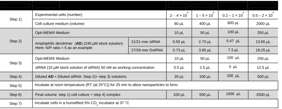
In this protocol, we firstly describe itemized steps for the synthesis and molecular characterization of the dendrimer AD. Using 21/21-mer siRNAs and 27/29-mer Dicer substrate siRNAs (DsiRNAs) as examples, we explain the procedure and the conditions for the AD-mediated transfection assays to deliver siRNA into primary immune cells such as T-cells, NK cells, macrophages and microglia in various plate formats, and the subsequent validation of gene knockdown. Typically, the chemical synthesis of AD takes 20 days including purification. The whole transfection procedure, from cell seeding, preparation of the siRNA/AD complex and cell transfection to validation tests and functional assays may take 7 days. Time frames for preparation of primary cell cultures may vary as described in the supplementary materials. Details about the isolation and maintenance or/and expansion of primary immune cells are also provided. Some key points relating to the protocols are discussed below.
Experimental design
6
Synthesis of AD is achieved by coupling the hydrophobic chain 1 and the hydrophilic PAMAM dendron 2 via “click” reaction using copper-catalyzed azide-alkyne cycloadditions (CuAAC), followed by amidation with ethylenediamine (EDA) (Figure 2). The starting materials 1 and 2 can be easily prepared using well-established protocols.16, 17 However, 1 is not well soluble in dimethylformamide (DMF), and it is necessary to raise the reaction temperature to 50 °C in order to solubilize 1 and drive the reaction to completion, thus offering 3 in high yield. It is also critical to keep the stoichiometric ratio of 1 and 2 at 1/1.05 for click reaction, because it is difficult to separate the remaining excess 1 and/or 2 from the product 3 if the reaction is performed without respecting the stoichiometry. For the amidation reaction, the amount and concentration of EDA must be controlled in order to maximally suppress the retro-Michael side reaction and cyclization byproducts.22-24 Different from our previously published protocol, we have used MeOH instead of MeOH/CH2Cl217 as solvent for the amidation. This is because CH2Cl2 can react with EDA to
generate oligomeric impurities,25 and at the same time, increase the formation of the defected dendrimer with cyclization terminals. The optimized conditions therefore allow more reliable amidation. The final dendrimer AD is purified using dialysis against water, and obtained as a pale powder after lyophilization.
Formation of the siRNA/AD complexes
As shown in Figure 3, the siRNA/dendrimer complexes are formulated by simply adding the siRNA solution to the dendrimer solution at 25 °C in PBS buffer or serum-free Opti-MEM®, followed by gentle vortexing. The complexes are then applied to the primary immune cell culture. It is preferable to use a freshly prepared solution of siRNA/AD complexes for transfection.
Calculation of the ratio of siRNA and the dendrimer AD in a transfection assay.
It should be noted that the formulation of siRNA/dendrimer complexes depends critically on the dendrimer-to-RNA charge ratio, which is defined as the “N/P ratio”. It is calculated as [total terminal amino groups in the cationic dendrimer] / [total phosphates in the siRNA]. We always calculate the amount of AD according to the amount of siRNA in the final volume of cell culture media and the N/P ratio, as follows:
7
Amount of AD = (amount of siRNA) × (number of phosphates in siRNA) × (N/P ratio) / (number of amino groups in AD).
In our previous experiment, we established an optimal N/P ratio of 5 or 10 for AD-mediated siRNA delivery into immune cells, including PBMC-CD4+ T cells, natural killer cells, macrophages and microglial cells. In Table 1, we present the steps for AD-mediated transfection of a siRNA or a Dicer-substrate siRNA (DsiRNA) in various plate formats. Different from the conventional siRNA which has oligonucleotide of 21-mer for both sense and antisense strains, the DsiRNA used in this work contains 27-mer oligonucleotide in the sense strain and 29-mer oligonucleotide in the antisense strand, which is also called 27/29-mer DsiRNA. Such DsiRNAs have been demonstrated to enhance RNAi potency and efficacy26, 27. For example, in a 24-well plate format, with 500 µL of cell culture medium in each well, and using a 21/21-mer siRNA or a 27/29-mer DsiRNA at 50 nM concentration, the amount of AD with 8 terminal amino groups is calculated at an N/P ratio of 5 as follows:
For a 21/21-mer siRNA, the amount of AD = (50 nM × 500 µL) × (21+21) × (5) / (8) = 656.25 pmol
For a 27/29-mer DsiRNA, the amount of AD = (50 nM × 500 µL) × (27+29) × (5) / (8) = 875 pmol
Consequently, 2.73 or 3.65 µL of 240 µM AD stock solution is needed for transfection of a 21-mer siRNA or a 27-21-mer DsiRNA, respectively, in a 24-well plate.
siRNA delivery and testing efficacy of gene silencing
For siRNA delivery using dendrimer AD, the prepared stock solution containing the siRNA/AD complexes is added to primary immune cells with an appropriate dilution to reach the required final siRNA concentrations. The effects of the transfection procedure on gene and protein expression are analyzed after 2 or 3 days by quantitative real-time PCR and western-blotting (WB) or FACS, respectively. To define an effective RNAi activity, several key controls should be included in the experimental design: 1) a cell alone control without any siRNA/AD transfection (as a mock control); 2) a cell control transfected with a unrelated or scrambled siRNA/AD complex (as a negative control); or 3) a cell control transfected with the experimental siRNA/commercial transfecting agent (as a positive control or as a comparison control). By setting these proper controls and comparing their effect on target gene and protein expression,
8
the AD-mediated delivery and siRNA-mediated knockdown activity is defined and validated. Generally, siRNAs
serving as negative controls that do not target any human’ or animal’ genome can be purchased from commercial vendors with the validation test.
Validation and applications
To demonstrate the validation and application of siRNA delivery into immune cells using the dendrimer AD, we have selected four examples (Figure 4) in which AD has been used to successfully deliver: 1) anti-HIV siRNAs into primary CD4+ T-cells for efficient suppression of HIV-1 replication;17 2) an anti-NKG2D siRNA into NK cells to reduce their cytotoxic activity towards tumor cells and motor neurons in a model of amyotrophic lateral sclerosis (ALS); 21 3) an siRNA targeting JAK1 gene in primary mouse macrophages to regulate their inflammatory activities; and 4) an siRNA targeting the transcription regulator Id1 into microglia for functional exploration of Id1.20 The procedure for formulation of the siRNA/dendrimer complexes is very easy, and requires only simple mixing of the siRNA with AD. The siRNA/AD complexes can then be readily applied to the primary immune cells, such as T-cells, NK cells, macrophages and microglia, for functional siRNA delivery. Therefore, this dendrimer AD constitutes the long-searched-for vector to delivery siRNA safely and effectively for the purpose of functional study and therapeutic applications in primary immune cells. Details for siRNA transfection and functional assays were presented below in the “Procedure” section and key results are summarized in “Anticipated Results” section.
Limitations
This protocol provides a facile and robust transfection agent, AD, for siRNA delivery into various primary immune cells. It will be important to determine whether the chemical composition of AD, or the size and surface charge of the siRNA/AD complexes, play a key role in the efficacy of siRNA delivery and gene expression interference in various types of immune cells. Furthermore, detailed bio-distribution studies in ex vivo white blood cells (WBCs) or in vivo animal models may be necessary to establish how cells at the different anatomic sites of the body would be exposed to the siRNA/AD complexes. Also, cell-specific targeting strategies can
9
be developed for AD with the goal of targeting immune cell subsets for specific siRNA delivery.19 We are actively working in this direction.
10 MATERIALS
REAGENTS
• Chemical reagents !CAUTION Because most of the chemicals and organic solvent used for dendrimer synthesis are potentially hazardous to human health, we recommend performing all reactions in a fume hood while wearing personal protective equipment (gloves, lab coat and goggles) to prevent exposure.
• 1 and 2 were synthesized according to the reported protocols16, 17
• N,N-Dimethylformamide (DMF; Sigma-Aldrich, cat. no. 227056) • Dichloromethane (CH2Cl2; Sigma-Aldrich, cat. no. 32222-M)
• Methanol (MeOH; Sigma-Aldrich, cat. no. 179337) • Triethylamine (TEA; Sigma-Aldrich, cat. no. 471283) • Copper(I) iodide (CuI; Sigma-Aldrich, cat. no. 215554)
• 1,8-Diazabicyclo[5.4.0]undec-7-ene (DBU; Sigma-Aldrich, cat. no. 139009) • Sodium sulfate anhydrous (Na2SO4; Sigma-Aldrich, cat. no. 798592)
• Magnesium sulfate monohydrate (Mg2SO4 *H2O; Sigma-Aldrich, cat. no. 434183)
• Ammonium chloride (NH4Cl; Sigma-Aldrich, cat. no. 213330)
• Sodium bicarbonate (NaHCO3; Sigma-Aldrich, cat. no. S6014)
• Sodium chloride (NaCl; Sigma-Aldrich, cat. no. S9888)
• Silica gel for flash chromatography (Sigma-Aldrich, cat. no. 227196)
• Ethylenediamine (EDA; Merck, cat. no. S7392647 745) !CAUTION To ensure the purity of the reagent, EDA should be distilled before using.
• Dialysis tubing, benzoylated (MWCO 2000; Sigma-Aldrich, cat. no. D7884-10FT) • Chloroform-d (99.8 atom % D; Sigma-Aldrich, cat. no. 416754)
• Methanol-d4 (99.8 atom % D; Sigma-Aldrich, cat. no. 151947)
• 1 × RBC lysis buffer solution (eBioscience, cat no 00433357) • Compressor (nessicare 30, cat. No. CNST-300D)
• Universal negative siRNA controls (Integrated DNA Technologies, IDT, Negative control DsiRNA, cat.no. 51-01-14-03; Scrambled negative control DsiRNA, cat.no. 51-01-19-08). ▲CRITICAL STEP It is crucial to have an appropriate negative control against which one can compare experimental results in gene silencing and functional studies using siRNA or
11
DsiRNA duplexes. A scrambled siRNA does not target any part of the human, mouse or rat transcriptomes.
• Custom-made siRNAs (Integrated DNA Technologies, IDT: HIV-1 tat/rev site I 27-mer DsiRNA, CD4 21-mer siRNA and 27-mer DsiRNA, TNPO3 21-mer siRNA and 27-mer DsiRNA) ▲CRITICAL STEP It is crucial to select an optimal siRNA sequence for effective gene silencing. According to our previous experience, we designed one 21-mer siRNA and one 27-mer DsiRNA per target sequence with appropriate 3’-overhangs and chemical modifications.
• HIV-1 tat/rev site I 27 mer DsiRNA. Sense: 5’ -GCG GAG ACA GCG ACG AAG AGC UCA UCA-3’; Antisense: 5’-UGA UGA GCU CUU CGU CGC UGU CUC CGC dTdT-3’. • CD4 21-mer siRNA. Sense: 5’-GAU CAA GAG ACU CCU CAG U dGdA-3’; Antisense:
5’-ACU GAG GAG UCU CUU GAU C dTdG-3’.
• CD4 27-mer DsiRNA. Sense: 5’-GAU CAA GAG ACU CCU CAG UGA GAA G-3’; Antisense: 5’-CUU CUC ACU GAG GAG UCU CUU GAU CUG-3’ (2′-OMe modified U was underlined.)
• TNPO3 21-mer siRNA. Sense: 5’-CGA CAU UGC AGC UCG UGU AUU-3’; Antisense: 5’-UAC ACG AGC UGC AAU GUC GUU-3’.
• TNPO3 27-mer DsiRNA. Sense: 5’-CGA CAU UGC AGC UCG UGU ACC AG dGdC-3’; Antisense: 5’-GCC UGG UAC ACG AGC UGC AAU GUC GUU-3’.
• Custom-made siRNAs (Horizon Discovery,JAK1 siRNA)
• JAK1 mouse siRNA. Sense: 5’-GAA UAA AUG CAG UAU CUA AAU-3’; Antisense:5’-UUA GAU ACU GCA UUU AUU CGG-3’.
• Rat Id1 siRNA (Dharmafect, ON-TARGET plus SMART pool; cat. no: L-080165-02-0005). • Custom made primers for qRT-PCR analysis (Integrated DNA Technologies, IDT or
Genescript).
• Negative control siRNA which does not target any human, mouse or rat gene products (Dharmafect, ON-TARGET plus non-targeting pool; cat. no: D-001810-10-05).
• AllStars Negative Control siRNA is currently the most thoroughly tested and validated negative control siRNA. This siRNA has no homology to any known mammalian gene (Qiagen, AllStars Neg. Control siRNA; Cat No./ID: 1027281).
12
• HIV-1 tat/rev forward primer: 5’-GGC GTT ACT CGA CAG AGG AG-3’; tat/rev reverse primer: 5’-TGC TTT GAT AGA GAA GCT TGA TG-3’;
• CD4 forward primer: GCT GGA ATC CAA CAT CAA GG-3’; CD4 reverse primer: 5’-CTT CTG AAA CCG GTG AGG AC-3’;
• TNPO3 forward primer: CCT GGA AGG GAT GTG TGC-3’; TNPO3 reverse primer: 5’-AAA AAG GCA AAG AAG TCA CAT CA-3’;
• GAPDH forward primer: 5’-CAT TGA CCT CAA CTA CAT G-3’; GAPDH reverse primer: 5’-TCT CCA TGG TGG TGA AGA C-3’.
• ID1 forward primer: AGTCTGAAGTCGCGACCGCC-3’; ID1 reverse primer 5’-CTGGAACACATGCCGCCTCGG-3’.
• NKG2D forward primer: 5’-TACTGTGGCCCATGTCCTAA-3’; NKG2D reverse primer 5’- CTTTCAGAAGGCTGGCATTT-3’.
• 18S RNA forward primer: 5’-CGGACATCTAAGGGCATCACA-3’; 18SRNA reverse primer 5’-AACGAACGAGACTCTGGCATG-3’.
• Milli-Q ultrapure water (Millipore)
• Opti-MEM® medium (Gibco, cat. no 31985-047)
• Dulbecco’s PBS without calcium and magnesium (DPBS, Corning, cat.no. 21-031-cv) • Dulbecco’s PBS with calcium and magnesium (Thermo Fisher, cat.no.14040091) • Thermo Fisher, cat.no.14040091)
• Ficoll-PAQUE plus (GE healthcare Pharmacia, cat.no. 17-1440-02) • Ficoll (Sigma-Aldrich (Milan, Italy)
• Trypsin inhibitor (Sigma-Aldrich, cat.no. T6522) • DNase (Sigma-Aldrich, cat. no. DN25)
• RPMI-1640 (Corning, cat.no. 15-040-cv)
• DMEM with GlutaMAX, high glucose (Thermo Fisher, cat.no 31966-021, 61965026) • MEM Non-Essential Amino Acids Solution (100×; Thermo Fisher, cat.no.11140050) • Sodium Pyruvate (100 mM,Thermo Fisher, cat.no. 11360070)
• 2-Mercaptoethanol (50 mM, Thermo Fisher, cat.no. 31350010). • HEPES (1 M, Thermo Fisher, cat.no. 11360070)
13
• L-Glutamine (200 mM, 100×, IrvineScientific, cat.no. 9317-100 mL)
• Penicillin/streptomycin, 10,000 units of penicillin per mL/10,000 μg of streptomycin per mL (Gibco, cat.no. 15140-122-100 mL)
• Phytohemagglutinin-L (PHA, Roche, cat. no. 11 249 738 001)
• Recombinant interleukin-2 (IL-2, Teceleukin, Hoffmann - La Roche Inc., cat.no. Ro 23-6019)
• ActiCyte® - TC Medium kit (CytoMedical Design Group, cat.no. TCM1000) • Trypan Blue Stain 0.4% (Invitrogen, cat. no. T10282)
• EasySep™ Human CD4+ T Cell Isolation Kit (StemCell Technologies, cat.no. 17952)
• RoboSep Buffer (StemCell Technologies, cat.no. 20104) !CAUTION Potential irritant to eyes, respiratory system and skin. Wear suitable protective clothing, glasses and gloves. • TRIZOL® agent (Thermo Fisher) !CAUTION Harmful if inhaled or comes in contact with
skin. Toxic if swallowed. Irritant to eyes. Evidence of a carcinogenic effect. Wear suitable protective clothing, glasses and gloves. This material and its container must be disposed of as hazardous waste.
• Chloroform/Isopropanol 24/1 solution (Sigma, cat. no. C0549) !CAUTION Harmful if inhaled or comes in contact with skin. Toxic if swallowed. Irritant to eyes. Evidence of a carcinogenic effect. Work under a fume hood and wear suitable protective clothing, glasses and gloves. This material and its container must be disposed of as hazardous waste.
• Glycogen (Roche, cat. no. 10 901 393 001)
• RNA isolation Kit (RNeasy Mini Kit, QIAGEN, cat. no. 74104)
• QuantiNova Reverse Transcription kit (QIAGEN, cat.no. 205411) or SuperScript™ III Reverse Transcriptase (Thermo Fisher, cat. no 18080-044) and a set of dNTPs (Promega, cat. no U1330)
• SsoAdvanced Universal SYBR Green Supermix (BIO-RAD, cat.no. 172-5271) or Fast SYBR Green Master Mix (Applied Biosystems, cat. no 4385612)
• Pacific Blue-conjugated CD4 antibody (clone RPA-T4) (BD Biosciences Cat# 558116, RRID:AB_397037)
• Id1 antibody (clone B-8) (Santa Cruz Biotechnology Cat# sc-133104, RRID:AB_2122863) • GAPDH antibody (Millipore Cat# MAB374, RRID:AB_2107445)
14
• Anti-mouse IgG peroxidase conjugated secondary antibody (Vector Laboratories Cat# PI-2000, RRID:AB_2336177)
• iNOS Antibody (Cell signaling Technology, cat.# 13120S, RRID: AB_2798613) • Arginase-1 Antibody (Cell signaling Technology, cat.# 93668S, RRID: AB_2800207) • α/β-Tubulin Antibody (Cell signaling Technology, cat.# 2148S, RRID: AB_2288042)
• Fixation/Permeabilization solution kit (BD Biosciences, cat. no. 554714) !CAUTION Limited evidence of a carcinogenic effect. May cause sensitization if it comes in contact with skin. Work under a fume hood and wear suitable protective clothing and gloves.
• Alliance HIV-1 p24 Antigen ELISA Kit (Perkin Elmer, cat. No. NEK050A001KT) • Recombinant interleukin-15 (IL-15, eBioscience Inc., San Diego, CA)
• BCA Protein Assay Kit (Thermo Scientific, Rockford, IL, USA, cat no 23225)
• 8-16% Tris-Glycine precast polyacrylamide gels (eg. Thermo Scientific) !CAUTION Harmful if inhaled, swallowed or comes in contact with eye or skin. Contains material which may cause damage to kidneys, the nervous system, liver, upper respiratory tract and skin. • TRIS (BioShop, Burlington, ON, Canada; cat. no. TRS001)
• Glycine (BioShop, Burlington, ON, Canada; cat. no. GLN001) • Bovine Serum Albumin (BSA; Sigma-Aldrich, cat. No. 9048-46-8)
• Sodium dodecyl sulfate (SDS; Sigma-Aldrich, cat. no. L3771) !CAUTION Harmful if inhaled. May cause damage to upper respiratory tract.
• 4-15% SDS-PAGE precasted gel (BioRad, cat. no. 64280120)
• Pre-stained protein markers for SDS-PAGE electrophoresis (eg. Thermo Scientific) • Nitrocellulose membrane (eg. Hybond™–ECL, Amersham)
• SuperSignal® West Pico Chemiluminescent Substrate (Thermo Scientific) or similar reagent containing HRP substrate for chemiluminescent protein detection in western blot analysis • RIPA Buffer (Sigma, R0278)
• cOmpletes, Mini Easypack (Roche, Ref:04693124001 )
• SuperSignalTM West Femto Maximum Sensitivity Substrate (Thermo Fisher, cat no. 34095)
• Accutase® (Biowest, Product code: L0950)
• CytoTox-ONE™ Homogeneous Membrane Integrity Assay (Promega, cat no. G7890) • MycoAlert Mycoplasma detection kit (Lonza, cat no. LT07-318)
15
• Plasmocin (Invivogen, cat code. ant-mpt-1) • BM-Cyclin (Sigma, cat no. 10799050001)
BIOLOGICAL MATERIALS
• Blood !CAUTION The research involves blood specimens from anonymous human subjects with no identifiers of age, race, ethnicity, or gender. Use of such specimens does not need to be approved nor does it need to undergo continuing review by the Institutional Review Board (IRB) in the authors’ Institute (J. H. Zhou and J.J. Rossi: City of Hope. REF#: 97071 / 075546). Human tissue was obtained and used in accordance with the Declaration of Helsinki, and the human subjects Ethical Committee of Sapienza University approved the selection process and technical procedures (reference 3314/25.09.14; protocol no. 1186/14). For patients’ samples, blood should be obtained according to a protocol approved by ethical committee and after obtaining consent from patients. All blood samples should be treated as infectious materials.
• Animal specimens from laboratory animals !CAUTION Ethics statement: All animal care and procedures were performed according to protocols reviewed and approved by the corresponding Institutional Animal Care and Use Committee (IACUC) held by the authors, if required. Experiments described in the present work were approved by the Italian Ministry of Health (authorization n. 78/2017-PR) in accordance with the guidelines on the ethical use of animals from the European Community Council Directive of September 22, 2010 (2010/63/EU), and from the Italian D.Leg 26/2014. All possible efforts were made to minimize animal suffering, and to reduce the number of animals used per condition by calculating the necessary sample size before performing the experiments.
• C57BL / 6J Mouse (Charles River, Ref. C57BL/6 JFEMELLESPF3)
• HIV-1 isolate !CAUTION HIV-1 IIIB and Bal (the NIH AIDS Research and Reference Reagent Program, Division of AIDS, IIIB cat. no. 398; Bal cat.no. 510). HIV-1 is a class 2/3 human pathogen and it should be handled in a BSL3 level facility.
• L929 Cell Line from mouse (Mouse C3H/An connective tissue, ECACC Cat# 85011425, RRID:CVCL_0462) !CAUTION The cell lines used in the research should be regularly checked to ensure they are authentic and are not infected with mycoplasma. Mycoplasma test: The cell lines will be check regularly using MycoAlert Mycoplasma detection kit from
16
Lonza or Universal Mycoplasma detection kit from ATCC. Cell lines that were mycoplasma contaminated will be discarded or cured if necessary using Plasmocin from InVivoGen or BM-Cyclin from Sigma-Aldrich.
▲CRITICAL STEP: L929 is a fibroblast cell line. These cells produce natural M-CSF (macrophage colony-stimulating factor) which is essential for the maturation of mouse bone marrow cells to macrophages. The use of L929-conditioned medium for mouse macrophage culture is well documented in the literature.28
EQUIPMENT
• All equipment for chemical synthesis / characterization: rotary evaporator / water bath / oil bath / reflux condenser
• Rotary evaporator (Heidolph) • Magnetic stirrer (IKA)
• Silica gel plates for thin-layer chromatography (Merck, cat. no. 1.0554.0001) • NMR spectrometer (JEOL, 400M; Bruker, 500M)
• NMR tube (5 mm; Wilmad) • MS–ESI (Waters)
• FTIR spectrometer (Bruker)
• HIV-1 BSL2/3 lab !CAUTION HIV-1 is a class 3 human pathogen and all the HIV-1-related procedures (HIV-1 infection, cell transfection and isolation of samples) should be carried out in a BSL2/3 level facility.
• EasySepTM Magnet (StemCell Technologies, cat.no. 18000)
• Flash-chromatography column
• 25 and 75 cm2 cell culture flask with vent cap
• Falcon® 100 mm x 15 mm Not TC-treated Bacteriological Petri Dish (Corning, Product Number 351029).
• 15 and 50 mL centrifuge tubes • 1.5 and 2 mL screw cap microtubes
• 1.7 mL Eppendorf safe-lock microcentrifuge tube • 14 mL Falcon Polystyrene Round-Bottom Tubes
17
• 5 mL round-bottom cytometer tube
• Flat-bottom 6-, 24-, 12-, 48- and 96-well tissue culture plates • Round-bottom 96-well tissue culture test plates
• Sterile 5, 10 and 25 mL disposable pipettes • Disposable glass Pasteur pipette
• Nuclease-free tips (0.5-10, 2-20, 20-200, 100-1000 µL) • Pipettes (0.5-10, 2-20, 20-200, 100-1000 µL)
• Multichannel pipette (50-200, 1000-1000 µL)
• Benchtop centrifuge with sealed buckets and plate carriers (Eppendorf, 5810R, 5424, 5425) or similar
• Mini vortex (Thermolyne, maxi Mix plus) or similar • Microbiological safety cabinet
• Plate shaker
• CO2 cell culture incubator (Thermal Fisher) or similar
• Hemocytometer (Fisher Scientific, cat.no.0267110) or similar
• NanoDrop Microvolume Spectrophotometers (Thermo Fisher, 2000) or similar • Optical microscope (Nikon Elipse, TE2000-S) or similar
• Confocal microscopy (ZEISS, LSM880) or similar
• ELISA microplate reader (with kinetic reading capabilities) (BioTek, Cytation 5 Cell Imaging Multi-Mode Reader, or Molecular Devices, SpetraMax iD5) or similar
• Multicolor Flow cytometry (BD Biosciences, LSRFortessa™) or similar
• Real-time PCR (Bio-Rad, CFX96 Touch Real-Time PCR Detection System) or similar • Thermocycler (Mastercycler gradient, Eppendorf or similar)
• MACS Separators for magnetic cell isolation (Miltenyi Biotec) • Large (LS) columns for magnetic cell isolation (Miltenyi Biotec)
• Protein electrophoresis and wet electroblotting system (Mini Trans–Blot® Electrophoretic Transfer Cell, Bio-Rad or similar) with power supply
• X-ray film processor (Fuji FPM 800A or similar) or chemiluminescence detection system (ChemiDoc, BioRad or similar)
18 REAGENT SETUP
• Solution of experimental AD Re-suspend the experimental AD in sterile, double-distilled, nuclease-free water as a 2.0 mM stock solution. Can be stored in aliquots at -80 °C for up to 12 months. Before assay, dilute with 1×PBS solution to 240 µM for transfection assay. Can be stored in aliquots at -20 °C for up to 4 weeks. !CAUTION Since freezing and thawing repeatedly (freeze-thaw cycles) can degrade AD, store the AD solution in aliquots.
• Solution of experimental RNAs Re-suspend the experimental RNA in sterile, double-distilled, nuclease-free water as a 500 µM stock solution. Dilute with 1×PBS solution to 10 µM for the transfection assay. Can be stored in aliquots at -20 °C for up to 4 weeks or at -80 °C for up to 6 months. !CAUTION Since freezing and thawing repeatedly (freeze-thaw cycles) can degrade RNA, store the RNA solution in aliquots.
• Separation buffer PBS 1× supplemented with 2.0 mM EDTA and FBS 0.5% (vol/vol). Can be stored at 4 °C for up to 3 weeks.
• Labeling buffer PBS 1×supplemented with FBS 1.0% (vol/vol). Can be stored at 4 °C for up to 2 months.
• Primary T-cell culture basic medium RPMI-1640 supplemented with 2 mM L-glutamine, penicillin-streptomycin (100 units per mL of penicillin /100 μg per mL of streptomycin) and FBS 10% (vol/vol). Can be stored at 4 °C for up to 4 weeks.
• Primary microglia culture basic medium DMEM with 4.5 g/L glucose supplemented with Glutamax, penicillin-streptomycin (100 units per mL of penicillin /100 μg per mL of streptomycin) and FBS 10% (vol/vol). Can be stored at 4 °C for up to 4 weeks.
• Activation/expansion medium Culture medium supplemented with PHA (1.0 µg/mL) and IL-2 (100 µL/mL). Should be freshly prepared or stored for no longer than 1 week at 4 °C. • IL-2 medium Culture medium supplemented with IL-2 (100 µL/mL). Can be stored at 4 °C
for up to 2 weeks.
• Solution of sodium acetate, pH 5.2, 3.0 M Dissolve 24.6 g of sodium acetate (anhydrous) in 70 mL of Milli-Q water in a 100 mL Duran bottle. Adjust the pH value to 5.2 by adding glacial acetic acid. Top up the solution to 100 mL with Milli-Q water. Filter the solution using a 0.20 µm filter membrane. Can be stored at room temperature (at 25°C) for several months.
19
• NK cell culture basic medium RPMI supplemented with Glutamax, penicillin-streptomycin (100 units per mL of penicillin /100 μg per mL of streptomycin) and FBS 10% (vol/vol). Can be stored at 4 °C for up to 4 weeks. Culture medium is supplemented with IL-15 (50 ng/mL). Can be stored at 4 °C for up to 2 weeks.
• BMDM cell culture basic medium DMEM supplemented with high glucose, GlutaMAX, (100 units per mL of penicillin /100 μg per mL of streptomycin), 1 % Non-Essential Amino Acid (vol/vol), 1 mM Sodium Pyruvate, HEPES 1% (vol/vol), 0.25 mM Beta-Mercaptoethanol and heat inactivated FBS 10% (vol/vol). Can be stored at 4 °C for up to 4 weeks. BMDM culture medium is supplemented with L929 conditioned media 20% (vol/vol). BMDM culture medium is prepared just before the cell culture and used immediately.
• MACS buffer PBS 1× supplemented with 2.0 mM EDTA and BSA 0.5% (vol/vol). Can be stored at 4 °C for up to 2 weeks.
• MgSO4 solution 3.8% w/v in PBS. Can be stored at -20 °C for up to 2 months.
• DNase solution 2.0 mg/mL in PBS. Aliquot and store at -20 °C for several months.
• Trypsin inhibitor solution 20 mg/mL in PBS. Aliquot and store at -20 °C for several months
• BSA solution 4.0% w/v in PBS. Aliquot and store at -20 °C for several months.
• Cell lysis buffer for protein isolation 20 mM Tris HCl, pH 6.8, 137 mM sodium chloride, 25 mM β-glycerophosphate, 2.0 mM sodium pyrophosphate, 2.0 mM EDTA, 1.0 mM sodium orthovanadate, 1% Triton X-100, 10% glycerol, 5.0 µg/mL leupeptin, 5.0 µg/mL aprotinin, 2.0 mM benzamidine, 0.5 mM DTT. Can be stored at -20 °C for several months. Add 1:100 of 100 mM PMSF just prior to use. !CAUTION PMSF is unstable in aqueous solutions.
• Sample loading buffer 1× 60 mM Tris-Cl pH 6.8, 2.0% SDS, 10% glycerol, 5.0% DTT, 0.01% bromophenol blue. Prepare as 4× concentrate without DTT and store at room temperature (at 25°C) for several months. Add DTT just prior to use. !CAUTION DTT and SDS powder are hazardous. Prepare solution in a ventilated fume hood. SDS can precipitate from the solution over time. Warm up the buffer for 3-5 min to at least 55oC to dissolve the
20
• Running buffer for SDS-polyacrylamide gel electrophoresis (SDS-PAGE) 1× 25 mM Tris, 192 mM glycine and 0.1% SDS, pH approx. 8.6. !CAUTION SDS powder is hazardous. Prepare solution in a ventilated fume hood.
• Electrophoretic transfer buffer 1× 25 mM Tris, 192 mM glycine, 0.05% SDS and 20% Methanol. !CAUTION SDS powder is hazardous. Prepare solution in a ventilated fume hood.
• Membrane wash buffer TBS-T Tris-buffered saline (TBS 1×: 25 mM TRIS, 130 mM NaCl, pH 7.6) with 0.1% Tween 20.
• Blocking buffer Dissolve 5.0 g skimmed milk powder (non-fat) in 100 mL membrane wash buffer (TBS-T).
EQUIPMENT SETUP
• Configure confocal microscopy as follows: fluorescent dye-labeled siRNA/AD complex is imaged in live cells by confocal microscopy through a high-magnification (40×) special water-immersion objective in a spectral window.
• Configure qRT-PCR system as follows: 10 min setting-up time per plate per parameter, 1.5-2 h run time.
• Configure flow cytometry as follows: 30 min pre-clean time, 15 min per parameter setting-up time, 30-60 min run time (will vary according to the number of samples), 30 min cleaning time.
• Configure ELISA microplate reader as follows: 15 min run time, 30 s of time interval to read at 490 nm wavelength.
• Configure chemiluminescence detection instrument (X-ray film processor or chemiluminescence detection system) as follows: 10 min pre-warming time, 5–30 min signal acquisition time.
PROCEDURE Synthesis of 3
● TIMING 0.5 h for set-up, 1.5 h for the reaction, 3 h for work-up and purification 1. Weigh 95 mg (0.102 mmol) of 1 in a 25-mL round-bottom flask.
21
2. Weight and add 5.7 mg (0.030 mmol) CuI to the round-bottom flask. !CAUTION The CuI is easily oxidized, weigh it quickly.
3. Add a magnetic stir bar, cap with a rubber septum and wrap it with Parafilm.
4. Create a vacuum inside the round-bottom flask and flush with argon from a balloon. ▲CRITICAL STEP As the Cu+ is easily oxidized, the argon protection is mandatory. 5. Inject a solution of 143 mg (0.10 mmol) 2 in 5.0 mL of DMF into the round-bottom flask
through a stainless-steel needle.
6. Inject 70 μL 1.8-diazabicyclo(5,4,0)undec-7-ene (DBU) through a stainless-steel needle into the round-bottom flask while stirring.
7. Place the flask in a preheated oil bath (60 °C) on a hot-plate magnetic stirrer and stir the reaction solution for 90 min under nitrogen atmosphere.
8. Monitor the progress of the reaction by taking an aliquot of it via a stainless-steel needle and running TLC using silica-gel-coated plates and MeOH/CH2Cl2 1/9 (vol/vol) (Rf is 0.6 for 1
(starting material)). Detect the eluted product and the starting material by I2.
9. After completion of the reaction, which generally takes 90 min, remove the stopper and the magnetic stir bar.
? TROUBLESHOOTING
10. Remove the DMF with a rotary evaporator at ~30 °C in a water bath. 11. Pour 15 mL CH2Cl2 into the round-bottom flask.
12. Transfer the mixed solution to a 125-mL separating funnel, and subsequently add 15 mL saturated NH4Cl solution.
13. Gently shake the mixed solution and let it sit for 5 min until clear layer appears.
14. Open the valve of the separating funnel and collect the lower organic phase (CH2Cl2 layer)
in a 250-mL conical flask.
15. Extract the aqueous phase with 15 mL CH2Cl2, and repeat the extraction twice.
16. Transfer the CH2Cl2 layer to a 125-mL separating funnel, then wash the organic phase twice
with 15 mL saturated NH4Cl solution, twice with 15 mL saturated NaHCO3 solution, and
once with 20 mL of brine solution.
17. Collect the CH2Cl2 layer in a 250-mL conical flask and dry the layer by adding anhydrous
Na2SO4 until the powder stops aggregating.
22
a 250-mL round-bottom flask.
19. Remove the solvent with a rotary evaporator at ~30 °C in a water bath. 20. Dissolve the residue in 1.0 mL of MeOH/CH2Cl2 1:9 (vol/vol).
21. Purify the compound by flash column chromatography (diameter of the column is 2.4 cm and height of the silica gel in the column is 20 cm), using MeOH/CH2Cl2 1:9 (vol/vol) and
MeOH/CH2Cl2 1:9 (vol/vol) with 1% (vol/vol) TEA.
▲CRITICAL STEP For the column chromatography, the possible unreacted starting materials and impurities can be easily removed by MeOH/CH2Cl2 1:9 (vol/vol) and the
product can be obtained by MeOH/CH2Cl2 1:9 (vol/vol) with 1% (vol/vol) TEA.
22. Collect eluents in 20 mL test tubes. Analyze the collected fractions by TLC using MeOH/CH2Cl2 1:9 (vol/vol) as the eluent.
23. Pool the pure fractions into a new 100 mL round-bottom flask. Evaporate the solvents with a rotary evaporator at ~30 °C in a water bath.
24. Remove the residual solvent under high vacuum overnight. Weigh the pure 3 and calculate its yield.
? TROUBLESHOOTING
PAUSE POINT 3 can be stored at -20 oC for a week.
Synthesis of AD
● TIMING 0.5 h for the set-up, 72 h for the reaction, 6 d for purification 25. Weigh 110 mg (0.046 mmol) of 3 in a 25-mL round-bottom flask.
26. Add a magnetic stir bar, cap with a rubber septum and wrap it with Parafilm.
27. Create a vacuum inside the round-bottom flask and flush with argon from a balloon.
▲CRITICAL STEP As there are amine functionalities in the reaction, the argon protection is mandatory.
28. Inject 4.0 mL of MeOH into the round-bottom flask through a stainless-steel needle.
? TROUBLESHOOTING
29. Wrap the round-bottom flask in black paper to protect the reaction from light.
▲CRITICAL STEP As there are amine functionalities in the reaction, it is suggested to avoid light to avoid unnecessary byproducts.
23
31. Place the flask in a preheated oil bath (30 °C) on a hot-plate magnetic stirrer and stir the reaction solution for 72 h under nitrogen atmosphere.
32. Monitor the progress of the reaction by taking an aliquot of it via a stainless-steel needle and running IR (look for disappearance of the peak at 1750 cm-1, which corresponds to the carbonyl function in the ester group). Generally, the expected product will show disappearance of the ester group peak at ~1750 cm-1 in IR spectrum.
33. After completion of the reaction, which generally takes 72 h, remove the stopper and the magnetic stir bar. Evaporate the MeOH and excess EDA with a rotary evaporator at ~30 °C in a water bath.
34. Dissolve the residue in 3.0 mL ultrapure water.
35. Transfer the solution to a dialysis tube (MW=2000). Before transferring the liquid, clamp one side of the tubing with a ‘heavy’ plastic clamp. After filling the tubing, close it with a ‘light’ plastic clamp. Attach a dialysis buoy to the top side of the tubing (the side with the light clamp), and suspend the tube in a separate 2-liter glass beaker filled with ultrapure water and containing a magnetic stirrer.
▲CRITICAL STEP The pore size of the dialysis membrane must be selected according to the molecular weight of the product. Generally, the product is subjected to dialysis three times to ensure its purity. The NMR spectrum is used to monitor the purity.
36. Dialyze the contents for 8 h at room temperature (at 25°C) with stirring. Replace the ultrapure water every hour to remove the EDA.
37. Transfer the contents of the dialysis tube into new 50-mL Falcon tubes (~10 mL per tube). Close the tube with aluminum film, and fasten with a rubber band.
38. Freeze the samples by keeping them immersed for 5 min in a tank of liquid nitrogen fitted with a tube rack. Transfer the frozen samples to a lyophilizer and dry them for 1 d.
39. Dissolve the lyophilates in 3.0 mL ultrapure water and repeat steps 35-38 twice to afford pure AD, which appears as a white to a faint-colored foam-like solid after lyophilization. ▲CRITICAL STEP In order to remove water completely, a lyophilization procedure is required. If there is still water/ice in the product after lyophilization, redissolve the product in ultrapure water again and repeat step 38.
24
AD-mediated siRNA transfection into HIV-1-infected CD4+ T cells
● TIMING 1 h for seeding the HIV-1-infected CD4+ T cells, 1.5 h for formulating the siRNA/AD complex, and 2-3 days for the complex-mediated gene silencing and HIV-1 suppression. (Figure 5)
40. Counting the HIV-1 infected CD4+ T cells: After the last wash, pipet off the supernatant and loosen the cell pellet by adding 1.0 mL PBS and gently resuspend cells with the 1.0 mL pipette. Mix 10 µL cells with 10 µL Trypan Blue Stain 0.4% and count cells (e.g. in a hemocytometer). Unstained cells are counted as live cells and the cell density is determined. Take the desired amount of cells, and add IL-2 medium to adjust the density as desired. ▲CRITICAL STEP Cell viability is calculated as the number of viable cells divided by the total number of cells within the grids on the hemocytometer. If cells take up trypan blue (turn blue), they are considered non-viable. Long processing time and poor technique may adversely affect and the cell viability.
41. Seeding the HIV-1-infected CD4+ T cells: Using a 24-well plate format as an example, seed 400 µL of cells at 5×10⁵ cells per mL into each well (2×10⁵ cells per well). Incubate the cells at 37 °C in a humidified incubator under 5% CO2.
42. Formulation of the siRNA/AD complex: As shown in Table 1, calculate the total amount of
AD and siRNA on the basis of experimental samples and design. Dilute AD solution (240
µM stock solution in PBS buffer) with Opti-MEM® medium and mix well by pipetting up and down for 15 seconds. Dilute siRNA solution (10 µM stock solution in PBS) and mix well by pipetting up and down for 15 seconds. Add the diluted siRNA solution to the diluted
AD solution, mix well by pipetting up and down for 20 seconds. Incubate the mixture for 25
min at room temperature (at 25°C) to allow the nanoparticles to form. In a 24-well plate format, 50 μL of diluted siRNA and 50 μL of AD is mixed to get 100 μL of complex in total. 43. AD-mediated siRNA transfection: Gently mix the complexes by pipetting up and down.
Carefully drop the solution containing the siRNA/AD complex into cells and mix them by gently rocking the plate back and forth. Incubate the cells at 37 °C in a humidified incubator under 5% CO2 for 24-72 h before further functional assays.
▲CRITICAL STEP Proceed to complex addition immediately once the complex is formed. Evenly drop the complex into the cells and do not vigorously mix the complex with the cells.
25
Functional evaluation of the gene silencing and anti-HIV activity
● TIMING 4 h for collecting the cell-free supernatant, collecting cells and isolating total RNA, 4 h for cDNA synthesis and qRT-PCR assay, 4 h for surface CD4+ staining and flow cytometry analysis, and 6 h for HIV-1 p24 ELISA assay.
44. Using a 1 mL pipette, carefully transfer the HIV-1-infected CD4+ T cells from wells to 1.7 mL Eppendorf Safe-Lock microcentrifuge tubes. Centrifuge at 400 g for 5 min at room temperature (at 25°C) and gently transfer the cell-free supernatant into a new 1.7 mL tube. Store the supernatant at -80 °C until the p24 ELISA test. Cells can be used for either total RNA extraction or antibody staining as described below.
▲CRITICAL STEP After centrifugation, the HIV-1-infected cells are concentrated at the bottom of the tube. For aspiration, tilt the tube and place the tip of the aspirator on the wall of the tube, well above the cell pellet, to avoid loss of cells.
45. Determination of siRNA-mediated silencing of target genes by qRT-PCR assay
i. Total RNA extraction by TRIZOL® reagent: Add an appropriate amount of TRIZOL® reagent and lyse the cells by gently pipetting up and down for 15 seconds. Incubate the homogenized samples for 5 min at room temperature (at 25°C) to permit the complete dissociation of nucleoprotein complexes. Add 0.2 mL of chloroform/isopropanol 24/1 solution per 1 mL of TRIZOL® reagent. Vortex tubes vigorously for 15 seconds and incubate them for 3 min at room temperature (at 25°C). Centrifuge the samples at more than 12000 g for 15 min at 2-8 °C. Carefully transfer the upper aqueous phase (clear) to a new 1.7 mL tube. The volume of this phase is about 60% of the volume of TRIZOL® reagent used for homogenization.
▲CRITICAL STEP 1 mL of TRIZOL® reagent is enough for up to 107 cells. To limit biohazard use and improve total RNA extraction, 0.5 mL of TRIZOL® reagent is used for one well of a 24-well plate.
ii. Precipitate the RNA by mixing with 0.5 mL of isopropanol per 1.0 mL of TRIZOL® reagent used for the initial homogenization. Incubate samples at room temperature (at 25°C) for 10 min and centrifuge at no more than 12000 g for 20 min at 2-8 °C.
▲CRITICAL STEP Prior to precipitating the RNA with isopropanol, add 5-10 µg RNase-free glycogen as carrier to the aqueous phase to improve RNA recovery.
26
iii. Wash the RNA pellet once with at least 1.0 mL of 75% ethanol per 1.0 mL of TRIZOL® reagent used for the initial homogenization. Centrifuge at no more than 7500 g for 5 min at 2-8 °C.
iv. Dissolve RNA in RNase-free water and quantify RNA concentration using a NanoDrop Microvolume Spectrophotometer.
v. cDNA synthesis: Equilibrate kit reagents to room temperature (at 25°C) before use. As recommended by the manufacturer (QuantiNova Reverse Transcription kit, QIAGEN, or similar), assemble 0.5–1.0 µg of total RNA with other reaction components in a 20 µL reaction on ice. Mix thoroughly by pipetting up and down several times. Incubate the complete reaction mix in a thermal cycler following the manufacturer’s protocol.
vi. Real-time qPCR: Thaw SsoAdvanced Universal SYBR Green Supermix (2*) (or similar) and gene-specific primers to room temperature (at 25°C). Determine the number of PCR reactions to use based on the total number of samples plus standards and controls. For a 20 µL PCR reaction, assemble 10 µL of SYBR Green Supermix (2*), 2.0 µL of cDNA reaction, forward and reverse primers (400 µM working concentration) and nuclease-free water on the ice. Mix thoroughly to ensure homogeneity and dispense into the wells of a PCR plate. Program the thermal cycling protocol into the real-time PCR instrument according to the manufacturer’s instructions (see EQUIPMENT SETUP).
▲CRITICAL STEP Set up a RT control with the same amount of total RNA and a no-template control for accurate detection of genomic DNA amplicons. The volume of cDNA synthesis reaction used must not exceed 10% of the qPCR volume. For optimal results, assemble the reaction components on ice.
? TROUBLESHOOTING
46. Determination of anti-HIV-1 (CD4 or HIV-1 Tat/Rev) siRNA-mediated knockdown of target protein by flow cytometry.
i. In the case that CD4 siRNA is used for the transfection experiment, the surface CD4 receptor can be assayed by flow cytometry.
ii. Cell-surface staining with anti-CD4 antibody: After 2-3 days of transfection, wash the cells twice with 500 µL of PBS buffer as described. Resuspend the cell pellet with 50
27
µL of labeling buffer for each tube. Add the fluorescent dye-labeled anti-CD4 antibody and gently mix by pipetting up and down for 15 seconds. Wrap the tubes in aluminum foil paper and incubate at room temperature (at 25°C) for 30 min. Add 1.0 mL of labeling buffer to each tube and centrifuge the tubes at 400 g for 5 min at room temperature (at 25°C). Discard supernatants.
iii. Fixation of HIV-1-infected CD4+ T cells: Loosen the cell pellets and add 250 µL of BD Cytofix/cytopermTM solution. Incubate in the dark at 4 °C for 20 min. Add 1.0 mL of BD Perm/Wash™ buffer to the tube and centrifuge the tubes at 400 g for 5 min at room temperature (at 25°C). Discard supernatants and resuspend cell pellets in 350 µL of labeling buffer. Store samples at 4 °C in the dark while setting up the flow cytometry with non-stained control and compensation controls in applied.
iv. Flow cytometry analysis: Proceed to acquisition of experimental data (surface CD4 expression level) by flow cytometry (see EQUIPMENT SETUP).
47. Determination of siRNA-mediated HIV-1 suppression by HIV-1 p24 ELSIA
i. Equilibrate kit reagents to room temperature (at 25°C) before use. Dilute plate wash concentrate 20 × to 1×, and determine the number of antibody-coated strips to use based on the total number of samples plus standards.
ii. Prepare p24 standards using POSITIVE CONTROL, which covers 6 concentrations from 4000 pg/mL to 12.5 pg/mL.
iii. Prepare samples by diluting the cell-free supernatant with RPMI medium to the appropriate concentration.
▲CRITICAL STEP It is recommended to set up a pre-test with non-transfected samples. This will help to determine the dilution ratio. If the supernatant is too diluted, the readout will be negative; if the supernatant is too concentrated, the readout will be saturated.
iv. Label plate and add 20 µL of Triton X-100 to all wells except substrate blank.
v. Add 200 µL of standards, negative control (RPMI medium) and diluted samples to appropriate wells. Seal plate and incubate for 2 h at 37 °C.
vi. Wash plate in cell washer. Add 100 µL of detector antibody to all wells, except blank. Seal plate and incubate for 1 h at 37 °C.
vii. Wash plate in cell washer. Add 100 µL of SA-HRP 1:100 working dilution to all wells, except blank. Seal plate and incubate for 0.5 h at room temperature (at 25°C).
28
viii. Wash plate in cell washer. Add 100 µL of OPD substrate solution to all wells, except blank. Seal plate and incubate for 0.5 h at room temperature (at 25°C).
▲CRITICAL STEP Add one OPD tablet to 11 mL of substrate diluent and protect from light. OPD should be freshly made and used.
ix. Stop the reaction by addition 100 µL of stop solution to all wells. Immediately read the plate at 490 nm wavelength on a preconfigured plate reader (see EQUIPMENT SETUP).
? TROUBLESHOOTING
AD-mediated siRNA transfection into NK cells
● TIMING 1.5 h for sorting NK cells from mouse spleen, cell counting and seeding, 1 day for resting of NK cells in medium supplemented with IL-15, 1.5 h for formulating the siRNA/AD complex, and 2 days for the complex-mediated gene silencing and functional studies.
48. Counting the NK cells: On the experimental day, count the NK cells with a hemocytometer. Add IL-15 medium to adjust the concentration as desired.
49. Seeding NK cells: As an example, for a 24-well plate format, seed 1.0 mL of cells at 5×10⁵ cells into each well. Incubate the cells at 37 °C in a humidified incubator under 5% CO2 for
24h.
50. Formulation of the siRNA/AD complex: follow the procedure as described for CD4+ T cells. 51. AD-mediated siRNA transfection: follow the procedure as described for CD4+ T cells.
Functional evaluation of NKG2D gene silencing in NK cells
● TIMING 4 h for cell collection and total RNA extraction, 4 h for cDNA synthesis and qRT-PCR assay, and 4 h for flow cytometry analysis.
52. Carefully transfer the NK cells from wells to 1.7 mL Eppendorf Safe-Lock microcentrifuge tubes with a 1.0 mL pipette. Centrifuge at 400 g for 5 min at room temperature (at 25°C), and proceed to total RNA extraction or antibody staining as described below.
▲CRITICAL STEP After centrifugation, the NK cells are concentrated at the bottom of the tube. For aspiration, tilt the tube and place the tip of the aspirator on the wall of the tube, well above the cell pellet, to avoid loss of cells.
29
i. RNA isolation: Follow the procedure as described for CD4+ T cells or alternatively the manufacturer’s instructions for the RNA isolation kit.
ii. Elute RNA with RNase-free water and quantify the RNA concentration using a NanoDrop Microvolume Spectrophotometer
iii. cDNA synthesis and qRT-PCR: follow the procedure as described for CD4+ T cells. 54. Determination of siRNA-mediated knockdown of NKG2D protein by flow cytometry
i. In the case that NKG2D siRNA is used for the transfection experiment, surface expression of the NKG2D receptor can be assayed by flow cytometry.
ii. Cell surface staining with anti-NKG2D antibody: After 2-3 days of transfection, wash the cells twice with 500 µL of PBS buffer as described. Re-suspend cell pellet with 50 µL of labeling buffer for each tube. Add the fluorescent dye-labeled anti-NKG2D antibody and gently mix by pipetting up and down for 15 seconds. Wrap the tubes in aluminum foil paper and incubate at room temperature (at 25°C) for 30 min. Add 1.0 mL of labeling buffer to each tube and centrifuge the tubes at 400 g for 5 min at room temperature (at 25°C). Discard supernatants.
iii. Fixation of NK cells: Loosen the cell pellets and add 250 µL of BD Cytofix/cytopermTM solution. Incubate in the dark at 4 °C for 20 min. Add 1 mL of BD Perm/Wash™ buffer to the tube and centrifuge the tubes at 400 g for 5 min at room temperature (at 25°C). Discard supernatants and resuspend cell pellets in 350 µL of labeling buffer. Store samples at 4 °C in the dark while setting up the flow cytometry with non-stained control and compensation controls in applied.
iv. Flow cytometry analysis: Proceed to acquisition of experimental data (surface NKG2D expression level) by flow cytometry (see EQUIPMENT SETUP).
AD-mediated siRNA transfection into primary mouse macrophages (BMDM, bone marrow derived macrophages)
● TIMING 1.5 h for detaching the BMDM from petri dish, cell counting and seeding them in a 12-well plate, 1 h for formulation of the siRNA/AD complex, and 2 days for the complex-mediated gene silencing.
55. Detaching the BMDM from Petri dish and cell counting: On day 7, detach cells from the culture petri dish using accutase as a detaching solution. Collect the cells in a 50 mL conical
30
tube and centrifuge at 150 g for 5 min at room temperature (at 25°C). Aspirate the supernatant and re-suspend the cells in DMEM base media and count the number of cells using a Countess™ II FL Automated Cell Counter using Trypan blue as a viability dye. 56. Seeding of macrophages: Seed 1×10⁶ cells per well in 1 mL culture medium on a 12-well
plate format. Incubate the cells at 37 °C in a humidified incubator under 5% CO2 for 1 h for
the attachment of the cells on the plate. After 1 h, centrifuge cells at 150 g for 5 min at room temperature (at 25°C) and remove the medium and refill with 800 µL of OPTI-MEM medium.
57. Formulation of the siRNA/AD complex: Follow the procedure as described for CD4+ T cells. 58. AD-mediated siRNA transfection: Gently mix the complexes by pipetting up and down. Carefully drop the solution containing the siRNA/AD complex into cells and mix them by pipetting. Incubate the cells at 37 °C in a humidified incubator under 5% CO2 for 8 h. Then
centrifuge the plate at 150 g for 5 min at room temperature (at 25°C) and remove the OPTI-MEM and replace with DOPTI-MEM base media and incubate for another 40 h before further functional assays.
Functional evaluation of JAK1 gene silencing in primary macrophages
● TIMING 1 h for collecting cell lysates and preparing protein samples, 6 h for SDS-PAGE and electrophoretic transfer, 16 h for protein detection by western blotting.
59. Wash the cell monolayer once with ice-cold DPBS with calcium and magnesium. For protein isolation, add 150 uL of complete supplemented RIPA buffer as cell lysis buffer to evenly cover the plate well surface.
▲CRITICAL STEP When collecting cell lysates for protein analyses, keep the culture plate and tubes on ice.
■ PAUSE POINT At this stage the culture plate can be stored at -20 °C until required for further processing for western blot analysis.
60. Determine siRNA-mediated knockdown of target protein in macrophages by western blotting
61. Remove the cell lysates from -20°C, defrost them on ice and centrifuge at 15,000 to 20,000 g for 15 min at 4°C. Transfer the supernatant to a fresh tube and discard the pellet for cytoplasmic protein of interest.
31
62. Determine the protein concentration with the BCA assay, adjust the concentration in each sample to equal value (e.g. 0.5 µg/mL) and add an appropriate volume of 4× LDS buffer and 10× sample reducing buffer. Boil at 95°C for 10 min to denature the proteins.
? TROUBLESHOOTING
63. Mount the precast polyacrylamide (4-15%) gels in the electrophoresis system and add the running buffer for SDS-PAGE. Load equal amounts of protein (e.g. 10 µg) into the wells of the gel along with the pre-stained protein marker. Run the gel for the initial 10 min at 80 V and then increase the voltage to 120-170 V and continue for 1-1.5 h until the protein samples are resolved in the gel.
64. Transfer the proteins from the gel to the nitrocellulose membrane in electrophoretic transfer buffer at 100 V for 60 min.
65. Block the membranes in blocking buffer for 1 h and incubate overnight at 2-8 °C with primary antibodies diluted in 5% BSA in TBS-T. The primary antibody reaction is followed by several TBS-T washes, then by incubation with the peroxidase-conjugated secondary antibody for 1 h.
? TROUBLESHOOTING
66. Detect the immunocomplexes using a chemiluminescence detection kit and instrument of choice. Estimate the molecular weight of the detected proteins according to pre-stained protein markers.
? TROUBLESHOOTING
AD-mediated siRNA transfection into primary microglia
● TIMING 1.5 h for separation of microglia from monolayer mixed glial culture, cell counting and seeding, 1-2 days for resting of the microglia culture, 1.5 h for formulation of the siRNA/AD complex, and 2-3 days for the complex-siRNA mediated gene silencing and functional studies. 67. Separation of microglia from a monolayer mixed glial culture and cell counting: Grow the
mixed glial cell cultures from newborn pups for 8-9 days. When bright microglial cells appear at the top of cell layer, shake the culture flasks on a vertical shaker at 100 rpm at 37 °C for 50 min, collect the medium and centrifuge at 150 g for 10 min at room temperature (at 25°C). Aspirate the supernatant leaving a small volume of culture medium to
32
re-suspend the cells, then count the cells using a Nucleocounter according to the manufacturer’s procedure.
68. Seeding of microglia cells: Using a 24-well plate format as an example, seed 1×10⁵ cells per well in 0.5 mL culture medium. Incubate the cells at 37 °C in a humidified incubator under 5% CO2 for 48 hours to reach quiescence.
69. Formulation of the siRNA/AD complex: follow the procedure as described for CD4+ T cells. 70. AD-mediated siRNA transfection: follow the procedure as described for CD4+ T cells.
Functional evaluation of Id1 gene silencing in primary microglia culture
● TIMING 3 h for collecting cell lysates and extracting total RNA or preparing protein samples, 4 h for cDNA synthesis and qRT-PCR assay, 6 h for SDS-PAGE and electrophoretic transfer, 4 - 16 h for protein detection by western blotting.
71. Wash the cell monolayer twice with ice-cold PBS. To collect the cell lysates for total RNA extraction, follow the procedure as described for CD4+ T cells or alternatively the manufacturer’s instructions for the RNA isolation kit. For protein isolation, scrape the cells into the cell lysis buffer containing phosphatase and protease inhibitors. Add a sufficient volume of the cell lysis buffer to evenly cover the plate well surface (for example, add 50 µL to each well of a 24-well plate). Transfer the lysate to a microcentrifuge tube and store the lysates at -80 °C until required for total RNA extraction or further processing for western blot analysis.
▲CRITICAL STEP When collecting cell lysates for protein analyses, keep the culture plate and tubes on ice.
72. Determination of siRNA-mediated silencing of the target gene by qRT-PCR assay
i. RNA isolation: Follow the procedure as described for CD4+ T cells or alternatively the manufacturer’s instructions for the RNA isolation kit.
ii. Elute RNA with RNase-free water and quantify RNA concentration using a NanoDrop Microvolume Spectrophotometer
iii. cDNA synthesis and qRT-PCR: follow the procedure as described for CD4+ T cells.
?TROUBLESHOOTING
73. Determination of siRNA-mediated knockdown of target protein in microglia by western blotting
33
i. Remove the cell lysates from -80oC, defrost them on ice and centrifuge at 16000 g for 15 min at 4 oC. Transfer the supernatant to a fresh tube and discard the pellet.
ii. Determine the protein concentration with the BCA assay, adjust the concentration in each sample to equal value (e.g. 0.5 µg/mL) and add an appropriate volume of 4× sample loading buffer. Boil at 98 oC for 5 min to denature the proteins.
iii. Mount the precast polyacrylamide gels in the electrophoresis system and add the running buffer for SDS-PAGE. Load equal amounts of protein (e.g. 20 µg) into the wells of the gel along with the pre-stained protein marker. Run the gel for the initial 10 min at 50 V and then increase the voltage to 100-150 V and continue for 1-1.5 h until the protein samples are resolved in the gel.
iv. Transfer the proteins from the gel to the nitrocellulose membrane in electrophoretic transfer buffer at 400 mA for 75 min.
v. Block the membranes in blocking buffer for 1 h and incubate for 2 h at room temperature (at 25°C) or overnight at 2-8 oC with primary antibodies diluted in 5% BSA in TBS-T. The primary antibody reaction is followed by incubation with the peroxidase-conjugated secondary antibody for 1 h.
vi. Detect the immunocomplexes using a chemiluminescence detection kit and instrument of choice. Estimate the molecular weight of the detected proteins according to pre-stained protein markers.
?TROUBLESHOOTING ● TIMING
Steps 1-39: 3 weeks for chemical synthesis, purification and characterization.
Steps 40-43: 2.5 hours for AD-mediated siRNA transfection into HIV-1-infected CD4+ T cells
Steps 44-47: 3 days for functional assays of siRNA-mediated gene silencing Steps 48-51: 1 day for AD-mediated siRNA transfection into NK cells Steps 52-54: 3 days for functional assays of siRNA-mediated gene silencing
Steps 55-58: 2.5 hours for AD-mediated siRNA transfection into primary macrophages cells