769
Antibodies to Lipopolysaccharides after Immunization of Humans with the
Rough Mutant
Escherichia coli
J5
J.
D. Baumgartner, D. Heumann, T. Calandra, and M. R GlauserDivision of Infectious Diseases, Department of Internal Medicine, Centre Hospitalier Universitaire Vaudois, Lausanne, Switzerland
To investigate whether immunization withEscherichia coliJS boiled cells induces antibodies directed at deep core structures, antibodies against JS lipopolysaccharide (LPS), Re LPSt and
Iipid A were measured in the serum of 70 volunteers before and 2 weeks after immunization. To improve the sensitivity and the specificity ofELISAtcomplexes of core LPS with high-density
lipoproteins were used instead of free core LPS as antigens. A median three-fold increasein anti-bodies directed against J5 LPS was observed, but no significant increaseinthe antibodies against Re LPS or lipid A was found. Since JS antiserum did not react with several smooth LPS or with Re LPS and lipidAtcross-reactivity could not be demonstrated. Thus, immunization of volun-teers withE. coliJS produced a modest specific antibody response against J5 LPS. The mecha-nism of protection previously observed with J5 antiserum remains unclear.
Two clinical trials have suggested that human serum from volunteers immunized with the rough mutantEscherichia coli J5 (J5 antiserum) is effective as an adjunct in the treatment [1] or prevention [2] of gram-negative shock. However, the mechanism of protection against gram-negative septic shock afforded by J5 antiserum remains unknown. Rough mutants of gram-negative bacilli expose on their surface various parts of the core region of the lipopolysaccharide (LPS) that are hidden on smooth bacteria by the O-side chains and are there-fore poorly accessible to' immunologic reactions. Since the core LPS is relatively conserved phylogenetically, a working hypothesis is that anti-core LPS 'antibodies may cross-react with various gram-negative bacteria and possibly be protec-tive against a wide range of gram-negaprotec-tive bacteria. However, the outcome of patients correlated weakly [1] or not at all (un-published data) with the levels
01
J5 LPS antibodies measured in the sera administered.J5 LPS is a complex molecule composed of core saccha-rides (heptose, glucose, N-acetylglucosamine, and 3-deoxy-o-manno-2-octulosonate [KDOD, of phosphate, ethanolamine, and of the toxic lipid A [3]. Immunization with J5 vaccine might therefore induce a mixture of antibodies against vari-ous epitopes of J5 LPS. A hypothesis raised to explain the lack of correlation between protection and levels of J5 LPS antibodies was that only antibodies directed to the immuno-dominant core sugars are detected when measuring J5 LPS
Received 5 July 1989; revised 18 October 1990. All study participants gave informed consent.
Grant support: Fonds National Suisse de la Recherche Scientifique (3.825-0.86, 32-25550.88).
Reprints or correspondence: Dr.1.D. Baumgartner, Division of Infec-tious Diseases, CHUV, CH-1011 Lausanne, Switzerland.
The Journal of Infectious Diseases 1991;163:769-772 © 1991 by The University of Chicago. All rights reserved. 0022-1899/91/6304-0015$01.00
antibodies with an ELISA or with an indirect hemagglutina-tion test. Ifthe postulated cross-protective antibodies are directed at deeper core structures, such as lipid A or the lipid A-KDO region, they may remain undetected when the whole J5 LPS molecule is used as antigen in serologic tests, because these deeper structures may be hidden by the outermost core
sugars. •
To investigate this hypothesis, we measured by ELISA the antibody response to lipid A and Re LPS (composed of lipid A and KDO) after 70 healthy volunteers were vaccinated with Escherichia coliJ5. Core LPS, especially Re LPS and lipid A, are hydrophobic structures with limited solubility and self-aggregating properties. They are difficult to use as antigens in ELISA because the coating of the plates may be poor and because nonspecific sticking of immunoglobulins to hydro-phobic structures may be observed [4]. We have noted that complexing LPS to high-density lipoproteins (HDL) , a natu-ral carrier of LPStmodified the physicochemical state of core
LPS and that use ofHDL-LPS complexes to coat ELISA plates increased both the sensitivity and the specificity of the test compared with coating with free-core LPS [4].
In the present study, we used complexes of LPS with HD L instead of free LPS as coating antigens [4]. To study whether J5 vaccine produces a polyclonal antibody increase, we also measured the levels of antibody directed against LPS from E.coli0111 (0111LPS) and against a mixture of LPS from seven different, smooth gram-negative strains and total IgG and IgM levels.
Materials and Methods
Immunization of volunteers. The procedure was performed as previously described [1].E. coliJ5vaccine (provided by E. J. Zie-gler, San Diego) was prepared from stationary-phaseE.coliJ5 bac-terial cells grown in trypticase soy broth, harvestedbycentrifugation, washed three times in sterile 0.15Msodium chloride, boiled for
770 Baumgartner et al. lID 1991;163(April)
2.5 h, adjusted to 5 x 10⁹bacteria/ml, and conserved with 5 gIl
of phenol. Healthy volunteers were immunized subcutaneously with two l-ml injections of E. coli J5 vaccine simultaneously in two sepa-rate sites and with two more I-ml injections 48 h later. Plasma sam-ples were collected before and 2 weeks after the first injections. The plasma were used during a study of prevention of gram-negative septic shock in high-risk surgical patients [2], and aliquots were kept at
-70°C for antibody determinations.
LPS. LPS from E. coli 0111:B4 (0111 LPS,) E. coli 026, E.
coli 0127, Salmonella. minnesota S128, Salmonella enteritidis,
Salmonella typhimurium, and Serratia marcescens were obtained from Sigma Chemical (St. Louis). LPS from E. coli J5 mutant (J5 LPS), LPS from S. minnesota R595 mutant (R595 LPS), and lipid A from S. minnesota R595 mutant (R595 lipid A) were obtained from Ribi Immunochem Research (Hamilton, MT). LPS fromE.
coliD3lm4 (D3lm4 LPS) (an Re-type rough mutant) and the lipid A extracted from it (D3lm4 lipid A) were obtained from List Bio-logical Laboratories (Campbell, CA). Lyophilized LPS were recon-stituted in pyrogen-free water to a concentration of 1 mglml. Triethylamine (0.5% vol/vol) was added for Re LPS and lipid A.
, Determination ofantibody levels by EliSA. Complexes of LPS with HDL were used instead of free LPS as coating antigens (see [4]). Controls with HDL alone were included and background values from these wells were subtracted from the readings. Hyperimmune rabbit antisera specific for E. coli 0111, J5, D3lm4 LPS, S.
min-nesotaR595 LPS, and for lipid A extracted both from S. minnesota R595 and from E. coli D3lm4 were used to demonstrate the ade-quate presence of antigens on the plates. The assay was standardized with immunopurified humana~ti-J5LPS IgG or IgM antibodies pre-pared as described [4]. A standard curve was established with the immunopurified antibodies for the determination of the levels of an-tibodies in the test samples. For the purpose of this study, the same standard curve was used to estimate the concentrations of all anti-LPS antibodies measured simultaneously with J5 anti-LPS antibodies. The sensitivity of the assay was 0.5~glmlfor IgG or IgM antibod-ies. For antibodies to E. coli D3lm4 LPS and lipid A, which were assayed later, the results were reported as arbitrary units (AU; opti-cal densities x dilutions).
IgG and IgM serum levels. Total IgG and IgM serum levels were measured by standard methods using a Behring nephelometer ana-lyzer (Hoechst-Behring).
Statistical methods. Antibody levels before and after immuni-zation were compared by the nonparametric Wilcoxon paired-sample test.
Results
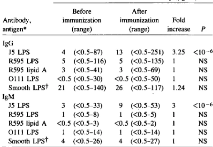
Antibodies to J5 LPS. Of the 70 volunteers before immu-nization, 60%and 96%had detectable levels of IgG and IgM antibodies, respectively, against J5 LPS. These values increased to 86%and 99%after vaccination with E. coli J5. The me-dian increase in the levels of antibodies directed to J5 LPS was 3-fold for IgG and a 3.25-fold for IgM (table 1). This slight increase contrasted with the several hundredfold increase ob-served in rabbits immunized intravenously: titers usually increased from 1:100-1:200 before immunization to 1:25,600-1:102,400 after immunization with boiled bacterial cells.
Table 1. Antibody levels in 70 volunteers immunized with
Escherichia coli J5.
Median levels of anti-LPS antibody (ug/rnl)
Before After
Antibody, immunization immunization Fold antigen* (range) <range) increase P
IgG J5LPS 4 «0.5-87) 13 «0.5-251) 3.25 <10-6 R595LPS 5 «0.5-116) 5 «0.5-135) 1 NS R595lipid A 3 «0.5-41) 3 «0.5-69) 1 NS 0111 LPS <0.5 «0.5-30) <0.5 «0.5-50) 1 NS Smooth LPSt 21 «0.5-140) 26 «0.5-117) 1.24 NS IgM J5LPS 3 «0.5-33) 9 «0.5-53) 3 <10-6 R595LPS 1 «0.5-8) 1 «0.5-5) 1 NS R595lipid A <0.5 «0.5-3) <0.5 «0.5-2) 1 NS 0111 LPS 1 «0.5-14) 1 «0.5-14) 1 NS Smooth LPSt 4 «0.5-26) 4 «0.5-27) 1 NS NOTE. LPS, lipopolysaccharide.
*Complexes of high-density lipoproteins with LPS or lipid A were used to coat plates.
tMixture of LPS from seven smooth bacterial strains (see text).
Antibodies to Re LPS and to lipid A. Of volunteers before immunization, 67 % and 69 %, respectively, had detectable IgG and IgM antibodies against S. minnesotaReLPS and 59 % and 16% against S. minnesota lipid A. Overall, these values did not increase significantly after vaccination, and there was no significant increase of the antibody levels in the subgroup of volunteers with detectable preimmune levels (table 1).
Since antigenic and structural differences exist between the core regions of S. minnesota and E. coli, we measured anti-bodies against the LPS of a Re mutant of E. coli (strain D3lm4) and against its lipid A in the sera of 21 volunteers randomly selected from our series. Before immunization with E. coli J5, the median (range) levels were 6 AU (0-64) for IgG and 2 (0-21) for IgM anti-D3lm4 LPS antibodies, and 2 (0-31) and 9 (0-55) AU, respectively, for D3lm4 lipid A anti-bodies. There was no statistically significant increase in any of these antibodies after immunization. In only four volun-teers was some increase (less than threefold) in the levels of one or several antibodies observed.
Antibodies to LPS from smooth bacteria and total immu-noglobulin levels. No significant increase in specific IgG or IgM antibodies against LPS from E. coli 0111 (the parent of the J5 mutant) was observed. A slight increase of the me-dian IgG antibody level against the mixture of LPS from seven smooth bacterial strains (1.24-fold) was not statistically significant and of doubtful biological importance. IgM anti-bodies directed against the same seven antigens did not in-crease (table 1).
The total serum IgG or IgM levels were measured in 10 volunteers randomly selected from our group: The mean (SD) IgG serum levels before and after immunization were 9.4 (1.4)
JID 1991;163 (April) Escherichia coli J5 Vaccine in Humans 771
gil and 9.2 (1.1) gil, and the mean (SD) IgM serum levels were 1.0 (.04) gil and 1.0 (.05) gil, respectively.
Discussion
The current knowledge of the protective activity of anti-body to endotoxin core was recently reviewed [5]. There is no direct and convincing proof so far that the protection ob-served with polyclonal antisera to rough mutants in animal experiments [6-8] is due to cross-reactive anti-LPS antibod-ies (see [9]). The protection reported with some monoclonal antibodies [10-12] needs confirmation [13-15]. In humans, there has been a discrepancy in clinical trials between the pro-tection affordedbyJ5 antiserum and the weak or lack of corre-lation with the levels of J5 LPS antibodies measured in sera administered to patients.
In the present study, we found that immunization of volun-teers withE.coli J5 did not result in a marked elevation of J5 LPS antibodies, or in an elevation of lipid A, anti-Re LPS, or anti-smooth LPS antibodies. This further raises the question of the mechanism of protection afforded by anti-J5 serum or plasma. One possible explanation for this dis-crepancy is that the antibody responsible for protection has not been accurately measured by the serologic methods used. Detection of antibodies to the various core LPS is difficult because of the limited solubility and self-aggregating proper-ties of hydrophobic core LPS, which may lead to nonspecific binding of antibodies and to irregular coating of ELISA plates. To avoid these problems, we modified the physicochemical state of core LPS by complexing it to HDL, a natural carrier of HDL in vivo, and used HDL-LPS complexes to coat the plates. This method has been shown to increase the sensitiv-ity and the specificsensitiv-ity of the detection of core LPS or lipid A antibodies [4]. In addition, the core region itself shows significant inter- and intraspecies differences in its composi-tion [13], and type-specific antibodies are elicited after im-munization with rough mutants. The structures most likely to induce broadly cross-reactive anti-LPS antibodies are the most conserved parts, that is, lipid A or the lipid A-KDO region. Therefore, if lipid A or lipid A-KDO antibodies are a subset of J5 LPS antibodies, their detection might be ob-scuredbythe quantitatively predominant type-specific J5 LPS antibodies. Moreover, since LPS in outer membranes is het-erogeneous [16], the innermost region of LPS may be exposed at the surface of the whole J5 bacteria, whereas only com-plete LPS with core sugars may be present in the J5 LPS ex-tractedbychemical methods. Thus, immunization with whole J5 bacteria would induce core LPS antibodies not detectable when using purified J5 LPS as antigen.
We investigated whether J5 vaccine induced the produc-tion of antibodies against lipid A or the lipid A-KDO region that would not be adequately detectedbyELISA using J5 LPS as antigen. We could not detect significant increases of anti-bodies to two lipid A and two Re LPS extracted both from
S.minnesota R595 or fromE.coli D3lm4. Since the use of HDL-LPS complexes instead of free LPS as antigen may mask epitopes of the core LPS, it remains possible that antibodies against these hidden epitopes may be missed. This seems un-likely, however, because monoclonal antibodies directed against well-defined epitopes of core LPS easily recognize their epitopes within HDL complexes [4]. Our results ques-tion the role of antibodies to lipid A or to lipid A-KDO as protective factors in J5 antiserum, although these negative findings must be compared with those of clinical studies (now underway) with anti-lipid A monoclonal antibodies and with purified intravenous immunoglobulins enriched in anti-Re LPS antibodies.
Another possible explanation for the discrepancy between the protection afforded by J5 antiserum and the weak anti-J5 LPS antibody increase observed is that the factor responsible for the protection is a nonspecific polyclonal antibody increase rather than cross-protective anti-core LPS antibodies. Indeed, studies in rabbits suggest that a polyclonal response against
o
antigens may occur after intravenous immunization with J5 bacteria [17]. However, the present data suggest that this is unlikely in man, because there was neither a significant increase in antibodies to LPS from smooth gram-negative bac-teria nor an increase in the total IgG or IgM serum levels. Alternative explanations may be that J5 vaccine increased nonspecifically some unrecognized acute-phase reactants capable of neutralizing LPS, altering its metabolism, or coun-teracting the biologic effects of humoral or cellular factors released by the stimulation of LPS. Additional studies are needed to explore these possibilities.References
1. Ziegler El, McCutchan JA, Fierer J, et al. Treatment of gram-negative bacteremia and shock with human antiserum to a mutantEscherichia coli. N Engl J Med 1982;307:1225-30.
2. Baumgartner JD, Glauser MP, McCutchan JA, et al. Prevention of gram-negative shock and death in surgical patients by prophylactic anti-body to endotoxin core glycolipid. Lancet 1985;2:59-63. 3. Jansson PE, Lindberg AA, Lindberg B, Wollin R. Structural studies
on the hexose region of the core in lipopolysaccharidesfrom enterobac-teraceae. Eur J Biochem 1981;115:571-7.
4. Heumann0,Baumgartner JD, Jacot-Guillarmod H, Glauser MP. Anti-bodies to core lipopolysaccharide determinants: absence of cross-reactivity with heterologous lipopolysaccharides. J Infect Dis 1991; 163:762-8.
5. ZieglerEl,Protective antibody to endotoxin core: the emperor's new clothes? [perspective]. J Infect Dis 1988;158:286-90.
6. Braude AI, Douglas H. Passive immunization against the local Shwartz-man reaction. J Immunol 1972;108:505-12.
7. McCabe WR. Immunization with R mutants of S.minnesota. I. Protec-tion against challenge with heterologous gram-negative bacilli. J Im-munol 1972;108:601-10.
8. Ziegler El, Douglas H, Braude AI. Human antiserum for prevention of the local Shwartzrnanreaction and death from bacterial lipopolysac-charides. J Clin Invest 1973;52:3236-8.
9. Baumgartner JD, Glauser MP. Immunoprophylaxis and immunother-apy of gram-negative bacterial infections. In: Sissons P, Borysiewicz
772 Baumgartner et al. ]ID 1991;163 (April)
L, CohenJ,eds. Immunology of infection. Norwell, MA: Kluwer Academic Publishers, 1990.
10. Dunn DL, Bogard WC, Cerra FB. Efficacy of type-specific and cross-reactive murine monoclonal antibodies directed against endotoxin dur-ing experimental sepsis. Surgery 1985;98:283-9.
11. Teng NNH, Kaplan HS, Hebert ]M. Protection against gram-negative bacteremia and endotoxemia with monoclonal IgM antibodies. Proc Nat! Acad Sci USA 1985;82:1790-4.
12. Young LS, Gascon R, Alam S, Bermudez LE. Monoclonal antibodies for treatment of gram-negative infections. Rev Infect Dis 1989;II(suppl 7):SI564-71.
13. Pollack M, Chia ]KS,·Koles NL, Miller M, Guelde G. Specificity and cross-reactivity of monoclonal antibodies reactive with the core and lipid A regions of bacterial lipopolysaccharide. ] Infect Dis 1989;159:168-88.
14. Baumgartner ]D, Heumann D, GerainJ,Weinbreck P, Grau GE, Glauser MP. Association between protective efficacy of anti-lipopolysaccharide (LPS) antibodies and suppression of LPS-induced tumor necrosis factor
exand interleukin 6: comparison of0 side chain-specific antibodies with core LPS antibodies. ] Exp Med 1990;171:889-96. 15. MinerKM, ManyakCL, WilliamsE. Characterization of murine
mono-clonal antibodies toEscherichia coli]5. Infect Immun 1986;52:56-62. 16. Munford RS, Hall CL, Rick PD. Size heterogeneity ofSalmonella typhimuriumlipopolysaccharides in outer membranes and culture su-pernatant membrane fragments. ] Bacteriol 1980;144:630-40. 17. Siber GR, Kania SA, Warren HS. Cross-reactivity of rabbit antibodies
to lipopolysaccharide ofEscherichiacoli]5 and other gram-negative bacteria. ] Infect Dis 1985;152:954-64.