A young woman with high blood pressure on haemodialysis: it is never too late to evaluate hypertension
4
0
0
Texte intégral
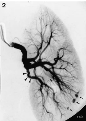
(2) Unilateral dysplasis of the renal artery. 2735. Fig. 1. Pre-operative angiogram of the left renal artery showing an occlusion 3 cm distal to the origin (arrowheads). Collateral vessels are well-developed.. Fig. 3. Fibromuscular dysplasia of the renal artery, perimedial type, with marked deposition of collagen in the outer vessel wall ( large asterisk). The lamina elastica interna (arrows) is segmentally missing (arrowheads). The intima consists of dense fibrous tissue (small asterisk). Elastica van Gieson stain; 20× original magnification.. Fig. 2. Post-operative angiogram demonstrating good arterial filling centrally and peripherally. Only one branch of the renal artery supplying the lower pole displays ‘beading’ along a short segment (arrowheads). This angiographic pattern is consistent with fibromuscular dysplasia. The small intraparenchymal ‘aneurysms’ (arrows) are a consequence of the renal biopsy.. severely altered by deposition of fibrous tissue, focally ‘replacing’ the outer half of the arterial wall, in particular the outer media ( Figure 3). Higher power magnification revealed that smooth muscle cells were not. eradicated, but highly atrophic and ‘strangulated’ by collagen ( Figure 4). Segmentally, connective tissue appeared to infiltrate from the outer portion of the media into the inner layers, sparing only small islands of muscle. In segments with better preserved media, rudimentary elastic lamellae were located amidst smooth muscle bundles separating the outer ‘longitudinal’ from the inner ‘circumferential’ portion ( Figure 4). Structural changes were also found along the internal elastic lamina with partial fragmentation and loss (Figures 3 and 5). Connective tissue expanded the intima which showed features of fibroplasia along the outer aspect (alpha smooth muscle actin positive myofibroblasts) and an organized thrombus with iron deposition and neo-vascularization centrally (Figures 3 and 5). The radiographically observed vascular occlusion was due to thrombus formation. Arterial wall thinning or aneurysms were not present. The renal biopsy contained seven normal glomeruli lacking any light microscopical or immunohistochemical evidence of glomerulonephritis. Tubules, arterioles and small arteries were unremarkable. The interstitium revealed only mild patchy inflammatory fibrosis and minute foci of non-specific mononuclear cell infiltrates. Arcuate type arteries were not sampled.. Comment Excessive persisting hypertension despite aggressive ultrafiltration prompted an intensive search for under-.
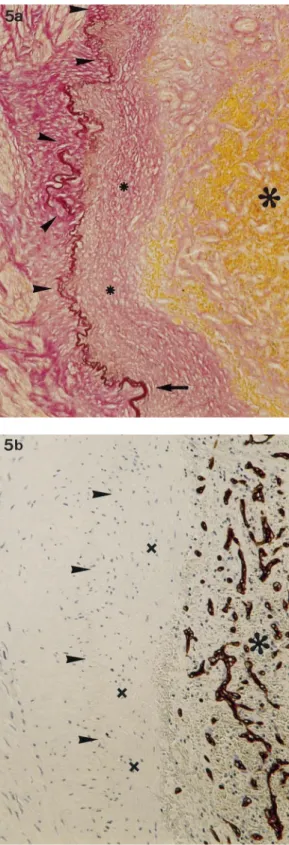
(3) 2736. V. Nickeleit et al.. Fig. 4. For legend see p. 2737.. Fig. 5. For legend see p. 2737.. lying renal vascular lesions which proved rewarding in this young lady who presented with (sub) acute renal failure. She had unilateral renal artery dysplasia with occlusion and became dialysis dependent because the contralateral kidney was hypoplastic. Renal artery dysplasia mostly affects patients in their second or third decades, is more common among. women and usually involves only one side. Renal veins remain unchanged [1]. The mechanisms leading to structural changes in the arterial wall are undetermined. It has been speculated that mechanical injury due to increased mobility of the kidney and stretching of the renal artery may lead to fibrous remodelling of the vessel wall [2,3]. However, this hypothesis seems.
(4) Unilateral dysplasis of the renal artery. 2737. Fig. 4. (a) Complex changes in the arterial wall: dense collagen is deposited in the outer media ( large asterisk). Connective tissue seems to infiltrate into the inner media (fat arrow) and accumulates along the lamina elastica interna (small asterisks), which is focally ruptured (thin arrow). Rudimentary elastic lamellae are located along the inner aspect of the thickened intima ( large arrowheads) as well as between smooth muscle bundles of the media (small arrowheads). The lumen is nearly completely occluded by an organized thrombus. Elastica van Gieson stain; 25× original magnification. (b) Highly atrophic smooth muscle cells (brown) in the outer media are surrounded by abundant collagen. Only small bundles of muscle in the inner media are spared (arrows). Immunohistochemistry, incubation to detect alpha-smooth muscle actin; 125× original magnification. Fig. 5. (a) Higher power view illustrating changes of the inner vascular wall: the lamina elastica interna (arrow) is segmentally fragmented and ruptured (arrowheads). Dense connective tissue, containing numerous alpha smooth muscle actin positive cells, is deposited on the medial and intimal side (small asterisks) of the elastica. A thrombus occludes the lumen ( large asterisk). Elastica van Gieson stain; 60× original magnification. (b) Neo-vascularization (dark brown) marks the area of the organized thrombus ( large asterisk). Clearly separated is the thickened intima (small asterisks; elastica interna marked by arrowheads). The thickened intima has features of fibroplasia. Same view as (a); immunohistochemistry, incubation to detect endothelial cells (CD34); 80× original magnification.. unlikely because the carotid or iliac arteries are occasionally dysplastic as well [4]. Renal artery dysplasia seems progressive [5,6 ]: both the degree of stenoses and the lengths of involved arterial segments tend to increase with time. On follow-up of untreated cases, a decrease of renal size was observed in 62% of patients [5]. The frequency of total renal infarction was reported to be as high as 24% [5]. Fibromuscular dysplasia can be subtyped according to the arterial wall layer being most affected [7,8,9,10]: i.. Intimal fibroplasia (rare; can be difficult to distinguish from non-specific fibrosis as seen in atherosclerosis) ii. Medial types; (A) Medial ‘muscular’ hyperplasia (rare). (B) Medial fibroplasia with aneurysms (common: 60–70%; bilateral in 60%) (C ) Perimedial fibroplasia (common, 15–25%) (D) Medial dissection (infrequent, 5–10%) iii. Adventitial fibroplasia (rare) Medial fibroplasia with aneurysms (iiB) and perimedial fibroplasia (iiC ) are the most common forms accounting for 75–95% of all cases. The histological changes observed in the case under discussion are consistent with a ‘perimedial’ type. The extensive fibrous replacement of the outer portion of the media, the lack of aneurysms, the unilateral occurrence, thrombosis, and angiographic ‘beading’ favour the perimedial variant [7]. Unusual features of the perimedial type in the current case are the segmental loss and fragmentation of the internal elastic lamina, elastic lamellae misplaced between smooth muscle bundles and fibroplastic thickening of the intima. Thus, our histological findings underline the great variability of patterns. The long-term prognosis of our patient seems excellent, since recurrence of dysplasia after renovascular repair is exceptional [4], and because her intrarenal vessels were free of radiologically evident dysplastic changes. Intraparenchymal arteries may occasionally be the site of dysplastic alterations causing therapy resistent hypertension [11]. Although, dysplastic changes were found in our patient postoperatively in a non-repaired branch of the renal artery, progressive stenosis might be managed in the future by percutaneous transluminal angioplasty [6 ].. Teaching Point 1.. 2.. 3.. The possibility of renovascular lesions should be considered in young patients with apparent endstage renal failure and severe hypertension despite aggressive ultrafiltration. Unilateral arterial dysplasia may cause renal failure if it induces hypertension and parenchymal damage in the contralateral kidney, or—as in this case—when the contralateral kidney is hypoplastic. Three categories of dysplastic arteries can be distinguished, but morphological subtyping is sometimes arbitrary.. References 1. Ekelund L, Gerlock AJ, Goncharenko V, Francis R. Renal venographic findings in 29 kidneys with fibromuscular dysplasia of the renal artery. Radiology 1977; 125: 631–632 2. Anderson GH Jr, Blackeman N, Streeten DH. The effect of age on prevalence of secondary forms of hypertension. J Hypertens 1994; 12: 609–615 3. Petit R, Delivigne J. Indication de la ne´phropexie: apports du ne´phrogramme isotopique et de l’arte´riographie ro¨nale. Acta Urol Belg 1973; 41: 386–395 4. Kelly TF, Morris GC. Arterial fibromuscular disease. Observations on pathogenesis and surgical management. Am J Surg 1982; 143: 232–236 5. Goncharenko V, Gerlock AJ Jr, Shaff MI, Hollifield JW. Progression of renal artery fibromuscular dysplasia in 42 patients as seen on angiography. Radiology 1981; 139: 45–51 6. Lorelius LE, Hemmingsson A, Hagg A, Morlin C, Aberg H. Progressive fibromuscular dysplasia of the renal artery. Angiographic and clinical follow-up. Acta Radiol [Diagn] (Stockh) 1985; 26: 705–708 7. Olson JL. Hypertension: essential and secondary forms. In: Jennette JC, Olson JL, Schwartz MM, Silva FG, eds. Heptinstall’s Pathology of the Kidney Lippincott-Raven, New York: 1998: 943–1001 8. Harrison EG Jr, McCormack LJ. Pathologic classification of renal arterial disease in renovascular hypertension. Mayo Clin Proc 1971; 46: 161–167 9. Youngberg SP, Sheps SG, Strong CG. Fibromuscular disease of the renal arteries. Med Clin North Am 1977; 61: 623–641 10. Luscher T, Lie JT, Stanson AW, Houser OW, Hollier LH, Sheps SG. Arterial fibromuscular dysplasia. Mayo Clin Proc 1987; 62: 931–952 11. Holm-Bentzen M, Gerstenberg T, Horn T, Larsen S. Medical fibroplasia: involvement of renal artery and small renal arteries in renal vascular hypertension. Scand J Urol Nephrol 27; 1993: 263–265.
(5)
Figure
Documents relatifs
The occurrence of mate change in females due to male evic- tion did not differ from zero (two treatments with addi- tional males present compared with the female treatment;
The massless one-loop vertex diagram is constructed by exploiting the causal structure of the diagram in configuration space, which can be translated directly into dis-
Parmi les avantages évidents de la technologie adoptée au CHA, notons : une utilisation de la technologie du code à barres pour la distribution de la grande majorité des
Results: (1) Self-initiated movements (SI-EC) revealed activations in the prefrontal cortex bilaterally, the right lateral premotor cortex, anterior cingulate cortex and cerebellum,
We tested for two additional contrasts an- alyzed by Smolka et al.: the difference in priming strength between Transparent and Opaque Deriva- tion (not significant in either
Buruli ulcer disability is preventable: go to hospital before it is too late!. Global Buruli
progenitor cells, stem cells, congenital heart defect, right ventricle failure, pulmonary arterial hypertension!. Date received: 16 November 2017; accepted: 9
In many developing nations, already beset with economic, social and other health problems, the rates of heart disease, diabetes and hypertension are as high as or
