Reversible accumulation of (1→3,1→4)‐β‐glucan endohydrolase in wheat leaves under sugar depletion
10
0
0
Texte intégral
(2) 2324. Roulin and Feller. also be detected during germination, but only proteins immunologically related to barley isoenzyme EI are present (Lai et al., 1993b). Nucleotide sequence analyses of cDNAs further confirm at the genetic level the similarity of the wheat (1£3,1£4)-b-glucanases with the isoenzyme EI from barley (Lai et al., 1993a). Previous work with barley (Slakeski and Fincher, 1992a, b) did not study in great detail the regulatory patterns of (1£3,1£4)-b-glucanase appearance in leaves of different types and at different developmental stages. The availability of monoclonal antibodies specific for a barley (1£3,1£4)-b-glucanase isoenzyme that react with the wheat enzymes allowed the investigation of the regulation of (1£3,1£4)-b-glucanase accumulation in leaves from young wheat plants. In this work, the impact of carbon status on both the activity and enzyme protein levels was studied. To this end, environmental and nutritional conditions increasing (light, nitrogen deficiency, glucose feeding) or decreasing (prolonged darkness, nitrogen supply) the leaf soluble carbohydrates were used.. Materials and methods Plant material and treatments. Wheat (Triticum aestivum L. cv. Arina) seeds were germinated on paper towels for 4 d in the dark at 25 8C. The germinated seeds were planted in gravel and further incubated under culture room conditions. On day 7 after imbibition the seedlings were transplanted into 1.0 l pots, six plants per pot, containing a halfstrength complete nutrient solution. On day 11 after the initiation of germination the plants were switched to a full-strength nutrient solution as described previously (Hildbrand et al., 1994). The nutrient medium was changed weekly. Plants were grown under controlled conditions using a 14 h photoperiod with 130 mmol m 2 s 1 photosynthetically active radiation (PAR). Day and night temperatures were 25 8C and 21 8C, respectively. For dark incubation experiments, intact plants were either switched to continuous illumination (85 mmol m 2 s 1 PAR) or to complete darkness when the second leaf was fully expanded (usually between day 17 and day 18 after imbibition). In some cases, 5 cm segments were cut from the middle part of fully expanded second leaves and were incubated at 25 8C, with the abaxial side down, in water or sugar solutions under continuous light (120 mmol m 2 s 1 PAR, measured at the level of the leaf segments) or in complete darkness. All sugar solutions were adjusted to pH 6.0 in 5 mM 2-morpholinoethanesulphonic acid (MES) buffer and were changed daily. Experiments were always started in the middle of the light phase and were carried out three times. At each sampling time two replicates, consisting of three intact leaves or five leaf segments from independent plants, were immediately frozen in liquid nitrogen and stored at 80 8C until analysis. Although the absolute values varied somewhat from experiment to experiment, the trends were always similar and results from one experiment are shown.. Protein extraction. The plant material was ground under liquid nitrogen and homogenized in 50 mM sodium acetate buffer (4 ml and 2 ml g 1 fresh weight [FW] for leaf and root material,. respectively), pH 5.0, containing 10 mM EDTA, 5 mM sodium azide, 3 mM 2-mercaptoethanol, 3 mM PMSF, and 0.1 M NaCl using a Polytron mixer (PT 2000, Kinematica, Littau, Switzerland) for 30 s at full speed. After a 30 min incubation on ice with repeated mixing, insoluble material was removed by filtration through Miracloth followed by centrifugation (10 min at 20 000 g) and the supernatant was stored at 80 8C prior to analysis. Under such conditions, the enzyme remained stable for at least 1 week.. Enzyme assays (1£3,1£4)-b-Glucan endohydrolase activity was measured viscometrically at 40 8C, using water-soluble barley (1£3, 1£4)-b-glucan (medium viscosity; Megazyme, Bray, Ireland) as substrate. A unit of activity is defined as the amount of enzyme causing an increase of 1.0 in the reciprocal specific viscosity (D1uhsp) per min of a 3 mg ml 1 (1£3,1£4)-b-glucan solution in 50 mM sodium acetate buffer, pH 5.0, containing 5 mM sodium azide and 400 mg ml 1 bovine serum albumin (Woodward and Fincher, 1982). Time-course experiments ensured that the activity was measured in the initial, linear response region of enzyme action. Since exo-b-glucanases release glucose from (1£3,1£4)-b-glucans, without having any significant effect on the viscosity of the substrate solution (Hrmova and Fincher, 1998), this assay is essentially specific for endohydrolase activity. For the estimation of cellulase activity, carboxymethyl cellulose with a degree of substitution of 0.4 (medium viscosity; Megazyme) was used as a substrate in viscometric assays. Exo-b-glucanase and b-glucosidase activities were measured by the determination of glucose released from the barley (1£3,1£4)-b-glucan and from the oligosaccharides produced by the action of (1£3,1£4)-b-glucan endohydrolases present in the leaf extracts. The reaction mixture contained 75 ml of a 3 mg ml 1 b-glucan solution in 50 mM sodium acetate buffer, pH 5.25, containing 160 mg ml 1 bovine serum albumin, 25 ml of leaf extract, and one drop of toluene to prevent microbial growth. The mixture was incubated at 37 8C for 5 h, and the glucose released was measured enzymatically with the GOD-Perid method (Boehringer Mannheim, Germany). One unit of activity is defined as the capacity to release 1 mmol glucose min 1. This assay did not discriminate between the exo-b-glucanase and b-glucosidase activities. All assays were performed in duplicate and mean values were used for the calculation of the activities.. Gel electrophoresis, antibodies, and immunoblotting Polyacrylamide gel electrophoresis was carried out according to Laemmli using 1.5 mm thick slab gels (12%) (Laemmli, 1970). Leaf and root soluble extracts were diluted with sample buffer containing 196 mM TRIS-HCl buffer, pH 6.8, 6.3% (wuv) SDS, 16% (vuv) 2-mercaptoethanol, 32% (vuv) glycerol, and 0.02% (wuv) bromphenol blue in a ratio 2 : 1 (vuv), and were heated at 95 8C for 5 min. Each lane was loaded with extract equivalent to 3 mg and 6 mg FW for leaf and root material, respectively. After electrophoresis, gels were stained with Coomassie Brilliant Blue R-250 or blotted onto nitrocellulose membranes (0.45 mm; BioRad, Glattbrugg, Switzerland) for the immunodetection of the (1£3,1£4)-b-glucanase proteins. Immunoblotting was performed as described earlier (Mitsuhashi and Feller, 1992) except that the membranes were exposed overnight at 4 8C to the primary monoclonal antibodies raised in mouse against barley (1£3,1£4)-b-glucanase isoenzymes EI and EII.
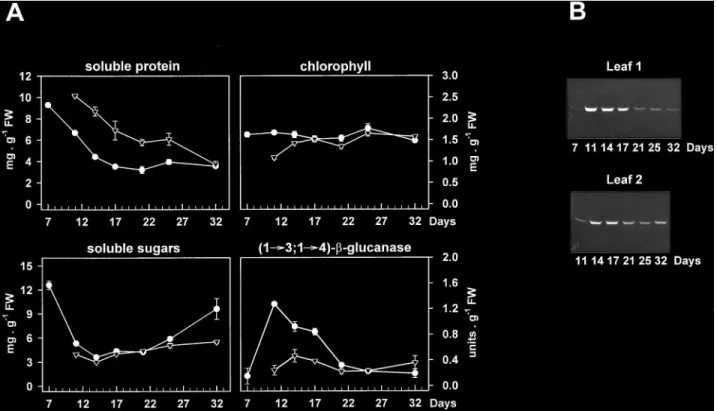
(3) Sugar starvation and accumulation of (1£3,1£4)-b-glucanase (Høj et al., 1990). The blots were incubated for 2 h with bridging antibodies (anti-mouse IgG developed in goat; Sigma) diluted 1 : 1000 (vuv) with 1% (wuv) ovalbumin in TRIS-buffered saline (50 mM TRIS-HCl, pH 7.5, containing 0.2 M NaCl) and afterwards for 2 h with peroxidase-anti-peroxidase complex (mouse; Sigma) diluted 1 : 400 (vuv) in the same buffer. The primary monoclonal antibodies were kindly provided by GB Fincher (Department of Plant Science, Waite Campus, University of Adelaide, South Australia) and were diluted 1 : 500 (vuv). Two replicate samples were analysed for each experiment and the results of one replicate are presented.. Measurement of chlorophyll, soluble protein and carbohydrates. Following homogenization of the leaves and filtration through Miracloth, the filtrate (20 ml) was mixed with 1 ml of 80% (vuv) acetone and chlorophyll was quantified as described earlier (Strain et al., 1971). Soluble protein was determined in the centrifuged filtrate using bovine serum albumin as a standard (Bradford, 1976). Buffer-soluble carbohydrates were measured according to Stieger and Feller (Stieger and Feller, 1994). The centrifugated filtrate (10–20 ml) was mixed with 1 ml anthrone reagent (20 ml ethanol, 200 mg anthrone, 100 ml H2SO4) and heated for 10 min in a vigorously boiling water bath. Colour development was stopped by incubating the sample on ice for 10 min, and the A623 was measured. Glucose (0–50 mg) was used for calibration. Individual analyses were carried out in duplicate and mean values were used for the calculation of chlorophyll, soluble protein and carbohydrate contents.. 2325. Results Appearance of (1£3,1£4)-b-glucanase during leaf development. The developmental pattern of (1£3,1£4)-b-glucanase accumulation in the first and second leaves from hydroponically-grown wheat plants was investigated by activity and Western blot analyses using monoclonal antibodies specific for barley isoenzymes EI and EII (Høj et al., 1990). Under the growing conditions applied, the first leaf emerged between days 5 and 6 after the initiation of germination and was fully expanded by day 11, while the second leaf started to develop at day 9 or 10 and was at full expansion at day 17 or 18. In both types of leaves the soluble protein content per unit FW decreased steadily throughout the experiment, whereas the chlorophyll levels remained quite stable until the later stages of leaf development (Fig. 1A). The decline in total soluble protein might be due initially to leaf expansion and subsequently to natural ageing. As observed with extracts of germinated wheat grain (Lai et al., 1993a, b), a single band (about 30 kDa) cross-reacted on immunoblots of leaf samples with antibodies raised against the barley isoenzyme EI. No proteins were detected in leaf extracts with the isoenzyme EII antibody (data not shown). Since the hexaploid wheat genome may carry at least two. Fig. 1. Developmental pattern of (1£3,1£4)-b-glucanase accumulation in the first (m) and second (,) leaves of wheat plants. The time scale represents the number of days after the initiation of germination. Leaf extracts were tested for their contents in soluble protein, chlorophyll, soluble sugars, and (1£3,1£4)-b-glucanase activity (A) and for the levels in (1£3,1£4)-b-glucanase protein (B) by immunoblotting with a monoclonal antibody specific for barley isoenzyme EI. Data are means"SD of two independent samples, each consisting of three leaves. SD bars are shown when larger than the symbols..
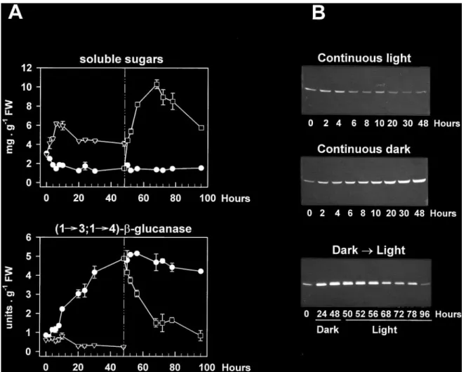
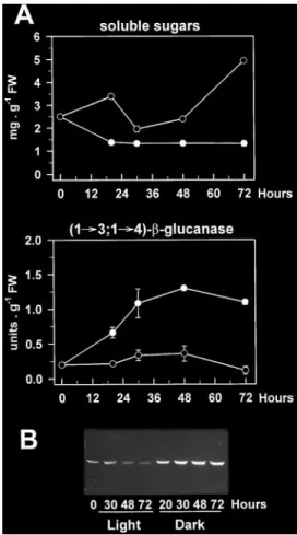
(4) 2326. Roulin and Feller. (1£3,1£4)-b-glucanase genes encoding enzymes with essentially identical primary structures (Slakeski et al., 1990; Lai et al., 1993a), the possibility that this band contained multiple isoforms of the enzyme with similar electrophoretic mobilities could not be excluded. The abundance of (1£3,1£4)-b-glucanase followed a similar developmental pattern in both the first and second leaves, with very low or undetectable levels in the early stages of development, maximum levels at maturation (day 11 and day 17 for the first and second leaves, respectively) and subsequent decreases upon leaf ageing (Fig. 1B). Such changes appeared to parallel the development of (1£3,1£4)-b-glucanase activity (Fig. 1A), indicating that the band detected on the immunoblot actually represented a wheat (1£3,1£4)-b-glucanase that was expressed in the leaves. The accumulation of this enzyme in the maturing first leaves was considerably greater than in the second leaves, and was correlated with a sharp decline in the soluble sugar content between days 7 and 11. (Fig. 1A). Conversely, the disappearance of (1£3,1£4)b-glucanase upon natural ageing seemed to be associated with increasing levels of soluble sugars. Dark induction of (1£3,1£4)-b -glucanase in intact leaves and its reversion by light. As discussed above (Fig. 1), the levels of (1£3,1£4)b-glucanase in mature second leaves were clearly lower than those in the first leaves. However, when 17-d-old intact plants with fully expanded second leaves were placed in complete darkness for up to 4 d, marked changes in the amounts of the enzyme could be observed. Thus, the band recognized on immunoblots by the barley isoenzyme EI-specific antibody started to accumulate after 10 h, peaked after about 48 h (Fig. 2B) and then remained high for up to 4 d of dark treatment (not shown). Again, these changes in protein abundance were paralleled by very similar changes in enzyme activity (Fig. 2A, B), which. Fig. 2. Time-course of (1£3,1£4)-b-glucanase development in the second leaf from young wheat plants subjected to different light conditions. Seventeen-day-old seedlings (second leaf fully expanded) grown under culture room conditions were incubated under continuous light (,) or set in complete darkness (m) for the times indicated. Some of the dark-incubated plants were transferred back to continuous light after 48 h (h). The photon fluence rate used was 85 mmol m 2 s 1 PAR. Soluble sugar and (1£3,1£4)-b-glucanase activity (A), as well as (1£3,1£4)-b-glucanase protein levels (B) were determined in leaf extracts. Data for enzyme activity and soluble sugar levels are means of two independent replicates (each consisting of three leaves) and SD bars are shown when exceeding the size of the symbols..
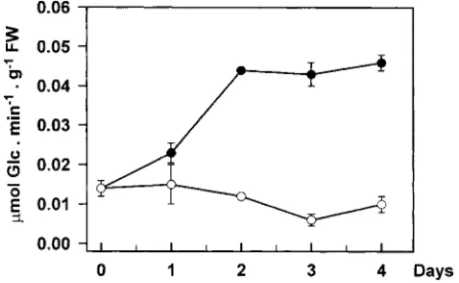
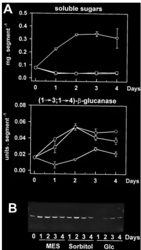
(5) Sugar starvation and accumulation of (1£3,1£4)-b-glucanase. underwent a 6-fold increase after 48 h. In control plants, that were incubated under continuous illumination, (1£3,1£4)-b-glucanase activity and protein levels were low at the beginning of the experiment and tended to further decline upon incubation. Re-exposure of darkincubated plants to continuous light after 48 h led to a marked decrease in (1£3,1£4)-b-glucanase abundance, within one day. Activity rapidly declined after re-illumination and was close to control levels within 20 h (Fig. 2A). Clear decreases in protein abundance could also be observed (Fig. 2B). Such changes in enzyme levels upon re-illumination were closely associated with reciprocal changes in the leaf soluble sugar content, which rapidly increased to high levels between 48 h (start of re-illumination) and 68 h, and decreased thereafter (Fig. 2A). This subsequent decline was most likely due to export processes to the developing third leaves. In contrast, the marked accumulation of (1£3,1£4)b-glucanase observed in the dark was correlated with a 2-fold decrease in leaf soluble sugars within 6 h (Fig. 2A). It should be noted that the effect of darkness on the enzyme levels in wheat leaves was not age-dependent since it could be observed in both first and second leaves of the same plants (data not shown). Although the assay used for detecting (1£3,1£4)b-glucanase activity is specific for endohydrolase activities, it might not be specific for the enzyme detected on the immunoblots. Besides (1£3,1£4)-b-glucanase, endoglucanase activity in cereals could relate to a number of enzyme families, yet only two of them have the potential to degrade the (1£3,1£4)-b-glucan substrate used in the activity assay as well. These include (1£4)-b-glucanase (cellulase) and, if adjacent (1£3)-b-linkages are present in the substrate, (1£3)-b-glucanase (Fincher, 1989). Cellulase activity has been tested in the present work but very low values were found in leaf extracts (not detectable in extracts from light-incubated plants; less than 0.3 units g 1 FW in extracts from dark-incubated plants as compared with about 5.0 units g 1 FW for (1£3,1£4)-b-glucanase). The good correlation between (1£3,1£4)-b-glucanase protein and activity levels further indicated that cellulase interference with the activity assay was minimal. In addition, the lack of evidence for the widespread occurrence of blocks of adjacent (1£3)-b-linkages in cereal (1£3,1£4)-b-glucans (Woodward et al., 1983) together with the inability of purified (1£3)-b-glucanases from barley leaves to hydrolyse the polysaccharide (Hrmova and Fincher, 1993) suggest that these enzymes were also unlikely to interfere. The activities of exo-b-glucanases and b-glucosidases also increased when wheat seedlings were incubated in the dark (Fig. 3). However, significant changes were not apparent until after 2 d in darkness, while marked increases in (1£3,1£4)-b-glucanase activity were already visible after 20 h (Fig. 2A).. 2327. Fig. 3. Changes in exo-b-glucanase and b-glucosidase activities in second leaves of 17-d-old wheat plants incubated under culture room conditions (k) or in complete darkness (m) for 4 d. Each point represents the mean"SD of two replicates, each consisting of three leaves. SD bars are shown when larger than the symbols.. Dark-induced accumulation of (1£3,1£4)-b-glucanase in leaf segments. Leaf segments were considered as a suitable system for manipulating the carbohydrate levels (e.g. by adding exogenous sugars or by varying the illumination conditions). Chlorophylls, Rubisco and other soluble proteins declined in leaf segments during incubation in the dark and, to a minor extent, also in the light indicating senescence (Fig. 4A, B). Rapid increases in (1£3,1£4)b-glucanase activity (Fig. 4A) and in the enzyme protein level (Fig. 4C) occurred upon dark incubation of the segments, with a maximum 3-fold increase in activity after 2 d. Although such changes were detected in darkness before the development of major senescence symptoms, they were maintained during the early stages of leaf senescence (2–3 d). Furthermore, while most soluble proteins had disappeared after 6 d of dark incubation (Fig. 4B), considerable amounts of (1£3, 1£4)-b-glucanase protein were still present in the extracts (Fig. 4C). Continuous illumination of the leaf segments led to the disappearance of (1£3,1£4)-b-glucanase protein (Fig. 4C), as well as to a marked increase in the soluble sugar content (Fig. 4A). Inhibition of dark-induced accumulation by glucose. The data reported in Figs 1 and 2 indicated that the abundance of (1£3,1£4)-b-glucanase in the light or in the dark might be controlled by the sugar levels in the leaves, with low levels in the dark leading to accumulation of the enzyme. Consequently, it should be possible to prevent or delay this accumulation in the dark by increasing the internal sugar concentrations. To test this possibility, the leaf segment system described previously was used to provide dark-incubated segments with glucose. As shown in Fig. 5, the dark-induced accumulation of the enzyme in leaf segments was strongly inhibited.
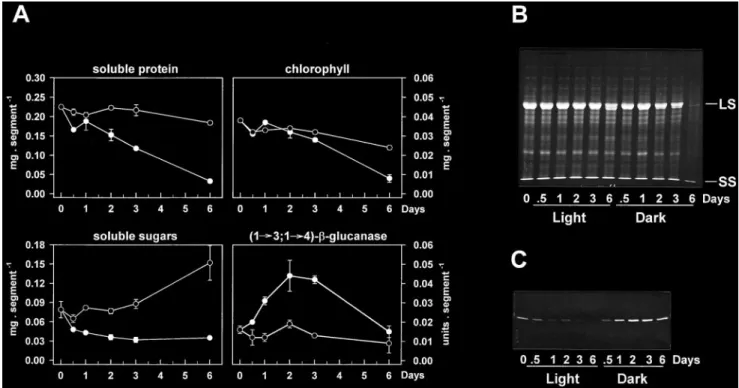
(6) 2328. Roulin and Feller. Fig. 4. Dark-induced senescence and accumulation of (1£3,1£4)-b-glucanase in wheat leaf segments. Segments were cut from fully expanded, second leaves from 17-d-old plants and were incubated at 25 8C in water under continuous light (k) or in complete darkness (m). The light intensity used was 120 mmol m 2 s 1 PAR, measured at the level of the leaf segments. Soluble protein, chlorophyll, soluble sugar and (1£3,1£4)-b-glucanase activity levels were determined in leaf extracts at the times shown (A), and mean values "SD (shown when larger than the symbols) are given for two independent samples, consisting each of five segments. Changes in the total soluble protein pattern were visualized by Coomassie-staining of the gel (B) and the amounts of (1£3,1£4)-b-glucanase protein were detected by immunoblotting (C). LS, large subunit of Rubisco; SS, small subunit of Rubisco.. by 0.2 M glucose for up to 4 d. Under such conditions, glucose was efficiently taken up by the leaf segments, as indicated by the sharp increase in the soluble sugar content (Fig. 5A). The addition of glucose initially caused a decline in (1£3,1£4)-b-glucanase protein levels (compare time 0 with days 1 and 2 on glucose in Fig. 5B) which increased slightly thereafter in spite of the high sugar levels still present in the leaf segments. The inhibitory effects of glucose were not due to changes in the osmoticum, because incubation of leaf segments in 0.2 M sorbitol did not alter the accumulation of the enzyme in the dark. Addition of sucrose to leaf segments in the dark triggered similar inhibitory effects (data not shown). Effects of nitrogen deficiency and sink strength. It might be argued that the carbohydrate levels in leaf segments incubated in the dark in 0.2 M glucose might be artificially high, leading to exaggerated or erroneous effects on enzyme levels. A system in which the sugar content could be manipulated in intact plants during a normal lightudark cycle was therefore considered. Investigations of the developmental pattern of (1£3, 1£4)-b-glucanase appearance in wheat leaves have shown that the enzyme accumulated strongly in expanding first leaves between days 7 and 11 after the initiation. of germination (Fig. 1). This coincided with the start of the hydroponic culture system on a nitrogen (N)containing nutrient solution and was associated with a large decrease in the leaf soluble sugar content. Since N-deficiency has been reported to increase the carbohydrate content of the leaves (Paul and Stitt, 1993; Paul and Driscoll, 1997), it was checked whether withdrawal of N from the beginning of the culture could modify the regulation pattern of (1£3,1£4)-b-glucanase accumulation in planta. Plants transferred to a N-containing medium quickly accumulated large amounts of the enzyme protein in the first leaves while the activity increased consistently over 4 d (Fig. 6). The levels of soluble sugars, on the other hand, dropped to 35% of the initial values after 2 d (Fig. 6A). Withdrawal of N considerably slowed the accumulation of the enzyme during the first two days and this was accompanied by a less pronounced decline in leaf soluble sugars (Fig. 6). In spite of the imposed N-stress, wheat plants continued to grow and develop new leaves which represent initially sinks for sugars and other metabolites. Removal of the developing leaves further enhanced the effect of N-deficiency on both the accumulation of (1£3,1£4)-bglucanase and on the levels of soluble sugars (Fig. 6). Identical treatment of N-sufficient plants had no effect (data not shown)..
(7) Sugar starvation and accumulation of (1£3,1£4)-b-glucanase. Fig. 5. Inhibition of dark-induced accumulation of (1£3,1£4)-bglucanase in wheat leaf segments by glucose (Glc). Leaf segments were prepared as described in the legend of Fig. 3 and were incubated at 25 8C in complete darkness in 5 mM MES buffer pH 6.0 (k), 0.2 M sorbitol (h) or 0.2 M Glc (,) for the times indicated. (A) Time-course of the changes in soluble sugar and (1£3,1£4)-b-glucanase activity levels (data points are means of duplicate samples containing five segments and SD bars are shown when exceeding the size of the symbols); (B) immunoblot showing the amounts of (1£3,1£4)-b-glucanase protein present in the leaf segments under the various incubation conditions.. (1£3,1£4)-b-Glucanase in the roots. A similar regulation pattern upon a light to dark transition was found in vegetative tissues that were not exposed to direct light, such as roots. In these tissues, low levels of (1£3,1£4)-b-glucanase in the light contrasted with clearly higher levels in the dark; the latter were associated with low soluble sugar contents (Fig. 7). This observation and previous results strongly support the notion that changes in the levels of photoassimilates, rather than any direct effects of light, participate in the regulation of (1£3,1£4)-b-glucanase levels in wheat leaves.. Discussion The presence of (1£3,1£4)-b-glucanase in leaves from young wheat plants has been demonstrated by Western. 2329. Fig. 6. Influence of nitrogen (N) supply and sink strength on the appearance of (1£3,1£4)-b-glucanase in the first leaf of young wheat seedlings. Following germination on paper towels and cultivation in gravel, 7-d-old seedlings were transplanted into 1.0 l pots and grown hydroponically in half-strength N-containing (k) or N-deficient (h) nutrient solution for 4 d. The final nitrate concentration in the N-containing medium was 1.8 mM. In some cases, developing second leaves (sink) were removed from plants grown in a N-free nutrient solution (,). Growing conditions were as described in ‘Materials and methods’. (A) Levels of soluble sugars and (1£3,1£4)-b-glucanase activity (each point represents the mean of two independent replicates, each containing five leaves, and SD bars are shown when larger than the symbols); (B) levels of (1£3,1£4)-b-glucanase protein visualized by immunoblotting.. analyses and activity measurements. The data showed that the levels of the enzyme in these tissues were modulated both by developmental cues and by light. The developmental pattern of (1£3,1£4)-b-glucanase appearance in wheat leaves indicated that the enzyme accumulated during the expansion phase of the tissues. In grass cell walls, the interaction between the (1£3,1£4)b-glucan molecules and the cellulose microfibrils may actually represent the tensile force-bearing structure that must be loosened to allow the wall to extend during growth of the tissues (Carpita and Gibeaut, 1993; Cosgrove, 1999). Such (1£3,1£4)-b-glucans are present in the cell walls of young wheat leaves, and their content and degree of polymerization decrease during development (Buchala and Wilkie, 1973). It remains open to discussion whether the enzyme identified in this work participates in (1£3,1£4)-b-glucan depolymerization.
(8) 2330. Roulin and Feller. Fig. 7. Dark induction of (1£3,1£4)-b-glucanase in the root system of young wheat plants. Seventeen-day-old seedlings were transferred to continuous light (k) or complete darkness (m). Roots were collected at the times shown and analysed for their soluble sugar content (A) as well as for (1£3,1£4)-b-glucanase activity (A) and protein levels (B). Data for soluble sugar and enzyme activity levels are means"SD of two individual samples, each consisting of the roots from two plants (SD bars shown only when exceeding the size of the symbols).. and might thus play a role in cell wall loosening and tissue elongation during leaf development. Evidence for a direct role of polysaccharide hydrolases in either wall loosening or cell expansion is, however, difficult to find and remains to be demonstrated unequivocally (Cosgrove, 1997, 1999). The results also indicated that the abundance of (1£3,1£4)-b-glucanase in wheat leaves was tightly controlled by the light conditions. Thus, dark incubation of intact plants or excised leaf segments led to a rapid and marked accumulation of the enzyme, whereas re-illumination of dark-grown plants completely reversed this effect, within one day. Negative effects of light on (1£3,1£4)-b-glucanase activities have been recently reported during light-induced inhibition of rice coleoptile growth (Chen et al., 1999), but the nature of the mechanisms involved has not been determined. The response of wheat leaves to dark treatment largely reflected de novo protein synthesis, because the increases in (1£3,1£4)b-glucanase activity closely paralleled similar changes in protein levels. On the other hand, the rapid light-induced. decreases in activity were followed by a marked decay of the enzyme protein, indicative of proteolytic degradation. Clearly, both de novo protein synthesis and proteolytic breakdown may be involved in the control of (1£3,1£4)b-glucanase levels in wheat leaves and it remains a task for the future to assess the relative contribution of these two processes under varying light conditions. However, regulation by additional, post-translational mechanisms is not ruled out. For instance, auxin stimulation of (1£3,1£4)-b-glucanases in maize coleoptile cell walls has been attributed to the control of the activity by a regulatory, non-enzymic wall protein (Inouhe and Nevins, 1997a, 1998). More recently, stimulation of (1£3,1£4)-b-glucanase isoform EI activity by acidification of the apoplast has been postulated during auxininduced elongation growth of barley coleoptiles (Kotake et al., 2000). Several lines of evidence indicated that the effects of light or darkness on the abundance of (1£3,1£4)b-glucanase were mainly mediated by changes in the leaf sugar levels. Hence, it appeared that whenever the leaves were depleted of sugars (such as during extended dark periods) the enzyme accumulated, whereas elevated sugar levels (such as upon continuous illumination of intact plants and leaf segments, or addition of glucose to leaf segments in the dark) caused a decrease in (1£3,1£4)b-glucanase abundance or prevented its accumulation. Although direct effects of light through phytochrome activation can not yet be excluded, the data suggested that regulation by sugars was more important, because it could be observed both in the light and in the dark. The activities of exo-b-glucanase and b-glucosidase also increased markedly under conditions of sugar starvation (Fig. 3). These enzymes release glucose from the non-reducing termini of (1£3,1£4)-b-glucans and oligosaccharides produced by the action of the endohydrolases (Hrmova and Fincher, 1998). Thus, the combined action of endo- and exohydrolase activities would lead to the complete depolymerization of cell wall (1£3,1£4)-b-glucans to glucose. Preliminary results using intercellular washing fluid isolated from wheat leaves further showed that (1£3,1£4)-b-glucanase was at least partially present in the extracellular space. However, it remains to be demonstrated whether the observed changes in enzyme activities under sugar starvation actually result in the degradation of the polymer and might thus represent a means to provide the tissues with glucose in order to sustain metabolism. Interestingly, the association of (1£3,1£4)-b-glucan hydrolysis with carbon reallocation to growing sink organs has been postulated in maize coleoptiles following growth cessation (Inouhe and Nevins, 1997b) and in germinated barley grain (Hrmova and Fincher, 1998). Experiments with leaf segments clearly showed that glucose provided exogenously was able to prevent the.
(9) Sugar starvation and accumulation of (1£3,1£4)-b-glucanase. accumulation of (1£3,1£4)-b-glucanase in the dark. In plants, sugar-regulated gene expression is a widespread phenomenon that is considered a central mechanism for adjusting to changes in nutrient availability and for controlling resource partitioning (Koch, 1996; Lalonde et al., 1999). Various lines of evidence suggest that the enzyme hexokinase is involved in sugar sensing (reviewed in Koch, 1996; Lalonde et al., 1999) although hexokinaseindependent glucose-signalling systems have also been reported (Lalonde et al., 1999). In addition, sugarmediated down-regulation of gene expression in plants may be modulated by complicated transcriptional and post-transcriptional processes involving specific ciselements and transfactors (Koch, 1996; Ngai et al., 1997; Urwin and Jenkins, 1997; Hwang et al., 1998), as well as specific determinants of mRNA stability (Chan and Yu, 1998). It remains a task for the future to identify the metabolic signal(s) regulating the expression of (1£3, 1£4)-b-glucanase in wheat leaves and the molecular mechanisms underlying the process. Acknowledgements The authors are grateful to Professor GB Fincher (Department of Plant Science, University of Adelaide, South Australia) for generously providing the monoclonal antibodies against barley (1£3,1£4)-b-glucanase isoenzymes EI and EII and for constructive comments on this work, and to Dr AJ Buchala (Department of Biology, University of Fribourg, Switzerland) for helpful discussions and for improving the English of the manuscript. The authors also thank Lisa Renfer, Fabienne Ja¨ggi and Bruno Hungerbu¨hler for assistance in some aspects of this work. This work was supported by a grant from the Swiss National Science Foundation (project no. 31-55289.98).. References Bradford MM. 1976. A rapid and sensitive method for the quantitation of microgram quantities of protein using the principle of protein–dye binding. Analytical Biochemistry 72, 248–254. Buchala AJ, Wilkie KCB. 1973. Total hemicellulose from wheat at different stages of growth. Phytochemistry 12, 499–505. Buchala AJ, Wilkie KCB. 1974. Total hemicellulose from Hordeum vulgare plants at different stages of maturity. Phytochemistry 13, 1347–1351. Carpita NC, Gibeaut DM. 1993. Structural models of primary cell walls in flowering plants: consistency of molecular structure with the physical properties of the walls during growth. The Plant Journal 3, 1–30. Chan M-T, Yu S-M. 1998. The 39 untranslated region of a rice a-amylase gene mediates sugar-dependent abundance of mRNA. The Plant Journal 15, 685–695. Chen L, Kamisaka S, Hoson T. 1999. Suppression of (1£3),(1£4)-b-D-glucan turnover during light-induced inhibition of rice coleoptile growth. Journal of Plant Research 112, 7–13. Cosgrove DJ. 1997. Relaxation in a high-stress environment: the molecular bases of extensible cell walls and cell enlargement. The Plant Cell 9, 1031–1041.. 2331. Cosgrove DJ. 1999. Enzymes and other agents that enhance cell wall extensibility. Annual Review of Plant Physiology and Plant Molecular Biology 50, 391–417. Fincher GB. 1989. Molecular and cellular biology associated with endosperm mobilization in germinating cereal grains. Annual Review of Plant Physiology and Plant Molecular Biology 40, 305–346. Hildbrand M, Fischer A, Feller U. 1994. Protein catabolism in bean leaf discs: accumulation of a soluble fragment of ribulose-1,5-bisphosphate carboxylaseuoxygenase under oxygen deficiency. Journal of Experimental Botany 45, 1197–1204. Høj PB, Hoogenraad NJ, Hartman DJ, Yannakena H, Fincher GB. 1990. Identification of individual (1£3,1£4)-bD-glucanase isoenzymes in extracts of germinated barley using specific monoclonal antibodies. Journal of Cereal Science 11, 261–268. Hoson T, Nevins DJ. 1989. b-D-Glucan antibodies inhibit auxin-induced cell elongation and changes in the cell wall of Zea coleoptile segments. Plant Physiology 90, 1353–1358. Hrmova M, Fincher GB. 1993. Purification and properties of three (1£3)-b-glucanase isoenzymes from young leaves of barley (Hordeum vulgare). Biochemical Journal 289, 453–461. Hrmova M, Fincher GB. 1998. Barley b-D-glucan exohydrolases. Substrate specificity and kinetic properties. Carbohydrate Research 305, 209–221. Hwang Y-S, Karrer EE, Thomas BR, Chen L, Rodriguez RL. 1998. Three cis-elements required for rice a-amylase Amy3D expression during sugar starvation. Plant Molecular Biology 36, 331–341. Inouhe M, Nevins DJ. 1991. Inhibition of auxin-induced cell elongation of maize coleoptiles by antibodies specific for cell wall glucanases. Plant Physiology 96, 426–431. Inouhe M, Nevins DJ. 1997a. Regulation of cell wall glucanase activities by non-enzymic proteins in maize coleoptiles. International Journal of Biological Macromolecules 21, 15–20. Inouhe M, Nevins DJ. 1997b. Changes in the autolytic activities of maize coleoptile cell walls during coleoptile growth. Plant and Cell Physiology 38, 161–167. Inouhe M, Nevins DJ. 1998. Changes in the activities and polypeptide levels of exo- and endoglucanases in cell walls during developmental growth of Zea mays coleoptiles. Plant and Cell Physiology 39, 762–768. Koch KE. 1996. Carbohydrate-modulated gene expression in plants. Annual Review of Plant Physiology and Plant Molecular Biology 47, 509–540. Kotake T, Nakagawa N, Takeda K, Sakurai N. 2000. Auxininduced elongation growth and expression of cell wall-bound exo- and endo-b-glucanases in barley coleoptiles. Plant and Cell Physiology 41, 1272–1278. Laemmli UK. 1970. Cleavage of structural proteins during the assembly of the head of bacteriophage T4. Nature 227, 680–685. Lai DML, Høj PB, Fincher GB. 1993a. Purification and characterization of (1£3,1£4)-b-glucan endohydrolases from germinated wheat (Triticum aestivum). Plant Molecular Biology 22, 847–859. Lai DML, Slade A, Fincher GB. 1993b. Development and regulation of (1£3,1£4)-b-glucan endohydrolases in germinating wheat (Triticum aestivum). Seed Science Research 3, 65–73. Lalonde S, Boles E, Hellmann H, Barker L, Patrick JW, Frommer WB, Ward JM. 1999. The dual function of sugar carriers: transport and sugar sensing. The Plant Cell 11, 707–726..
(10) 2332. Roulin and Feller. Mitsuhashi W, Feller U. 1992. Effects of light and external solutes on the catabolism of nuclear-encoded stromal proteins in intact chloroplasts isolated from pea leaves. Plant Physiology 100, 2100–2105. Ngai N, Tsai F-Y, Coruzzi G. 1997. Light-induced transcriptional repression of the pea AS1 gene: identification of ciselements and transfactors. The Plant Journal 12, 1021–1034. Paul MJ, Driscoll SP. 1997. Sugar repression of photosynthesis: the role of carbohydrates in signalling nitrogen deficiency through source : sink imbalance. Plant, Cell and Environment 20, 110–116. Paul MJ, Stitt M. 1993. Effects of nitrogen and phosphorus deficiencies on levels of carbohydrates, respiratory enzymes and metabolites in seedlings of tobacco and their response to exogenous sucrose. Plant, Cell and Environment 16, 1047–1057. Sakurai N, Masuda Y. 1978. Auxin-induced changes in barley coleoptile cell wall composition. Plant and Cell Physiology 19, 1217–1223. Slakeski N, Baulcombe DC, Devos KM, Ahluwalia B, Doan DNP, Fincher GB. 1990. Structure and tissue-specific regulation of genes encoding barley (1£3,1£4)-b-glucan endohydrolases. Molecular and General Genetics 224, 437–449.. Slakeski N, Fincher GB. 1992a. Developmental regulation of (1£3,1£4)-b-glucanase gene expression in barley. Plant Physiology 99, 1226–1231. Slakeski N, Fincher GB. 1992b. Barley (1£3,1£4)-b-glucanase isoenzyme EI gene expression is mediated by auxin and gibberellic acid. FEBS Letters 306, 98–102. Stieger PA, Feller U. 1994. Senescence and protein remobilisation in leaves of maturing wheat plants grown on waterlogged soil. Plant and Soil 166, 173–179. Strain HH, Cope BT, Svec WA. 1971. Analytical procedures for the identification, estimation and investigation of the chlorophylls. Methods in Enzymology 23, 452–487. Urwin NAR, Jenkins GI. 1997. A sucrose repression element in the Phaseolus vulgaris rbcS2 gene promoter resembles elements responsible for sugar stimulation of plant and mammalian genes. Plant Molecular Biology 35, 929–942. Woodward JR, Fincher GB. 1982. Purification and chemical properties of two (1£3,1£4)-b-glucan endohydrolases from germinating barley. European Journal of Biochemistry 121, 663–669. Woodward JR, Fincher GB, Stone BA. 1983. Water-soluble (1£3),(1£4)-b-D-glucans from barley (Hordeum vulgare) endosperm. II. Fine structure. Carbohydrate Polymers 3, 207–225..
(11)
Figure
+3
Documents relatifs
In contrast, at 4 wpi, when the AM fungal sink is fully established, all MtSUTs are significantly upregulated in source leaves (Fig 45) suggesting higher long distance
In fact, FWHM positions were used to define the corresponding temperature range of glass
Involvement of the HXK-dependent sugar-sensing pathway was tested using 2-DOG or mannose, which are phosphorylated by HXK but poorly metabolized further (37). Neither compound
(1968) Factors influencing expression of oxidant damage to plants. Micronutrients and cancer prevention. Dietary antioxidant flavonoids and risk of coronary heart disease:
We aimed to identify the metabolite fingerprint in sugarcane leaves using LC-MS during plant development, and correlate it with sucrose production and
In order to investigate metabolic differences between the internodes in one individual, it was proposed a study of the metabolome internodes 1, 5 and 9 to search
Give examples of complete, redundant and indeterminate systems in Whittaker classification
After elementarily studying the above mentioned class of Heegner points, upon imposing certain conditions (see Theorem 2.3 and Proposition 2.4), given a complex elliptic curve E,
