ERa, ERb, and gpER: novel aspects of oestrogen receptor
signalling in atherosclerosis
Matthias R. Meyer and Matthias Barton*
Departement fu
¨r Innere Medizin, Klinik und Poliklinik fu
¨r Innere Medizin, Universita
¨tsspital Zu
¨rich, Ra
¨mistrasse 100,
CH-8091 Zu
¨rich, Switzerland
Online publish-ahead-of-print 18 June 2009
Cardiovascular disease represents the leading cause of
mor-bidity and mortality in women. Epidemiological and
exper-imental
evidence
indicates
several
atheroprotective
effects of endogenous oestrogens, which interfere with
atherosclerosis progression and inflammation. Nevertheless,
oestrogen receptor function across different stages of
ather-osclerosis development remains poorly characterized. This
viewpoint editorial discusses the current knowledge of
oes-trogen receptor function in physiology and within different
stages of atherogenesis. Increasing our understanding of
the molecular mechanisms determining oestrogen action in
humans, with and without established atherosclerosis, may
help to develop new strategies for the treatment of
cardio-vascular disease in women, and possibly also in men.
1. Endogenous oestrogens and cardiovascular
risk: role of menopause
Despite substantial efforts to improve education and public
awareness and despite the use of effective medications and
life-style changes for controlling the associated risk
factors—hypertension, tobacco use, hypercholesterolemia,
obesity, and diabetes—cardiovascular disease remains the
leading cause of death in women worldwide.
1,2In contrast
to age-matched men, the incidence of clinical manifestations
of coronary artery disease is considerably lower in
premeno-pausal women; indeed, about 95% of women develop
cardio-vascular
disease
after
menopause
when
endogenous
oestrogen levels are low.
3–5Accordingly, endogenous
oestro-gen protects from accelerated atherooestro-genesis due to obesity
in female teenagers and young adults compared with their
male counterparts.
6Cardiovascular risk increases after
bilat-eral ovariectomy and in conditions associated with impaired
ovarian function.
3,7Thus, ovarian dysfunction and either
natural or surgical menopause have been recognized as a
major risk factor for accelerated atherosclerotic vascular
disease development. In addition, cardiovascular risk
factors and certain polymorphisms in oestrogen receptor
genes may also determine an earlier onset of menopause.
8Given that the number of postmenopausal women is expected
to rise by 1 billion worldwide within the next 40 years,
9under-standing
the
impact
of
menopause
on
women’s
cardiovascular health has become increasingly important.
In addition to the epidemiological evidence, numerous
in vitro studies have identified mechanisms whereby
oestro-gens
exert
beneficial
effects
on
the
cardiovascular
system.
10,11In vivo, the presence of endogenous oestrogens
and their effect on cardiovascular homeostasis appear to be
closely related to the degree of atherosclerosis progression
throughout a woman’s life.
12Reversible fatty streak
lesions are present in utero,
13and 13% of US women aged
30–34 years already display advanced coronary artery
lesions susceptible to rupture.
14In stages of disrupted
ovu-latory cycling, low levels of endogenous oestrogens during
premenopausal years accelerate the progression of
athero-sclerosis,
12,15which can be reversed by oestrogen therapy
in animals.
12In addition, results from experimental studies
and recent clinical trials indicate that oestrogen therapy
started within few years after menopause, i.e. before the
development of severe atherosclerosis, may in fact reduce
cardiovascular risk.
5,16–20In contrast, initiation of oestrogen
therapy many years after menopause, i.e. when advanced
and multiple atherosclerotic lesions are present, may have
no or even deleterious cardiovascular effects.
5,16–20Thus,
the vascular response to endogenous and exogenous
oestro-gens appears to change with ageing and the presence or
progression of atherosclerosis: while oestrogen could
prevent the development of atherosclerotic plaques in
newly menopausal women, it would have no or even
harmful effects in older postmenopausal women with
advanced atherosclerotic disease and vulnerable plaques.
This has been referred to as the ‘timing hypothesis’.
5,16–202. Vascular oestrogen receptor signalling
in women and men
Biological effects of oestrogens are mediated by three
oestrogen receptors: oestrogen receptor-a (ERa), oestrogen
receptor-b (ERb), and the intracellular, transmembrane G
protein-coupled oestrogen receptor (gpER).
21The ‘classical’
ERa and ERb receptors act as ligand-activated transcription
factors that reside in the cytosol and translocate into the
nucleus upon ligand binding. Subsequently, oestrogen
*
Corresponding author. Tel: þ41 44 255 56 63; fax: þ41 44 255 87 47.E-mail address: [email protected]
The opinions expressed in this article are not necessarily those of the Editors of Cardiovascular Research or of the European Society of Cardiology.
Published on behalf of the European Society of Cardiology. All rights reserved.
&
The Author 2009.For permissions please email: [email protected].
receptors interact with oestrogen response elements in the
promoter region of target genes.
11In addition, oestrogens
bind to plasma membrane-associated subpopulations of
ERa and ERb, thereby activating a variety of rapid
intra-cellular signalling cascades.
21,22Oestrogens also activate gpER, previously termed GPR30,
which is located to the endoplasmic reticulum and mediates
rapid oestrogen signalling.
23gpER is widely expressed in
human tissues, including the cardiovascular system.
24,25Interestingly, oestrogen signalling in cardiomyocytes involves
ERa- and ERb-independent pathways,
26and treatment with
the oestrogen receptor antagonists ICI 182,780 and
tamoxi-fen, which are agonists of gpER,
27inhibits cardiac cell
growth.
28Although genetic linkage studies indicated a
poten-tial role of the gpER locus on human chromosome 7p22 in the
susceptibility to low-renin hypertension,
29the role of gpER
for vascular homeostasis remained unclear. We recently
found that selective gpER activation acutely dilates human
arteries, and markedly reduces blood pressure even under
normotensive conditions, indicating that this receptor
indeed controls vascular tone.
30Moreover, selective gpER
activation potently inhibits human vascular smooth muscle
cell growth,
30(Figure 1) consistent with the
growth-inhibitory effect of ICI 182,780 and tamoxifen in
cardiomyo-cytes.
28The clinical significance of gpER as a mediator of
the vascular protective effects of oestrogens in humans
remains to be determined;
21however, a recent subanalysis
of the Raloxifene Use for The Heart (RUTH) trial suggests
that treatment with a gpER agonist (raloxifene) may reduce
cardiovascular risk in younger postmenopausal women.
31ERa, ERb, and gpER are expressed in the arterial wall of
both women and men,
11,25and the non-selective oestrogen
receptor agonist 17b-estradiol has potent dilator effects
on vascular tone of human coronary and internal mammary
arteries obtained from either female or male patients.
25,32These findings suggest a potential role for oestrogen
recep-tor function also in the male cardiovascular system.
Inter-estingly, 80% of plasma-bound
17b-estradiol in men
originates from aromatization of testosterone and
androste-nedione in peripheral tissues by the enzyme aromatase,
33which is highly expressed in the male vasculature.
34More-over, aromatase is expressed at high levels in the
endo-plasmic
reticulum,
35to
which
gpER
is
located.
23Pharmacological inhibition of aromatase accelerates the
for-mation of atherosclerotic lesions in male animals,
36and
impairs flow-mediated vasodilation in young men.
37More-over, male mice lacking functional ERb develop
hyperten-sion.
38In line with the finding that ERa mediates
atheroprotective effects,
39impaired vascular function and
premature coronary artery disease were noted in a man
with a disruptive mutation in the ERa gene.
40Thus, not
only the female but also the male cardiovascular system
appears to be an important source and target for oestrogens
affecting vascular disease development.
11,41Nevertheless,
studies in humans comparing oestrogen plasma
concen-trations and the progression of cardiovascular disease
revealed conflicting results.
41At present, there is doubt
that treatment of male patients with oestrogen
receptor-activating compounds, as shown experimentally in mice
42and
previously
unsuccessfully
attempted
in
male
patients,
43,44represents a treatment option to interfere
with atherosclerosis progression. However, 17b-estradiol
has been successfully used in male patients with coronary
artery disease as a coating agent for endovascular stents,
as indicated by the promising results of the Estrogen And
Stents To Eliminate Restenosis (EASTER) trial.
453. Does the development of atherosclerosis
affect oestrogen receptor function?
Experimental studies suggest that at least in mice ERa
appears to be largely responsible for the protective effects
of oestrogens against atherosclerotic vascular disease.
39On the other hand, expression of ERb in humans correlates
with coronary calcification and atherosclerosis independent
of age.
46Indeed, ERb is expressed at higher levels than ERa
in postmenopausal women with advanced atherosclerosis.
46Moreover, ERb expression is up-regulated following balloon
angioplasty of structurally normal arteries of mice.
47However, the absence of atherosclerosis makes the clinical
relevance of this experimental model questionable.
Inter-estingly, ERb exhibits an inhibitory action on ERa-dependent
gene expression and may oppose the actions of ERa,
includ-ing its effects on facilitatinclud-ing nitric oxide-dependent,
endothelium-mediated relaxation.
48,49This could explain
the loss of oestrogen responsiveness of arteries with
athero-sclerosis. Conversely, both ERb deficiency in laboratory
animals as well as functionally relevant mutations of the
ERb gene in women have been linked to the development
of hypertension,
38,50and associations of two ERb
poly-morphisms with an increased cardiovascular risk in women
have been reported.
51It has also been recently
demon-strated that ERa and ERb regulate distinct and largely
non-overlapping sets of genes in the arterial vascular wall.
52Thus, both ERa and ERb may have distinct roles ensuring
vascular homeostasis that may, however, be affected by
the presence of atherosclerosis.
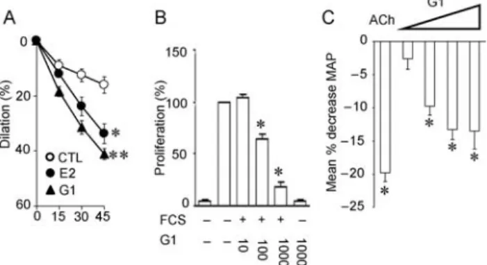
Figure 1 Involvement of gpER in regulation of vascular function, vascular smooth muscle cell growth, and blood pressure. The non-selective oestrogen receptor agonist 17b-estradiol (E2) and the selective gpER agonist G-1 acutely induce a relaxant response in human internal mammary arteries when compared with solvent control (CTL). However, the dilator effect of G-1 is even more potent than E2 (A). gpER activation also potently and concentration-dependently inhibits serum-stimulated cell proliferation in human vascular smooth muscle cells (B). Intravenous infusion of G-1 at increasing concentrations reduces mean arterial blood pressure (MAP) in normotensive Sprague–Dawley rats. For comparison, the pressure response to acetylcholine (ACh) is shown (C ). Figure reproduced in part from Haas, E., Bhattacharya, I., Brailoiu, E., Damjanovic, M., Brailoiu, G.C, Gao, X., Mueller-Guerre, L., Marjon, N.A., Gut, A., Minotti, R., Meyer, M.R, Amann, K., Ammann, E., Perez-Dominguez, A., Genoni, M., Clegg, D.J., Dun, N.J., Resta, T.C., Prossnitz, E.R., Barton, M., 2009. Regulatory role of G protein-coupled estrogen receptor for vascular function and obesity. Circulation Research. 104, 288–291.30
&
2009 American Heart Association.According to some studies, the abundance of both
oestro-gen receptor subtypes ERa and ERb in the aorta of humans
decreases with the progression of atherosclerosis.
53,54Inac-tivation of ERa and ERb genes in the vasculature may be
caused by DNA methylation of the promoter region of
these genes; DNA methylation, an important epigenetic
mechanism,
increases
with
ageing
and
plaque
pro-gression.
55–57Thus, structural changes of oestrogen
recep-tors may result in functional loss of responsiveness to
oestrogens. Indeed, proliferating vascular smooth muscle
cells, which have undergone phenotypic modulation upon
migrating to the intima,
58display a high degree of ERa
methylation.
56However,
we
currently
do
not
know
whether and by which mechanisms changes in oestrogen
receptor expression and structure in vascular cells affect
atherogenesis, or whether vascular cells without functional
oestrogen receptors become more abundant and prevent
effects of oestrogens from becoming functional.
It has been suggested that the local formation of
biologi-cally active estrogens is higher in human aortas with mild
atherosclerotic changes than in those with more advanced
lesions.
59Moreover,
the
ERb-associated
co-regulatory
protein NM23-H2, which activates ERb signalling in vitro,
is down-regulated with progression of atherosclerosis in
coronary arteries.
60The notion that the protective effects
of vascular oestrogen signalling are lost with the progression
of atherosclerosis is further supported by the finding that
27-hydroxycholesterol, an abundant cholesterol metabolite
found in atherosclerotic lesions, acts as an oestrogen
recep-tor antagonist.
61In addition, the aforementioned
inacti-vation of oestrogen receptor genes, an altered balance of
ERa and ERb protein expression, and changes in oestrogen
receptor signalling during different steps of atherogenesis
may add up to a different regulation of oestrogen-sensitive
genes, as well as of vascular homeostasis in general
(Figure 2). Oestrogen therapy may also have beneficial
effects on the expression and function of genes regulating
calcium homeostasis that are limited to early, but not
advanced, stages of atherosclerotic lesions.
19Moreover,
oestrogen-dependent induction of matrix metalloproteinases,
proteolytic enzymes capable of degrading components of
the extracellular matrix expressed in fibrous caps, may
contribute to maintenance of lumen size in early
athero-sclerosis, but could increase the likelihood of plaque erosion
or rupture in more advanced stages of the disease.
19,62Animal studies by Oparil’s group have found that—unlike
in young ovariectomized rats—oestrogen therapy in aged
animals fails to prevent neointima formation and
inflamma-tory responses following balloon angioplasty in structurally
normal arteries.
63This indicates that ageing—even in the
absence
of
atherosclerosis—appears
to
modulate
the
chronic vascular response to natural oestrogens.
18It has
also been suggested that oestrogens, under certain
con-ditions such as prolonged oestrogen deficiency, and
particu-larly when used in the context of vascular inflammation as
seen in atherosclerosis, may attenuate vasodilator capacity,
enhance inflammatory activity, and improve plaque
instabil-ity.
64,65Accordingly, a prolonged period of hypoestrogenicity
disrupts both neuroprotective and anti-inflammatory actions
of
oestrogens
in
animals.
66In
addition,
oestrogen-dependent direct and endothelium-mediated relaxation as
well as up-regulation of ERa and eNOS protein levels in
ovariectomized rats is maintained when 17b-estradiol
treat-ment is initiated 4 months, but not 8 months after surgery.
67Together, these experimental studies provide evidence to
support the concept of the ‘timing hypothesis’.
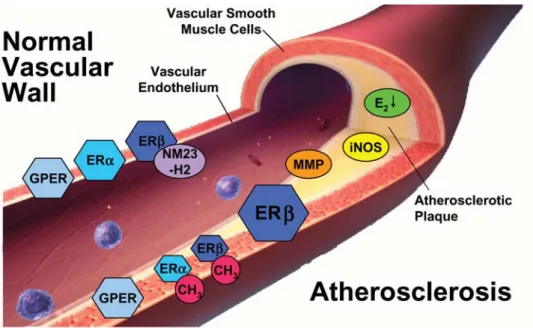
Figure 2 Molecular targets of oestrogens in the normal (left) and atherosclerotic vascular wall (right). With the progression of an atherosclerotic lesion as well as with ageing, expression of ERa and ERb becomes a target of methylation (CH3)-associated inactivation. Moreover, expression of oestrogen receptor-associated co-regulatory proteins, such as NM23-H2, is altered, and ERb expression changes in advanced atherosclerosis. Local formation of biologically active oestrogens through aromatase and other pathways appears to be reduced (E2#) under these conditions. Based on these changes of oestrogen receptor signalling in advanced atherosclerosis, it is currently not clear how endogenous oestrogens affect oestrogen-dependent proteins involved in inflammation and structure of the athero-sclerotic plaque, such as inflammatory nitric oxide synthase (iNOS) and matrix metalloproteinases (MMP). This illustrates the complexity of action of oestrogens and how equine oestrogen mixtures containing hormonal substances of unidentified activity affect oestrogen receptor function and regulation of oestrogen-sensitive genes in the presence of atherosclerosis. gpER, G protein-coupled estrogen receptor.
4. Clinical implications and perspectives
Although there is now substantial evidence suggesting that
vascular functionality of oestrogen receptors depends on
the length of a period of hypoestrogenicity and the
degree of atherosclerosis progression, it is still not known
how the cessation of endogenous oestrogen production
after menopause affects oestrogen receptor signalling in
human vascular cells in vivo. Therefore, future basic
research should include studies that will help identifying
the nature of changes of oestrogen receptor function in
dis-eased arteries. Such changes were not considered in earlier
randomized hormone therapy trials such as the Heart and
Estrogen/progestin Replacement Study (HERS) and the
Women’s Health Initiative (WHI), which demonstrated
adverse cardiovascular outcomes of hormone therapy in
women late after menopause.
68,69The medical regimen
used in these studies consisted of conjugated equine
oes-trogens derived from horse urine, and of synthetic
proges-tins such as medroxyprogesterone acetate.
68,69Unlike
17b-estradiol, the major endogenous human oestrogen
lost
after
menopause,
conjugated
equine
estrogens
contain a mixture of at least 10 different oestrogens,
several androgens, progestins, and other substances of
yet unknown vascular and procoagulatory activity.
5,18,41These substances display different or even unknown
selectivity and binding affinity for oestrogen receptors,
5which result in unpredictable effects on multiple oestrogen
signalling pathways evoking random vascular responses in
vivo, especially in the presence of atherosclerosis.
More-over, medroxyprogesterone acetate has been reported to
abrogate beneficial effects of oestrogens on vascular
smooth muscle cell growth, lipid profile, and vascular
function, even turning oestrogen-induced vasodilation
into coronary vasospasm.
70–72Currently, prospective clinical trials are underway to test
whether natural oestrogen has therapeutic potential in
women with atherosclerosis: The Kronos Early Estrogen
Pro-tection
Study
(KEEPS;
ClinicalTrials.gov
number,
NCT00154180), and the Early vs. Late Intervention Trial
With
Estradiol
(ELITE;
ClinicalTrials.gov
number,
NCT00114517). Moreover, the use of natural oestrogens
such as 17b-estradiol at low doses, and possibly intermittent
administration mimicking the normal menstrual cycle,
18,41potentially will help to preserve activating signalling
cas-cades, which are responsible for the beneficial vascular
effects of oestrogens in premenopausal women. Finally,
the use of selective oestrogen receptor modulators such as
raloxifene, which act as gpER agonists,
27might be suitable
for treatment and prevention of atherosclerotic vascular
disease. Results from the RUTH trial support this notion, at
least in younger postmenopausal women.
31At present, hormone therapy is not an option for primary
or secondary prevention of cardiovascular disease in
women. There are proven therapies such as simple
changes in lifestyle including maintaining a normal body
weight, a diet rich in fruits and vegetables, and regular
physical exercise. These measures—although still
under-used—should be initiated as early as possible to reduce
the burden of atherosclerotic vascular disease later in
life.
2,18Conflict of interest: none declared.
Funding
Original work by the authors is supported by the Swiss National
Science Foundation [grant numbers 3200-108258/1, and
K-33KO_122504/1].
References
1. Yusuf S, Reddy S, Ounpuu S, Anand S. Global burden of cardiovascular diseases: part I: general considerations, the epidemiologic transition, risk factors, and impact of urbanization. Circulation 2001;104:2746–2753. 2. Libby P. The forgotten majority: unfinished business in cardiovascular risk
reduction. J Am Coll Cardiol 2005;46:1225–1228.
3. Witteman JC, Grobbee DE, Kok FJ, Hofman A, Valkenburg HA. Increased risk of atherosclerosis in women after the menopause. Br Med J 1989; 298:642–644.
4. Barrett-Connor E. Sex differences in coronary heart disease. Why are women so superior? The 1995 Ancel Keys Lecture. Circulation 1997;95: 252–264.
5. Dubey RK, Imthurn B, Barton M, Jackson EK. Vascular consequences of menopause and hormone therapy: importance of timing of treatment and type of estrogen. Cardiovasc Res 2005;66:295–306.
6. McGill HC Jr, McMahan CA, Herderick EE, Zieske AW, Malcom GT, Tracy RE et al. Obesity accelerates the progression of coronary atherosclerosis in young men. Circulation 2002;105:2712–2718.
7. Punnonen R, Jokela H, Aine R, Teisala K, Salomaki A, Uppa H. Impaired ovarian function and risk factors for atherosclerosis in premenopausal women. Maturitas 1997;27:231–238.
8. Bittner V. Menopause and cardiovascular risk cause or consequence? J Am Coll Cardiol 2006;47:1984–1986.
9. Barton M, Meyer MR. Postmenopausal hypertension: mechanisms and therapy. Hypertension 2009;54:11–18.
10. Mendelsohn ME, Karas RH. The protective effects of estrogen on the car-diovascular system. N Engl J Med 1999;340:1801–1811.
11. Meyer MR, Haas E, Barton M. Gender differences of cardiovascular disease: new perspectives for estrogen receptor signaling. Hypertension 2006;47:1019–1026.
12. Clarkson TB. Estrogen effects on arteries vary with stage of reproductive life and extent of subclinical atherosclerosis progression. Menopause 2007;14:373–384.
13. Napoli C, D’Armiento FP, Mancini FP, Postiglione A, Witztum JL, Palumbo G et al. Fatty streak formation occurs in human fetal aortas and is greatly enhanced by maternal hypercholesterolemia. Intimal accumulation of low density lipoprotein and its oxidation precede mono-cyte recruitment into early atherosclerotic lesions. J Clin Invest 1997; 100:2680–2690.
14. McMahan CA, Gidding SS, Fayad ZA, Zieske AW, Malcom GT, Tracy RE et al. Risk scores predict atherosclerotic lesions in young people. Arch Intern Med 2005;165:883–890.
15. Bairey Merz CN, Johnson BD, Sharaf BL, Bittner V, Berga SL, Braunstein GD et al. Hypoestrogenemia of hypothalamic origin and coron-ary artery disease in premenopausal women: a report from the NHLBI-sponsored WISE study. J Am Coll Cardiol 2003;41:413–419. 16. Salpeter SR, Walsh JM, Greyber E, Salpeter EE. Brief report: coronary
heart disease events associated with hormone therapy in younger and older women. A meta-analysis. J Gen Intern Med 2006;21:363–366. 17. Rossouw JE, Prentice RL, Manson JE, Wu L, Barad D, Barnabei VM et al.
Postmenopausal hormone therapy and risk of cardiovascular disease by age and years since menopause. J Am Med Assoc 2007;297:1465–1477. 18. Barton M, Meyer MR, Haas E. Hormone replacement therapy and
athero-sclerosis in postmenopausal women: does aging limit therapeutic benefits? Arterioscler Thromb Vasc Biol 2007;27:1669–1672.
19. Mendelsohn ME, Karas RH. HRT and the young at heart. N Engl J Med 2007;356:2639–2641.
20. Manson JE, Bassuk SS. Invited commentary: hormone therapy and risk of coronary heart disease why renew the focus on the early years of meno-pause? Am J Epidemiol 2007;166:511–517.
21. Meyer MR, Haas E, Prossnitz ER, Barton M. Non-genomic regulation of vas-cular cell function and growth by estrogen. Mol Cell Endocrinol 2009; 308:9–16.
22. Moriarty K, Kim KH, Bender JR. Minireview: estrogen receptor-mediated rapid signaling. Endocrinology 2006;147:5557–5563.
23. Revankar CM, Cimino DF, Sklar LA, Arterburn JB, Prossnitz ER. A trans-membrane intracellular estrogen receptor mediates rapid cell signaling. Science 2005;307:1625–1630.
24. Owman C, Blay P, Nilsson C, Lolait SJ. Cloning of human cDNA encoding a novel heptahelix receptor expressed in Burkitt’s lymphoma and widely distributed in brain and peripheral tissues. Biochem Biophys Res Commun 1996;228:285–292.
25. Haas E, Meyer MR, Schurr U, Bhattacharya I, Minotti R, Nguyen HH et al. Differential effects of 17beta-estradiol on function and expression of estrogen receptor alpha, estrogen receptor beta, and GPR30 in arteries and veins of patients with atherosclerosis. Hypertension 2007;49: 1358–1363.
26. Ullrich ND, Krust A, Collins P, MacLeod KT. Genomic deletion of estrogen receptors ERalpha and ERbeta does not alter estrogen-mediated inhi-bition of Ca2þinflux and contraction in murine cardiomyocytes. Am J Physiol Heart Circ Physiol 2008;294:H2421–H2427.
27. Thomas P, Pang Y, Filardo EJ, Dong J. Identity of an estrogen membrane receptor coupled to a G protein in human breast cancer cells. Endocrin-ology 2005;146:624–632.
28. Mercier I, Mader S, Calderone A. Tamoxifen and ICI 182,780 negatively influenced cardiac cell growth via an estrogen receptor-independent mechanism. Cardiovasc Res 2003;59:883–892.
29. Lafferty AR, Torpy DJ, Stowasser M, Taymans SE, Lin JP, Huggard P et al. A novel genetic locus for low renin hypertension: familial hyperaldosteron-ism type II maps to chromosome 7 (7p22). J Med Genet 2000;37:831–835. 30. Haas E, Bhattacharya I, Brailoiu E, Damjanovic M, Brailoiu GC, Gao X et al. Regulatory role of G protein-coupled estrogen receptor for vascular function and obesity. Circ Res 2009;104:288–291.
31. Collins P, Mosca L, Geiger MJ, Grady D, Kornitzer M, Amewou-Atisso MG et al. Effects of the selective estrogen receptor modulator raloxifene on coronary outcomes in the Raloxifene Use for The Heart trial: results of subgroup analyses by age and other factors. Circulation 2009;119: 922–930.
32. Mu¨gge A, Riedel M, Barton M, Kuhn M, Lichtlen PR. Endothelium indepen-dent relaxation of human coronary arteries by 17 beta-oestradiol in vitro. Cardiovasc Res 1993;27:1939–1942.
33. Liu PP, Fukuoka M. Sex hormones as novel risk biomarkers for athero-sclerosis in peripheral vascular disease. J Am Coll Cardiol 2007;50: 1077–1079.
34. Murakami H, Harada N, Sasano H. Aromatase in atherosclerotic lesions of human aorta. J Steroid Biochem Mol Biol 2001;79:67–74.
35. Hojo Y, Hattori TA, Enami T, Furukawa A, Suzuki K, Ishii HT et al. Adult male rat hippocampus synthesizes estradiol from pregnenolone by cyto-chromes P45017alpha and P450 aromatase localized in neurons. Proc Natl Acad Sci USA 2004;101:865–870.
36. Nathan L, Shi W, Dinh H, Mukherjee TK, Wang X, Lusis AJ et al. Testoster-one inhibits early atherogenesis by conversion to estradiol: critical role of aromatase. Proc Natl Acad Sci USA 2001;98:3589–3593.
37. Lew R, Komesaroff P, Williams M, Dawood T, Sudhir K. Endogenous estro-gens influence endothelial function in young men. Circ Res 2003;93: 1127–1133.
38. Zhu Y, Bian Z, Lu P, Karas RH, Bao L, Cox D et al. Abnormal vascular func-tion and hypertension in mice deficient in estrogen receptor beta. Science 2002;295:505–508.
39. Hodgin JB, Krege JH, Reddick RL, Korach KS, Smithies O, Maeda N. Estro-gen receptor alpha is a major mediator of 17beta-estradiol’s atheropro-tective effects on lesion size in ApoE2/2 mice. J Clin Invest 2001;107: 333–340.
40. Sudhir K, Chou TM, Chatterjee K, Smith EP, Williams TC, Kane JP et al. Premature coronary artery disease associated with a disruptive mutation in the estrogen receptor gene in a man. Circulation 1997;96: 3774–3777.
41. Meyer MR, Haas E, Barton M. Need for research on estrogen receptor function: importance for postmenopausal hormone therapy and athero-sclerosis. Gend Med 2008;5(Suppl. A):S19–S33.
42. Bourassa PA, Milos PM, Gaynor BJ, Breslow JL, Aiello RJ. Estrogen reduces atherosclerotic lesion development in apolipoprotein E- deficient mice. Proc Natl Acad Sci USA 1996;93:10022–10027.
43. Marmorston J, Magdison O, Kuzma O, Moore FJ. Estrogen therapy in men with myocardial infarction. Side-effects with increasing dosage and time. J Am Med Assoc 1960;174:241–244.
44. Stamler J, Pick R, Katz LN, Pick A, Kaplan BM, Berkson DM et al. Effec-tiveness of estrogens for therapy of myocardial infarction in middle-age men. J Am Med Assoc 1963;183:632–638.
45. Abizaid A, Albertal M, Costa MA, Abizaid AS, Staico R, Feres F et al. First human experience with the 17-beta-estradiol-eluting stent: the Estrogen And Stents To Eliminate Restenosis (EASTER) trial. J Am Coll Cardiol 2004; 43:1118–1121.
46. Christian RC, Liu PY, Harrington S, Ruan M, Miller VM, Fitzpatrick LA. Intimal estrogen receptor (ER)beta, but not ERalpha expression, is corre-lated with coronary calcification and atherosclerosis in pre- and postme-nopausal women. J Clin Endocrinol Metab 2006;91:2713–2720. 47. Lindner V, Kim SK, Karas RH, Kuiper GG, Gustafsson JA, Mendelsohn ME.
Increased expression of estrogen receptor-beta mRNA in male blood vessels after vascular injury. Circ Res 1998;83:224–229.
48. Matthews J, Gustafsson JA. Estrogen signaling: a subtle balance between ER alpha and ER beta. Mol Interv 2003;3:281–292.
49. Traupe T, Stettler CD, Li H, Haas E, Bhattacharya I, Minotti R et al. Distinct roles of estrogen receptors alpha and beta mediating acute vasodilation of epicardial coronary arteries. Hypertension 2007;49: 1364–1370.
50. Ogawa S, Emi M, Shiraki M, Hosoi T, Ouchi Y, Inoue S. Association of estro-gen receptor beta (ESR2) estro-gene polymorphism with blood pressure. J Hum Genet 2000;45:327–330.
51. Rexrode KM, Ridker PM, Hegener HH, Buring JE, Manson JE, Zee RY. Poly-morphisms and haplotypes of the estrogen receptor-beta gene (ESR2) and cardiovascular disease in men and women. Clin Chem 2007;53: 1749–1756.
52. O’Lone R, Knorr K, Jaffe IZ, Schaffer ME, Martini PG, Karas RH et al. Estrogen receptors alpha and beta mediate distinct pathways of vascular gene expression, including genes involved in mitochondrial electron transport and generation of reactive oxygen species. Mol Endocrinol 2007;21:1281–1296.
53. Losordo DW, Kearney M, Kim EA, Jekanowski J, Isner JM. Variable expression of the estrogen receptor in normal and atherosclerotic coronary arteries of premenopausal women. Circulation 1994;89: 1501–1510.
54. Nakamura Y, Suzuki T, Miki Y, Tazawa C, Senzaki K, Moriya T et al. Estro-gen receptors in atherosclerotic human aorta: inhibition of human vascu-lar smooth muscle cell proliferation by estrogens. Mol Cell Endocrinol 2004;219:17–26.
55. Post WS, Goldschmidt-Clermont PJ, Wilhide CC, Heldman AW, Sussman MS, Ouyang P et al. Methylation of the estrogen receptor gene is associated with aging and atherosclerosis in the cardiovascular system. Cardiovasc Res 1999;43:985–991.
56. Ying AK, Hassanain HH, Roos CM, Smiraglia DJ, Issa JJ, Michler RE et al. Methylation of the estrogen receptor-alpha gene promoter is selectively increased in proliferating human aortic smooth muscle cells. Cardiovasc Res 2000;46:172–179.
57. Kim J, Kim JY, Song KS, Lee YH, Seo JS, Jelinek J et al. Epigenetic changes in estrogen receptor beta gene in atherosclerotic cardiovascular tissues and in-vitro vascular senescence. Biochim Biophys Acta 2007;1772: 72–80.
58. Campbell GR, Campbell JH, Manderson JA, Horrigan S, Rennick RE. Arter-ial smooth muscle. A multifunctional mesenchymal cell. Arch Pathol Lab Med 1988;112:977–986.
59. Nakamura Y, Suzuki T, Sasano H. Estrogen actions and in situ synthesis in human vascular smooth muscle cells and their correlation with athero-sclerosis. J Steroid Biochem Mol Biol 2005;93:263–268.
60. Rayner K, Chen YX, Hibbert B, White D, Miller H, Postel EH et al. NM23-H2, an estrogen receptor beta-associated protein, shows dimin-ished expression with progression of atherosclerosis. Am J Physiol Regul Integr Comp Physiol 2007;292:R743–R750.
61. Umetani M, Domoto H, Gormley AK, Yuhanna IS, Cummins CL, Javitt NB et al. 27-Hydroxycholesterol is an endogenous SERM that inhibits the car-diovascular effects of estrogen. Nat Med 2007;13:1185–1192. 62. Libby P, Aikawa M. Stabilization of atherosclerotic plaques: new
mechan-isms and clinical targets. Nat Med 2002;8:1257–1262.
63. Miller AP, Xing D, Feng W, Fintel M, Chen YF, Oparil S. Aged rats lose vaso-protective and anti-inflammatory actions of estrogen in injured arteries. Menopause 2007;14:251–260.
64. Mendelsohn ME, Karas RH. Molecular and cellular basis of cardiovascular gender differences. Science 2005;308:1583–1587.
65. Cann JA, Register TC, Adams MR, St Clair RW, Espeland MA, Williams JK. Timing of estrogen replacement influences atherosclerosis progression and plaque leukocyte populations in ApoE2/2 mice. Atherosclerosis 2008 doi:10.1016/j.atherosclerosis.2008.01.018.
66. Suzuki S, Brown CM, Dela Cruz CD, Yang E, Bridwell DA, Wise PM. Timing of estrogen therapy after ovariectomy dictates the efficacy of its neuro-protective and antiinflammatory actions. Proc Natl Acad Sci USA 2007; 104:6013–6018.
67. Pinna C, Cignarella A, Sanvito P, Pelosi V, Bolego C. Prolonged ovarian hormone deprivation impairs the protective vascular actions of estrogen receptor alpha agonists. Hypertension 2008;51:1210–1217.
68. Hulley S, Grady D, Bush T, Furberg C, Herrington D, Riggs B et al. Random-ized trial of estrogen plus progestin for secondary prevention of coronary heart disease in postmenopausal women. Heart and Estrogen/progestin Replacement Study (HERS) Research Group. J Am Med Assoc 1998;280: 605–613.
69. Rossouw JE, Anderson GL, Prentice RL, LaCroix AZ, Kooperberg C, Stefanick ML et al. Risks and benefits of estrogen plus progestin in healthy postmenopausal women: principal results From the Women’s Health Initiative randomized controlled trial. J Am Med Assoc 2002; 288:321–333.
70. Effects of estrogen or estrogen/progestin regimens on heart disease risk factors in postmenopausal women. The Postmenopausal Estrogen/Proges-tin Interventions (PEPI) Trial. The WriEstrogen/Proges-ting Group for the PEPI Trial. J Am Med Assoc 1995;273:199–208.
71. Miyagawa K, Rosch J, Stanczyk F, Hermsmeyer K. Medroxyprogesterone interferes with ovarian steroid protection against coronary vasospasm. Nat Med 1997;3:324–327.
72. Dubey RK, Jackson EK, Gillespie DG, Zacharia LC, Wunder D, Imthurn B et al. Medroxyprogesterone abrogates the inhibitory effects of estradiol on vascular smooth muscle cells by preventing estradiol metabolism. Hypertension 2008;51:1197–1202.