Electron microscopical study to assess the in vitro effects
of the synthetic trioxolane OZ78 against the liver fluke,
Fasciola hepatica
L. HALFERTY1, J. F. O’NEILL1, G. P. BRENNAN1, J. KEISER2and I. FAIRWEATHER1* 1
Parasite Therapeutics Research Group, School of Biological Sciences, The Queen’s University of Belfast, Medical Biology Centre, 97 Lisburn Road, Belfast BT9 7BL, Northern Ireland
2
Department of Medical Parasitology and Infection Biology, Swiss Tropical Institute, P.O. Box, CH-4002 Basel, Switzerland (Received 26 April 2009; revised 29 May 2009; accepted 30 May 2009; first published online 7 August 2009)
S U M M A R Y
Adult Fasciola hepatica were incubated for 48 h in vitro in the synthetic peroxide, OZ78 at a concentration of 100mg/ml and then prepared for scanning and transmission electron microscopy. There was limited disruption to the external fluke surface, with only slight swelling and blebbing of the interspinal tegument in the midbody and ventral tail regions. By contrast, significant disruption was observed to the ultrastructure of the tegument and subtegumental tissues. There was severe swelling of the basal infolds in the tegumental syncytium and the flooding spread internally to affect the subtegu-mental tissues. In the tegusubtegu-mental system, there was swelling of the cisternae of granular endoplasmic reticulum and of the mitochondria, with the latter showing signs of breaking down. Autophagic vacuoles and lipid droplets were present and the synthesis of tegumental secretory bodies was much reduced. The gastrodermal cells were severely affected, with swelling and degeneration of the mitochondria and the presence of autophagic vacuoles and lipid droplets. The granular endoplasmic reticulum was swollen and vesiculated and the cells contained few secretory bodies. Both the vitelline and testis follicles showed evidence of extensive cellular disruption and degeneration. This study confirms previous data indicating the potential flukicidal activity of OZ78.
Key words : Fasciola hepatica, liver fluke, synthetic artemisinin derivative, OZ78, scanning electron microscopy, trans-mission electron microscopy, in vitro culture.
I N T R O D U C T I O N
The liver fluke, Fasciola hepatica is a major parasite of livestock in temperate regions of the world. There has been a dramatic rise in the incidence of disease in recent years, which has been attributed to climate change (Mitchell, 2002). Furthermore, fascioliasis is now recognized as a major zoonotic infection of increasing importance (Mas-Coma et al. 2005 ; WHO, 2007). Current control methods effectively centre round the use of anthelmintic drugs, of which the most commonly employed is the benzimid-azole derivative, triclabendbenzimid-azole (TCBZ, ‘ Fasinex ’) (Fairweather, 2009). It is favoured due to its re-markably high efficacy against both adult and juv-enile stages of F. hepatica (Boray et al. 1983). Unfortunately, due to the over-dependence on this drug and its continued use as a monotherapy, resist-ance is steadily on the increase. Resistresist-ance to tricla-bendazole was first noted on Australian farms in 1995 (Overend and Bowen, 1995). Since then, resistance
has been detected throughout much of Europe (Fairweather, 2005, 2009). Resistance in the UK and Ireland is believed to be spreading and is probably significantly underestimated (Wolstenholme et al. 2004). This means that there is an urgent need to find novel fluckicidal compounds.
One group of drugs that has attracted considerable attention in the last few years is the artemisinins. The artemisinins are a family of natural or semi-synthetic compounds derived from the plant, Artemisia annua which is grown in eastern Asia (O’Neill, 2004) and have established uses as malarial and anti-schistosomal drugs (Borstnik et al. 2002 ; Woodrow et al. 2005 ; Xiao, 2005 ; Utzinger et al. 2007). The main semi-synthetic derivatives of artemisinin are artemether, artesunate and the active metabolite, di-hydroartemisinin (Woodrow et al. 2005). Artemether and artesunate have been shown to be effective fascio-licides in the treatment of rodent infections (Keiser et al. 2006 a). Due to the unfavourable pharmaco-kinetic properties of the artemisinins, many synthetic anti-malarial peroxides have been prepared, includ-ing the synthetic 1,2,4-trioxolanes, such as OZ277 and OZ78 (Vennerstrom et al. 2004). Synthetic peroxides retain the peroxide pharmacophore es-sential for anti-parasitic activity, yet are more simple molecules, easier to synthesize and have improved
* Corresponding author : School of Biological Sciences, The Queen’s University of Belfast, Medical Biology Centre, 97 Lisburn Road, Belfast BT9 7BL, Northern Ireland. Tel : +44 28 90972298. Fax: +44 28 90975877. E-mail : i.fairweather@qub.ac.uk
pharmacokinetic properties (Jefford, 2007). Worm burden reductions of 100 % have been achieved with both juvenile and adult F. hepatica following a single oral dose of 100 mg/kg OZ78 (Keiser et al. 2006 b). Significantly, high efficacy has also been recorded for OZ78 and artemether in treatment of tricla-bendazole-resistant fluke infections in rats (Keiser et al. 2007 a). These results suggest that OZ78 has a high potential as a flukicidal drug and warrants further evaluation. Relatively little work has been performed so far in relation to the morphological effects of artemisinin-type compounds on F. hepatica and it has been confined to surface changes (Keiser et al. 2006 a, b ; Keiser and Morson, 2008 a, b).
The present study was carried out to determine the changes induced by treatment in vitro with the syn-thetic peroxide OZ78 on the surface morphology of adult F. hepatica, as visualized by scanning electron microscopy (SEM), and on the internal ultrastruc-ture by means of transmission electron microscopy (TEM). The aim was to identify possible tissue tar-gets of drug action.
M A T E R I A L S A N D M E T H O D S
The experiment was carried out in 2 parts : the rat infections, in vitro incubation in OZ78 and initial fixation of fluke material was performed at the Swiss Tropical Institute, Basel and then the fluke material was sent to The Queen’s University of Belfast for further processing and examination by scanning and transmission electron microscopy.
Female Wistar rats (purchased from RCC, Itingen, Switzerland) were experimentally infected with metacercarial cysts of the Cullompton isolate, shown in previous experiments to be susceptible to TCBZ in vivo and to its sulphoxide metabolite in vitro (Robinson et al. 2002 ; McCoy et al. 2005 ; Halferty et al. 2008, 2009 ; McConville et al. 2006, 2009 a ; Meaney et al. 2006, 2007 ; Devine et al. 2009 ; Toner et al. 2009). All animals were housed in environ-mentally-controlled conditions in groups of 4 and afforded 1 week to acclimatize. Aforementioned con-ditions were : temperature, 25 xC ; humidity, 70 % ; 12 h light/dark cycle. Free access to water and rodent diet was also maintained. Twelve rats were infected intragastrically with 25 cysts each and, after a period of 9–10 weeks, adult flukes were recovered from the bile ducts at necropsy. Recovered flukes were then incubated in 6-well plates (Costar, Corning, USA), placing 2 fluke specimens per well. Each well con-tained 5 ml of NCTC 135 culture medium (In-vitrogen, Carlsbad, USA) containing streptomycin and penicillin at concentrations of 50mg/ml and 50 IU/ml, respectively. A stock solution of OZ78 (synthesized at the College of Pharmacy, University of Nebraska Medical Centre, USA) was prepared in 60 % (v/v) DMSO and flukes were incubated with the drug for a period of 48 h at a concentration of 100mg/
ml. The concentration used was based on the results of previous studies with OZ78 involving F. hepatica, Echinostoma caproni and Clonorchis sinensis (Keiser et al. 2006 b, 2007 a, b). Fluke motility was observed at regular intervals by way of a dissecting microscope. Specimens were judged dead if no movement was observed for 2 min. The experiment was terminated at 48 h, when the flukes were considered dead. Con-trols consisted of incubating flukes for 48 h at 37 xC in NCTC 135 culture medium in the presence of streptomycin and penicillin. The concentration of DMSO in the control medium (0.05 %) was matched to that of the drug medium. The control flukes were still active and appeared normal after 48 h incu-bation. Controls at 0 h were also prepared. Following treatment, the flukes were fixed with 2.5 % (v/v) glutaraldehyde in a PBS buffer (pH 7.3) for 24 h at room temperature. After rinsing and storing in PBS buffer, the specimens were sent to The Queen’s University of Belfast for further processing and examination.
For scanning electron microscopy (SEM), 8 flukes were washed in 0.1M sodium cacodylate buffer
(pH 7.4) containing 3 % (w/v) sucrose. Following a post-fixation in 1 % osmium tetroxide for 1 h, the flukes were given 3 brief washes in 70 % (v/v) ethanol and dehydrated in an ascending series of ethanol. After this, they were dried in hexamethyldisilazane, mounted on aluminium stubs and sputter-coated with gold-palladium. The specimens were viewed in an FEI Quanta 200 scanning electron micro-scope, operating at an accelerating voltage of 10 keV.
For transmission electron microscopy (TEM), 5 flukes were washed in 0.1Msodium cacodylate buffer
(pH 7.4) containing 3 % (w/v) sucrose. The apical cones and tail regions of the flukes were discarded and the remaining midbody region was divided transversely into 2 parts ; each part was further sub-divided transversely into 6 pieces. The pieces were washed in 0.1M sodium cacodylate buffer (pH 7.4)
containing 3 % (w/v) sucrose. Following post-fixation in 1 % osmium tetroxide for 1 h, the tissue was washed in fresh buffer, dehydrated in an ascending series of ethanol and infiltrated and embedded in Agar 100 resin. Ultrathin sections, 60–70 nm in thickness, were cut on a Reichert Ultracut E ultramicrotome, mounted on bare 200-mesh copper grids, double-stained with alcoholic uranyl acetate (5 min) and aqueous lead citrate (3 min) and viewed in a FEI CM 100 transmission electron microscope operating at an accelerating voltage of 100 keV.
R E S U L T S
Scanning electron microscopy
The surface architecture of the control flukes ap-peared normal (Fig. 1A). It matched the images
presented by Bennett (1975), Fairweather et al. (1999) and McConville et al. (2009 b).
Following treatment with OZ78, little change was observed anteriorly (Fig. 1B). Spines were grouped in a regular, tight-knit arrangement and clearly pro-truded from the inter-spinal surface. The spinelets at the tips of the spines (Fig. 1B) were distinct and there was no spine disruption or swelling of the tegument.
The ventral midbody region showed slight swell-ing of the interspinal tegument (Fig. 1C). This swelling lent an uneven appearance to the surface of the fluke. At higher magnification (Fig. 1C inset), the spines were seen to be slightly sunken, indicating some swelling of the tegument surrounding them. The interspinal surface appeared rough and was covered with small blebs. The dorsal surface of the
Fig. 1. Scanning electron micrographs (SEMs) of adult Fasciola hepatica incubated in vitro for 48 h. (A) Control, incubated in NCTC culture medium ; (B–D) flukes treated with OZ78 at a concentration of 100mg/ml. (A) Control fluke. Low-power image, showing the oral sucker (OS), ventral sucker (VS), midbody region (MB) and tail region (TR). Inset shows spines (S) from the oral cone region, which bear distinct spinelets (arrow). (B) Low-power image, showing the oral sucker (OS), ventral sucker (VS) midbody region (MB) and tail region (TR). Inset shows spines (S) from the oral cone region, which bear distinct spinelets (arrow). (C) Ventral midbody region. The inter-spinal tegument appears swollen (broken arrows). The arrangement of the spines (S) is more irregular than normal. Inset shows that the spines (S) are slightly sunken due to the swelling of the inter-spinal tegument (arrows). The tegumental surface has a roughened appearance (*). (D) Dorsal midbody region. Swelling of the inter-spinal surface is evident (broken arrows). The arrangement of the spines (S) appears irregular. Inset is a higher-power image showing that the spinelets (broken arrow) are less well-defined. The inter-spinal surface, although swollen (arrows), appears smooth. S, spine.
midbody region exhibited general swelling of the interspinal tegument (Fig. 1D). The swelling again lent an uneven appearance to the surface of the fluke. Spines in this region appeared sunken, due to the swelling of the tegument around them (Fig. 1D inset) and the spinelets were less clearly defined (Fig. 1D inset).
The ventral surface of the tail region (Fig. 2A) showed a relatively normal morphology and the tegument was not swollen. At higher magnification (Fig. 2A inset), the interspinal surface was seen to be roughened. Spines were prominent and identical in shape and structure to those of control specimens. The dorsal tail (Fig. 2B) showed a regular pattern of spines protruding from a roughened tegumental surface. There was no evidence of swelling of the tegument.
Figures 2C and D demonstrate unusual features seen in 2 of the specimens. A bulbous swelling was observed in the dorsal anterior midbody region of 1 specimen, the identity of which is not clear (Fig. 2C and inset). Apart from this swelling, the surface of the fluke appeared normal. More dramatic disrup-tion was evident in another specimen, in which the posterior end of the fluke was covered with large blebs, many of which had burst (Fig. 2D). One of the burst blebs was seen to contain a spine (Fig. 2D inset).
Transmission electron microscopy
The ultrastructure of the tegumental syncytium, tegumental cells and reproductive structures in the control specimens remained normal. For images, the reader is referred to the papers by Threadgold (1963, 1967) for tegument and parenchyma ; Irwin and Threadgold (1970) and Colhoun et al. (1998) for vitellaria ; and Stitt and Fairweather (1990) for testis. Following drug treatment, severe alterations were evident throughout the tegumental syncytium, sub-tegumental region, gut and reproductive tissues, in sections taken from the midbody region and as il-lustrated in Figs 3 and 4. In the syncytium, the basal infolds were severely swollen and occupied the entire basal region of the syncytium (Fig. 3A and C). Blebbing of the apical plasma membrane was ob-served (Fig. 3B) and autophagic vacuoles were present in the syncytium (Fig. 3B). The numbers of T1 and T2 secretory bodies in the syncytium were considerably less than normal. The mitochondria were swollen and electron-lucent (Fig. 3C). The crystalline structure of the spines was seen to be disrupted : it had a rippled and striated appearance (Fig. 3B).
Beneath the basal lamina, large spaces separated the muscle blocks belonging to the circular and longitudinal muscle layers (Fig. 3D). Large spaces surrounded the tegumental cell bodies, too (Fig. 3E and F). Branches of the excretory ducts contained
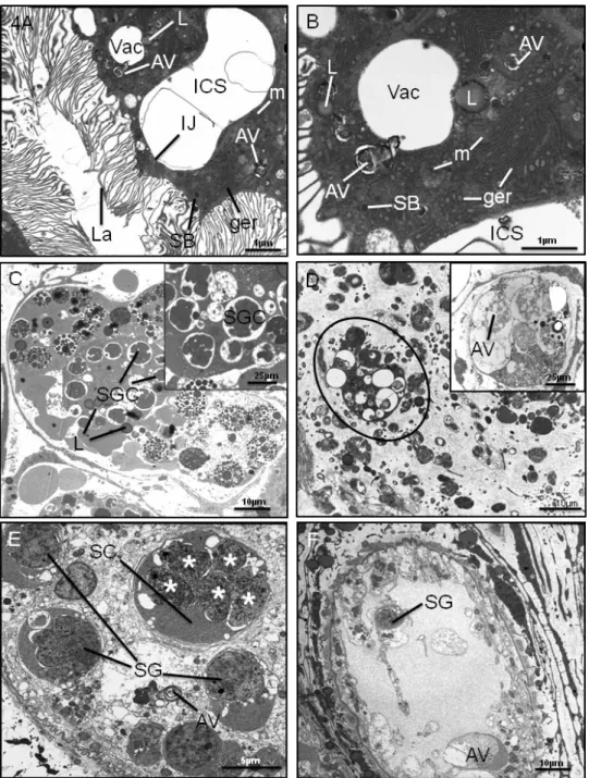
many lipid droplets (Fig. 4A). Within some of the tegumental cell bodies themselves, there were large numbers of lipid droplets as well (Fig. 3E). In all of the cells, the cytoplasm was filled with swollen and electron-lucent mitochondria, with distinct cristae and some of the mitochondria appeared to be in the process of breaking down (Fig. 3F). The cisternae of granular endoplasmic reticulum (ger) were swollen as well (Fig. 3F). Autophagic vacuoles were present in the cells (Fig. 3F). Type 1 and type 2 tegumental secretory bodies were generally absent from the cells and there were no discernable Golgi complexes present.
There was severe disruption to the gastrodermal cells. Large spaces were present between adjacent cells, the cells only being held together by the tight junction at their apices (Fig. 4A). Within the cells themselves, there were large empty vacuoles, together with autophagic vacuoles and lipid droplets, but few secretory bodies (Fig. 4A and B). The cis-ternae of granular endoplasmic reticulum were swollen and vesiculated (Fig. 4B).
The condition of the vitelline follicles is shown in Fig. 4C and D. Within the follicles it was difficult to distinguish any stem and early immature cells and the follicle was loosely-packed. The predominant cell type was the mature cell, although it displayed a number of abnormalities. That is, the shell globule clusters were either loosely-packed or the shell pro-tein globules appeared to have coalesced (Fig. 3C). The cisternae of the ger were swollen and a number of lipid droplets were present (Fig. 3C). In some cells, the cytoplasm appeared to be breaking down (Fig. 3C). In more extreme cases, the vitelline fol-licles were greatly reduced in size and contained no recognizable cell types, as the cells had broken down and were full of autophagic vacuoles (Fig. 3D). In these situations, the surrounding tissue was very disorganized and empty looking (Fig. 3D).
The testis follicles are pictured in Fig. 4E and F. Only spermatogonia and primary spermatocytes were present. The spermatogonia were rounded, the space between the inner and outer nuclear mem-branes was swollen and vacuoles were present in the cytoplasm (Fig. 4E). The spermatocytes appeared to have undergone nuclear division without undergoing cytoplasmic division, giving rise to multinucleate cells (Fig. 4E). In the most extreme cases, the follicle was almost totally devoid of spermatogenic cells and the tissue that remained was very disorganized and autophagic (Fig. 4F).
D I S C U S S I O N
Following treatment in vitro of F. hepatica with the synthetic peroxide OZ78 for 48 h at a concentration of 100mg/ml, there was a marked difference between what disruption was evident externally and what disruption occurred internally. That is, changes to
the surface morphology were quite limited. In stark contrast to the SEM observations, however, there was severe internal disruption of the tegumental, subtegumental and reproductive tissues. The basal infolds of the tegumental syncytium were swollen, flooding the basal region of the syncytium. Flooding was also evident internally, with large spaces separ-ating the subtegumental cells. The gastrodermal cells were severely affected, with evidence of metabolic disruption and breakdown. In both the vitelline and testis follicles, the normal development of the cells
was disrupted and, in more extreme cases, the vitel-line follicles appeared to be in the process of breaking down. These findings will now be discussed in detail and compared with the findings of previous, related studies.
Previous morphological studies in vitro and in vivo with OZ78 and other artemisinins have concentrated solely on the tegument of F. hepatica using SEM (Keiser et al. 2006 a, b, 2007 b ; Keiser and Morson, 2008 a, b ; O’Neill et al. 2009). While the changes seen can vary between flukes, they are generally relatively
Fig. 2. Scanning electron micrographs (SEMs) of the posterior region of adult Fasciola hepatica following 48 h treatment in vitro with OZ78 (100mg/ml). (A) Ventral tail region. The spines (S) project from the surface of the
tegument, which is not swollen. Inset shows the roughened appearance of the inter-spinal surface, due to the covering of small blebs (*). S, spine. (B) Dorsal tail region. The spines (S) are regularly-arranged and protrude from the surface. Inset shows that the tegumental surface appears roughened (*) at a higher magnification. S, spine. (C) The main image shows the presence of a bulbous swelling (*) on the ventral surface in the posterior midbody region. EP, excretory pore. Inset shows the swelling (*) at higher magnification. (D) Severe disruption to the tail end of the fluke, which appears to be covered with large blebs, many of which have burst (oval outline). Inset shows the large and burst blebs (*) at high magnification. One of the burst blebs contains a spine (S).
mild, with swelling, localized areas of blebbing and occasional sloughing. The results of the present study were consistent with this pattern, with swelling and limited blebbing over part of the body surface.
There was not the severe and extensive disruption seen with other fasciolicides (e.g. Fairweather et al. 1987 ; Skuce and Fairweather, 1990 ; Stitt and Fairweather, 1993 ; McKinstry et al. 2003 ; Meaney
Fig. 3. Transmission electron micrographs (TEMs) of the tegumental syncytium and subtegumental region of adult F. hepatica following 48 h treatment in vitro with OZ78 (100mg/ml). (A) The basal infolds (BI) are severely swollen. Beneath the basal lamina (BL), the circular (CM) and longitudinal (LM) muscle layers can be seen. APM, apical plasma membrane. (B) High-power image of the apical region of the tegumental syncytium. Small blebs (B) can be seen on the surface. An autophagic vacuole (AV) is present. APM, apical plasma membrane. Inset shows disruption (broken arrows) to the crystalline structure of a spine (S). (C) High-power image of the swollen basal infolds (BI) above the basal lamina (BL). The mitochondria (m) are swollen and electron-lucent. (D) Circular muscle (CM) layer showing the muscle bundles surrounded by large spaces (*). Inset shows the large spaces (*) between the longitudinal muscle (LM) bundles. (E) Low-power image of the subtegumental region. The tegumental cell bodies (TCB) contain large numbers of lipid droplets (L) and are separated from each other by large spaces (*). (F) High-power image of a tegumental cell body (TCB). The irregularly-shaped nucleus (N) can be seen located centrally within the cell. Inset i shows the swollen and electron-lucent appearance of the mitochondria (m). The cisternae of the granular endoplasmic reticulum (ger) are also distended. Inset ii shows the severe swelling of the cisternae of the granular endoplasmic reticulum (ger) at higher magnification. An autophagic (AV) vacuole is present in the cell.
Fig. 4. Transmission electron micrographs (TEMs) of adult Fasciola hepatica following 48 h treatment in vitro with OZ78 (100mg/ml). (A) Gastrodermal cells, showing a large intercellular space (ICS) between two adjacent cells. Within the cells, are vacuoles (Vac), autophagic vacuoles (AV), lipid droplets (L) and a small number of secretory bodies (SB). The cisternae of granular endoplasmic reticulum (ger) are swollen. The mitochondria (m) are swollen and
electron-lucent. La, gut lamellae. (B) Gastrodermal cell, containing empty vacuoles (Vac), autophagic vacuoles (AV) and lipid droplets (L). Few secretory bodies (SB) are present. The mitochondria (m) are swollen and some are electron-lucent and appear to be breaking down. The cisternae of granular endoplasmic reticulum (ger) are swollen and vesiculated. ICS, intercellular space. (C) Vitelline follicle. The follicle is heavily-disrupted, with no definable early developmental stages present. The shell protein globules in the shell globule clusters (SGC) show signs of coalescence. Lipid droplets (L) are present in the cells. Inset is a high-power image showing the abnormal shell globule clusters (SGC). (D) Vitelline follicle (oval outline) is severely disrupted and is surrounded by widespread degeneration of the parenchymal tissue. Inset is a high-power image showing a follicle in the final stages of breakdown. Almost no discernible structures remain, and autophagic vacuoles (AV) are present throughout. (E) Testis follicle, showing disruption of the spermatogenic cells. The spermatogonial cells (SG) appear rounded and abnormal and the primary spermatocytes (SC) show division of the nuclei (*) without cytoplasmic division of the cells themselves. General degeneration of the follicular tissue is evident throughout the follicle and autophagic vacuoles (AV) are present. (F) Testis follicle, which is almost completely devoid of spermatogenic cells. There is widespread disruption
throughout. SG, spermatagonia ; AV, autophagic vacuole.
et al. 2003 ; McConville et al. 2006 ; Halferty et al. 2009). More severe changes were seen in 2 of the flukes. The large swelling observed in one of them is similar to that described previously (Keiser et al. 2006 b), but it is not known what internal structure it relates to. Such individual variation in response to artemisinin treatment seems to be typical of F. hepa-tica, as it has been described in other studies (Keiser et al. 2006 b ; Keiser and Morson, 2008 a, b). Surface changes with artemisinins are greater when haemin is included in the in vitro culture medium (Keiser and Morson, 2008 a, b) and following in vivo treat-ment, which may be related to proposals concerning the activation of the drug following its ingestion by the fluke, as has been suggested for schistosomes (Xiao et al. 2003). That is, interaction with haemin or another iron-containing compounds derived from the haemoglobin in the blood that the worms feed upon may lead to activation of the artemisinin com-pound. Activation leads to cleavage of the peroxide bridge in the artemisinin molecule (which is the es-sential moiety required for activity) and generation of free radicals which are damaging to the parasite (Xiao et al. 2003). In relation to the role of haemin in drug action, an interesting observation has been made that, although disruption is greater in the presence of haemin, it does not accelerate the ‘ death ’ of the fluke (Keiser and Morson, 2008 b). A similar observation has been made for artemether and artesunate (Keiser and Morson, 2008 a). This suggests that cleavage of the peroxide bridge is not an absolute pre-requisite for drug action and that an iron-independent mech-anism may be involved, views expressed by Xiao et al. (2007) and Keiser and Morson (2008 a). Indeed, it has been suggested that the trioxolanes have dif-ferent actions from the semi-synthetic artemisinins (Uhlemann et al. 2007).
In a separate in vivo study on F. hepatica involving artemether, the gut was consistently more severely affected than the tegument, lending support to the notion of oral uptake of the artemisinin compounds (O’Neill et al. 2009). In the present study, the gut was severely disrupted and this would make the condition of the fluke worse. There was little secretory activity and the ger was swollen and vesiculated. Vesiculation of the ger has been linked to a state of starvation or stress in the fluke (Robinson and Threadgold, 1975 ; Skuce and Fairweather, 1990 ; Meaney et al. 2004, 2005 ; McKinstry et al. 2007). Moreover, there were signs that the cells were beginning to break down, as evidenced by the pres-ence of autophagic vacuoles and degenerating mito-chondria.
In the tegumental system, the main changes seen were a severe swelling of the basal infolds, surface blebbing, swelling of the mitochondria and cisternae of ger, the presence of autophagic vacuoles and lipid droplets and a lack of secretory bodies in the tegu-mental cells. Similar changes to the tegument,
together with the widespread internal oedema, have been observed in schistosomes following treatment with artemether (Xiao et al. 2002 a, b). In the present study, the blebs present on the apical plasma mem-brane were also seen with SEM and the swelling of the basal infolds (together with the internal flooding) will explain the tegumental swelling visible exter-nally. Greater swelling of the basal infolds would lead to the sloughing of the tegument, as has been observed following treatment with other fasciolicides (e.g. Fairweather et al. 1986 ; Skuce and Fairweather, 1990 ; Stitt and Fairweather, 1993 ; Anderson and Fairweather, 1995 ; Meaney et al. 2004). Clearly, there is disruption of the tegumental osmoregulatory system, which spreads internally to affect the sub-tegumental tissues. This may be due to inhibition of the energy-dependent ion pumps on the tegumental membranes and is evidenced by the swollen mito-chondria and cisternae of ger and the presence of lipid droplets, together with the swelling of the basal infolds. Similar changes have been seen following treatment with fasciolicides that are believed to target energy metabolism, including clorsulon, closantel and nitroxynil (Skuce and Fairweather, 1990 ; Meaney et al. 2004, 2005, 2007 ; McKinstry et al. 2007, 2009). Alternatively, the flooding may stem from damage to the surface membrane as a re-sult of impairment of production of secretory bodies in the tegumental cells. Maintenance of the apical membrane is dependent on the continual production and release of secretory bodies (Fairweather et al. 1999). The lack of secretory bodies in the tegumental cells and reduced numbers in the syncytium sug-gest that they are no longer being produced, with the consequence that the surface membrane will be affected, leading to the changes observed. Similar morphological changes have been seen with fascio-licides that are not believed to affect energy meta-bolism, such as TCBZ and diamphenethide (Fairweather et al. 1986 ; Skuce et al. 1987 ; Stitt and Fairweather, 1994 ; Halferty et al. 2009). These two drugs have been shown to inhibit protein synthesis in the fluke (Anderson et al. 1993 ; Stitt et al. 1995), an action also attributed to artemisinin in the malarial parasite (Gu et al. 1983).
The testes and vitelline follicles were severely disrupted by treatment with OZ78. In the testes, only spermatogonia and primary spermatocytes were present and in low numbers. The fluke isolate used for this experiment was the Cullompton isolate : in this isolate, spermatogenesis is known not to proceed beyond the primary spermatocyte stage, with the consequence that this isolate is aspermic and par-thenogenetic (Fletcher et al. 2004 ; Hanna et al. 2008). Following OZ78 treatment, the gonia were abnormal and the primary spermato-cytes were multinucleate, as a result of nuclear, but not cytoplasmic, division. Eosinophilic and apoptotic multinucleate bodies equivalent to aberrant
secondary spermatocytes have been described by Hanna et al. (2008), but were not seen in the present study. So, even within the limited amount of sperm development that this isolate undergoes, there was disruption of the two early stages and the follicle became very empty.
In those vitelline follicles that could still be rec-ognized, there appeared to be a preponderance of mature cells, suggesting that the normal develop-mental sequence from stem to mature cell had been disrupted. A similar shift in cell population within the follicle was observed after treatment with TCBZ and diamphenethide (Fairweather et al. 1988 ; Stitt and Fairweather, 1996). Within the mature cells, the shell globule clusters were loosely-packed ; in some clusters the shell protein globules had coalesced ; the cisternae of ger were swollen ; lipid droplets were present and the cytoplasm appeared to be breaking down. Again, similar changes have been observed in the vitelline cells of Schistosoma mansoni following treatment with artemether (Xiao et al. 2002 b). The presence of lipid and the disruption of the ger could be indicative of metabolic disruption by OZ78, whilst the loosely-packed clusters could be the result of a decrease in shell protein synthesis, as occurred with TCBZ and diamphenethide (Fairweather et al. 1988 ; Stitt and Fairweather, 1996). Premature fusion of the shell protein globules has been described in drug-treated F. hepatica before, but may be related more to pH changes than to alterations in calcium ion levels, as it did not occur after treatment with the calcium ionophore, lasalocid (Colhoun et al. 1998). A potential target of artemisinin action in malarial parasites is the sarco(endo)plasmic reticulum Ca++ -ATPase (SERCA), a membrane-bound enzyme that regulates calcium levels within the parasite (Eckstein-Ludwig et al. 2003 ; Jung et al. 2005 ; Uhlemann et al. 2007) : swelling of the cisternae of ger in the vitelline (and tegumental) cells may be linked to this action, but comparable changes to the sarcoplasmic reticulum in the muscle cells was not seen. The disruption of the ger would severely affect the synthetic activity of the cells.
In extreme instances, the vitelline follicles were greatly reduced in size, contained very few cells (which were in the process of breaking down) and were barely recognizable. There is an old maxim, first advanced by Dawes (1968) that, in order to survive, flukes will shut down non-essential organs (such as reproductive organs) when under stress – such as that induced by drug treatment. It is possible that the changes to the testes and vitelline follicles simply reflect that maxim. The vitelline follicles oc-cupy a large proportion of the body of the fluke and the vitelline cells produce the shell protein that underpins the production of large numbers of eggs (y25 000 eggs per fluke per day: Happich and Boray, 1969) ; the cells have a high rate of turnover and are very sensitive to drug action (Fairweather et al. 1988 ;
Skuce and Fairweather, 1990 ; Stitt and Fairweather, 1996 ; Colhoun et al. 1998). However, the changes seen were considerable and may be indicative of a more general action of OZ78. A severe degeneration of the vitelline follicles and atrophy (along with almost complete disappearance) of the testes and ovaries have been described following treatment of Schistosoma mansoni with artemether (Xiao and Catto, 1989 ; Xiao et al. 2004). Remarkably, the changes were reversible. The changes seen in the current study could be attributed simply to pro-longed incubation in vitro, but this is unlikely as they were not observed in the control flukes.
So far, the morphological changes seen following treatment with OZ78 have been discussed largely in relation to those observed with existing fasciolicides. As indicated above, it is believed that artemisinins may act – at least in part – via the production of free radicals and it may be possible to explain some of the changes on this basis. For example, artemisinins and free radicals are known to induce the peroxidation of membrane lipids and oxidation of membrane pro-teins and membrane-bound enzymes, such as ion pumps (Rohn et al. 1996 ; Berman and Adams, 1997 ; Mason et al. 1997 ; Sumegi et al. 2005). Na+/K+ -ATPase pumps are associated with the apical and basal tegumental membranes (Skuce et al. 1987) and their inhibition by OZ78 could lead to the severe swelling of the basal infolds and the internal tissue flooding observed. The swelling of the ger cisternae could be due to a similar mechanism. Treatment of other organisms and tissues with artemisinins and free radicals also causes collapse of the membrane potential of mitochondria, leading to their swelling and inhibition of electron transfer and oxidative phosphorylation (Dubin and Stoppani, 2000 ; Wakabayashi and Karbowski, 2001 ; Li et al. 2005). This action could explain the mitochondrial changes seen and would have a major impact on energy pro-duction by the fluke. Such changes contribute to the process leading to cell death, or apoptosis (Dubin and Stoppani, 2000 ; Wakabayashi and Karbowski, 2001 ; Li et al. 2005). The degenerative changes in the tegumental and vitelline cells (and the vitelline fol-licles) may be indicative of this phenomenon.
Finally, the present study has highlighted an in-teresting anomaly between the SEM and TEM data : indeed, the lack of significant surface changes belies the widespread disruption evident internally. A similar observation has been made by O’Neill et al. (2009). The severe disruption of the tegumental syn-cytium and subtegumental tissues might contribute to the efficacy of this particular synthetic peroxide against F. hepatica (Keiser et al. 2006 b, 2007 a ; Keiser and Morson, 2008 b). It is difficult to see how the fluke could survive such major impairment to the tegumental system that protects the fluke against immunological, enzymatic, bile and drug attack. Moreover, it is unlikely that the affected flukes could
produce any viable eggs, given the marked de-generation of the vitellaria. The present results have revealed some of the morphological changes induced by OZ78 and some of its tissue targets. It will be interesting to see if similar changes take place when haemin is incorporated into the culture medium in vitro and following drug treatment in vivo. Further research on the efficacy and actions of artemisinins and synthetic peroxides against F. hepatica is war-ranted, to consolidate and extend the promising re-sults obtained to date.
In part, this study was supported by a grant from the European Union (DELIVER grant, no. FOOD-CT-200X-023025). J. Keiser (Project No. PPOOA—114941) is grateful to the Swiss National Science Foundation for financial support.
R E F E R E N C E S
Anderson, H. R. and Fairweather, I. (1995). Fasciola hepatica : ultrastructural changes to the tegument of juvenile flukes following incubation in vitro with the deacetylated (amine) metabolite of diamphenethide. International Journal for Parasitology25, 319–333. Anderson, H. R., Fairweather, I., Bamford, D. R.
and Montgomery, W. I. (1993). The effect of diamphenethide on protein synthesis by the liver fluke, Fasciola hepatica. International Journal for Parasitology 23, 1053–1062.
Bennett, C. E. (1975). Scanning electron microscopy of Fasciola hepatica during migration in the mouse. Journal of Parasitology61, 892–898.
Berman, P. A. and Adams, P. A. (1997). Artemisinin enhances heme-catalysed oxidation of lipid membranes. Free Radical Biology and Medicine22, 1283–1288. Boray, J. C., Crowfoot, P. D., Strong, M. B., Allison,
J. R., Schellenbaum, M., Von Orelli, M. and Sarasin, G. (1983). Treatment of immature and mature Fasciola hepatica infections in sheep with
triclabendazole. Veterinary Record113, 315–317. Borstnik, K., Paik, I.-H., Shapiro, T. A. and Posner,
G. H. (2002). Antimalarial chemotherapeutic peroxides : artemisinin, yinghaosu A and related compounds. International Journal for Parasitology32, 1661–1667. Colhoun, L. M., Fairweather, I. and Brennan, G. P.
(1998). Observations on the mechanism of eggshell formation in the liver fluke, Fasciola hepatica. Parasitology116, 555–567.
Dawes, B. (1968). Further evidence on the effect of bithionol (‘‘ Actamer ’’) on Fasciola hepatica. Wiadomosci Parazytologiczne14, 575–577.
Devine, C., Brennan, G. P., Lanusse, C. E., Alvarez, L. I., Trudgett, A., Hoey, E. and Fairweather, I. (2009). Effect of the metabolic inhibitor, methimazole on the drug susceptibility of a triclabendazole-resistant isolate of Fasciola hepatica. Parasitology136, 183–192. Dubin, M. and Stoppani, A. O. M. (2000). Programmed
cell death and apoptosis. The role of mitochondria. Medicina60, 375–386.
Eckstein-Ludwig, U., Webb, R., van Goethem, I., East, J., Lee, A., Kimura, M., O’Neill, P., Bray, P., Ward, S. and Krishna, S. (2003). Artemisinins target
the SERCA of Plasmodium falciparum. Nature, London 424, 957–961.
Fairweather, I. (2005). Triclabendazole : new skills to unravel an old(ish) enigma. Journal of Helminthology79, 227–234.
Fairweather, I. (2009). Triclabendazole progress report, 2005–2009 : an advancement of learning ? Journal of Helminthology83, 139–150.
Fairweather, I., Anderson, H. R. and Baldwin, T. M. A. (1987). Fasciola hepatica : tegumental surface alterations following treatment in vitro with the deacetylated (amine) metabolite of diamphenethide. Parasitology Research73, 99–106.
Fairweather, I., Anderson, H. R. and Threadgold, L. T. (1986). Fasciola hepatica : tegumental changes induced in vitro by the deacetylated (amine) metabolite of diamphenethide. Experimental Parasitology62, 336–348.
Fairweather, I., Anderson, H. R. and Threadgold, L. T. (1988). Fasciola hepatica : morphological changes in vitelline cells following treatment in vitro with the deacetylated (amine) metabolite of diamphenethide (DAMD). International Journal for Parasitology18, 1061–1069.
Fairweather, I., Threadgold, L. T. and Hanna, R. E. B. (1999). Development of Fasciola hepatica in the mammalian host. In Fasciolosis (ed. Dalton, J. P.), pp. 47–111. CAB International, Wallingford,
Oxon., UK.
Fletcher, H. F., Hoey, E. M., Orr, N., Trudgett, A., Fairweather, I. and Robinson, M. W. (2004). The occurrence and significance of triploidy in the liver fluke, Fasciola hepatica. Parasitology128, 69–72. Gu, H. M., Warhurst, D. C. and Peters, W. (1983).
Rapid action of qinghaosu and related drugs on incorporation of (3H) isoleucine by Plasmodium falciparum in vitro. Biochemical Pharmacology32, 2463–2466.
Halferty, L., Brennan, G. P., Hanna, R. E., Edgar, H. W., Meaney, M. M., McConville, M., Trudgett, A., Hoey, L. and Fairweather, I. (2008). Tegumental surface changes in juvenile Fasciola hepatica in response to treatment in vivo with triclabendazole. Veterinary Parasitology155, 49–58. Halferty, L., Brennan, M., Trudgett, A., Hoey, L.
and Fairweather, I. (2009). Relative activity of triclabendazole metabolites against the liver fluke, Fasciola hepatica. Veterinary Parasitology159, 126–138. Hanna, R. E. B., Edgar, H., Moffett, D., McConnell, S.,
Fairweather, I., Brennan, G. P., Trudgett, A., Hoey, E. M., Cromie, L., Taylor, S. M. and Daniel, R. (2008). Fasciola hepatica : histology of the testis in egg-producing adults of several laboratory-maintained isolates of flukes grown to maturity in cattle and sheep and in flukes from naturally infected hosts. Veterinary Parasitology157, 222–234.
Happich, F. A. and Boray, J. C. (1969). Quantitative diagnosis of chronic fasciolosis. 2. The estimation of daily total egg production of Fasciola hepatica and the number of adult flukes in sheep by faecal egg counts. Australian Veterinary Journal45, 329–331.
Irwin, S. W. B. and Threadgold, L. T. (1970).
The development of the vitelline cells. Experimental Parasitology28, 399–411.
Jefford, C. W. (2007). New developments in synthetic peroxide drugs as artemisinin mimics. Drug Discovery Today12, 487–495.
Jung, J., Kim, H., Nam, K. Y. and No, K. T. (2005). Three-dimensional structure of Plasmodium falciparum Ca2+-ATPase (PfATP6) and docking of artemisinin derivatives to PfATP6. Bioorganic and Medicinal Chemistry Letters15, 2994–2997.
Keiser, J. and Morson, G. (2008 a). Fasciola hepatica : tegumental alterations in adult flukes following in vitro and in vivo administration of artesunate and artemether. Experimental Parasitology 118, 228–237.
Keiser, J. and Morson, G. (2008 b). Fasciola hepatica : surface tegumental responses to in vitro and in vivo treatment with the experimental fasciolicide OZ78. Experimental Parasitology119, 87–93.
Keiser, J., Utzinger, J., Tanner, M., Dong, Y. and Vennerstrom, J. L. (2006 b). The synthetic peroxide OZ78 is effective against Echinostoma caproni and Fasciola hepatica. Journal of Antimicrobial Chemotherapy 58, 1193–1197.
Keiser, J., Utzinger, J., Vennerstrom, J. L., Dong, Y., Brennan, G. P. and Fairweather, I. (2007 a). Activity of artemether and OZ78 against triclabendazole-resistant Fasciola hepatica.
Transactions of the Royal Society of Tropical Medicine and Hygiene101, 1219–1222.
Keiser, J., Xiao, S., Tanner, M. and Utzinger, J. (2006 a). Artemether and artesunate are effective fasciolicides in the rat model and in vitro. Journal of Antimicrobial Chemotherapy57, 1139–1145. Keiser, J., Xiao, S.-H., Dong, Y., Utzinger, J.
and Vennerstrom, J. L. (2007 b). Clonorchicidal properties of the synthetic trioxolane OZ78. Journal of Parasitology93, 1208–1213.
Li, W., Mo, W., Shen, D., Sun, L., Wang, J., Lu, S., Gitschier, J. M. and Zhou, B. (2005). Yeast model uncovers dual roles of mitochondria in the action of artemisinin. PloS Genetics1, 329–334.
Mas-Coma, S., Bargues, M. D. and Valero, M. A. (2005). Fascioliasis and other plant-borne trematode zoonoses. International Journal for Parasitology35, 1255–1278.
Mason, R. P., Walter, M. F. and Mason, P. E. (1997). Effect of oxidative stress on membrane structure : small-angle X-ray diffraction analysis. Free Radical Biology and Medicine23, 419–425.
McConville, M., Brennan, G. P., Flanagan, A., Edgar, H. W. J., Hanna, R. E. B., McCoy, M., Gordon, A. W., Castillo, R., Herna´ ndez-Campos, A. and Fairweather, I. (2009 a). An evaluation of the efficacy of compound alpha and triclabendazole against two isolates of Fasciola hepatica. Veterinary Parasitology 162, 75–88.
McConville, M., Brennan, G. P., Flanagan, A., Hanna, R. E. B., Edgar, H. W. J., Castillo, R., Herna´ ndez-Campos, A. and Fairweather, I. (2009 b). Surface changes in adult Fasciola hepatica following treatment in vivo with the experimental fasciolicide, compound alpha. Parasitology Research (in the Press).
McConville, M., Brennan, G. P., McCoy, M., Castillo, R., Herna´ ndez-Campos, A., Ibarra, F. and
Fairweather, I. (2006). Adult triclabendazole-resistant Fasciola hepatica : surface and subsurface tegumental responses to in vitro treatment with the sulphoxide metabolite of the experimental fasciolicide compound alpha. Parasitology133, 195–208.
McCoy, M., Fairweather, I., Brennan, G. P., Kenny, J. M., Ellison, S. and Forbes, A. B. (2005). The efficacy of nitroxynil and triclabendazole administered synchronously against juvenile triclabendazole-resistant Fasciola hepatica in sheep. Research in Veterinary Science 78 (Suppl. A), 33.
McKinstry, B., Brennan, G. P., Halferty, L., Forbes, A. B. and Fairweather, I. (2007). Ultrastructural changes induced in the tegument and gut of Fasciola hepatica following in vivo and in vitro drug treatment with nitroxynil (Trodax). Parasitology Research101, 929–941.
McKinstry, B., Fairweather, I., Brennan, G. P. and Forbes, A. B. (2003). Fasciola hepatica : tegumental surface alterations following treatment in vivo and in vitro with nitroxynil (Trodax). Parasitology Research91, 251–263.
McKinstry, B., Halferty, L., Brennan, G. P. and Fairweather, I. (2009). Morphological response of susceptible and triclabendazole-resistant isolates of Fasciola hepatica to treatment in vitro with nitroxynil (Trodax). Parasitology Research104, 645–655.
Meaney, M., Allister, J., McKinstry, B., McLaughlin, K., Brennan, G. P., Forbes, A. B. and Fairweather, I. (2006). Fasciola hepatica : morphological effects of a combination of triclabendazole and clorsulon against mature fluke. Parasitology Research99, 609–621. Meaney, M., Allister, J., McKinstry, B.,
McLaughlin, K., Brennan, G. P., Forbes, A. B. and Fairweather, I. (2007). Fasciola hepatica : ultrastructural effects of a combination of
triclabendazole and clorsulon against mature fluke. Parasitology Research100, 1091–1104.
Meaney, M., Fairweather, I., Brennan, G. P. and Forbes, A. B. (2004). Transmission electron microscope study of the ultrastructural changes induced in the tegument and gut of Fasciola hepatica following in vivo drug treatment with clorsulon. Parasitology Research92, 232–241.
Meaney, M., Fairweather, I., Brennan, G. P.,
McDowell, L. S. L. and Forbes, A. B. (2003). Fasciola hepatica : effects of the fasciolicide clorsulon in vitro and in vivo on the tegumental surface, and a comparison of the effects on young- and old-mature flukes. Parasitology Research91, 238–250.
Meaney, M., Haughey, S., Brennan, G. P. and Fairweather, I. (2005). Ultrastructural observations on oral ingestion and trans-tegumental uptake of clorsulon by the liver fluke, Fasciola hepatica. Parasitology Research95, 201–212.
Mitchell, M. (2002). Update on fascioliasis in cattle and sheep. In Practice24, 378–385.
O’Neill, P. (2004). A worthy adversary for malaria. Nature, London430, 838–839.
O’Neill, J. F., Johnston, R. C., Halferty, L., Brennan, G. P., Keiser, J. and Fairweather, I.
(2009). Adult triclabendazole-resistant Fasciola hepatica : morphological changes in the tegument and gut following in vivo treatment with artemether in the rat model. Journal of Helminthology 83, 151–163.
Overend, D. J. and Bowen, F. L. (1995). Resistance of Fasciola hepatica to triclabendazole. Australian Veterinary Journal72, 275–276.
Robinson, G. and Threadgold, L. T. (1975).
Electron microscope studies of Fasciola hepatica : XII. The fine structure of the gastrodermis. Experimental Parasitology37, 20–36.
Robinson, M. W., Trudgett, A., Hoey, E. M. and Fairweather, I. (2002). Triclabendazole-resistant Fasciola hepatica :b-tubulin and response to in vitro treatment with triclabendazole. Parasitology124, 325–338.
Rohn, T. R., Hinds, T. R. and Vincenzi, F. F. (1996). Inhibition of Ca2+-pump ATPase and the Na+/K+ -pump ATPase by iron-generated free radicals. Protection by 6,7-dimethyl-2,4-di-1-pyrrolidinyl-7H-pyrrol0[2,3-d]pyrimidine sulfate (U-89843D), a potent, novel, antioxidant/free radical scavenger. Biochemical Pharmacology51, 471–476.
Skuce, P. J., Anderson, H. R. and Fairweather, I. (1987). The interaction between the deacetylated (amine) metabolite of diamphenethide (DAMD) and cytochemically demonstrable Na+/K+-ATPase activity in the tegument of Fasciola hepatica. Parasitology Research74, 161–167.
Skuce, P. J. and Fairweather, I. (1990). The effect of the hydrogen ionophore closantel upon the pharmacology and ultrastructure of the adult liver fluke Fasciola hepatica. Parasitology Research76, 241–250.
Stitt, A. W. and Fairweather, I. (1990). Spermatogenesis and the fine structure of the mature spermatozoon of the liver fluke, Fasciola hepatica. Parasitology101, 395–407.
Stitt, A. W. and Fairweather, I. (1993). Fasciola hepatica : tegumental surface changes in adult and juvenile flukes following treatment in vitro with the sulphoxide metabolite of triclabendazole (Fasinex). Parasitology Research79, 529–536.
Stitt, A. W. and Fairweather, I. (1994). The effect of the sulphoxide metabolite of triclabendazole
(‘‘ Fasinex ’’) on the tegument of mature and immature stages of the liver fluke, Fasciola hepatica. Parasitology 108, 555–567.
Stitt, A. W. and Fairweather, I. (1996). Fasciola hepatica : disruption of the vitelline cells in vitro by the sulphoxide metabolite of triclabendazole. Parasitology Research82, 333–339.
Stitt, A. W., Fairweather, I. and Mackender, R. O. (1995). The effect of triclabendazole (‘‘ Fasinex ’’) on protein synthesis by the liver fluke, Fasciola hepatica. International Journal for Parasitology25, 421–429.
Sumegi, B., Kovacs, K., Veres, B., Radnai, B., Varbiro, G., Bognar, Z., Toth, A. and Gallyas, F. (2005). Oxidative stress and the endoplasmic reticulum. In Endoplasmic Reticulum : a Metabolic Compartment (NATO Science Series, Vol. 363) (ed. Benedetti, A., Banhegyi, G. and Burchell, A.), pp. 121–130. I.O.S. Press, Amsterdam, The Netherlands.
Threadgold, L. T. (1963). The tegument and associated structures of Fasciola hepatica. Quarterly Journal of Microscopical Science104, 505–512.
Threadgold, L. T. (1967). Electron-microscope studies of Fasciola hepatica. III. Further observations on the tegument and associated structures. Parasitology57, 633–637.
Toner, E., McConvery, F., Brennan, G. P., Meaney, M. and Fairweather, I. (2009). A scanning electron microscope study on the route of entry of triclabendazole into the liver fluke, Fasciola hepatica. Parasitology136, 523–535.
Uhlemann, A.-C., Wittlin, S., Matile, H., Bustamante, L. Y. and Krishna, S. (2007). Mechanism of antimalarial action of the synthetic trioxolane RBX1160 (OZ277). Antimicrobial Agents and Chemotherapy51, 667–672.
Utzinger, J., Xiao, S.-H., Tanner, M. and Keiser, J. (2007). Aremisinins for schistosomiasis and beyond. Current Opinion in Investigational Drugs8, 105–116. Vennerstrom, J. L., Arbe-Barnes, S., Brun, R.,
Charman, S. A., Chiu, F. C., Chollet, J., Dong, Y., Dorn, A., Hunziker, D., Matile, H., McIntosh, K., Padmanilayam, M., Tomas, J. S., Scheurer, C., Scorneaux, B., Tang, Y., Urwyler, H., Wittlin, S. and Charman, W. N. (2004). Identification of an antimalarial synthetic trioxolane drug development candidate. Nature, London430, 900–904.
Wakabayashi, T. and Karbowski, M. (2001).
Structural changes of mitochondria related to apoptosis. Biological Signals and Receptors10, 26–56.
World Health Organization (2007). Report of the WHO Informal Meeting on Use of Triclabendazole in Fascioliasis Control, held at WHO Headquarters, Geneva,
Switzerland, October 2006. World Health Organization, Geneva, Switzerland.
Wolstenholme, A., Fairweather, I., Prichard, R., von Samson-Himmelstjerna, G. and Sangster, N. (2004). Drug resistance in veterinary helminths. Trends in Parasitology20, 469–476.
Woodrow, C., Haynes, R. and Krishna, S. (2005). Artemisinins. Postgraduate Medical Journal 81, 71–78.
Xiao, S.-H. (2005). Development of antischistosomal drugs in China, with particular consideration to praziquantel and the artemisinins. Acta Tropica96, 153–167.
Xiao, S.-H., Binggui, S., Utzinger, J., Chollet, J. and Tanner, M. (2002 a). Transmission electron
microscopic observations on ultrastructural damage in juvenile Schistosoma mansoni caused by artemether. Acta Tropica81, 53–61.
Xiao, S.-H., Binggui, S., Utzinger, J., Chollet, J. and Tanner, M. (2002 b). Ultrastructural alterations in adult Schistosoma mansoni caused by artemether. Memorias do Instituto Oswaldo Cruz97, 717–724.
Xiao, S.-H. and Catto, B. A. (1989). In vitro and in vivo studies of the effect of artemether on Schistosoma mansoni. Antimicrobial Agents and Chemotherapy33, 1557–1562.
Xiao, S.-H., Guo, J., Chollet, J., Wu, J.-T., Tanner, M. and Utzinger, J. (2004). Effect of artemether on Schistosoma mansoni : dose-efficacy relationship, and changes in worm morphology and histopathology.
Chinese Journal of Parasitology and Parasitic Diseases22, 148–153.
Xiao, S.-H., Keiser, J., Chollet, J., Utzinger, J., Dong, Y., Endriss, Y., Vennerstrom, J. L. and Tanner, M. (2007). In vitro and in vivo activities of synthetic trioxolanes against major human schistosome species. Antimicrobial Agents and Chemotherapy51, 1440–1445.
Xiao, S.-H., Wu, Y.-L., Tanner, M., Wu, W.-M., Utzinger, J., Mei, J.-Y., Scorneaux, B., Chollet, J. and Zhai, Z. (2003). Schistosoma japonicum :
in vitro effects of artemether combined with haemin depend on cultivation media and appraisal of artemether products appearing in the media. Parasitology Research 89, 459–466.