This paper is available online free of all access charges (see http://jxb.oxfordjournals.org/open_access.html for further details)
ReseaRch PaPeR
The NPR1-dependent salicylic acid signalling pathway is pivotal
for enhanced salt and oxidative stress tolerance in Arabidopsis
Maheswari Jayakannan1,2,3, Jayakumar Bose2, Olga Babourina1, Sergey Shabala2,Amandine Massart4,5, Charlotte Poschenrieder3 and Zed Rengel1,*
1 School of Earth and Environment, Faculty of Science, University of Western Australia, Crawley WA 6009, Australia 2 School of Land and Food, University of Tasmania, Hobart TAS 7001, Australia
3 School of Biological Sciences, University of Tasmania, Hobart TAS 7001, Australia
4 Fisiología Vegetal, Facultad de Biociencias, Universidad Autónoma de Barcelona, E-08193 Bellaterra, Spain
5 Environmental Science and Engineering Section, Ecole Polytechnique Fédérale de Lausanne, CH 1015 Lausanne, Switzerland * To whom correspondence should be addressed. E-mail: [email protected]
Received 31 July 2014; Revised 6 December 2014; Accepted 9 December 2014
Abstract
The role of endogenous salicylic acid (SA) signalling cascades in plant responses to salt and oxidative stresses is unclear. Arabidopsis SA signalling mutants, namely npr1-5 (non-expresser of pathogenesis related gene1), which lacks dependent SA signalling, and nudt7 (nudix hydrolase7), which has both constitutively expressed NPR1-dependent and NPR1-inNPR1-dependent SA signalling pathways, were compared with the wild type (Col-0) during salt or oxidative stresses. Growth and viability staining showed that, compared with wild type, the npr1-5 mutant was sensi-tive to either salt or oxidasensi-tive stress, whereas the nudt7 mutant was tolerant. Acute salt stress caused the strong-est membrane potential depolarization, highstrong-est sodium and proton influx, and potassium loss from npr1-5 roots in comparison with the wild type and nudt7 mutant. Though salt stress-induced hydrogen peroxide production was low-est in the npr1-5 mutant, the reactive oxygen species (ROS) stress (induced by 1 mM of hydroxyl-radical-generating copper-ascorbate mix, or either 1 or 10 mM hydrogen peroxide) caused a higher potassium loss from the roots of the
npr1-5 mutant than the wild type and nudt7 mutant. Long-term salt exposure resulted in the highest sodium and the
lowest potassium concentration in the shoots of npr1-5 mutant in comparison with the wild type and nudt7 mutant. The above results demonstrate that NPR1-dependent SA signalling is pivotal to (i) controlling Na+ entry into the root
tissue and its subsequent long-distance transport into the shoot, and (ii) preventing a potassium loss through depo-larization-activated outward-rectifying potassium and ROS-activated non-selective cation channels. In conclusion, NPR1-dependent SA signalling is central to the salt and oxidative stress tolerance in Arabidopsis.
Key words: ROS, membrane potential, oxidative stress, potassium fluxes, proton fluxes, salinity, salicylic acid, sodium fluxes, viability staining.
Introduction
Soil salinity is one of the major abiotic stresses that threaten sustainable food production worldwide. About 831 mil-lion ha of land is affected by natural salinization worldwide
(Rengasamy, 2006). In addition, secondary salinization,
resulting from poor irrigation and/or drainage practices, affects more than 50% of productive irrigated land globally
This is an Open Access article distributed under the terms of the Creative Commons Attribution License (http://creativecommons.org/licenses/by/3.0/), which permits unrestricted reuse, distribution, and reproduction in any medium, provided the original work is properly cited.
© The Author 2015. Published by Oxford University Press on behalf of the Society for Experimental Biology.
(Martinez-Beltran and Manzur, 2005), increasing on average by up to 0.5 M ha each year. Remediation of salt-affected arable lands is very expensive, time consuming, and hard to implement on a large scale. Thus, increasing the salt toler-ance of crop plants through molecular and plant breeding approaches is the most attractive and viable option to sus-tain food production in salt-affected environments (Ondrasek
et al., 2011). In this regard, salicylic acid (SA) has gained
importance as an important signalling phytohormone that can marshal salt tolerance in plants (Borsani et al., 2001; Horváth et al., 2007). However, the exact SA signalling cas-cades during salt stress remain elusive.
Endogenous SA is synthesised from a primary metabo-lite, chorismate, by two distinct pathways: the phenylalanine ammonia-lyase pathway in the cytoplasm, and the isochoris-mate pathway in the chloroplast (reviewed in Dempsey et al., 2011; Rivas-San Vicente and Plasencia, 2011). The latter pathway is responsible for the bulk of the pathogen-induced SA synthesis in diverse plant species (reviewed in Vlot et al., 2009). An Arabidopsis sid2 (SA induction deficient 2) mutant defective in the expression of the isochorismate synthase (ICS1) gene is hypersensitive to salt stress (Lee et al., 2010;
Asensi-Fabado and Munné-Bosch, 2011), implying that this
pathway is essential for salinity tolerance in plants. In con-trast, some studies have found that a SA-deficient Arabidopsis mutant exhibited higher salinity stress tolerance compared with the wild type and SA-hyper-accumulating mutants (Borsani et al., 2001; Cao et al., 2009; Hao et al., 2012). However, opposite to the aforementioned results were also reported by some other authors (Asensi-Fabado and Munné-Bosch, 2011; Miura et al., 2011). The reason for such discrep-ancy is due to the use of mutants that were not altered in the isochorismate-synthase-mediated SA synthesis causing subsequent changes in SA accumulation. Instead, the SA levels were altered by SA hydroxylase (NahG) activity, allow-ing for the possibility that SA signallallow-ing might be turned on before NahG converts SA into catechol (Borsani et al., 2001). Moreover, among the SA biosynthesis pathways, only the isochorismate-synthase-mediated SA synthesis pathway is stress inducible (see above); hence, it is imperative to evaluate specifically the isochorismate-synthase-mediated SA-hyper-accumulating mutants during salt stress to decipher SA signalling.
The Arabidopsis genome contains 25–32 Nudix (nucleoside diphosphates linked to moiety X) hydrolases (AtNUDTs) that hydrolyse nucleoside derivatives (Kraszewska, 2008); however, the work on estimating the number of Nudix genes is ongoing. Among the members, AtNUDT7 (At4g12720) was identified as a gene induced by multiple stresses, includ-ing salinity (Jambunathan and Mahalingam, 2006), and its knockout mutant, nudt7-1 (SALK_046441; formerly known as growth factor gene 1; hereafter described as nudt7) was found to have three- to four-fold higher concentration of SA than the wild type under control growth conditions (Bartsch
et al., 2006; Straus et al., 2010; Wang et al., 2012). This SA
concentration increase is absent in the double mutant nudt7 sid2-1 (Bartsch et al., 2006; Straus et al., 2010), suggesting that isochorismate-synthase-mediated SA biosynthesis is
responsible for high SA in nudt7 mutant. Hence, characteri-zation of nudt7 mutant under salt stress may be a useful tool to answer whether isochorismate-synthase-mediated SA bio-synthesis and SA accumulation are essential for salt tolerance in plants.
To activate a defence response, SA should bind to some specific receptors. The NPR1 (non-expresser of pathogene-sis-related gene 1) protein was identified as one of these (Wu
et al., 2012). Simultaneous studies revealed that SA also binds
with NPR1 prologues NPR3 and NPR4, which in turn trig-ger the reduction of inactive oligomeric NPR1 into active monomeric NPR1 (a master regulator of SA-induced defence genes) in the cytoplasm (Fu et al., 2012). The monomeric NPR1 enters the nucleus and functions as a transcriptional co-activator of defence genes (Attaran and He, 2012; Fu
et al., 2012). Microarray analysis in Arabidopsis reported that
among SA-induced defence genes, more than 90 percent were NPR1-dependent genes (Wang et al., 2006; Blanco et al., 2009). In particular, the Atnudt7 mutant has been reported to mediate both NPR1-dependent and NPR1-independent defence response against pathogens (Ge et al., 2007). Moreover, defence genes that control programmed cell death and osmotic and oxidative stress tolerance (all important for salt tolerance) fall under either pathway (Blanco et al., 2009).
Recently, an Arabidopsis NPR1 knockout mutant (npr1-1) accumulated SA upon salt stress and showed enhanced salt tolerance (Hao et al., 2012). On the other hand, an NPR1-hyper-accumulating Arabidopsis double mutant (npr3npr4) failed to undergo programmed cell death (Attaran and He, 2012; Fu et al., 2012), suggesting NPR1-mediated preven-tion of programmed cell death may be beneficial for salt tol-erance. Overall, it seems that salt tolerance in plants can be controlled by both NPR1-independent and NPR1-dependent mechanisms. Comparison of a nudt7 mutant (which has both constitutively expressed independent and NPR1-dependent SA-mediated pathways) with a NPR1 knockout mutant (without SA-mediated NPR1-dependent pathway) will pave the way for characterizing a SA-mediated defence response against salt stress.
Salt stress increases the production of various forms of reac-tive oxygen species (ROS) namely superoxide (O2−), singlet oxygen (1O
2), hydrogen peroxide (H2O2), and hydroxyl radi-cal (˙OH) in plants (reviewed in Parida and Das, 2005). Some of these ROS species (˙OH, O2−, and H2O2) can induce K+ loss via ROS-activated channels and trigger programmed cell death during salt stress (e.g Shabala et al., 2007; Demidchik
et al., 2010; Poór et al., 2012; Tran et al., 2013). Several
inde-pendent studies confirmed that Atnudt7 mutant participated in redox homeostasis maintenance (Ge et al., 2007; Ishikawa
et al., 2009; Jambunathan et al., 2010; Straus et al., 2010) and
delayed programmed cell death (Straus et al., 2010). However, it needs to be tested whether delayed programmed cell death in the nudt7 mutant is due to prevention of K+ loss through ROS-activated channels. Exploring this issue was one of the aims of this study.
The present study hypothesized that the elevated SA con-centration may mediate adaptive responses against salt and oxidative stresses through both NPR1-independent and
NPR1-dependent pathways. This hypothesis was tested by characterizing roots of Arabidopsis mutants, namely nudt7, and npr1-5 under saline and oxidative stresses. The nudt7 contains the constitutively expressed SA-mediated NPR1-independent and NPR1-dependent defence genes, whereas npr1-5 (formerly known as sai1, salicylic acid-insensitive1), is a knockout mutant without the SA-mediated NPR1-dependent defence response (Shah et al., 1997; Shah et al., 1999). The reported results confirm that SA-mediated salt and oxidative stress tolerance is NPR1-dependent. Particularly, NPR1-dependent SA signalling helps plants to (i) prevent Na+ loading into root tissue and its subsequent transport into shoots, and (ii) retain K+ both in the roots and shoots by controlling K+ loss through depolarization-activated out-ward-rectifying K+ channels (KOR) and ROS-activated non-selective cation channels (NSCC).
Materials and methods
Plant materialSeeds of Arabidopsis thaliana L. wild type (Col-0) and mutant seeds of loss-of-function of NPR1 gene npr1-5 (Salk_CS3724, Col-0) and
NUDT7 gene nudt7 (Salk_046441, Col-0) were obtained from the Arabidopsis Biological Resource Centre (http://www.Arabidopsis. org/abrc/). Arabidopsis seeds were surface sterilized with 1 % v/v sodium hypochlorite (commercial Bleach) plus 0.01 % v/v Triton (wetting agent) for 10 min followed by at least three rinses with ster-ile deionized water.
Long-term growth experiments
For genotype comparison, 15 surface-sterilized seeds of each geno-type (Col-0, nudt7, and npr1-5) were sown on the surface of 90-mm Petri dishes containing solid 0.35 % w/v phytogel, full strength Murashige and Skoog medium (MS; Sigma-Aldrich, Castle Hill, NSW, Australia), 1% w/v sucrose, and various concentrations of NaCl (0, 50, 100, or 150 mM). Media pH was adjusted to 5.7 by adding either KOH or HCl. The Petri dishes were divided into three equal parts to accommodate three genotypes per dish (Fig. 1). The Petri dishes containing seeds were sealed with Parafilm strips, kept at 4 °C for 2 d, and then transferred into a growth chamber with 16/8 h day/night photoperiod, 150 µmol m–2 s–1 photon flux density and 23 °C temperature. The Petri dishes were placed in a horizontal position, allowing the roots to grow through the phytogel MS media for 25 d. To assess radicle emergence during salt stress, Arabidopsis seeds were sown on the MS media containing 150 mM NaCl. Seeds were then vernalized (as above), and the germination percentage was assessed after 7 d in the growth chamber. These experiments were repeated at least twice, with four replicates each time.
At the end of the experiment, plants were harvested and thor-oughly rinsed with ice-cold 0.5 mM CaSO4 solution; excess water was removed by blotting shoots with paper towels, and fresh weight was measured immediately. Plants were then dried at 65 °C for 2 d in a Unitherm Dryer (Birmingham, UK) and weighed. Shoot water content (%) was calculated as the difference between fresh and dry weight.
Short-term experiments
Surface-sterilized seeds were sown on the surface of 90-mm Petri dishes containing 0.4 % w/v agar, 1.0 mM KCl plus 0.1 mM CaCl2 at pH 5.7 (Jayakannan et al., 2011; Jayakannan et al., 2013). The Petri dishes containing seeds were sealed, vernalized, and grown under controlled conditions as described above. In the short-term experi-ments, the Petri dishes were placed vertically, allowing the roots to
grow down along the agar surface without penetrating it, but being anchored in it via root hairs. The 4- to 5-day-old seedlings were used for all the short-term experiments (measurements of ion fluxes, membrane potential, and root viability).
Ion flux measurements
The Microelectrode Ion Flux Estimation (MIFETM, University of Tasmania, Hobart, Australia) technique was used to measure net fluxes of H+, K+, and Na+. The principles and methods of this MIFETM technique can be found in Newman (2001). The details pertinent to microelectrode fabrication, conditioning, and calibra-tion were detailed in previous publicacalibra-tions (Jayakannan et al., 2011; Bose et al., 2013; Jayakannan et al., 2013).
Preparation of Arabidopsis seedlings for MIFE measurements
The roots of an intact Arabidopsis seedling were immobilized and conditioned in a Petri dish containing 30 ml of BSM (basal salt medium; 1 mM KCl and 0.1 mM CaCl2, pH 5.5) for at least 30 min
a c f a b e a d g 0 200 400 600 800 1000 1200
Control 50 mM NaCl 100 mM NaCl
Col-0
nudt7 npr1-5 Fresh weight (mg pot
-1) Control 50 mM NaCl 100 mM NaCl (i) (ii) (iii) Radicle emergence at 150 mM NaCl b a c 0 10 20 30 40 50 60 col-0 nudt7 npr1-5 Col-0 (i) (ii) (iii) (i) (ii) (iii)
Number (% of seeds sown
)
A B
C D
E
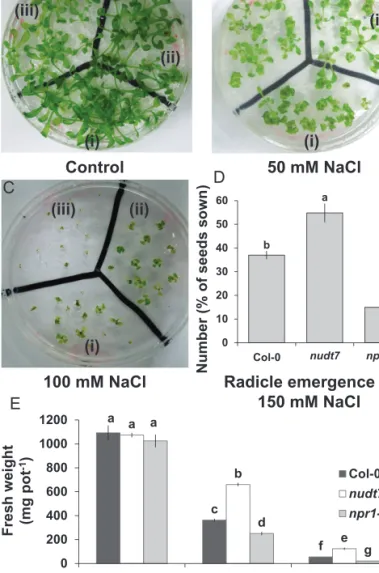
Fig. 1. Growth and radicle emergence of Arabidopsis thaliana grown in full-strength MS medium with 2% w/v phytogel infused with different concentration of salt. (A–C) Photographs of radicle emergence in (i) Col-0, (ii)
nudt7, (iii) npr1-5 at the indicated NaCl concentrations 7 d after sowing. (D)
Quantification of radicle emergence out of 20 seeds shown under 150 mM NaCl treatment at 7 d after sowing. (E) Fresh weight of the three genotypes under indicated NaCl concentrations 2 weeks after sowing. Each bar in the graphs represents mean±SEM. Different letters in bar graphs indicate significant differences. (This figure is available in colour at JXB online.)
before commencing MIFE measurements (Jayakannan et al., 2011; Jayakannan et al., 2013). The Petri dish was then placed on the microscope stage of the MIFE system. Electrodes were positioned at either the distal elongation zone (180–300 µm from the root cap) or mature root zone (>2 mm from the root cap) as described in Bose
et al. (2010a, b). Ion fluxes were measured under control conditions
for 5 min before treatment application. Treatments (100 mM NaCl; 1 mM copper-ascorbate mix; 1 or 10 mM hydrogen peroxide) were applied by pipetting the required volume of treatment stock solu-tions into the bathing solution in the Petri dish. After addition, the bathing solution was thoroughly mixed by sucking into, and expel-ling from, a pipette approximately five times. The bathing solution was allowed to equilibrate for 1 min before recording ion fluxes under treatment conditions; hence, the time required for the stock addition and the establishment of the diffusion gradients is about 40 s (Shabala and Hariadi, 2005). Accordingly, flux measurements dur-ing the first minute after treatment applications were discarded from the analysis and appear as gaps in the figures. Transient flux kinetics of K+, H+, and Na+ were measured for specified times.
Membrane potential measurements
The roots of an intact Arabidopsis seedling were gently secured in a measuring chamber in a horizontal position using a Parafilm strip and small plastic blocks. The seedling was then placed in a 10-ml Perspex measuring chamber filled with 7 ml of BSM and pre-conditioned as described above. The specific details pertinent to microelectrode preparation, impalement into the epidermal cells of mature root zone, and data recording can be found in previous publications (Bose et al., 2013; Jayakannan et al., 2013). Once a stable membrane potential measurement was obtained for 1 min, salt treatment (100 mM NaCl) was imposed. The transient mem-brane potential kinetics was recorded up to 30 min after treatment commencement. The membrane potential values of eight indi-vidual seedlings were averaged for every genotype and treatment combination.
Viability staining
Root viability was assessed by fluorescein diacetate/propidium iodide double staining method as described in a previous publica-tion (Bose et al., 2014).
In vivo hydrogen peroxide imaging
The H2O2 imaging of root tissue was done by following the stand-ard procedure adopted in a previous publication (Bose et al., 2014). The 4- to 5-day-old Arabidopsis seedlings were treated with 100 mM NaCl in BSM background. At 4 h and 24 h after salt treatment, the roots were washed with 10 mM Tris-HCl buffer and incubated in 25 µM 2′,7′-dichlorofluorescein diacetate (DCF-DA, D6883; Sigma) for 30 min at 30 °C. Following DCF-DA incubation, the amount of H2O2 produced in roots was assessed by visualizing fluores-cence intensity using a confocal microscope (Leica TCS SP5, Leica Microsystems). The Argon, visible laser power was set at 20%. Given that the H2O2 fluorescence intensity at 4 h was stronger than at 24 h time point, two different settings (and, hence, two different sets of controls) were used to resolve the signal. The acousto-optic tuneable filter (AOTF-488) was set at 10 % and 40 %, and the hybrid detector (HyD) gain was set at 19 and 120 for 4-h and 24-h time points, respectively. The software Leica Application Suite Advanced Fluorescence (LAS AF, Leica Microsystems) used to acquire images, and ImageJ (National Institutes of Health) was used to calculate the mean fluorescence intensity.
Statistical analysis
Data are reported as means±SEM. Statistical significance of mean values was determined using the standard LSD test at P≤0.05 level.
Results
nudt7 and npr1-5 plants differ in salt sensitivity
Similar to a previous report (Bose et al., 2013), 2 weeks of salt stress had a strong effect on plant growth, with fresh mass, dry mass, and water content all declining significantly and in a dose-dependent manner for all three Arabidopsis genotypes tested (Fig. 1 and Supplementary Fig. S1). This decline was smallest in nudt7 plants, followed by the wild type, and then by npr1-5 (most sensitive to salinity; Fig. 1 and Supplementary Fig. S1). Furthermore, under control conditions (i.e. no salt), the fresh (Fig. 1) and dry mass (Supplementary Fig. S1) were slightly lower in npr1-5 plants than the wild type and nudt7, but the difference was not statistically significant. At 150 mM NaCl, salt-sensitive npr1-5 had fewer radicles emerging than nudt7 and the wild type (Fig. 1).
The extent of salt-induced loss of cell viability was more severe in npr1-5 than nudt7 roots
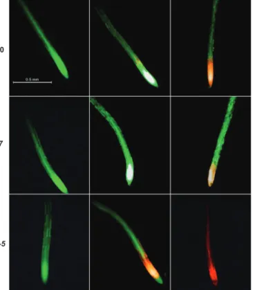
To determine the effect of salinity on root cell viability, 4- to 5-day-old Arabidopsis seedlings were exposed to 100 mM NaCl for 1 or 12 h and then double stained with fluorescein diacetate– propidium iodide (FDA–PI; Fig. 2). Under the fluorescence microscope, viable cells fluoresced bright green, whereas dead/ damaged cells fluoresced bright red (Fig. 2). The Arabidopsis seedlings incubated in BSM alone (control) showed green fluo-rescence even after 12 h, suggesting the control roots were via-ble and healthy in our experimental solutions (Fig. 2).
An hour of salt stress severely affected the viability of npr1-5 root cells in the elongation and meristematic regions, with the wild-type roots also showing a few dead cells in the elon-gation zone (Fig. 2). However, no damage was observed in the roots of nudt7 mutant (Fig. 2). Prolonged salt exposure (12 h) increased the extent of the damage in the following order npr1-5 > Col-0 > nudt7. These results were consistent with the long-term salinity exposure data (Fig. 1 and Supplementary Fig. S1) and imply that roots of npr1-5 were sensitive to salt stress, whereas nudt7 was salt-tolerant.
NaCl-induced ion flux responses varied between nudt7 and npr1-5
Consistent with our previous observations on Arabidopsis roots (Jayakannan et al., 2011; Bose et al., 2013), salin-ity (100 mM NaCl) caused significant changes in net ion fluxes measured from the elongation and mature zones of Arabidopsis roots (Figs 3, 4 and 5).
Acute salt stress caused significant K+ efflux from elonga-tion and mature root zones in all genotypes tested (Fig. 3). The peak K+ efflux was reached within 2 min after imposition of salt stress, followed by gradual recovery and stabilization 20 min later. Nearly a 4-fold difference in peak K+ fluxes was found between the elongation and the mature root zones in each Arabidopsis genotype (Fig. 3), implying the root elon-gation zone is more sensitive to salt stress than the mature root zone.
Among the three genotypes, the highest NaCl-induced K+ efflux was measured from npr1-5 roots in both the elongation and the mature root zones (–9269 ± 574 and –2096 ± 367 nmol m–2 s–1, respectively), whereas nudt7 showed about a 3-fold smaller peak K+ efflux (Fig. 3). The wild type had a peak K+ efflux in between the two mutants. In addition, the average K+ efflux over the first 60 min of salt treatment was about 9-fold (elongation zone) and 6-fold (mature zone) higher in salt-sen-sitive npr1-5 than salt-tolerant nudt7 mutant (Fig. 3 insets).
Salinity-induced H+ fluxes also showed genotypic differ-ences, in both the elongation and mature root zones (Fig. 4). Under control conditions (no salt), a significantly higher net H+ influx was observed in the root elongation zone of the npr1-5 mutant in comparison with Col-0 and the nudt7 mutant (Fig. 4 top panel). Addition of 100 mM NaCl caused a significant increase in net H+ influx in the elongation zone of npr1-5 (58 ± 8.5 nmol m–2 s–1) and Col-0 (7.4 ± 4.4 nmol m–2 s–1; Fig. 4 top panel). By contrast, 100 mM NaCl addi-tion induced an initial H+ efflux in the elongation zone of the nudt7 mutant followed by recovery towards the steady state before salt treatment (Fig. 4 top panel). In the mature root zone (Fig. 4, bottom panel), NaCl increased H+ influx for all three genotypes with the following magnitude npr1-5 > Col-0 > nudt7 (Fig. 4, bottom panel). Similarly, the aver-age H+ influx (over the first 60 min after salt application) at
both the elongation and mature root zones was highest in the npr1-5 mutant followed by Col-0 and was least in nudt7 (Fig. 4 insets).
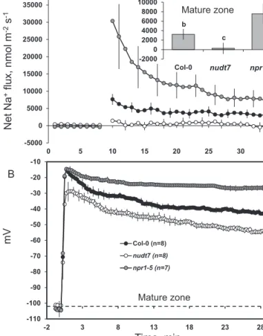
Na+ fluxes were measured in the mature root zone of the three Arabidopsis genotypes (Fig. 5A) using an improved Na+-selective resin (Jayakannan et al., 2011). Acute salt stress caused an immediate Na+ influx in Col-0 and npr1-5 (Fig. 5A). The peak Na+ influx was observed within minutes of salt addition and declined thereafter, but remained positive (influx) throughout the measurement period in npr1-5 and the wild type, while hovering around zero in nudt7 (Fig. 5A). The average Na+ flux measured during 1-h salt stress was about 28-fold higher in npr1-5 than nudt7 (Fig. 5A inset).
nudt7 and npr1-5 differ in the magnitude of NaCl-induced depolarization of the plasma membrane
The resting membrane potential in the mature zones of Arabidopsis roots was not significantly different among the three genotypes under control conditions (Fig. 5B). Adding 100 mM NaCl to the bathing medium resulted in highly significant (P≤0.01) membrane depolarization in all three Arabidopsis genotypes tested. The time-course of membrane potential changes (Fig. 5B) mirrored both Na+ (Fig. 5a) and K+ flux (Fig. 3) data, with the maximum membrane Col-0
npr1-5 nudt7
Fig. 2. Viability staining images of 4- to 5-day-old Arabidopsis thaliana roots exposed to 100 mM salt stress. The seedlings were grown in basal salt medium (BSM) containing 0.4% (w/v) agar for 4–5 d, then pre-treated with 100 mM NaCl in BSM for 1 or 12 h, and double stained with fluorescein diacetate–propidium iodide for imaging under a fluorescence microscope. The control plants were treated only with BSM; the image shown is the control plant after 12 h in BSM. (This figure is available in colour at JXB online.) -10000 -8000 -6000 -4000 -2000 0 0 5 10 15 20 25 30 35 Col-0 nudt7 npr1-5 -2200 -1700 -1200 -700 -200 0 5 10 15 20 25 30 35 Net K +flux, nmol m -2s -1 Mature zone Elongation zone Time, min b a c -900 -700 -500 -300 -100
Col-0 nudt7nudt7 npr1-5npr1-5 b a c -2000 -1600 -1200 -800 -400
0 Col-0 nudt7nudt7 npr1-5npr1-5
A
B
Fig. 3. Transient K+ fluxes measured at the root elongation and the mature zones of 4- to 5-day-old Arabidopsis thaliana seedlings exposed to 100 mM salt stress. The insets were average K+ fluxes during 1-h exposure to 100 mM NaCl stress. Each point or bar represents mean±SEM of 8–12 seedlings. Different letters below the bars in the insets indicate significant differences.
depolarization observed within minutes of NaCl treatment; approximately at the same time as the peak Na+ influx and K+ efflux (the magnitude of the former being greater than that of the latter) (Figs 3 and 5). Initial depolarization was followed by a substantial (10–20 mV) membrane repolari-zation, with the membrane potential reaching new steady-state values in all three Arabidopsis genotypes 20–30 min after salt application (Fig. 5B). Among the genotypes, the salt-sensitive npr1-5 showed the highest magnitude of mem-brane depolarization (to –15 ± 1 mV), whereas salt-tolerant nudt7 showed the least membrane depolarization (to –30 ± 1 mV) (Fig. 5B). A ≈25mV difference between nudt7 and npr1-5 plants was maintained throughout the measurement period (Fig. 5B).
Salt-induced H2O2 production was higher in nudt7
than npr1-5
In vivo imaging of H2O2 production in root tissue was done 4 h and 24 h after 100 mM NaCl addition (Fig. 6). The salt-induced H2O2 production was several folds higher at 4 h than 24 h in all the genotypes tested, necessitating specific settings (described in the Materials and methods section) to acquire images for each time point to avoid oversaturation and pho-tobleaching. Among the genotypes, mutant npr1-5 with SA
signalling blockage had lower capacity to increase H2O2 pro-duction under salt stress, whereas nudt7 mutant showed sus-tained elevation in H2O2 production under salt stress at both time points.
Shoot Na and K concentrations differed between nudt7 and npr1-5 during long-term salt exposure
As expected, 25 d of growth in NaCl-supplemented MS media caused a substantial increase in the shoot Na+ concentration and a decrease in the shoot K+ concentration in all three
Arabidopsis genotypes tested (Fig. 7). Under salt stress, nudt7 showed the lowest Na+ concentration in shoots followed by the wild type, whereas the npr1-5 mutant had the highest con-centration (Fig. 7A). In contrast, the shoot K+ concentration was the highest in the nudt7 mutant followed by the wild type and was lowest in the npr1-5 mutant (Fig. 7B) under either 50 or 100 mM NaCl stress.
nudt7 and npr1-5 mutants vary in their oxidative stress tolerance
The viability staining was used to evaluate the responses of Arabidopsis genotypes during oxidative stress by
-110 -100 -90 -80 -70 -60 -50 -40 -30 -20 -10 -2 3 8 13 18 23 28 Col-0 (n=8) nudt7 (n=8) npr1-5 (n=7) Time, min Net Na +flux, nmol m -2s -1 -5000 0 5000 10000 15000 20000 25000 30000 35000 40000 0 5 10 15 20 25 30 35 Col-0 nudt7 npr1-5 b c a -2000 0 2000 4000 6000 8000 10000
Col-0 nudt7nudt7 npr1-5npr1-5
mV
A
B
Mature zone
Mature zone
Fig. 5. Transient (A) Na+ fluxes and (B) membrane potential dynamics measured at the mature root zone of 4- to 5-day-old Arabidopsis thaliana seedlings exposed to 100 mM salt stress. The inset was average Na+ fluxes during 1-h exposure to 100 mM NaCl stress. Each point or bar represents mean±SEM of 8–12 seedlings. Different letters above the bars in the inset indicate significant differences.
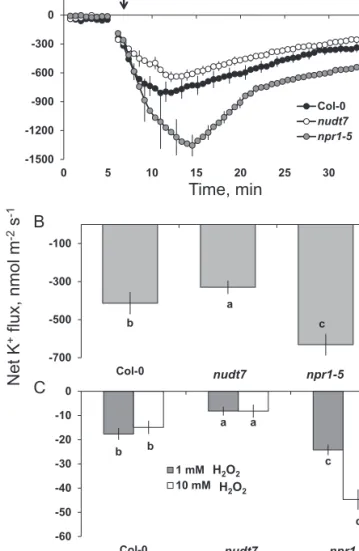
-10 0 10 20 30 40 50 60 70 0 5 10 15 20 25 30 35 Col-0 nudt7 npr1-5 0 5 10 15 20 25 30 35 40 0 5 10 15 20 25 30 35 Col-0 nudt7 npr1-5 Elongation zone Mature zone Time, min Net H +flux, nmol m -2 s -1 b b a 0 5 10 15 20 25 30
Col-0 nudt7nudt7 npr1-5npr1-5
b c a 0 5 10 15
Col-0 nudt7nudt7 npr1-5npr1-5
A
B
Fig. 4. Transient H+ fluxes measured at the root elongation and the mature zones of 4- to 5-day-old Arabidopsis thaliana seedlings exposed to 100 mM salt stress. The insets were average H+ fluxes during 1-h exposure to 100 mM NaCl stress. Each point or bar represents mean±SEM of 8–12 seedlings. Different letters above the bars in the insets indicate significant differences.
treating 4- to 5-day-old seedlings in a hydroxyl-radical-producing medium (1 mM copper-ascorbate or 10 mM H2O2) for 1 h (Fig. 8). Fluorescence microscopy showed that 1-h exposure to hydroxyl radicals caused severe dam-age to the roots of npr1-5 and less so to the wild type Col-0 (Fig. 8). No damage was found in nudt7 mutant (Fig. 8). Furthermore, in npr1-5 treated with copper-ascorbate the damage was detected in the root tips as well as in the mature root part, whereas in Col-0 plants only the mature zone showed damage symptoms (Fig. 8). With respect to H2O2, the damage was smaller in Col-0 and nudt7 in comparison to npr1-5 (Fig. 8). The damage was detected only in the cortex of the mature roots of Col-0 and nudt7 (Fig. 8), whereas the whole roots were severely affected by H2O2 stress in npr1-5.
Net ion fluxes influenced by oxidative stress differ between nudt7 and npr1-5 mutants
Application of 1 mM of hydroxyl-radical-generating cop-per-ascorbate mix caused a large K+ efflux from the mature root zone of all three Arabidopsis genotypes (Fig. 9A). This hydroxyl-radical-induced K+ efflux was not instantaneous, but increased gradually over time, reaching a peak value 5 min after the commencement of the oxidative stress treatment in Col-0 and nudt7 and 10 min for npr1-5 (Fig. 9A). The magni-tude of K+ efflux was the lowest in nudt7 and the highest in
npr1-5 (Fig. 9A; 2-fold difference; significant at P≤0.05). The K+ flux gradually recovered after reaching the peak, although it remained negative for the treatment duration in all three Arabidopsis genotypes (Fig. 9A). The average K+ efflux
d b d a d c 0.0 0.2 0.4 0.6 0.8 1.0 1.2 1.4 1.6 control 100 mM NaCl Col-0 nudt7 npr1-5 b b b a b b 0 1 2 3 4 5 6 Control 100 mM NaCl Col-0 nudt7 npr1-5 Av erage fluorescenc e (arb. units) 4 h 24 h
A
B
C
D
Fig. 6. In vivo detection of hydrogen peroxide production in the root tissue of Arabidopsis thaliana seedlings after NaCl treatment. (A, B) Images of Arabidopsis thaliana seedling roots, after being exposed to the indicated salt concentrations for 4 or 24 h. Samples were stained with
2′,7′-dichlorofluorescein diacetate for imaging under a fluorescence microscope. Roots for treatments were taken from 4- to 5-day-old seedlings grown in basal salt medium (BSM) containing 0.4% (w/v) agar. Because the hydrogen peroxide fluorescence was much higher at 4 h than at 24 h, different settings were used to acquire images to show difference between genotypes at each time point. (C, D) Quantification of fluorescence in the roots of the different genotypes after exposure to salt stress for the indicated times. Each bar represents mean±SEM of 8–12 seedlings. Different letters above the bars in the bar graphs indicate significant differences. (This figure is available in colour at JXB online.)
measured over a 60-min Cu-ascorbate treatment period was 2-fold higher in npr1-5 than nudt7 (Fig. 9B).
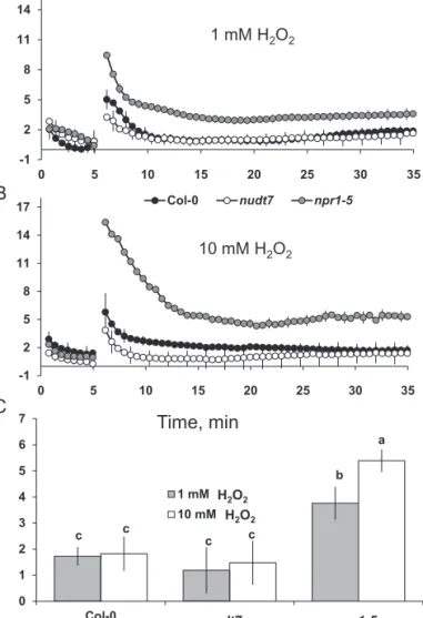
The average K+ fluxes during 1-h exposure to either 1 or 10 mM H2O2 treatment revealed no significant dose-depend-ency in Arabidopsis genotypes nudt7 and Col-0 (Fig. 9C). However, the npr1-5 mutant had 2-fold greater K+ efflux at 10 than at 1 mM H2O2 (Fig. 9C). This mutant had greater K+ efflux than nudt7 and Col-0 regardless of the H2O2 concentra-tion used (Fig. 9C).
Though the initial H+ flux from the mature root zone of
Arabidopsis was higher at 10 mM H2O2 than 1 mM H2O2, the steady state H+ flux (from 10 min onwards) is similar for differ-ent genotypes exposed to either concdiffer-entration of H2O2 (Fig. 10). In general, the salt-sensitive npr1-5 mutant showed significantly higher (4- to 5-fold) H+ influx compared with the other two gen-otypes (nudt7 and Col-0) in either 1 or 10 mM H2O2 (Fig. 10).
Discussion
The NPR1-dependent SA signalling is pivotal for Na+
exclusion from roots and shoots
Maintaining relatively low Na+ concentration in shoots is an important trait for salt tolerance in glycophytes (Colmer et al., Col-0
npr1-5 nudt7
Fig. 8. Viability staining of 4- to 5-day-old Arabidopsis thaliana roots exposed to 1 mM Cu-ascorbate or 10 mM H2O2 for 1 h. The seedlings were grown in basal salt medium (BSM) containing 0.4 % w/v agar for 4–5 d, were pre-treated with either 1 mM CuCl2+1 mM ascorbate or 10 mM H2O2 in the BSM background for 1 h and then stained with fluorescein diacetate–propidium iodide for observations under a fluorescence microscope. (This figure is available in colour at JXB online.)
-1500 -1200 -900 -600 -300 0 0 5 10 15 20 25 30 35 Col-0 nudt7 npr1-5 1 mM Cu/Asc mix b a c -700 -500 -300 -100 Col-0 nudt7 npr1-5 Net K +flux, nmol m -2s -1 Time, min A B C b a c b a d -60 -50 -40 -30 -20 -10 0 Col-0 nudt7 npr1-5 1 mM H2O2 10 mM H2O2 nudt7 npr1-5 H2O2 H2O2 nudt7 npr1-5
Fig. 9. K+ fluxes in response to 1 mM Cu-ascorbate. (A) Transient K+ fluxes in response to 1 mM Cu-ascorbate applied after 5 min. (B) Average K+ fluxes during 1-h exposure to 1 mM Cu-ascorbate or (C) 1 or 10 mM H2O2 stress. K+ fluxes measured at the mature root zone of 4- to 5-day-old Arabidopsis
thaliana seedlings. Each point or bar represents mean±SEM of 8–12
seedlings. Different letters below the bars indicate significant differences. a c f a b e a d g 0 3 6 9 12
Control 50 mM NaCl 100 mM NaCl
Col-0 nudt7 npr1-5 g e b g f c g d a 0 2 4 6 8 Shoot K (g kg -1D W) Shoot Na (g kg -1D W)
A
B
Fig. 7. Effect of different NaCl treatment on concentrations of Na+ (A) and K+ (B) in Arabidopsis shoots after 25 d of growth in the full-strength MS medium with 2% w/v phytogel. Each bar represents mean±SEM. Different letters above the bars indicate significant differences.
2005; Munns and Tester, 2008). The main mechanisms employed by the glycophytes to minimize Na+ accumulation in shoots are linked to the enhanced capacity of plants to (i) restrict the ini-tial entry of Na+ ions into the root tissue, (ii) excrete Na+ from root tissue back into the rhizosphere, (iii) sequester Na+ inside the root vacuoles, and (iv) reduce the long-distance transport of Na+ into the shoots (Cuin et al., 2011). Given that Arabidopsis is a glycophyte, shoot Na+ concentration analysis and root Na+ flux measurements were employed to ascertain the operation of the above mechanisms in two SA-signalling mutants. The npr1-5 mutant lacking NPR1-dependent SA-signalling recorded the highest Na+ influx into root tissue in comparison with the wild type and nudt7 mutant (Fig. 5A). If npr1-5 was efficient in sequestering Na+ in root vacuoles or excluding Na+ out of root cells, there would have been significant improvement in growth accompanied by reduction in the shoot Na+ concentration. However, poor growth (Fig. 1 and Supplementary Fig. S1) and viability of root cells (Fig. 2) along with the highest shoot Na+ concentration (Fig. 7A) in comparison with the wild type and nudt7 mutant implied that the npr1-5 mutant was defective in
preventing the entry of Na+ into root tissue and its subsequent transport into the shoots.
In contrast to npr1-5, the nudt7 mutant had the lowest Na+ influx into root tissue (Fig. 5a). This may be attributable to either decreased Na+ entry or enhanced Na+ extrusion via H+ -ATPase-energized SOS1 (a Na+/H+ exchanger) activity in the plasma membrane (Cuin et al., 2011). Four lines of evidence favour the latter explanation for the nudt7 mutant. First, the initial Na+ entry into the epidermis of root tissue during acute salt stress is ther-modynamically passive and is poorly controlled in glycophytes (Tester and Davenport, 2003). Second, the inherent stability of SOS1 mRNA is poor (with a half-life of only 10 min), and it was shown that exogenous H2O2 application increased the stability of
SOS1 in a rapid (within 30 min) concentration-dependent man-ner (Chung et al., 2008). If this is the case, sustained elevation of H2O2 production in the root tissue of nudt7 mutant (Fig. 6) dur-ing salt stress is expected to result in improved SOS1 mRNA sta-bility. Thirdly, SOS1 transcripts were found to be higher in roots of the salt-tolerant mutant over-expressing haem oxygenase (EC 1.14.99.3) (Bose et al., 2013). Indeed, a 3-fold higher induc-tion of putative haem oxygenase (At1g69720) was found in the nudt7 mutant when grown under nutrient stress (Jambunathan
et al., 2010). Finally, the nudt7 mutant showed either H+ efflux or
reduced net H+ influx during acute salt stress (Fig. 4) in compari-son with the wild type and npr1-5 mutant, which is usually the result of enhanced H+-ATPase activity fuelling SOS1 operation (Bose et al., 2013; Jayakannan et al., 2013). Overall, the above results suggest that the nudt7 mutant has enhanced capacity to decrease both the loading of Na+ into the root tissue and the transport of Na+ into the shoot (Fig. 7A).
The NPR1-dependent SA signalling assists plants in retaining K+ during salt stress by controlling both
depolarization-activated KOR and ROS-activated NSCC channels
Salinity stress has ionic, hyperosmotic, and oxidative stress components that severely hamper plant growth and produc-tivity. Apart from hyperosmotic stress, both the ionic stress through depolarization-activated KOR and the oxidative stress through ROS-activated non-selective cation channels (NSCC) exacerbate K+ loss, thereby depleting the cytosolic K+ pool available for metabolic functions, which eventually leads to cell death (Shabala and Cuin, 2008; Shabala, 2009). Hence, the magnitude of salt-induced K+ loss can be used as a measure of salt tolerance of diverse plant species, including Arabidopsis (Bose et al., 2013; Jayakannan et al., 2013). Acute salt stress in the study presented here resulted (as expected) in a K+ loss from both the elongation and mature root zones of all three genotypes tested (Fig. 3). However, the salt-induced K+ loss was lowest in the nudt7 mutant and highest in the
npr1-5 mutant (Fig. 3), suggesting NPR1-dependent SA sig-nalling is critical for decreasing the K+ loss during salt stress. In Arabidopsis, comparison of the depolarization-acti-vated KOR knock-out mutant gork1-1 with rbhoD (a mutant lacking ROS production via NADPH oxidase) during acute 100 mM NaCl stress revealed that 3/4 of K+ loss were medi-ated by depolarization-activmedi-ated KOR and the remaining -1 2 5 8 11 14 17 0 5 10 15 20 25 30 35 Col-0 nudt7 npr1-5 -1 2 5 8 11 14 17 0 5 10 15 20 25 30 35 Col-0 nudt7 npr1-5 Time, min Net H +flux, nmol m -2 s -1 1 mM H2O2 10 mM H2O2 c c b c c a 0 1 2 3 4 5 6 7 Col-0 nudt7 npr1-5 1 mM H2O2 10 mM H2O2 nudt7 npr1-5 H2O2 H2O2 A B C
Fig. 10. H+ fluxes in response to treatment with H
2O2. (A, B) Transient H+ fluxes measured at the mature root zone of 4- to 5-day-old Arabidopsis
thaliana seedlings in response to 1 or 10 mM H2O2. (C) Average H+ fluxes during 1-h exposure to 1 or 10 mM H2O2. Each point or bar represents mean±SEM of 8–12 seedlings. Different letters above the bars in bar graph indicate significant differences.
1/4 through H2O2-activated channels (Jayakannan et al.,
2013). Superoxide (Tran et al., 2013) and hydroxyl radicals (Demidchik et al., 2010) can also induce K+ loss through the GORK channel. Thus, the contrasting capacity of nudt7 and npr1-5 mutants to retain K+ in roots (Fig. 3) and shoots (Fig. 7B) during salt stress may be underpinned by their dif-ferential K+ loss through KOR and/or ROS-activated NSCC channels.
The entry of positively charged Na+ (Fig. 5A) and H+ (Fig. 4) ions into root tissue during acute 100 mM NaCl stress resulted in net depolarization of the plasma membrane in all three genotypes tested (Fig. 5b), implying that the bulk of the NaCl-induced K+ loss (Fig. 3) might have been through depolarization-activated KOR channels. Among the genotypes, H+ and Na+ uptake (Figs 4, 5A) as well as NaCl-induced membrane depolarization were highest in the npr1-5 mutant followed by the wild type, and were lowest in the nudt7 mutant. Moreover, approximately a 15–25 mV difference was observed between npr1-5 and nudt7 mutants (the latter being less depolarized) throughout the measurement period (Fig. 5B). Such a difference in depolarization voltage may be associated with a lower NaCl-induced K+ loss in nudt7 com-pared with npr1-5. It is evident that NPR1-mediated SA sig-nalling plays a key role in regulating the membrane potential during salt stress.
An increase in the production of superoxide (Borsani et al., 2001), hydrogen peroxide (Xie et al., 2011), and hydroxyl rad-icals (Demidchik et al., 2010) was noted in Arabidopsis roots exposed to salt stress. These ROS species can promote K+ loss through NSCC channels (Demidchik et al., 2003; Zepeda-Jazo et al., 2011) and/or through KOR channels (Demidchik
et al., 2010; Tran et al., 2013). The results here (Fig. 9) showed
that hydroxyl radicals caused a severe K+ loss (about 15- to 20-fold higher) compared with up to 10 mM H2O2. Among the genotypes, the npr1-5 mutant showed a higher K+ loss than the wild type and nudt7 mutant under hydroxyl radical and hydrogen peroxide treatments (Fig. 9), suggesting npr1-5 was more sensitive to these ROS species in comparison with the wild type and nudt7 mutant. The viability staining results confirmed this, whereby a 1-h treatment with either hydroxyl radicals or 10 mM hydrogen peroxide affected root cells more severely in npr1-5 than in the nudt7 mutant (Fig. 8). The nudt7 mutant was able to increase the salt-induced H2O2 produc-tion in root tissue over a 24 h period, but the npr1-5 mutant was not (Fig. 6) suggesting NPR1 is a key regulator of salt-induced H2O2 production in plants. Because the nudt7 mutant produced more ROS than wild type and npr1-5 during salt stress, it is reasonable to assume that H2O2-induced K+ efflux would be greater in nudt7. However, in the exogenous H2O2 treatment (1 and 10 mM), the K+ efflux of nudt7 mutant did not differ from the wild type, and was lower than in the npr1-5 mutant (Fig. 9C). This suggests that the presence of an NPR1-mediated SA signalling component in the nudt7 mutant makes K+-efflux transporters insensitive to elevated H2O2 concentration during salt stress. Overall, these results provide evidence that (i) NPR1-mediated SA signalling is pivotal for H2O2 production during salt stress, and also for decreasing K+ loss through the NSCC and KOR channels
activated by hydrogen peroxide and hydroxyl radicals, and (ii) the nudt7 mutant shows no response to hydrogen peroxide and is tolerant to hydroxyl radicals.
In summary, an npr1-5 mutant lacking the NPR1-dependent SA signalling was unable to control both the entry of Na+ into roots and its long-distance transport into the shoot, and to pre-vent K+ loss via depolarization-activated KOR and the ROS-activated NSCC channels during salt stress. As a result, the npr1-5 mutant was sensitive to salt stress. On the other hand, the constitutive expression of NPR1-dependent SA signalling enhanced the salt tolerance of a nudt7 mutant by controlling Na+ entry into the root tissue and subsequent transport to the shoot, as well as minimizing K+ loss during salt stress. In con-clusion, NPR1-dependent SA signalling is a crucial compo-nent of salt and oxidative stress tolerance in Arabidopsis.
Supplementary data
Supplementary data are available at JXB online
Figure S1. Effect of salt stress on dry weight and water content of Arabidopsis thaliana seedlings grown in the full-strength MS medium with 2% w/v phytogel for two weeks.
Acknowledgements
Maheswari Jayakannan is a recipient of the Australian Postgraduate Award (APA) and University of Western Australia Postgraduate Award (UPA). This work was supported by the Australian Research Council grants to Z. Rengel (DP0988193 and DP130104825) and S. Shabala (DP0987402 and DP1094663), and Spanish MICINN grant (BFU2010-14873) to C. Poschenrieder.
References
Asensi-Fabado M, Munné-Bosch S. 2011. The aba3-1 mutant of Arabidopsis thaliana withstands moderate doses of salt stress by modulating leaf growth and salicylic acid levels. Journal of Plant Growth
Regulation 30, 456–466.
Attaran E, He SY. 2012. The long-sought-after salicylic acid receptors.
Molecular Plant 5, 971–973.
Bartsch M, Gobbato E, Bednarek P, Debey S, Schultze JL, Bautor J, Parker JE. 2006. Salicylic acid–independent ENHANCED DISEASE SUSCEPTIBILITY1 signaling in Arabidopsis immunity and cell death is regulated by the monooxygenase FMO1 and the nudix hydrolase NUDT7.
The Plant Cell 18, 1038–1051.
Blanco F, Salinas P, Cecchini N, Jordana X, Hummelen P, Alvarez M, Holuigue L. 2009. Early genomic responses to salicylic acid in
Arabidopsis. Plant Molecular Biology 70, 79–102.
Borsani O, Valpuesta V, Botella MA. 2001. Evidence for a role of salicylic acid in the oxidative damage generated by NaCl and osmotic stress in Arabidopsis seedlings. Plant Physiology 126, 1024–1030. Bose J, Babourina O, Shabala S, Rengel Z. 2010a. Aluminium-induced ion transport in Arabidopsis: the relationship between Al tolerance and root ion flux. Journal of Experimental Botany 61, 3163–3175.
Bose J, Babourina O, Shabala S, Rengel Z. 2010b. Aluminum-dependent dynamics of ion transport in Arabidopsis: specificity of low pH and aluminum responses. Physiologia Plantarum 139, 401–412. Bose J, Shabala L, Pottosin I, Zeng F, Velarde-Buendía A-M, Massart A, Poschenrieder C, Hariadi Y, Shabala S. 2014. Kinetics of xylem loading, membrane potential maintenance, and sensitivity of K+-permeable channels to reactive oxygen species: physiological traits that differentiate salinity tolerance between pea and barley. Plant, Cell and
Bose J, Xie Y, Shen W, Shabala S. 2013. Haem oxygenase modifies salinity tolerance in Arabidopsis by controlling K+ retention via regulation of the plasma membrane H+-ATPase and by altering SOS1 transcript levels in roots. Journal of Experimental Botany 64, 471–481.
Cao Y, Zhang ZW, Xue LW, Du JB, Shang J, Xu F, Yuan S, Lin HH. 2009. Lack of salicylic acid in Arabidopsis protects plants against moderate salt stress. Zeitschrift fur Naturforschung C, Journal of
Biosciences 64, 231–238.
Chung JS, Zhu JK, Bressan RA, Hasegawa PM, Shi H. 2008. Reactive oxygen species mediate Na+-induced SOS1 mRNA stability in
Arabidopsis. The Plant Journal 53, 554–565.
Colmer TD, Munns R, Flowers TJ. 2005. Improving salt tolerance of wheat and barley: future prospects. Australian Journal of Experimental
Agriculture 45, 1425–1443.
Cuin TA, Bose J, Stefano G, Jha D, Tester M, Mancuso S, Shabala S. 2011. Assessing the role of root plasma membrane and tonoplast Na+/H+ exchangers in salinity tolerance in wheat: in planta quantification methods. Plant, Cell and Environment 34, 947–961.
Demidchik V, Cuin TA, Svistunenko D, Smith SJ, Miller AJ, Shabala S, Sokolik A, Yurin V. 2010. Arabidopsis root K+-efflux conductance activated by hydroxyl radicals: single-channel properties, genetic basis and involvement in stress-induced cell death. Journal of Cell Science 123, 1468–1479.
Demidchik V, Shabala SN, Coutts KB, Tester MA, Davies JM. 2003. Free oxygen radicals regulate plasma membrane Ca2+- and K+-permeable channels in plant root cells. Journal of Cell Science 116, 81–88.
Dempsey DMA, Vlot AC, Wildermuth CM, Klessig FD. 2011. Salicylic acid biosynthesis and metabolism. The Arabidopsis Book 9, e0156. Fu ZQ, Yan S, Saleh A, Wang W, Ruble J, Oka N, Mohan R, Spoel SH, Tada Y, Zheng N. 2012. NPR3 and NPR4 are receptors for the immune signal salicylic acid in plants. Nature 486, 228–232.
Ge X, Li GJ, Wang SB, Zhu H, Zhu T, Wang X, Xia Y. 2007. AtNUDT7, a negative regulator of basal immunity in Arabidopsis, modulates two distinct defense response pathways and is involved in maintaining redox homeostasis. Plant Physiology 145, 204–215.
Hao L, Zhao Y, Jin D, Zhang L, Bi X, Chen H, Xu Q, Ma C, Li G. 2012. Salicylic acid-altering Arabidopsis mutants response to salt stress. Plant
and Soil 354, 81–95.
Horváth E, Szalai G, Janda T. 2007. Induction of abiotic stress tolerance by salicylic acid signaling. Journal of Plant Growth Regulation 26, 290–300.
Ishikawa K, Ogawa T, Hirosue E, Nakayama Y, Harada K, Fukusaki E, Yoshimura K, Shigeoka S. 2009. Modulation of the poly (ADP-ribosyl) ation reaction via the Arabidopsis ADP-ribose/NADH pyrophosphohydrolase, AtNUDX7, is involved in the response to oxidative stress. Plant Physiology 151, 741–754.
Jambunathan N, Mahalingam R. 2006. Analysis of Arabidopsis
growth factor gene 1 (GFG1) encoding a nudix hydrolase during oxidative
signaling. Planta 224, 1–11.
Jambunathan N, Penaganti A, Tang Y, Mahalingam R. 2010. Modulation of redox homeostasis under suboptimal conditions by
Arabidopsis nudix hydrolase 7. BMC Plant Biology 10, 173. Jayakannan M, Babourina O, Rengel Z. 2011. Improved measurements of Na+ fluxes in plants using calixarene-based microelectrodes. Journal of Plant Physiology 168, 1045–1051. Jayakannan M, Bose J, Babourina O, Rengel Z, Shabala S. 2013. Salicylic acid improves salinity tolerance in Arabidopsis by restoring membrane potential and preventing salt-induced K+ loss via a GORK channel. Journal of Experimental Botany 64, 2255–2268.
Kraszewska E. 2008. The plant Nudix hydrolase family. Acta Biochimica
Polonica 55, 663–671.
Lee S, Kim SG, Park CM. 2010. Salicylic acid promotes seed germination under high salinity by modulating antioxidant activity in
Arabidopsis. New Phytologist 188, 626–637.
Martinez-Beltran J, Manzur CL. 2005. Overview of salinity problems in the world and FAO strategies to address the problem. Proceedings of the
International Salinity Forum. California: Riverside.
Miura K, Sato A, Ohta M, Furukawa J. 2011. Increased tolerance to salt stress in the phosphate-accumulating Arabidopsis mutants siz1 and
pho2. Planta 234, 1191–1199.
Munns R, Tester M. 2008. Mechanisms of salinity tolerance. Annual
Review of Plant Biology 59, 651–681.
Newman IA. 2001. Ion transport in roots: measurement of fluxes using ion-selective microelectrodes to characterize transporter function. Plant,
Cell and Environment 24, 1–14.
Ondrasek G, Rengel Z, Veres S. 2011. Soil salinisation and salt stress in crop production. In: Shanker A, Venkateswarlu B, eds. Abiotic stress in
plants—mechanisms and adaptations. Rijeka: InTech, 171–190.
Parida AK, Das AB. 2005. Salt tolerance and salinity effects on plants: a review. Ecotoxicology and Environmental Safety 60, 324–349.
Poór P, Szopkó D, Tari I. 2012. Ionic homeostasis disturbance is involved in tomato cell death induced by NaCl and salicylic acid. In Vitro
Cellular and Developmental Biology—Plant 48, 377–382.
Rengasamy P. 2006. World salinization with emphasis on Australia.
Journal of Experimental Botany 57, 1017–1023.
Rivas-San Vicente M, Plasencia J. 2011. Salicylic acid beyond defence: its role in plant growth and development. Journal of Experimental Botany 62, 3321–3338.
Shabala S. 2009. Salinity and programmed cell death: unravelling mechanisms for ion specific signalling. Journal of Experimental Botany 60, 709–711.
Shabala S, Cuin TA. 2008. Potassium transport and plant salt tolerance.
Physiologia Plantarum 133, 651–669.
Shabala S, Cuin TA, Prismall L, Nemchinov LG. 2007. Expression of animal CED-9 anti-apoptotic gene in tobacco modifies plasma membrane ion fluxes in response to salinity and oxidative stress. Planta 227, 189–197. Shabala S, Hariadi Y. 2005. Effects of magnesium availability on the activity of plasma membrane ion transporters and light-induced responses from broad bean leaf mesophyll. Planta 221, 56–65.
Shah J, Kachroo P, Klessig DF. 1999. The Arabidopsis ssi1 mutation restores pathogenesis-related gene expression in npr1 plants and renders defensin gene expression salicylic acid dependent. The Plant Cell Online 11, 191–206.
Shah J, Tsui F, Klessig DF. 1997. Characterization of salicylic acid-insensitive mutant (sai1) of Arabidopsis thaliana, identified in a selective screen utilizing the SA-inducible expression of the tms2 gene. Molecular
Plant–Microbe Interactions 10, 69–78.
Straus MR, Rietz S, Ver Loren van Themaat E, Bartsch M, Parker JE. 2010. Salicylic acid antagonism of EDS1-driven cell death is important for immune and oxidative stress responses in Arabidopsis. The Plant
Journal 62, 628–640.
Tran D, El-Maarouf-Bouteau H, Rossi M, Biligui B, Briand J, Kawano T, Mancuso S, Bouteau F. 2013. Post-transcriptional regulation of GORK channels by superoxide anion contributes to increases in outward-rectifying K+ currents. New Phytologist 198, 1039–1048. Tester M, Davenport R. 2003. Na+ tolerance and Na+ transport in higher plants. Annals of Botany 91, 503–527.
Vlot AC, Dempsey DMA, Klessig DF. 2009. Salicylic acid, a multifaceted hormone to combat disease. Annual Review of Phytopathology 47, 177–206.
Wang D, Amornsiripanitch N, Dong X. 2006. A genomic approach to identify regulatory nodes in the transcriptional network of systemic acquired resistance in plants. PLoS Pathol 2, e123.
Wang H, Lu Y, Liu P, Wen W, Zhang J, Ge X, Xia Y. 2012. The ammonium/nitrate ratio is an input signal in the temperature-modulated,
SNC1-mediated and EDS1-dependent autoimmunity of nudt6-2 nudt7. The Plant Journal , doi: 10.1111/tpj.12032.
Wu Y, Zhang D, Chu JY, Boyle P, Wang Y, Brindle ID, De Luca V, Després C. 2012. The Arabidopsis NPR1 protein is a receptor for the plant defense hormone salicylic acid. Cell Reports 1, 639–647.
Xie Y-J, Xu S, Han B, Wu M-Z, Yuan X-X, Han Y, Gu Q, Xu D-K, Yang Q, Shen W-B. 2011. Evidence of Arabidopsis salt acclimation induced by up-regulation of HY1 and the regulatory role of RbohD-derived reactive oxygen species synthesis. The Plant Journal 66, 280–292.
Zepeda-Jazo I, Velarde-Buendía AM, Enríquez-Figueroa R, Bose J, Shabala S, Muñiz-Murguía J, Pottosin I. 2011. Polyamines interact with hydroxyl radicals in activating Ca2+ and K+ transport across the root epidermal plasma membranes. Plant Physiology 157, 2167–2180.