Morphologic characteristics, location, and associated
complications of maxillary and mandibular supernumerary teeth
as evaluated using cone beam computed tomography
Jessica Mossaz*, Dimitrios Kloukos*, Nikolaos Pandis*, Valerie G. A. Suter**,
Christos Katsaros* and Michael M. Bornstein**
*Department of Orthodontics and Dentofacial Orthopedics and **Department of Oral Surgery and Stomatology, School of Dental Medicine, University of Bern, Switzerland
Correspondence to: Michael M. Bornstein, Department of Oral Surgery and Stomatology, School of Dental
Medicine, University of Bern, Freiburgstrasse 7, CH-3010 Bern, Switzerland. E-mail: [email protected] summary
OBJECtiVES: to evaluate the location and morphologic characteristics of supernumerary teeth and to assess
the frequency and extent of root resorption of adjacent teeth using cone beam computed tomography (CBCt).
MAtEriAlS AND MEtHODS: CBCt scans of 82 patients with supernumerary teeth in the maxilla and mandible
were evaluated by two orthodontists independently. Data regarding the type, shape, and three-dimen-sional (3D) location of the supernumeraries including the frequency and extent of root resorption of adja-cent teeth were recorded and evaluated for possible associations.
rESUltS: the study comprised a total of 101 supernumerary teeth. Most of the patients (80.5 per cent)
exhibited one single supernumerary tooth, while 15.8 per cent had two and 3.7 per cent had three super-numeraries. Males were affected more than females with a ratio of 1.65:1. Mesiodentes were the most fre-quently diagnosed type of supernumerary teeth (48.52 per cent), followed by supernumerary premolars (23.76 per cent) and lateral incisors (18.81 per cent). Supernumeraries were most commonly conical in shape (42.6 per cent) with a normal or inclined vertical position (61.4 per cent). root resorption of adjacent teeth was detected for 22.8 per cent of the supernumerary teeth, most frequently for supernumerary pre-molars. there was a significant association between root resorption of adjacent teeth and type and shape of tooth. interrater agreement for the measurements performed showed kappa values ranging from 0.55 to 1 with a kappa value of 1 for type and shape of the supernumerary teeth.
CONClUSiONS: CBCt provides 3D information about location and shape of supernumerary teeth as well
as prevalence and degree of root resorption of neighbouring teeth with moderate to high interrater correlation.
Introduction
The term ‘supernumerary’ defines teeth that form addition-ally to the normal dentition. Several theories have been postulated regarding their aetiology, including atavism (evolutionary throwback), dichotomy of the tooth germ, hyperactivity of the dental lamina, and genetic and environ-mental factors. Localized and independent hyperactivity of the dental lamina is the most widely accepted cause for the development of supernumerary teeth today (Primosch, 1981; von Arx, 1990; Stellzig et al., 1997; Rajab and Hamdan, 2002; Russell and Folwarczna, 2003; Ferrés-Padró et al., 2009; Wang and Fan, 2011). Supernumerary teeth are usually detected incidentally during routine radi-ographic examination or when normal tooth eruption is delayed or fails.
The literature lists several studies assessing the preva-lence of supernumerary teeth. Percentages ranging from
0.07 to 0.6 per cent for the primary dentition (Ravn, 1971;
Järvinen and Lehtinen, 1981; Magnússon, 1984; Skrinjarić and Barac-Furtinović, 1991; Yonezu et al., 1997; Chen
et al., 2010) and 0.3 to 3.2 per cent for the permanent denti-tion have been reported with geographic variadenti-tion (Luten, 1967; Bäckman and Wahlin, 2001; Salcido-García et al., 2004; Leco Berrocal et al., 2007; Gündüz et al., 2008;
Yagüe-García et al., 2009; Schmuckli et al., 2010; Fardi
et al., 2011). Supernumerary teeth have been reported to be more prevalent in men than women with a ratio ranging from 1.18:1 to 4.5:1 (Rajab and Hamdan, 2002; Fernández Montenegro et al., 2006; Gündüz et al., 2008; Wang and Fan, 2011). They may occur as single supernumerary teeth or in multiple. Cases involving one or two supernumerary teeth are usually located in the anterior maxilla, followed by the mandibular premolar region (Rajab and Hamdan, 2002;
Fernández Montenegro et al., 2006).
Supernumerary teeth can remain asymptomatic. However, complications such as failure of eruption, displacement, root resorption, and crowding of adjacent teeth, as well as cyst formation have been reported. Therefore, surgical removal of supernumerary teeth may be necessary (Garvey et al., 1999; Rajab and Hamdan, 2002). Proper localization of the supernumerary teeth is very important for the diagnosis, treatment planning, and operative removal. Traditionally, supernumerary teeth were diagnosed and located using two-dimensional (2D) radiographic methods such as pano-ramic views, cephalometric, apical, or occlusal radiographs (Garvey et al., 1999; Rajab and Hamdan, 2002). Using 2D radiographic imaging modalities, determining the precise three-dimensional (3D) location of the tooth in relationship to neighbouring structures and adjacent teeth remained dif-ficult. In order to overcome the shortcomings of 2D imag-ing, cone beam computed tomography (CBCT) for 3D evaluation and location of supernumerary teeth has recently been advocated (Kapila et al., 2011).
Only one retrospective study (Liu et al., 2007) and one series of three cases (Gurgel et al., 2012) have used CBCT for the assessment of the location and complications associ-ated with supernumerary teeth. There still is insufficient data regarding the frequency of root resorption of neighbouring teeth and little information regarding the reliability of these assessments when using CBCT. The aim of the present study was to evaluate the 3D location of supernumerary teeth, to assess the frequency, extent, and influencing factors of asso-ciated root resorption of adjacent teeth, and to analyse inter-rater agreement of the observers using CBCT imaging.
Materials and methods Patients
The design of the present study was retrospective and included CBCT images from 82 consecutive patients referred for radiographic localization of one or more super-numerary teeth between January 2010 and December 2012 to the Section of Dental Radiology and Stomatology, Department of Oral Surgery and Stomatology, University of Bern. Patients suffering from a syndrome or a cleft lip and palate were excluded from the study. Due to the retrospec-tive nature of the study, it was exempt from formal approval by the ethical committee of the Canton of Bern.
CBCT imaging and analysis
All CBCT images were taken using a dentoalveolar field of view (FOV: 4 × 4, 6 × 6, or 8 × 8 cm; 3D Accuitomo XYZ Slice View Tomograph, Morita Corp., Kyoto, Japan). A basic voxel size of 0.08 mm was used for evaluation of all FOVs included. The operating parameters were set at 5.0 mA and 80 kV, and the exposure time was 17.5 seconds. The data were recon-structed in slices and examined slice by slice in all three dimen-sions (sagittal, coronal, and axial) using a specialized software
program (i-Dixel, Morita Corp., Kyoto, Japan). When needed, the magnifying tool and the ruler of the viewer were used.
The following analyses (qualitative) and measure-ments (quantitative; in mm) were performed for every supernumerary tooth:
1. Type and location of the supernumerary tooth:
(a) Type: the supernumerary teeth were grouped into supernumerary mesiodentes, lateral incisors, canines, premolars, paramolars, or distomolars. (b) Vertical: location of the cusp tip of the
supernumer-ary tooth in relation to the long axis of the closest erupted adjacent tooth divided into apical to the root tip, apical third of the root, middle third of the root, cervical third of the root, or coronal.
(c) Bucco-oral: location of the crown of the supernu-merary tooth classified as labial/buccal, median/ within arch, or oral (palatal/lingual).
2. Shape of the supernumerary tooth: classified into coni-cal, tuberculate, supplemental, odontoma, or develop-ing tooth bud (accorddevelop-ing to the definition proposed by
Garvey et al., 1999; see Figures 1–5).
3. Position (in relation to normal tooth eruption) of the supernumerary tooth: divided into normal, inclined, transverse, inverted, or undefinable.
4. State of eruption of the supernumerary tooth: erupted or impacted.
5. Follicle size measurement was performed (in millime-tres) at the widest area of the follicle perpendicular to the crown of the impacted tooth. For a qualitative descrip-tion, distances greater than 3 mm were considered to be enlarged follicles (Ericson and Bjerklin, 2001).
6. Root resorption of adjacent teeth and its location in rela-tion to the long axis of the involved tooth, classified as the cervical, middle, or apical third of the root or the tip of the root. If root resorption was suspected, the degree of resorption was graded according to the classification based on clinical and CT data by Ericson and Kurol (2000) for each tooth separately into: no resorption, slight resorption (resorption up to half of the dentine thickness), moderate resorption (resorption of the den-tine midway to the pulp or more, the pulp lining being unbroken), and severe resorption (resorption reaches the pulp).
7. The closest distance between the supernumerary tooth and the nearest adjacent tooth was measured (in milli-metres). For a qualitative assessment, tooth proximity was defined by ≤0.5 mm distance between the two teeth (Walker et al., 2005; Lai et al., 2013b).
8. Presence of associated local aberrations/complications such as inclusion, malposition, or dismorphia of the adjacent permanent tooth.
9. For the mesiodens only, the relationship of the supernu-merary tooth to the cortex of the nasal floor (no contact;
in contact with cortical bone; partially perforated corti-cal bone; located within corticorti-cal bone), the nasopalatine canal (no contact; in contact with the canal; partially per-forated the canal; located within the canal), and the labial cortical bone (no relation; in contact with labial cortical plate; labial perforation) was additionally evaluated. Two orthodontists blinded to patient history, diagnosis, and therapy reviewed all CBCT scans. First, a calibration exercise took place with 10 random scans. Then, all scans were evaluated independently. Interrater agreement was evaluated for all measurements taken. After calculation of interrater agreement, disagreement in data was solved by discussion, and thereafter descriptive and statistical analy-ses on one data set were performed.
Statistical analysis
Descriptive statistics were calculated for all recorded param-eters. To assess interrater agreement, unweighted Cohen’s kappa values were calculated. Initially, univariable and mul-tivariable random effects logistic regression models were applied using the patient as the cluster with the objective to assess potential associations between root resorption, and gender, patient age, shape, position, root proximity, follicular width (quantitative values), and associated local aberrations of the supernumerary teeth. However, the models were unsta-ble possibly due to low sample sizes as indicated by the large standard errors and the results of the quadrature evaluation of the conditional logistic model. Based on these models, only the effect of age on root resorption could be calculated. Due
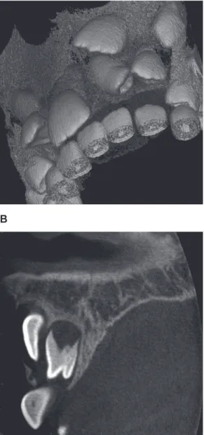
A
B
Figure 1 Representative example of a cone beam computed tomography
(CBCT) scan exhibiting a conically shaped supernumerary tooth in the anterior maxilla (A: volume rendered image; B: axial CBCT image).
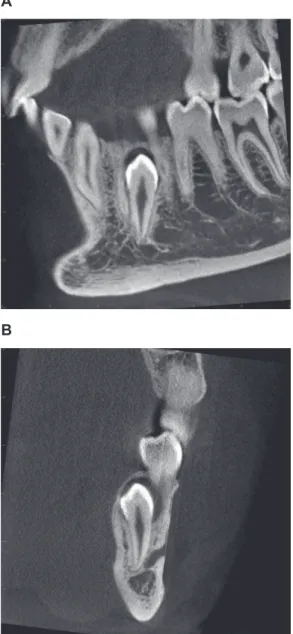
A
B
Figure 2 Representative example of a cone beam computed tomography
(CBCT) scan exhibiting a tuberculate type of supernumerary tooth in the anterior maxilla (A: volume rendered image; B: sagittal CBCT image).
to the inability to fit the random effects models on the present data, the association between follicular width and root prox-imity on root resorption were calculated using unconditional logistic regression. Chi-square tests accounting for clustered data were used to evaluate the association between the other variables of interest and root resorption of adjacent teeth. All statistical tests were performed using STATA 13.1 (Stata Corp., College Station, Texas, USA).
Results
Patients and sample characteristics
Of the 82 patients included, 51 (62.2 per cent) were male and 31 (37.8 per cent) were female, resulting in a gender ratio
of 1.65:1. The age of the patients ranged from 6 to 72 years with a mean age of 15.8 years. Most of the patients were 20 years or younger with 23 patients aged 6–10 years (28.05 per cent), 46 patients aged 10–20 years (56.10 per cent), and only 13 patients older than 20 years (15.85 per cent).
In the 82 patients included, a total of 101 supernumer-ary teeth were diagnosed resulting in an average number of 1.23 teeth per person. Two-thirds of the supernumerary teeth (67/101, 66.34 per cent) were found in males and 34 in females (33.66 per cent). Most of the patients had one supernumerary tooth (66/82, 80.5 per cent), 13 patients (15.8 per cent) had two supernumeraries, and 3 patients (3.7 per cent) had three. The mesiodentes were the most frequently diagnosed type of super-numerary tooth (49/101, 48.52 per cent), followed by supernu-merary premolars (24/101, 23.76 per cent) and supernusupernu-merary lateral incisors (19/101, 18.81 per cent). Of the 35 multiple supernumerary teeth, 42.9 per cent (15 teeth) were mesi-odentes and 40 per cent (14 teeth) were premolars. Qualitative and quantitative data are presented in detail in Tables 1 and 2.
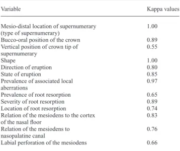
Interrater agreement
The Cohen’s kappa for interrater agreement ranged from 0.55 to 1 (Table 3). The highest kappa values (1) were found for type and shape of the supernumerary teeth.
Descriptive analysis of mesiodentes
Of the 49 diagnosed mesiodentes, most had a conical (34/49, 69.4 per cent) or tuberculate shape (13/49, 26.5 per cent;
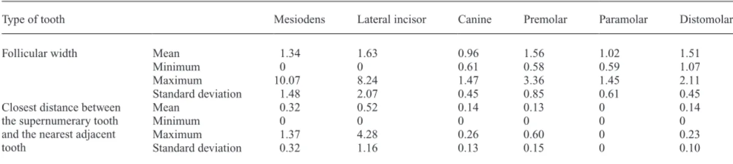
Table 1). The position of the tooth was normal or inclined in 53 per cent (26 teeth) and inverted in 36.75 per cent (18 teeth). The analysis of the 3D location revealed that most mesiodentes were located palatally or within the alveolar arch (47/49, 95.9 per cent). In most cases, the vertical loca-tion of the mesiodens cusp tip in relaloca-tion to the long axis of the closest erupted adjacent tooth was in the cervical third of the root (19/49, 38.8 per cent). More than two-thirds of the mesiodentes (33/49, 67.3 per cent) exhibited local aber-rations of the permanent dentition such as inclusion of the permanent neighbouring tooth or dental malposition and the mean follicular width was 1.34 mm (Table 2). Of the 49 mesiodentes, 10 (20.5 per cent) were in contact with the cortical bone of the nasal floor (Supplementary figure 1) and 24 (49 per cent) with the nasopalatine canal (Table 4 and
Supplementary figure 2). In seven cases, the mesiodentes were in close relation to the labial cortical plate or exhibited a labial perforation. In most cases (38/49, 77.6 per cent), the mesiodentes were located 0.5 mm or less from the roots of the neighbouring central incisors (mean distance: 0.32 mm).
Descriptive analysis of supernumerary lateral incisors
Of the 19 supernumerary lateral incisors, 13 (68.4 per cent) were located in the maxilla (Table 1). The shape and position of the teeth were distributed evenly within the different cat-egories. As with the mesiodentes, most of the supernumerary
A
B
Figure 3 Representative example of a cone beam computed tomography
(CBCT) scan exhibiting a supplemental type of supernumerary teeth in the left posterior mandible (A: sagittal CBCT image; B: coronal CBCT image).
lateral incisors (18/19, 94.7 per cent) were located palatally or within the alveolar arch. The cusp of the supernumerary incisors was mostly located in the middle third of the root of the closest adjacent tooth (7/19, 36.8 per cent), and two lateral incisors (10.5 per cent) were erupted. Of the 19 supernumer-ary lateral incisors, 14 (73.7 per cent) were associated with local aberrations of the permanent dentition. The mean follic-ular width was 1.63 mm (Table 2) and a follicular enlargement (greater than 3 mm) was present in two cases (10.5 per cent).
Descriptive analysis of supernumerary premolars
Of the 24 supernumerary premolars, 20 (83.3 per cent) were located in the mandible (Table 1). Most of them were of the supplemental type (17/24, 70.8 per cent), and they all had a normal or inclined position of eruption. The crown was
usually located orally (19/24, 79.2 per cent), whereas the roots were often located in the middle of the arch. Vertically, the supernumerary cusp tip was mostly located in the mid-dle third of the root of the adjacent tooth (14/24, 58.3 per cent). All the supernumerary premolars were located at or less than 0.6 mm away from the closest adjacent tooth (mean distance of 0.13 mm; Table 2). Associated local aberrations were diagnosed for six teeth (25.1 per cent). The mean fol-licular width was 1.56 mm and a folfol-licular enlargement was detected in only one of the cases (4.2 per cent).
Descriptive analysis of supernumerary canines, paramolars, and distomolars
The supernumerary canines (three teeth) were all located in the mandible, the paramolars (two teeth) in the maxilla, and
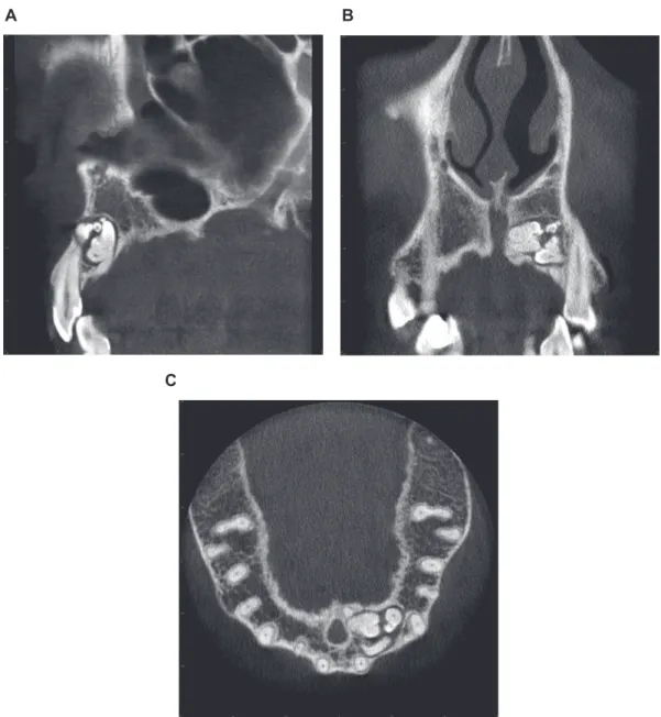
A B
C
Figure 4 Representative example of a cone beam computed tomography (CBCT) scan exhibiting an odontoma type of supernumerary tooth in the anterior
the distomolars (four teeth) were evenly distributed in both jaws. For further details, see Tables 1 and 2.
Descriptive analysis of root resorption of neighbouring permanent teeth
Root resorption of the adjacent teeth was detected for 22.8 per cent (23 teeth) of the supernumerary teeth. Root resorp-tion of the adjacent teeth was most frequent in supernumer-ary premolars (62.5 per cent; Tables 1 and 5). Four premolar supernumeraries resorbed two adjacent teeth simultane-ously. For mesiodentes, root resorption was detected in five cases (10.2 per cent), and for the 19 supernumerary lat-eral incisors, in three cases (15.8 per cent). No resorption of adjacent teeth was detected for supernumerary canines, paramolars, and distomolars.
Association of root resorption with patient and CBCT data
Logistic regression showed no association between patient age (P = 0.72) and root resorption. Some evidence of an association between root resorption and proximity of the supernumerary tooth with the adjacent tooth [odds ratio (OR): 0.01; 95 per cent confidence interval (CI): 0.0, 0.56;
P = 0.02) and follicular width (OR: 2.12; 95 per cent CI:
1.16, 3.38; P = 0.02) was observed. However, results from the unconditional logistic regression should be interpreted with caution, as standard errors are likely to be underes-timated. The chi-square tests showed a statistically sig-nificant association between root resorption and type of supernumerary teeth (P = 0.001) with most resorptions found for supernumerary premolars. There was also a
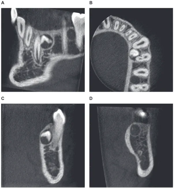
A B
C D
Figure 5 Representative example of a cone beam computed tomography (CBCT) scan exhibiting two supernumerary teeth classified as developing tooth
buds in the left posterior mandible (between canine and first premolar, and second premolar and first molar; A: sagittal CBCT image; B: axial CBCT image; C: coronal CBCT image between canine and first premolar; D: coronal CBCT image between second premolar and first molar).
Table 2 Quantitative data (in millimetres) regarding follicular width of supernumerary teeth and the distance between the
supernumerary tooth to the closest adjacent tooth in the maxilla and mandible in 82 patients.
Type of tooth Mesiodens Lateral incisor Canine Premolar Paramolar Distomolar
Follicular width Mean 1.34 1.63 0.96 1.56 1.02 1.51
Minimum 0 0 0.61 0.58 0.59 1.07
Maximum 10.07 8.24 1.47 3.36 1.45 2.11
Standard deviation 1.48 2.07 0.45 0.85 0.61 0.45
Closest distance between the supernumerary tooth and the nearest adjacent tooth
Mean 0.32 0.52 0.14 0.13 0 0.14
Minimum 0 0 0 0 0 0
Maximum 1.37 4.28 0.26 0.60 0 0.23
Standard deviation 0.32 1.16 0.13 0.15 0 0.10
Table 1 Descriptive data regarding location, morphology, and associated complications of supernumerary teeth in the maxilla and
mandible in 82 patients.
Type of tooth Mesiodens Lateral incisor Canine Premolar Paramolar Distomolar
Number 49 19 3 24 2 4
Mean age of patients 12.38 19.3 13.89 18.99 12.84 25.92
Jaw Maxilla 49 (100%) 13 (68.4%) 0 4 (16.7%) 2 (100%) 2 (50%)
Mandible 0 6 (31.6%) 3 (100%) 20 (83.3%) 0 2 (50%)
Shape Developing tooth bud 0 2 (10.5%) 1 (33.3%) 5 (20.8%) 0 0
Conical 34 (69.4%) 6 (31.6%) 0 1 (4.2%) 1 (50%) 1 (25%) Tuberculate 13 (26.5%) 4 (21.05%) 0 1 (4.2%) 0 1 (25%) Supplemental 1 (2.05%) 3 (15.8%) 1 (33.3%) 17 (70.8%) 0 1 (25%) Odontoma 1 (2.05%) 4 (21.05%) 1 (33.3%) 0 1 (50%) 1 (25%) Position/inclination Normal 13 (26.5%) 3 (15.8%) 1 (33.3%) 9 (37.5%) 1 (50%) 1 (25%) Inclined 13 (26.5%) 4 (21.05%) 1 (33.3%) 15 (62.5%) 0 1 (25%) Transverse/horizontal 4 (8.2%) 3 (15.8%) 1 (33.3%) 0 0 1 (25%) Inverted 18 (36.75%) 5 (26.3%) 0 0 0 0 Indefinable 1 (2.05%) 4 (21.05%) 0 0 1 (50%) 1 (25%) Bucco-oral position of crown Labial/buccal 2 (4.1%) 1 (5.3%) 1 (33.3%) 1 (4.2%) 1 (50%) 0 Within arch/median 23 (46.9%) 10 (52.6%) 0 4 (16.7%) 1 (50%) 3 (75%) Oral (palatal/lingual) 24 (49%) 8 (42.1%) 2 (66.7%) 19 (79.1%) 0 1 (25%)
Vertical position of the tip of the supernumerary tooth (compared with the closest erupted adjacent tooth)
Apical to the adjacent teeth
10 (20.4%) 1 (5.3%) 0 0 0 0
Apical third of the root of the adjacent tooth
10 (20.4%) 4 (21.05%) 0 1 (4.2%) 0 0
Middle third of the root of the adjacent tooth
5 (10.2%) 7 (36.8%) 1 (33.3%) 14 (58.3%) 0 2 (50%)
Cervical third of the root of the adjacent tooth
19 (38.8%) 4 (21.05%) 2 (66.7%) 6 (25%) 2 (100%) 2 (50%)
Coronal region of the adjacent tooth
5 (10.2%) 3 (15.8%) 0 3 (12.5%) 0 0
Proximity ≤0.5 mm to adjacent
tooth 38 (77.6%) 17 (89.5%) 3 (100%) 23 (95.8%) 2 (100%) 4 (100%)
State of eruption Impacted 45 (91.8%) 17 (89.5%) 3 (100%) 23 (95.8%) 2 (100%) 4 (100%)
Erupted 4 (8.2%) 2 (10.5%) 0 1 (4.2%) 0 0 Associated local aberrations/complications Asymptomatic 16 (32.7%) 5 (26.3%) 0 18 (74.9%) 0 2 (50%) Inclusion of perma-nent teeth 15 (30.6%) 6 (31.6%) 0 1 (4.2%) 2 (100%) 2 (50%) Dental malposition 18 (36.7%) 8 (42.1%) 3 (100%) 4 (16.7%) 0 0 Delayed/abnormal root development of permanent incisor 0 0 0 0 0 0 Other 1* (2.05%) 0 0 1** (4.2%) 0 0
Follicle size Follicular width
>3 mm 2 (4.1%) 2 (10.5%) 0 1 (4.2%) 0 0
Root resorption Yes 5 (10.2%) 3 (15.8%) 0 15 (62.5%) 0 0
*Geminated 21 (one patient who also had dental malposition). **Only three lower incisors.
statistically significant association between the shape of the supernumerary tooth and root resorption (P = 0.01) with resorptions being more frequent in the proximity of supplemental supernumeraries. No significant association was found between root resorption and gender (P = 0.43), 3D location of the supernumerary teeth in the vertical (P = 0.17) and oro-buccal plane (P = 0.74), and associated local aberration (P = 0.08).
Discussion
This study resulted in an almost perfect interrater agree-ment (kappa values) with regards to the type (1) and shape of the supernumerary teeth (1), and the prevalence of asso-ciated local aberrations (0.97; Landis and Koch, 1977). Furthermore, there was a high agreement between the two orthodontists in assessing the location, prevalence, and
the severity of root resorption on adjacent teeth. This was similar to results reported by Lai and co-workers (2013b)
for root resorption of adjacent teeth in the anterior maxilla caused by impacted canines.
In this study, the male:female ratio was 1.61:1 for super-numerary teeth, a value that corroborates the gender ratios previously reported (Rajab and Hamdan, 2002; Fernández Montenegro et al., 2006; Gündüz et al., 2008; Wang and Fan, 2011) but lower than the 2.6:1 (von Arx, 1990) and 2.75:1 (Schmuckli et al., 2010) ratios reported previously for Switzerland. In the present study, 16 patients (out of 82) had two or more supernumerary teeth, which is in accordance with previous studies (von Arx, 1990; Rajab and Hamdan, 2002;
Fernández Montenegro et al., 2006; Liu et al., 2007; Gündüz
et al., 2008; Ferrés-Padró et al., 2009; Hyun et al., 2009). In the current investigation, 61.4 per cent of the supernumer-ary teeth were located in the anterior maxilla. Several studies have reported higher (Liu et al., 2007/92 per cent; Rajab and Hamdan, 2002/89.6 per cent; Schmuckli et al., 2010/86 per cent; Ferrés-Padró et al., 2009/72.2 per cent) and two lower percentages (Fernández Montenegro et al., 2006/48.3 per cent; Salcido-García et al., 2004/46.8 per cent). Nevertheless, it has to be taken into consideration that the population inves-tigated in the present study was specifically referred for fur-ther diagnosis and treatment of supernumerary teeth, which may explain some of the differences found.
Supernumeraries appeared in a variety of shapes, with conical being the most common in this study (42.6 per cent). This confirms data reported in other studies (Rajab and Hamdan, 2002; Liu et al., 2007; Gündüz et al., 2008;
Ferrés-Padró et al., 2009; Hyun et al., 2009; Schmuckli
et al., 2010). In the present study, approximately one-third of the supernumeraries in the anterior maxilla were inverted, which was similar to findings from other studies (Koch
et al., 1986; Liu et al., 2007; Gündüz et al., 2008). On the contrary, Rajab and Hamdan (2002) reported that most supernumerary teeth were normally oriented. A significant number of mesiodentes were located in close contact to the cortex of the nasal floor (20.5 per cent) or the nasopalatine canal (49 per cent) and within a 0.5 mm distance to the roots of one of the central incisors (77.6 per cent). These find-ings underline the importance of a thorough 3D analysis of supernumerary teeth for treatment planning to decide upon the appropriate surgical approach prior to removal.
In this study, approximately two-thirds (67.3 per cent) of the mesiodentes were associated with local aberrations of the den-tition such as dental malposition or impaction of permanent teeth. Higher (77.7–80.9 per cent; Tay et al., 1984; Gündüz
et al., 2008) and lower (46.9–59 per cent; von Arx, 1990; Liu
et al., 2007; Hyun et al., 2009) percentages have previously been reported in the literature. These findings demonstrate that in approximately one-third of the patients, supernumerary teeth are an incidental finding during a routine radiographic examination without clinical symptoms. This also explains the wide range of patient ages (6–72 years) included.
Table 3 Interrater agreement between the two experienced
orthodontists using Cohen’s kappa values.
Variable Kappa values
Mesio-distal location of supernumerary (type of supernumerary)
1.00
Bucco-oral position of the crown 0.89
Vertical position of crown tip of supernumerary
0.55
Shape 1.00
Direction of eruption 0.80
State of eruption 0.85
Prevalence of associated local aberrations
0.97
Prevalence of root resorption 0.65
Severity of root resorption 0.89
Location of root resorption 0.74
Relation of the mesiodens to the cortex of the nasal floor
0.83 Relation of the mesiodens to
nasopalatine canal
0.76
Labial perforation of the mesiodens 0.66
Kappa values: no agreement, <0; slight, 0–0.2; fair, 0.21–0.40; moderate, 0.41–0.60; substantial, 0.61–0.80; almost perfect, 0.81–1 (Landis and Koch, 1977).
Table 4 Detailed descriptive analysis of three-dimensional
location and position of the 49 mesiodentes.
Relation to the cortical bone of the nasal floor
No contact 39 (79.5%)
In contact with cortical bone 4 (8.2%) Partially perforated cortical bone 4 (8.2%) Located within the cortical bone 2 (4.1%) Relation to
nasopalatine canal No contactIn contact with the canal 25 (51%)19 (38.8%) Partially perforated the canal 4 (8.2%)
Located within the canal 1 (2%)
Relation to labial/ buccal bone of the alveolar process
No contact 42 (85.7%)
In contact with labial cortical plate 3 (6.1%)
Surgical removal of a supernumerary tooth is indicated when eruption of the adjacent tooth has been delayed or inhibited, when altered eruption or displacement of the adjacent tooth is evident, when the supernumerary tooth interferes with active orthodontic treatment, if associated pathology (neighbouring root resoprtion, cyst formation, etc.) exists, or if spontaneous eruption of the supernu-merary has occurred (Garvey et al., 1999). The ideal time point for removal, particularly in the premaxillary region, remains controversial. Whereas some authors recommend immediate removal of the supernumerary teeth following diagnosis (Primosch, 1981; Nazif et al., 1983; Mason et al., 2000), others prefer to postpone surgery until the age of at least 8–10 years, when the root development of the central and lateral incisor is nearly complete in order to minimize injury to those teeth (Koch et al., 1986). In some instances, unerupted and asymptomatic supernumerary teeth are left in place and kept under clinical and radiographic obser-vation (Kurol, 2006). Garvey and co-workers (1999) rec-ommended monitoring of supernumerary teeth without surgical removal where satisfactory eruption of the neigh-bouring teeth has occurred, no active orthodontic treatment is planned, no associated pathology is diagnosed, and where removal would risk damaging teeth in the vicinity.
In the present investigation, root resorption of adjacent teeth was detected for 22.8 per cent of the supernumerary teeth. A much lower frequency of root resorption of adja-cent teeth (1.6 per adja-cent) was reported in a study from China evaluating CBCT scans (Liu et al., 2007). In contrast to the present study, root resorption was not a primary out-come variable of this study, and no information was given on the extent of resorption. Therefore, this study could
have omitted slight or moderate root resorption, which could at least in part explain the differences in the percent-ages reported. Studies evaluating panoramic views for root resorption caused by mesiodentes reported resorption rates between 4.7 (Gündüz et al., 2008) and 7.6 per cent (Hyun
et al., 2009). Tyrologou and co-workers (2005) even reported no resorption found. However, these studies only looked at mesiodentes, where root resorption of adjacent teeth seems to be less frequent. Furthermore, the reported incidence of root resorption also depends on the radiographic imaging method used. 2D radiographs have been demonstrated to be inaccurate for diagnosing root resorption and overlook-ing pathosis in 50 per cent of the cases (Ericson and Kurol, 1987; Heimisdottir et al., 2005; Botticelli et al., 2011;
Alqerban et al., 2011a).
By using 3D visualization, CBCT provides information in all three planes and thus enhances diagnostic accuracy (Becker et al., 2010; Pazera et al., 2011; Lai et al., 2013b), which is quite an important parameter when considering sur-gical removal (Becker et al., 2010). When taking into account the proximity of supernumerary teeth to vital anatomical structures such as the nasal floor, the nasopalatine canal, or the floor of the mouth, the need for accurate diagnostic 3D information prior to surgical and interdisciplinary interven-tions becomes evident. Recent studies comparing traditional radiography with CBCT concluded that 3D imaging provides more information on the presence and severity of root resorp-tion (Alqerban et al., 2009; Katheria et al., 2010; Botticelli
et al., 2011; Alqerban et al., 2011a; Lai et al., 2013a). Another study (Haney et al., 2010) reported that orthodontists had a substantially different perception of localization and sever-ity of root damage, and a significantly higher confidence in
Table 5 Descriptive data regarding location and extent of root resorption.
Type of tooth Mesiodens Lateral incisor Premolar
Number of supernumerary causing resorption
5 3 15
Number of supernumerary causing resorption on two adjacent teeth
1 (20%) 0 4 (26.7%)
Total of resorbed adjacent teeth
6 3 19
Direct contact between supernumerary and adjacent tooth at resorption location
3 (60%) 2 (66.6%) 15 (100%)
Extent of resorption Slight (up to half of the
dentine thickness to the pulp) 4 (66.7%) 0 16 (84.2%)
Moderate (resorption midway to the pulp or more, the pulp lining being unbroken)
2 (33.3%) 1 (33.3%) 3 (15.8%)
Severe (the pulp is exposed by
the resorption) 0 2 (66.7%) 0
Location of resorption Apical tip of the root 0 1 (33.3%) 2 (10.5%)
Apical third of the root 3 (50%) 0 8 (42.1%)
Middle third of the root 1 (16.7%) 1 (33.3%) 8 (42.1%)
Coronal third of the root 2 (33.3%) 0 1 (5.3%)
diagnosis and treatment planning using CBCT images com-pared with conventional radiographs alone. A recent in vitro study on human skulls has shown no significant differences between different CBCT systems for assessing the severity of root resorptions (Alqerban et al., 2011b).
The aetiology of root resorption following ectopic eruption of a tooth is still unclear. In earlier studies, it has been postulated that an enlarged dental follicle, as well as the pressure caused by an erupting tooth, may be responsible for root resorption of adjacent teeth (Bidwell
et al. 1995; Marks et al., 1997). However, Ericson and co-workers (2001) have concluded, based on CT exami-nations of impacted canines, that it is probably the direct physical contact of the ectopic canine with the permanent adjacent tooth and the direct pressure from the canine as a part of the eruption process that cause root resorption, and not the dental follicle. In the present study, there was some evidence of an association between enlarged follicles and supernumerary proximity to adjacent teeth and the occurrence of root resorption. However, these results should be interpreted with caution due to small sample size.
There was no association between age or gender of the patient and risk of root resorption. Root resorption showed a statistically significant association with the type of super-numerary tooth with root resorption being more prevalent in supernumerary premolars (15 out of 24). This could be explained by the fact that most premolar supernumeraries were found in the lower jaw (83.3 per cent), where they are often in close contact with adjacent teeth due to the anatomy of the mandible. The association between shape of the supernumer-ary tooth and root resorption is probably partially due to the fact that supernumerary premolars had predominantly a sup-plemental shape. In one-third of the cases, dental malposition of adjacent permanent teeth was detected when root resorption was present (8 out of 23 supernumerary teeth). On the other hand, root resorption was never detected in cases where the supernumerary teeth caused the inclusion of the adjacent tooth probably because in these cases the supernumerary teeth were mostly located coronally to the permanent tooth.
Conclusions
On the basis of the data from the present study, the follow-ing can be concluded:
• Supernumerary teeth occur alone or in multiple in any region of the jaws. They are most often located in the anterior maxilla (mesiodentes) and are conical in shape. • CBCT provides 3D information with regards to the type,
shape, and position of the supernumerary tooth as well as local aberrations and root resorption of adjacent per-manent teeth with moderate to high interrater correlation. • Root resorption of adjacent teeth is most likely to be
diag-nosed in the premolar region.
• Root resorption of adjacent teeth was associated to tooth type (premolars) and shape (supplemental) of supernu-merary teeth.
• A significant number of mesiodentes were located either in close contact to the cortical bone of the nasal floor, the nasopalatine canal, and the roots of one of the adjacent central incisors. Therefore, a radiographic 3D analysis may be important for exact localization and treatment planning including surgical removal of mesiodentes.
Supplementary material
Supplementary material is available at European Journal of Orthodontics online.
References
Alqerban A, Jacobs R, Fieuws S, Willems G 2011a Comparison of two cone beam computed tomographic systems versus panoramic imaging for localization of impacted maxillary canines and detection of root resorption. European Journal of Orthodontics 33: 93–102
Alqerban A, Jacobs R, Souza P C, Willems G 2009 In-vitro comparison of 2 cone-beam computed tomography systems and panoramic imaging for detecting simulated canine impaction-induced external root resorp-tion in maxillary lateral incisors. American Journal of Orthodontics and Dentofacial Orthopedics 136: 764.e1–e11
Alqerban A, Jacobs R, Fieuws S, Nackaerts O, The SEDENTEXCT Project Consortium, Willems G 2011b Comparison of 6 cone-beam computed tomography systems for image quality and detection of simulated canine impaction-induced external root resorption in maxillary lateral incisors. American Journal of Orthodontics and Dentofacial Orthopedics 140: e129–e139
Bäckman B, Wahlin Y B 2001 Variations in number and morphology of permanent teeth in 7-year-old Swedish children. International Journal of Paediatric Dentistry 11: 11–17
Becker A, Chaushu S, Casap-Caspi N 2010 Cone-beam computed tomog-raphy and the orthosurgical management of impacted teeth. Journal of the American Dental Association 141: 14S–18S
Bidwell, J P, Fey E G, Marks S C Jr 1995 Nuclear matrix-intermediate fila-ment proteins of the dental follicle/enamel epithelium and their changes during tooth eruption in dogs. Archives of Oral Biology 40: 1047–1051 Botticelli S, Verna C, Cattaneo P M, Heidmann J, Melsen B 2011 Two-
versus three-dimensional imaging in subjects with unerupted maxillary canines. European Journal of Orthodontics 33: 344–349
Chen Y H, Cheng N C, Wang Y B, Yang C Y 2010 Prevalence of congenital dental anomalies in the primary dentition in Taiwan. Pediatric Dentistry 32: 525–529
Ericson S, Bjerklin K 2001 The dental follicle in normally and ectopi-cally erupting maxillary canines: a computed tomography study. Angle Orthodontist 71: 333–342
Ericson S, Kurol J 1987 Radiographic examination of ectopically erupting maxillary canines. American Journal of Orthodontics and Dentofacial Orthopedics 91: 483–492
Ericson S, Kurol J 2000 Resorption of incisors after ectopic eruption of maxillary canines: a CT study. Angle Orthodontist 70: 415–423 Ericson S, Bjerklin K, Falahat B 2001 Does the canine dental follicle cause
resorption of permanent incisor roots? A computed tomographic study of erupting maxillary canines. Angle Orthodontist 72: 95–104
Fardi A, Kondylidou-Sidira A, Bachour Z, Parisis N, Tsirlis A 2011 Incidence of impacted and supernumerary teeth-a radiographic study in a North Greek population. Medicina Oral Patologia Oral y Cirugia Bucal 16: e56–e61
Fernández Montenegro P, Valmaseda Castellón E, Berini Aytés L, Gay Escoda C 2006 Retrospective study of 145 supernumerary teeth. Medicina Oral Patologia Oral y Cirugia Bucal 11: 339–344
Ferrés-Padró E, Prats-Armengol J, Ferrés-Amat E 2009 A descriptive study of 113 unerupted supernumerary teeth in 79 pediatric patients in Barcelona. Medicina Oral Patologia Oral y Cirugia Bucal 14: 146–152 Garvey T, Barry H J, Blake M 1999 Supernumerary teeth - an overview
of classification, diagnosis and management. Journal of the Canadian Dental Association 65: 612–616
Gündüz K, Celenk P, Zengin Z, Sümer P 2008 Mesiodens: a radiographic study in children. Journal of Oral Science 50: 287–291
Gurgel C V et al. 2012 Cone beam computed tomography for diagnosis and treatment planning of supernumerary teeth. General Dentistry 60: e131–e135
Haney E, Gansky S A, Lee J S, Johnson E, Maki K, Miller A J, Huang J C 2010 Comparative analysis of traditional radiographs and cone-beam computed tomography volumetric images in the diagnosis and treatment planning of maxillary impacted canines. American Journal of Orthodontics and Dentofacial Orthopedics 137: 590–597
Heimisdottir K, Bosshardt D, Ruf S 2005 Can the severity of root resorp-tion be accurately judged by means of radiographs? A case report with histology. American Journal of Orthodontics and Dentofacial Orthopedics 128: 106–109
Hyun H K, Lee S J, Lee S H, Hahn S H, Kim J W 2009 Clinical characteris-tics and complications associated with mesiodentes. Journal of Oral and Maxillofacial Surgery 67: 2639–2643
Järvinen S, Lehtinen L 1981 Supernumerary and congenitally miss-ing primary teeth in Finnish children. An epidemiologic study. Acta Odontologica Scandinavica 39: 83–86
Kapila S, Conley R S, Harrell W E Jr 2011 The current status of cone beam computed tomography imaging in orthodontics. Dentomaxillofacial Radiology 40: 24–34
Katheria B C, Kau C H, Tate R, Chen J W, English J, Bouquot J 2010 Effectiveness of impacted and supernumerary tooth diagnosis from tra-ditional radiography versus cone beam computed tomography. Pediatric Dentistry 32: 304–309
Koch H, Schwartz O, Klausen B 1986 Indications for surgical removal of supernumerary teeth in the premaxilla. International Journal of Oral and Maxillofacial Surgery 15: 273–281
Kurol J 2006 Impacted and ankylosed teeth: why, when, and how to inter-vene. American Journal of Orthodontics and Dentofacial Orthopedics 129: S86–S90
Lai C S, Suter V G A, Katsaros C, Bornstein M M 2013a Localization of impacted maxillary canines and root resorption of neighbouring teeth: a study assessing the diagnostic value of panoramic radiographs in two groups of observers. European Journal of Orthodontics 36: 450–456 Lai C S, Bornstein M M, Mock L, Heuberger B M, Dietrich T, Katsaros C
2013b Impacted maxillary canines and root resorptions of neighbouring teeth: a radiographic analysis using cone-beam computed tomography. European Journal of Orthodontics 35: 529–538
Landis J R, Koch G G 1977 The measurement of observer agreement for categorical data. Biometrics 33: 159–174
Leco Berrocal M, Martín Morales J F, Martínez González J M 2007 An observational study of the frequency of supernumerary teeth in a popu-lation of 2000 patients. Medicina Oral Patologia Oral y Cirugia Bucal 12: E134–E138
Liu D G, Zhang W L, Zhang Z Y, Wu Y T, Ma X C 2007 Three-dimensional evaluations of supernumerary teeth using cone-beam computed tomog-raphy for 487 cases. Oral Surgery, Oral Medicine, Oral Patholology, Oral Radiology, and Endodontology 103: 403–411
Luten J R Jr 1967 The prevalence of supernumerary teeth in primary and mixed dentitions. Journal of Dentistry for Children 34: 346–353 Magnússon T E 1984 Hypodontia, hyperodontia, and double formation of
primary teeth in Iceland. An epidemiological study. Acta Odontologica Scandinavica 42: 137–139
Marks S C, Schroeder H E, Andreasen J O 1997 Theories and mechanism of tooth eruption. In: Andreasen J O, Kölsen-Pedersen J, Laskin D M (eds). Textbook and color atlas of tooth impactions. Mosby, St Louis, pp. 20–65 Mason C, Azam N, Holt R D, Rule D C 2000 A retrospective study of
unerupted maxillary incisors associated with supernumerary teeth. British Journal of Oral and Maxillofacial Surgery 38: 62–65
Nazif M M, Ruffalo R C, Zullo T 1983 Impacted supernumerary teeth: a sur-vey of 50 cases. Journal of the American Dental Association 106: 201–204 Pazera P, Bornstein M M, Pazera A, Sendi P, Katsaros C 2011 Incidental
maxillary sinus findings in orthodontic patients: a radiographic analy-sis using cone-beam computed tomography (CBCT). Orthodontics & Craniofacial Research 14: 17–24
Primosch R E 1981 Anterior supernumerary teeth--assessment and surgi-cal intervention in children. Pediatric Dentistry 3: 204–215
Rajab L D, Hamdan M A 2002 Supernumerary teeth: review of the lit-erature and a survey of 152 cases. International Journal of Paediatric Dentistry 12: 244–254
Ravn J J 1971 Aplasia, supernumerary teeth and fused teeth in the pri-mary dentition. An epidemiologic study. Scandinavian Journal of Dental Research 79: 1–6
Russell K A, Folwarczna M A 2003 Mesiodens--diagnosis and manage-ment of a common supernumerary tooth. Journal of the Canadian Dental Association 69: 362–366
Salcido-García J F, Ledesma-Montes C, Hernández-Flores F, Pérez D, Garcés-Ortíz M 2004 Frequency of supernumerary teeth in Mexican population. Medicina Oral Patologia Oral y Cirugia Bucal 9: 407–409 Schmuckli R, Lipowsky C, Peltomäki T 2010 Prevalence and
morphol-ogy of supernumerary teeth in the population of a Swiss community. Short communication. Schweizer Monatsschrift für Zahnmedizin 120: 987–993
Skrinjarić I, Barac-Furtinović V 1991 Anomalies of deciduous teeth and findings in permanent dentition. Acta Stomatologica Croatica 25: 151–156
Stellzig A, Basdra E K, Komposch G 1997 Mesiodentes: incidence, mor-phology, etiology. Journal of Orofacial Orthopedics 58: 144–153 Tay F, Pang A, Yuen S 1984 Unerupted maxillary anterior supernumerary
teeth: report of 204 cases. ASDC Journal of Dentistry for Children 51: 289–294
Tyrologou S, Koch G, Kurol J 2005 Location, complications and treat-ment of mesiodentes--a retrospective study in children. Swedish Dental Journal 29: 1–9
von Arx T 1990 Mesiodens. Klinische, radiologische und chirurgisch-therapeutische Aspekte. Schweizer Monatsschrift für Zahnmedizin 100: 433–442
Walker L, Enciso R, Mah J 2005 Three-dimensional localization of maxil-lary canines with cone-beam computed tomography. American Journal of Orthodontics and Dentofacial Orthopedics 128: 418–423
Wang X P, Fan J 2011 Molecular Genetics of Supernumerary tooth forma-tion. Genesis 49: 261–277
Yagüe-García J, Berini-Aytés L, Gay-Escoda C 2009 Multiple supernumer-ary teeth not associated with complex syndromes: a retrospective study. Medicina Oral Patologia Oral y Cirugia Bucal 14: E331–E336 Yonezu T, Hayashi Y, Sasaki J, Machida Y 1997 Prevalence of congenital
dental anomalies of the deciduous dentition in Japanese children. The Bulletin of Tokyo Dental College 38: 27–32