Differential effects of tissue plasminogen activator
and streptokinase on infarct size and on rate of
enzyme release: influence of early infarct related
artery patency
The GUSTO Enzyme Substudy
T. Baardman, W. Th. Hermens*, T. Lenderink, G. P. Molhoekt, G. Grollier±,
M. Pfisterer§ and M. L. Simoons
Thoraxcenter, Erasmus University Rotterdam, * Cardiovascular Research Institute Maastricht, Maastricht, \Medhch Spectrum Twente, Enschede, The Netherlands, \Centre Hospitalier Universitaire, Caen, France and
§ University Clinics, Basel, Switzerland
Background The recent international GUSTO trial of 41 021 patients with acute myocardial infarction demon-strated improved 90-min infarct related artery patency as well as reduced mortality in patients treated with an accel-erated regimen of tissue plasminogen activator, compared to patients treated with streptokinase. A regimen combin-ing tissue plasminogen activator and streptokinase yielded intermediate results. The present study investigated the effects of treatment on infarct size and enzyme release kinetics in a subgroup of these patients.
Methods A total of 553 patients from 15 hospitals were enrolled in the study. Four thrombolytic strategies were compared: streptokinase with subcutaneous heparin, strep-tokinase with intravenous (i.v.) heparin, tissue plasminogen activator with i.v. heparin, and streptokinase plus tissue plasminogen activator with i.v. heparin. The activity of alpha-hydroxybutyrate dehydrogenase (HBDH) in plasma was centrally analysed and infarct size was defined as cumulative HBDH release per litre of plasma within 72 h of the first symptoms (Q(72)). Patency of the infarct-related vessel was determined by angiography in 159 patients, 90 min after treatment.
Results Infarct size was 3-72 g-eq. 1- 1 in patients with
adequate coronary perfusion (TIMI-3) at the 90 min
angi-ogram and larger in patients with TIMI-2 (4-35 g-eq . 1 ') or TIMI 0-1 (507 g-eq . 1" ') flow (P=0-024). In this subset of the GUSTO angiographic study, early coronary patency rates (TIMI 2 + 3) were similar in the two streptokinase groups (53 and 46%). Higher, but similar, patency rates were observed in the tissue plasminogen activator and combination therapy groups (87 and 90%). Median infarct size for the four treatment groups, expressed in gram-equivalents (g-eq) of myocardium, was 4-4, 4-5, 3-9 and 3-9 g-eq per litre of plasma (P=0-04 for streptokinase vs tissue plasminogen activator). Six hours after the first symptoms, respectively 5-3, 6-6, 140 and 13-6% of total HBDH release was complete (/><00001 for streptokinase vs tissue plasminogen activator).
Conclusions Rapid and complete coronary reperfusion salvages myocardial tissue, resulting in limitation of infarct size and accelerated release of proteins from the myo-cardium. Treatment with tissue plasminogen activator, re-sulting in earlier reperfusion was more effective in reducing infarct size than the streptokinase regimens, which con-tributes to the differences in survival between treatment groups in the GUSTO trial.
(Eur Heart J 1996; 17: 237-246)
Key Words: Thrombolysis, infarct size, enzymes, GUSTO.
Revision submitted 7 July 1995, and accepted 7 August 1995. Correspondence: Maarten L. Simoons, MD, Thoraxcenter, BD 434, Erasmus University/Dijkzigt Hospital, Dr. Molewaterplein 40, 3015 GD Rotterdam, The Netherlands.
Introduction
Prognosis of patients with myocardial infarction is markedly improved by timely reperfusion of the ischaemic myocardium. The GUSTO trial recently dem-onstrated differences in outcome from four treatment
regimens1'1, which could be explained by different rates of early coronary reperfusion, varying between 81% for accelerated tissue plasminogen activator with i.v. heparin and 54 and 60% for streptokinase with subcuta-neous or intravenous heparin'21. Early reperfusion of the ischaemic myocardium allows recovery and salvage of myocytes and preserves left ventricular function'31. This concept was confirmed by the angiographic study in GUSTO, which demonstrated small but significant improvements in regional and global left ventricular function with the accelerated tissue plasminogen activa-tor regimen'2'. Infarct size can be assessed from regional left ventricular function, and measured directly by assessing cumulative protein release from the infarcted myocardium'4'51. The cumulative amount of myocardial enzymes or other proteins that can be recovered from plasma samples correlates closely with infarct size, as determined by pathology examinations'6"91.
In the setting of thrombolytic therapy, the pattern of protein release from the infarct changes. First, proteins such as creatine kinase appear earlier after symptom onset, reflecting either rapid washout or (more likely) accelerated disruption of myocytes after coronary reperfusion'101. Secondly, later release of proteins is reduced after thrombolysis. Thus, in spite of an early and often high peaking of creatine kinase and other proteins after coronary reperfusion, the total amount of proteins released from the infarcted tissue is reduced by thrombolytic therapy. Limitation of infarct size by different regimens of thrombolytic therapy ranged from 20% to 35% when compared with conventional therapy1"1 or placebo1'21.
Within the GUSTO trial, a substudy was con-ducted to assess the patterns of enzyme release in the four treatment groups. In particular, this substudy was designed to verify the relationships among early cor-onary patency, infarct size, left ventricular function and survival. A priori it was postulated that more rapid reperfusion in patients receiving tissue plasminogen activator (either the accelerated regimen or in combi-nation with streptokinase) would result in smaller infarct size when compared with the streptokinase regimens.
Methods
Patients and thrombolytic strategies
Additional data for the enzyme substudy were collected from 553 GUSTO patients enrolled in 15 hospitals in four European countries. Inclusion criteria have been described in detail'11. In short, patients with onset of symptoms less than 6 h before enrolment, chest pain lasting at least 20 min and ST segment elevation ^ 0 1 mV in two or more limb leads, or ^>0-2 mV in two or more precordial leads were eligible. Patients with a history of stroke, recent trauma or major surgery, active bleeding or previous treatment with streptokinase or anistreplase were excluded. After informed consent,
patients were randomized to one of four intravenous thrombolytic regimens: (1) streptokinase, 15 million U in 60 min, with subcutaneous heparin, 12 500 U twice daily, beginning 4 h after the start of thrombolytic therapy until day 7 or hospital discharge; (2) strepto-kinase 1-5 million U in 60 min, with intravenous heparin in a bolus dose of 5000 U followed by 1000 U per hour for at least 48 h, with the dose adjusted to prolong the activated partial thromboplastin time to 2—2-5 times normal; (3) accelerated alteplase, bolus of 15 mg, fol-lowed by 0-75mg.kg"' (not to exceed 50 mg) over 30 min and 0 5 mg . kg~ ' (not to exceed 35 mg) over the next 60 min, along with intravenous heparin; (4) the combination of alteplase (10 mg . kg" ', not to exceed 90 mg, over 60 min with 10% given as an initial bolus) and streptokinase (10 million U over 60 min), along with intravenous heparin. All patients received aspirin. Patients without contraindication to beta-blockade received intravenous atenolol ( 2 x 5 mg), followed by 50 to 100 mg orally per day. All other medication was left to the discretion of the attending physician.
Angiographic substudy
In 10 out of the 15 participating hospitals, patients were also enrolled in the Angiographic Substudy*21 and were randomly assigned to angiography at 90 min, 180 min, 24 h or 5-7 days after the start of thrombolysis. Patients randomized to angiography at 90 min were also assigned to follow-up angiography 5-7 days later. The infarct-related artery was identified by ECG and angiographic data combined. The first injection into this artery was used to determine coronary perfusion according to TIMI criteria'13': no flow (grade 0), minimal filling (grade 1), incomplete or delayed filling (grade 2) and normal flow (grade 3). Global left ventricular function and regional wall motion were measured'2'.
Collection of plasma samples and enzyme
determination
Participating hospitals received instructions on blood collection and sample handling. Twelve out of the 15 participating hospitals obtained 11 blood samples at 0, 1, 3, 6, 12, 18, 24, 36, 48, 72 and 96 h after the start of thrombolytic therapy. The first six samples were drawn within 30 min of the intended time, later samples could be synchronized to routine phlebotomy. Three hospitals (not participating in the Angiographic Substudy) were not able to adhere to this frequent sampling schedule and collected five samples: at enrolment and between 0800 and lOOOh on the following 4 days. In all cases, the exact time of sampling was recorded in the GUSTO Enzyme Case Report Form. Venous blood samples of 5-10 ml were collected in predistributed tubes contain-ing dry heparin to prevent clottcontain-ing. Blood samples from indwelling catheters were allowed provided that the first
few drops were discarded to prevent dilution with infu-sion fluids. After centrifugation for 10-15 min at 1000-2000 x g, the supernatant plasma was collected and stored at - 20 °C, leaving some plasma on the sediment to prevent contamination by blood cells.
Within 8 weeks, plasma samples were trans-ported in polystyrene boxes with dry ice to the Enzyme Core Laboratory at Maastricht, The Netherlands, for central analysis. Haemolytic samples, as detected by visual inspection, and those arriving thawed were discarded. The remaining were stored at — 80 °C. Plasma activity of alpha-hydroxybutyrate dehydroge-nase (HBDH) was determined by spectrophotometry, using a centrifugal analyser (Cobas Bio System, Hoffmann la Roche, Basle, Switzerland) and a commer-cial test kit (Boehringer Mannheim, Germany). The HBDH test mainly measures the myocardial isoforms LDH1 and LDH2 of lactate dehydrogenase (EC 1.1.1.27), by means of their higher catalytic activities in the conversion of alpha-ketobutyrate. Determinations were performed in duplicate at 25 °C, within 6 weeks after arrival of the samples in Maastricht. Activities were expressed in micromoles of substrate converted per minute per litre of plasma [U . P '].
Determination of infarct size
Cumulative release of HBDH activity per litre of plasma between the onset of symptoms (t = 0) and time t, indicated as Q(t), was calculated as described'4'5' from the expression:
Q(t) = C(t) + TER
}<
C(T)dT+FCR
r
C(T)dT
t
r
with C(t) the plasma activity of HBDH, TER the fractional transcapillary escape rate constant, ERR the fractional extravascular return rate constant and FCR the fractional catabolic rate constant for the elimina-tion of HBDH activity from plasma. Values of C(t) were obtained by subtraction of the normal steady-state plasma activity C, from the actually measured activities. If the first measurement of plasma HBDH activity was performed within 3 h of the first symp-toms, the obtained value was used for C5; otherwise a fixed mean value of 82 U . 1 ~ ' was used. Values used for TER, ERR and FCR were respectively 0 0 1 4 h "1, 0018 h "1 and 0-015 h"1'4-51. Infarct size was defined as Q(t) calculated over 72 h, that is Q(72). Q(72) was divided by the normal HBDH content of human myo-cardium determined with the same assay, that is, 123 U per gram of wet weight'14'151 to obtain infarct size in gram-equivalents of myocardium per litre of plasma (g-eq. I"1).To assess differences in release pattern, the proportion of early HBDH release in the first 6 or 12 h was calculated as Q(6)/Q(72) and Q(12)/Q72.
Statistical analysis
Results were analysed on an intention-to-treat basis with imputation for missing data. To this end, a value of 11 0 g-eq . 1 ~ ' for Q(72) was allotted to patients who died early, and a value of 4-9 g-eq . 1~' to 39 remaining cases with missing data. The first value was the mean infarct size found in six patients who died within 4 days after the first symptoms, and the second value was the overall mean infarct size in the 502 cases that allowed calculation of infarct size. At the start of the Enzyme Substudy it was appreciated that the sample size would not allow detection of differences in infarct size among all four GUSTO treatment groups. Therefore, it was decided to combine the two tissue plasminogen activator groups as well as the two streptokinase regimens in the primary analysis. Furthermore, the intention was to enroll consecutive patients in order to avoid systematic bias in subsequent comparisons of the enzyme results.
Data analysis was performed with standard soft-ware packages (BMDP Statistical Softsoft-ware, Inc., Los Angeles, version 1990 and SAS Institute Inc., Cary release 6 08). Median values were calculated. The signifi-cance of differences was tested two-tailed according to Kruskal-Wallis or Mann-Whitney. Multiple linear re-gression analysis was performed with BMDP routine
1R. Regression lines were computed in SAS.
Results
A total of 553 consecutive patients were enrolled into the GUSTO Enzyme Substudy, of whom 502 patients had enzyme data allowing calculation of Q(72). Twelve patients died before Q(72) could be determined and 39 cases could not be analysed because of haemolytic plasma samples, arrival of thawed plasma samples in Maastricht, or administrative errors. Baseline character-istics of the 553 patients did not differ from the 41 021 patients in the GUSTO main trial (Table 1). There were also no significant differences in baseline characteristics among the four treatment groups within the Enzyme Substudy.
Coronary patency, infarct size and
enzyme release
Coronary angiography was performed in 159 patients within 2 h of the start of treatment (Table 1). In this subgroup of the GUSTO angiographic study, complete perfusion (TIMI-3) was observed in similar proportions of patients receiving accelerated alteplase as the combi-nation regimen. As in the larger study, TIMI scores were worse in patients treated with the streptokinase regimens. A marked association between early TIMI scores and infarct size was observed. Median infarct size was 3-72 (25th and 75th percentile respectively 1 -43 and 6-48) g-eq . 1~' in patients with normal coronary
Table 1 Baseline data and selected outcome parameters
Baseline characteristics Number of patients Age (years) Female sex (%)
Infarct location Anterior (%) Inferior (%) Other (%) Previous infarction (%) Previous coronary surgery (%) Time to treatment (h) Selected clinical events
24 h mortality (%) 30 day mortality (%) All strokes (%)
Coronary angiography at 90 min Number of patients TIM I 3 (patients) 2 0-1 patent TIMI 3 (%) TIMI 2 + 3 (%) Main trial 41 021 62 25 39 58 3 16 4 2-8 2-7 7 0 1-5 Enzyme cohort 553 62 (54-69) 23 40 56 4 15 2 2-8 (20-3-8) 1-6 4-5 11 159 65 50 44 41 72 SK s.c. hep. 139 61 (52-69) 21 40 56 4 17 3 2 8 (2-2-3-8) 1-4 2-9 0-7 40 7 14 19 18 53 SK i.v. hep 136 63 (55-70) 28 36 60 4 10 4 2-6 (1-8-3-8) 2-9 81 1-5 26 7 5 14 27 46 tPA i.v. hep. 137 62 (52-70) 22 43 54 4 19 2 2-8 (2 1-3-7) 0-7 2-9 0-7 52 29 16 7 56 87 tPA + SK i.v. hep. 141 62 (55-69) 19 42 54 4 15 1 2-8 (2-0-3-8) 1-4 4-3 1-4 41 22 15 4 54 90
SK = streptokinase; s.c. hep = subcutaneous heparin; i.v. hep=intravenous heparin; tPA = alteplase. Values followed by numbers in parentheses are medians, with the 25th and 75th percentiles shown inside the parentheses.
72
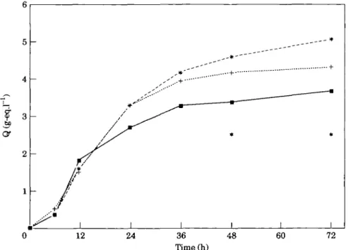
Figure 1 HBDH release patterns (Q) over 72 h for patients witb TIMI 3 ( • , n = 65), 2 (+, n = 50) or 0/1 (*, m=44) at the 90 min angiogram. *Indicates a significant difference between the groups (y<0-05).
perfusion (TIMI-3), 4-35 (2-63, 640) g-eq . 1~' in patients with incomplete perfusion (TIMI-2) and 5-07 (3-73, 8-22) g-eq . 1 ~ ' in those with minimal or no per-fusion (TIMI-0,1) (Fig. 1). The differences among the
three groups were significant at 48 h (/>=0047) and at 72 h (/>=f>024).
The overall pattern of enzyme release varied among the three patient groups. In patients with normal
12
Figure 2 Percentage of Q72 released for patients with TIMI 3 ( • , n = 65), 2 ( + ,
n=41), and 0/1 (*, n=42) at the 90 min angiogram. Q72 denotes the cumulative release of HBDH at 72 h. indicates a significant difference between the groups
Table 2 Effect of treatment on median infarct size (Q(72)) expressed in g-eq. I '
SK + s.c. hep SK+i.v. hep tPA + i.v. hep tPA + SK + i.v. hep SK combined tPA combined /"-value for SK vs tPA n=125 4 36 2-5-6-7 With imputation n=139 4-90 2-6-6-5 n=120 4-52 2-6-7-1
for missing cases (see n=136 4-90 2-8-7-1 n=130 3-92 1-6-6-4 text): n = 137 4-39 1-7-6-3 n=127 3-88 1-9-6-6 n=141 4-17 2-1-6-7 n = 245 4-39 2-5-6-8 n = 275 4-90 2-7-6-8 n = 257 3-88 1-7-6-6 n = 278 4-35 2-0-6-6 0043 0034
Abbreviations as in Table 1, n = number of patients. Medians with the 25th and 75th percentiles are given.
perfusion at early angiography a greater proportion was released within the first 6 or 12 h (Fig. 2, P<, 0002). Because the proportion of early HBDH release can be meaningfully calculated only in patients with a complete series of early samples, only 343 patients who had at least 10 samples and Q(72) exceeding 0-5 g-eq . 1 ~ ' were used for this analysis.
Effect of treatment on infarct size and
enzyme release kinetics
Table 2 presents the median values of Q(72) for the four treatment groups, without and with imputation for missing cases, and for the combined tissue plasminogen activator and streptokinase groups. Patients treated with
accelerated tissue plasminogen activator or combination therapy had infarcts that were 12% smaller then those who received streptokinase therapy. These differences were apparent, but not statistically significant in a four-group comparison (Kruskal-Wallis, />=0-19). However, the pooled patients receiving streptokinase only differed significantly (,P=0043) from the patients receiving either tissue plasminogen activator or tissue plasminogen activator plus streptokinase. Because of the high value of 110 g-eq . 1 ~ ' used for the patients who died early (see Methods), imputation raised the median values by 10-15%, but these trends remained un-changed. Due to the higher number of deaths in the streptokinase patients (see Table 1) imputation resulted in a slightly more significant difference (P^O-034) be-tween the combined tissue plasminogen activator and the combined streptokinase groups.
Table 3 Effect of treatment 0(3) 0(4) 0(6) 0(9) Q(12) Q(6VQ(72) Q(12)/Q(72) SK+s.c. hep 5 n = 87 0-00 001 0 1 5 0-74 1-29 0053 0-282
on early HBDH release expressed in g-eq. I '
>K + i.v. hep n = 77 0-00 002 0-30 0-87 1-34 0066 0-318
tPA + i.v. hep
n=95 0 0 2 0 0 9 0-41 0-88 1 35 0140 0-372
tPA + SK + i.v. hep
n = 84 0 0 3 0 1 3 0 41 1-20 1 76 0136 0-423
JSL.
n=164 0 0 0 001 0-23 0-77 1-32 0062 0-295 tPA n = 179 0-02 O i l 0-41 0-97 1 57 0140 0-319 /"-value for SK. vs tPA 0-002 00001 00001 0-07 0-55 <00001 <0-001 Abbrevations as in Tables 1 and 2. Medians of cumulative HBDH release (Q) are given for the first 3, 4, 6, 9 and 12 h, and ratios of early release are given (Q(6)/Q(72) and Q(12)/Q(72)).12 24 38 48 60
1 -72 0
Time(h)
12 38 48 60 72
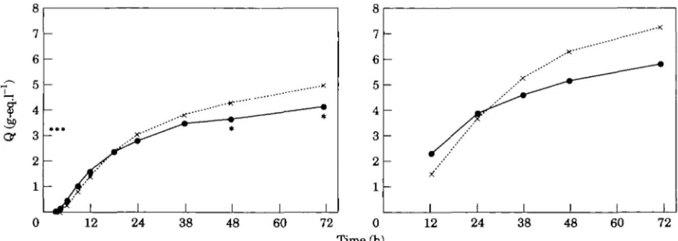
Figure 3 HBDH release patterns (Q) of patients treated with streptokinase (X) (and intravenous or
subcutaneous heparin) or with alteplase ( • ) (and intravenous heparin or with streptokinase and intravenous heparin). * indicates a significant difference between the groups (/><0-05). For comparison, data of the European
Cooperative Study Group (rt-PAVplacebo trial), are presented (right panel). Note accelerated (early) release for tissue plasminogen activator ( • ) in comparison with both placebo ( x , right panel) as well as streptokinase ( x , left panel).
Early release of HBDH, expressed in g-eq .1 ', within the first 12 h after onset of symptoms is presented in Table 3 for 343 patients with at least 10 samples. In spite of the larger ultimate infarct size in the strepto-kinase groups (Table 2), release up to 6 h after first symptoms was significantly lower in the combined strep-tokinase groups, compared with the combined tissue plasminogen activator groups. Differences between ratios Q6/Q72 and Q12/Q72 were more significant than differences between absolute values of Q6 and Q12 (Table 3). This reflects two distinct effects on release kinetics: enhancement of early release and reduction of late release in patients treated with tissue plasminogen activator vs patients treated with streptokinase. This is also illustrated in Fig. 3.
In order to verify whether the effect of treatment could be caused by factors other than infarct-related artery patency, multiple linear regression analysis was performed using Q(72) as the dependent variable and treatment (streptokinase vs tissue plasminogen
activa-tor) as well as TIMI score (3 vs 2 vs 0 or 1) as independent variables. In this analysis, the contribution of treatment became totally insignificant (P=0-97) while the effect of TIMI score remained highly significant (P<00l). This indicates that the effect of treatment on infarct size may be explained by its effect on 90-min infarct-related artery patency. Similarly, multiple linear regression analysis with Q(12)/Q(72) as the dependent variable and treatment (streptokinase vs tissue plasminogen activator) and TIMI score (3 vs 2 vs 0 or 1) as independent variables demonstrated that the effect of treatment on the acceleration of enzyme release was explained by early coronary patency.
Infarct size and left ventricular function
Both infarct size and left ventricular function measure-ments at 5-7 days after allocation were available in 156 patients with a first myocardial infarction. There was an
100 7 5 -§3 •5?
23
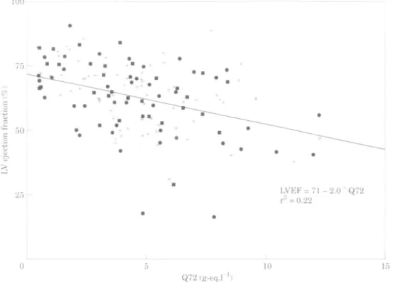
25 r LVEF = 11- 2.0 Q72r" = 0.22 10 15 Q72(g-eq.rFigure 4 Left ventricular (LV) ejection fraction (LVEF) 5-7 days after a first myocardial
infarction against Q72. Q72 denotes the cumulative release of HBDH at 72 h. The linear regression line for all patients is given in the figure. The regression lines for the four treatment groups did not differ (intercept and slope for alteplase+intravenous heparin ( • ) were, respec-tively, 70 and —1-9; for steptokinase+intravenous heparin ( + ) 72, —2-2; for alteplase+ streptokinase+intravenous heparin ( • ) 72, - 2 - 2 and for streptokinase and subcutaneous heparin ( x ) 72, - 1-8).
inverse relationship between infarct size and both left ventricular ejection fraction and regional wall motion of the infarct zone. This relationship for left ventricular ejection fraction is shown in Fig. 4. Regression lines were computed as shown in Fig. 4, indicating consistent relationships between infarct size and global or regional left ventricular function, independent of the treatment regimens.
Discussion
In GUSTO, treatment with accelerated tissue plasmino-gen activator yielded improved survival compared with two streptokinase regimens. The absolute difference in 30 day mortality was 1%, which corresponded to a 14% relative mortality reduction1'1. Intermediate results were obtained with the combined regimen of tissue plasmino-gen activator with streptokinase. These differences could be explained by more frequent early reperfusion in patients receiving tissue plasminogen activator, as docu-mented by the GUSTO angiographic substudy121. The present GUSTO enzyme substudy extends these findings and confirms the concept that the improved survival after treatment with accelerated tissue plasminogen ac-tivator in comparison with streptokinase11' is related to
myocardial salvage by more rapid coronary reperfusion. Indeed, infarct size in patients treated with tissue plas-minogen activator appeared to be 12% smaller than in those receiving streptokinase.
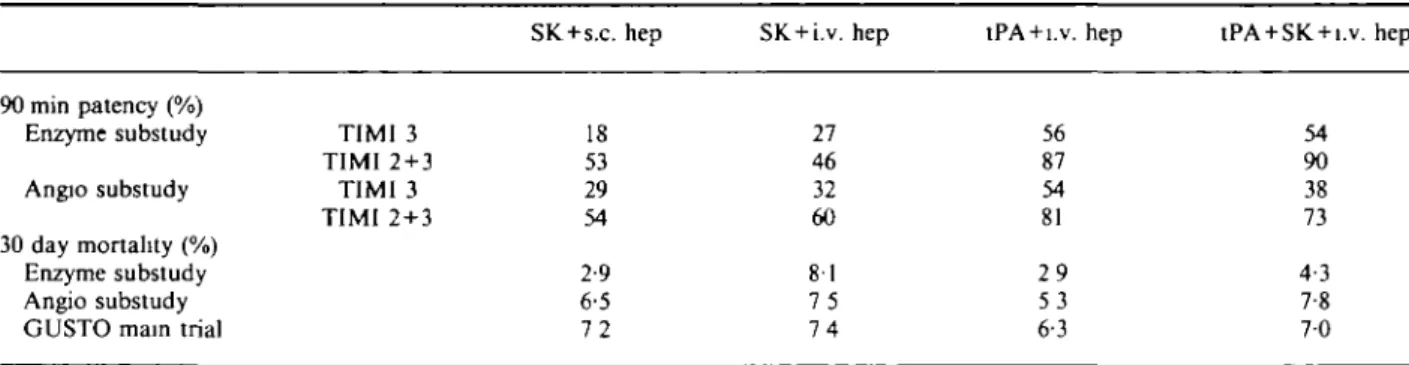
It should be appreciated that the mortality and patency results in this substudy are compatible with, but not identical to, those in the larger trial (Table 4). In particular, early coronary patency was almost identical among patients receiving accelerated tissue plasminogen activator or the combination regimen (tissue plasmino-gen activator within streptokinase) in this subset of the angiographic substudy. This is in agreement with the infarct size measurements, which were almost identical in these two groups. Accordingly, the predefined inten-tion to compare the two tissue plasminogen activator regimens (accelerated tissue plasminogen activator and combined tissue plasminogen activator and strepto-kinase) with the two streptokinase regimens appeared justified. In contrast, in the larger angiographic substudy and in the mortality study, both early patency and survival for the combination regimen were intermediate between the figures for accelerated tissue plasminogen activator and the streptokinase groups, and combina-tion of the two regimens containing tissue plasminogen activator would not be appropriate.
Table 4 Comparison 90 min patency (%) Enzyme substudy Angio substudy 30 day mortality (%) Enzyme substudy Angio substudy GUSTO main trial
of outcomes in TIMI 3 TIMI 2 + 3 TIMI 3 TIMI 2 + 3 different GUSTO SK + s.c. hep 18 53 29 54 2-9 6-5 7 2 cohorts SK + i.v. hep 27 46 32 60 81 7 5 7 4
tPA + i.v. hep
56 87 54 81 2 9 5 3 6-3
tPA + SK + i.v. hep
54 90 38 73 4-3 7-8 7 0 Abbreviations as in Table 1.
Estimation of infarct size in patients
receiving thrombolytic therapy
The validity of using myocardial enzyme release into plasma for estimation of infarct size has been demon-strated in several clinical studies16"91 and in experimental studies on permanent coronary artery ligation in dogs'16"18' using histological staining of the infarcted area as the gold standard. These studies have shown highly significant relationships between total infarcted muscle volume and peak values or cumulative release of enzymes such as creatine kinase, lactate dehydrogenase and aspartate aminotransferase. In patients treated with thrombolytic therapy, however, some clinical studies have indicated altered relationships between the release of creatine kinase, or creatine kinase-MB into plasma and myocardial injury as estimated from an independent parameter such as left ventricular ejection fraction1'9"2'1 or thallium-201 defect1221. In these studies, thrombolysis caused extra release of creatine kinase, creatine kinase-MB compared with control patients for the same degree of damage as determined from the independent variable. Reperfusion also resulted in enhanced creatine kinase recovery in plasma after experimental infarction in the dog1231. Currently, this remains a controversial issue because complete recovery of creatine kinase in plasma has been reported after permanent coronary occlu-sion'241, while recovery was found to be decreased after reperfusion1251. It should be appreciated that this contro-versy does not invalidate the conclusion of the present study, that infarcts remain smaller after tissue plasmino-gen activator therapy. All clinical studies suggesting an effect of thrombolytic therapy on enzyme release have claimed reperfusion-induced enhancement of release. This implies that the 12% reduction of infarct size in patients treated with tissue plasminogen activator could only be under-estimated, and that the true benefits might be greater.
For HBDH, complete recovery in plasma after permanent coronary occlusion has been reported'241. In a large clinical trial, the relationship between release of HBDH and left ventricular ejection fraction remained unaltered by thrombolytic therapy1261, indicating that no reperfusion-induced extra release of HBDH occurs. The
relationship between left ventricular ejection fraction and enzymatic infarct size was identical in patients treated with streptokinase or direct angioplasty1271. Simi-larly, in the present study the relationship between HBDH infarct size and left ventricular function was independent of the therapeutic regimen (Fig. 4). This supports the validity of HBDH infarct size measure-ments for comparison of different reperfusion regimens.
Effect of infarct artery patency on
infarct size
The reduction of infarct size by the tissue plasminogen activator regimens, compared to streptokinase, could be explained by improved 90 min patency of the infarct-related artery. It is also interesting to note that reduction of infarct size was not restricted to patients with early complete perfusion (TIMI-3). Patients with partial per-fusion (TIMI-2) also had smaller infarcts than those with an occluded infarct related artery (TIMI-01). Others have reported a lack of infarct size reduction in patients with TIMI-2 flow'281. A possible explanation for this discrepancy could be the somewhat later angiogra-phy in the TEAM-2 study: a mean delay of 2-4 h vs 1-6 h in the present study. This is supported by data from 72 patients in the present study who had angiography between 2 and 4 h after initiation of thrombolytic therapy. In these patients, infarct size was 3-41 g-eq . 1 ~', 5-29 g-eq . 1"' and 4-99 eq . 1"' for TIMI perfusion grades, 3, 2 and 0-1 respectively (P=042), so no benefit was apparent in patients with TIMI-2 flow between 2 and 4 h. Thus, early incomplete perfusion (TIMI-2) might be adequate for some recovery of ischaemic myocytes. Alternatively, some of these patients may have further clot resolution with complete perfusion (TIMI-3) before necrosis has been completed.
Effect of infarct artery patency on
acceleration of enzyme release
Earlier release of myocardial enzymes into plasma was already noted in the first study on thrombolysis after
acute myocardial infarction'291 and was thought to re-flect enhanced washout of enzymes due to thrombolysis-induced restoration of blood flow or possible reperfusion injury1301. Since then, large trials have confirmed that reperfusion does accelerate myocardial enzyme release131-321. The difference in enzyme release pattern between patients treated with tissue plasminogen activator vs streptokinase in the present study is strikingly similar to, although smaller than, the differ-ence between tissue plasminogen activator and placebo in an earlier study as shown in Fig. 3. This supports the notion that both the benefit of accelerated tissue plas-minogen activator vs streptokinase and the larger benefit of tissue plasminogen activator vs placebo are caused by more rapid coronary reperfusion by accelerated tissue plasminogen activator.
The more rapid evolution of infarction during thrombolytic therapy has been confirmed independently by serial ECG analysis in a trial by the European Cooperative Study Group1331. In that trial, limitation of infarct size was apparent since patients receiving tissue plasminogen activator had fewer Q waves after 10-20 days than the placebo group. However, at 6 h after initiation of therapy, Q waves were more pronounced although the ST-segment had resolved more rapidly in patients allocated to thrombolytic therapy. Early release of various myocardial marker proteins is now widely used as a non-invasive indicator of successful reper-fusion in individual patients, although the sensitivity and specificity of this method are not established. In the evaluation of thrombolytic therapy, acceleration of enzyme release is a more sensitive parameter than reduction of infarct size.
Limitation of infarct size and reduced
mortality
Collectively, the large placebo-controlled or open trials of thrombolytic therapy have demonstrated a marked reduction in mortality for patients presenting to hospital within 6 to 12 h of the onset of symptoms'381. In trials that studied the release of HBDH (or LDH1), reduc-tions of infarct size between 20% and 35% were consist-ently found'1'-12-34-351. Both reduction of mortality111 as well as reduction of HBDH release136'371 were most impressive in patients treated within 1 h of the first symptoms. Similar trends were observed in the GUSTO study, with a 14% reduction in mortality for patients treated with tissue plasminogen activator reported in the main trial'11 which corresponded to a 12% reduction in infarct size as shown in this study.
To conclude: the presented data are in full agree-ment with and support the concept that more rapid reperfusion (by accelerated tissue plasminogen activator, in comparison with the streptokinase regimens in GUSTO) salvages myocytes, resulting in limitation of infarct size, preservation of left ventricular function and improved survival. The observed association between
limitation of infarct size and improved survival in this and other studies'"12-351 indicates that infarct size measurements may be applied to compare the clinical efficacy of different therapeutic regimens in medium size trials.
References
[1] The GUSTO Investigators: An international randomized trial comparing four thrombolytic strategies for acute myocardial infarction. N Engl J Med 1993; 329: 673-82.
[2] The GUSTO Angiographic Investigators: The effects of tissue plasminogen activator, streptokinase, or both on coronary-artery patency, ventricular function, and survival, after acute myocardial infarction. N Engl J Med 1993; 329: 1615-22. [3] Granger ChB, CahffRM, Topol EJ. Thrombolytic therapy for
acute myocardial infarction. Drugs 1992; 44: 293-325. [4] Willems GM, Muijtjens AMM, Lambi FHH, Hermens WTh.
Estimation of circulatory parameters in patients with acute myocardial infarction. Significance of calculation of enzymatic infarct size. Cardiovasc Res 1979; 13: 578-87.
[5] Willems GM, Visser MP, Krill MTA, Hermens WTh. Quan-tative analysis of plasma enzyme levels based on simultaneous determination of different enzymes. Cardiovasc Res 1982; 16:
120-31.
[6] Erhardt LR Clinical and pathological observations in differ-ent types of acute myocardial infarction. Acta Med Scand
1974; Suppl. 560. 1-78.
[7] Bleifeld W, Mathey D, Hanrath P, Buss H, Effert S. Infarct size estimated from serial serum creatine phosphokinase in relation to left ventricular dynamics. Circulation 1977; 55: 303-11.
[8] Grande P, Hansen BF, Christiansen C, Naestoft J. Estimation of acute myocardial infarct size in man by serum CK-MB measurements. Circulation 1982; 65: 756-64.
[9] Hackel DB, Reimer KA, Ideker RE el al. for the MILIS Study Group. Comparison of enzymatic and anatomic estimates of myocardial infarct size in man. Circulation 1984; 70: 824-35. [10] Van der Uarse A, Van der Wall EE, Van den Pol RC el al. Rapid enzyme release from acutely infarcted myocardium after early thrombolytic therapy: Washout or reperfusion damage? Am Heart J 1988; 115: 711-16.
[11] Simoons ML, Serruys PW, van den Brand M el al. Early thrombolysis in acute myocardial infarction: Limitation of infarct size and improved survival. J Am Coll Cardiol 1986; 7: 717-28.
[12] Van de Werf F, Arnold AER for the European Cooperative Study group for recombinant tissue type plasminogen activa-tor. Intravenous tissue plasminogen activator and size of infarct, left ventricular function, and survival in acute myo-cardial infarction. Br Med J 1988; 297: 1374-9.
[13] Chesebro JH, Knatterud G, Roberts R, Braunwald E for the TIMI investigators. Thrombolysis in myocardial infarction (TIMI) Trial, Phase 1: a comparison between intravenous tissue plasminogen activator and intravenous streptokinase. Circulation 1987; 76: 142-57.
[14] Van der Laarse A, Dijkshoorn NJ, Hollar L, Caspers Th. The (iso)enzyme activities of lactate dehydrogenase, alpha-hydroxybutyrate dehydrogenase, creatine kinase and aspar-tate amino-transferase in human myocardial biopsies and autopsies. Clin Chim Acta 1980; 104: 381-91.
[15] Van der Veen FH, Visser R, Willems GM, Kop-Klaassen B, Hermens WTh. MyocardiaJ enzyme depletion in infarcted human hearts: infarct size and equivalent tissue mass. Cardio-vasc Res 1988; 22: 611-19.
[16] Ruegsegger P, Nydick I, Freiman A, La Due JS. Scrum activity patterns of glutamic oxaloacetic transaminase, glutamic pyruvic transaminase and lactic dehydrogenase fol-lowing graded myocardial infarction in dogs. Circ Res 1959; VI: 4-10.
[17] Nachlas MM, Friedman MM, Cohen SP. A method for the quantitation of myocardial infarcts and the relation of serum levels to infarct size. Surgery 1964; 55: 700-8.
[18] Swain JL, Cobb FR, McHale PH, Roe CR. Nonlinear rela-tionship between creatine kinase estimates and histologic extent of infarction in conscious dogs: Effects of regional myocardial blood flow. Circulation 1980; 62: 1239^*7. [19] Ong L, Reiser P, Coromilas J, Scherr L, Morrison J. Left
ventricular function and rapid release of creatine kinase MB in acute myocardial infarction. N Engl J Med 1983; 309: 1-6. [20] Blanke H, von Hardenberg D, Cohen M et al. Patterns of
creatine kinase release during acute myocardial infarction after nonsurgical treatment and correlation with infarct size. J Am Coll Cardiol 1984; 3: 675-80.
[21] Isobe M, Nagai R, Ueda S et al. Quantitative relationship between left ventricular function and serum cardiac myosin light chain I levels after coronary reperfusion in patients with acute myocardial infarction Circulation 1987, 76: 1251-61. [22] Tamaki S, Murakami T, Kadota K et al. Effects of coronary
artery reperfusion on relation between creatine kinase-MB release and infarct size estimated by myocardial emission tomography with thallium-201 in man. J Am Coll Cardiol 1983; 2: 1031-8.
[23] Vatner SF, Baig H, Manders WT, Maroko PR. Effects of coronary artery reperfusion on myocardial infarct size calcu-lated from creatine kinase. J Clin Invest 1978; 61: 1048-56. [24] Hermens WTh, van der Veen FH, Willems GM,
Mullers-Boumans ML, Schrijvers-van Schendel A, Reneman RS. Complete recovery in plasma of enzymes lost from the heart after permanent coronary artery occlusion in the dog. Circu-lation 1990; 81: 649-59.
[25] Jarmakani JM, Limbird L, Graham ThC, Marks RA. Effect of reperfusion on myocardial infarct, and the accuracy of estimating infarct size from serum creatinine phosphokinase in the dog. Cardiovasc Res 1976; 10: 245-53.
[26] Van der Laarse A, Kerkhof PLM, Vermeer F et al. Relation between infarct size and left ventricular performance assessed in patients with first acute myocardial infarction randomized to intracoronary thrombolytic therapy or to conventional treatment. Am J Cardiol 1988; 61. 1-7.
[27] de Boer MJ, Suryapranata H, Hoorntje JCA et al. Limitation of infarct size and preservation of left ventricular function after primary coronary angioplasty compared with intrave-nous streptokinase in acute myocardial infarction. Circulation 1994; 90: 753-61.
[28] Karagounis L, Sorensen SG, Menlove RL, Moreno F, Anderson JL. Does Thrombolysis in Myocardial Infarction (TIMI) perfusion grade 2 represent a mostly patent artery or a mostly occluded artery? Enzymatic and electrocardiographic evidence from the TEAM-2 study. J Am Coll Cardiol 1992;
19: 1-10.
[29] Fletcher AP, Sherry S, Alkjaeng N, Smyrniotis FE, Jick S. The maintenance of a sustained thrombolytic state in man. II Clinical observations on patients with myocardial infarction and other thrombo-embolic disorders. J Clin Invest 1959; 35: 1546-51.
[30] Braunwald E, KJoner RA. Myocardial reperfusion: A double-edged sword? J Clin Invest 1985; 76: 1713-19.
[31] Van der Laarse A, Vermeer F, Hermens WTh et al. Effects of early intracoronary streptokinase on infarct size estimated
from cumulative enzyme release rate. A randomized trial on 533 patients with acute myocardial infarction. Am Heart J 1986; 112: 672-81
[32] Hackworthy RA, Sorensen SG, Fitzpatrick PG et al. Effect of reperfusion on electrocardiographic and enzymatic infarct size: Result of a randomized multicenter study of intravenous amsolated plasminogen streptokinase activator complex (APSAC) versus intracoronary streptokinase in acute myo-cardial infarction. Am Heart J 1988; 116: 903-14.
[33] Willems JL, Willems RJ, Bijnens I, Doerr R, Verstraete M for the European Cooperative Study Group for Recombinant Tissue-Type Plasminogen Activator. Value of electrocardio-graphic scoring systems for the assessment of thrombolytic therapy in acute myocardial infarction. Eur Heart J 1991; 12: 378-88.
[34] Anderson JL, Marschall HW, Bray BE et al. A randomized trial of intracoronary streptokinase in the treatment of acute myocardial infarction N Eng J Med 1983; 308: 1312-18. [35] The Thrombolysis Early in Acute Heart Attack Trial Study
Group. Very early thrombolytic therapy in suspected acute myocardial infarction. Am J Cardiol 1990; 65: 401-7 [36] Vermeer F, Simoons ML, Bar FW et al. Which patients
benefit most from early thrombolytic therapy with intra-coronary streptokinase? Circulation 1986; 74: 1379-89. [37] Hermens WTh, Willems GM, Nijssen KM, Simoons ML.
Effect thrombolytic treatment delay on myocardial infarct size. Lancet 1992; 340: 1297.
[38] Fibrinolytic Therapy Trialists' (FTT) Collaborative Group. Indications for fibrinolytic therapy in suspected acute myo-cardial infarction: collaborative overview of early mortality and major morbidity results from all randomised trials of more than 1000 patients. Lancet 1994; 343: 311-22.
Appendix
The following centres and investigators collaborated (alphabetical order).
France: Caen, Centre Hospitalier Universitaire: Grollier G, Valette B; Paris, Hopital Boucicaut-Vaugirard: Guerot G, Grenier O; Paris, Hopital Broussais: Guermonprez J, Guize L, Iliou M; Paris, Hopital Lariboisiere: Beaufils P, Rapoport P; Paris, Hopital Tenon: Vahanian A, Nallet O; Strasbourg, Hopital de Hautepierre: Mossard M, Arbogast R. Germany: Lubeck, Medizinische Universitat zu Lubeck: Sheikzadeh A, Djonlagic H, Kurowski V. The Netherlands: Alkmaar, MCA: Arnold AER; Apeldoom, Juliana Ziekenhuis: Cozijnsen L; Assen, Wilhelmina Ziekenhuis: de Leeuw MJ; Emmen, Scheper Ziekenhuis: Engbers JG; Enschede, Medisch Spectrum: Molhoek GP, Lalisang R; Rotterdam, Dijkzight: Simoons ML, van den Brand M, Kint P; Rotterdam, Ikazia: Kerker J.
Switzerland: Basel, University Hospital: Pfisterer M, Hammerli R.