Nephrol Dial Transplant (2011) 26: 1413–1416 doi: 10.1093/ndt/gfq752
Advance Access publication 10 January 2011
Preliminary communications
Protein level expression of Toll-like receptors 2, 4 and 9 in renal disease
Stephen Batsford
1, Ursula Duermueller
1, Christian Seemayer
2, Christoph Mueller
3, Helmut Hopfer
1and
Michael Mihatsch
11Institute for Pathology, University Hospital Basel, Basel, Switzerland2Institute of Pathology, Centre Hospitalier Universitaire
Vaudois, Lausanne, Switzerland and3Institute of Pathology, University Bern, Bern, Switzerland
Correspondence and offprint requests to: Stephen Batsford; E-mail: [email protected]
Abstract
Background. Toll-like receptors (TLR) recognize a variety of ligands, including pathogen-associated molecular pat-terns and link innate and adaptive immunity. Individual receptors can be up-regulated during infection and inflam-mation. We examined the expression of selected TLRs at the protein level in various types of renal disease. Methods. Frozen sections of renal biopsies were stained with monoclonal antibodies to TLR-2, -4 and -9.
Results. Up-regulation of the three TLRs studied was seen, although the extent was modest. TLR-2- and -4-positive cells belonged to the population of infiltrating inflamma-tory cells; only in the case of TLR-9 were intrinsic glomer-ular cells positive in polyoma virus infection and haemolytic uraemic syndrome (HUS).
Conclusions. Evidence for the involvement of the three TLRs tested in a variety of human renal diseases was found. These findings add to our understanding of the role of the innate immune system in kidney disease.
Keywords: renal biopsy; Toll-like-receptors (TLR); TREM1
Introduction
The innate immune system possesses Toll-like receptors (TLR) and this versatile receptor system senses invasion of mi-crobial pathogens. TLR recognize a variety of ligands, includ-ing pathogen-associated molecular patterns (PAMP) and link innate and adaptive immunity [1,2]. Mammalian TLRs com-prise a large family of at least 11 members. TLR-2 recognizes a variety of microbial components, most prominent being di-and tri-acyl lipopeptides [3]. TLR-4 is the essential long sought receptor for lipopolysaccharide recognition [2]. TLR-9 recog-nizes unmethylated CpG motifs in bacterial DNA [2]. Activa-tion of innate immunity is a critical step in the development of antigen-specific acquired immunity and individual receptors can be up-regulated during infection and inflammation.
TLR up-regulation may be involved in renal disease [4] and there is increasing data supporting a role for TLRs in
infectious autoimmune and inflammatory disorders of the kidney [5].
We examined expression of selected TLRs at the protein level in various types of renal disease; the range was re-stricted by the limited availability of antibodies suitable for immune histochemistry. Triggering receptors expressed by myeloid cells (TREMs) activate myeloid cells, in particular TREM1 triggers phagocytic secretion of pro-inflammatory chemokines and cytokines amplifying inflammation in-duced by microorganisms [6]. Since TLRs and TREMs are believed to cooperate closely [6], the expression of TREM1 in selected cases was also looked at.
Materials and methods
The classification and numbers of cases studied are listed in Table 1. Frozen sections of renal biopsies were fixed in acetone for 10 min and then stained with monoclonal antibodies to TLR-2 (clone TL2.3, diluted 1:50; Alexis ALX-804-324) and TLR-9 (clone 5G5, diluted 1:80; Serotec MCA 2265) using the Vector ABC kit (VECTASTAIN AK-5000) and TLR-4 (clone HTA 125, diluted 1:60; HyCult HM 2068) using the R.T.U. VECASTAIN Universal Quick kit (PK 8800). All these antibodies have been subjected to stringent specificity testing, including immunoprecipi-tation and western blotting, as described in the suppliers’ data sheets (see appropriate Web sites).
As positive control sections, frozen sections of Epstein-Barr virus-infected tonsils were employed for TLR-2 and frozen sections of normal placenta were used for TLR-4 and -9 (examples of positive and negative controls for all antibodies shown as supplementary data).
As a negative control, the specific antibody was replaced with nonim-mune mouse IgG2a (DAKO X0943) at the appropriate concentration. Staining with all antibodies was also performed on sections from six samples of normal human kidney. Sections were evaluated by two to three observers independently.
Staining for TREM1 was performed with the anti TREM1 hybridoma 21C7 and the Vector ABC kit, as detailed above. A total of 10 renal biopsies (5 positive and 5 negative for TLRs) were studied (polyoma nephropathy n = 3, haemolytic uraemic syndrome (HUS) n = 2, M. Wegener n = 2, interstitial rejection n = 2 and a single case of p-ANCA-positive GN).
Results
Antibodies to TLR-2, -4 and -9 did not produce noteworthy staining on six different samples of normal human kidney. The results obtained on 66 renal biopsy specimens from
© The Author 2011. Published by Oxford University Press on behalf of ERA-EDTA. All rights reserved. For Permissions, please e-mail: [email protected]
selected renal diseases are shown in Table 1. Any unspe-cific staining seen with the isotype control was taken into account for the final judgment.
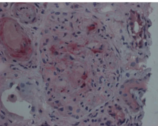
As can be seen in Table 1, TLR-2 and -4 were only seen on infiltrating inflammatory cells (granulocytes/mono-cytes), mainly in cases of HUS, M. Wegener, Polyoma virus nephropathy (PVN) (Figure 1) and p-ANCA GN (Figure 2); TLR-4 was more frequently positive than TLR-2 and intrinsic renal cells were always negative. TLR-9 was, with a single exception, seen only on glo-merular cells (probably mesangial/podocytes) in four of eight cases (50%) of HUS (Figure 3) and 2/12 (17%) cases of PVN. Further pictures from the various disease entities studied are shown in the supplementary data. An example of unspecific tubular staining in a negative control is shown in Figure 4.
In the 10 cases studied, staining for TREM1 was nega-tive, and weak staining of tubular structures was considered to be unspecific. For this reason, the series was not extended.
Conclusions
Positive results were most often found in cases of polyoma virus nephropathy, HUS and M. Wegener. TLR-2 and -4-positive cells belonged to the population of infiltrating inflammatory cells, which were often sparsely distributed; only in the case of TLR-9 were intrinsic glomerular cells positive in polyoma virus infection and HUS. These findings may reflect aetiopathogenetic mechanisms; for
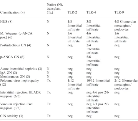
Table 1. Results of staining for TLR-2, -4 and -9 in selected renal diseases
Classification (n) Native (N), transplant (Tx) TLR-2 TLR-4 TLR-9 HUS (8) N 1/8 Interstitial infiltrate 3/8 Interstitial infiltrate 4/8 Glomerular mesangium/ podocytes M. Wegener (c-ANCA pos.) (6) N 3/6 Interstitial infiltrate 4/6 Interstitial infiltrate 1/6 Interstitial infiltrate Postinfectious GN (4) N neg 2/4 Interstitial infiltrate neg p-ANCA GN (6) N neg 5/6 Interstitial infiltrate neg
Acute interstitial nephritis (3) N neg neg neg
IgA-GN (3) N neg neg neg
Membranous GN (3) N neg neg neg
Polyoma virus nephropathy (12) Tx 1/12 Interstitial infiltrate 10/12 Interstitial infiltrate 2/12 Glomerular mesangium/ podocytes Interstitial rejection HLADR
neg/posa (6/6)
Tx neg neg 4/6 pos 2/6
interstitial infiltrate
neg
Vascular rejection C4d neg/posa (3/3)
Tx neg neg 2/3 pos 2/3
interstitial infiltrate
neg
CIN toxicity (3) Tx neg neg neg
aNo obvious difference between positive and negative cases. neg, negative; pos, positive.
Fig. 1. TLR2: PVN with minor mononuclear/histiocytic infiltrates around affected tubules, tubular epithelium and glomeruli are negative.
Fig. 2. TLR-4: p-ANCA-associated glomerulonephritis with dense focal histiocytic infiltrates around tubules, tubular epithelium completely negative.
example, infections have long been thought to be involved in cases of Wegener’s disease and HUS [7,8].
Little is known about innate immunity and polyoma virus infection, a serious problem in renal transplantation. Our finding of up-regulation of TLR-2, -4 and -9 in some cases is intriguing; it possibly marks efforts to control viral replication. A recent study in mice found that the TLR signalling adapter protein MyD88 is involved in the devel-opment of humoral immunity to polyoma virus [9]. More intensive studies of innate immune mechanisms in polyo-ma virus infections are clearly warranted.
Renal TLR expression has been studied by many inves-tigators, mostly at the messenger RNA expression level [10]. However, most of these studies were done on samples of renal tissue compartments [11] and therefore contribu-tions of infiltrating and intrinsic cells cannot be differen-tiated. TLR-2, but not TLR-4 expression in tubules from renal graft recipients, has been reported using immuno-histochemistry [12]. Another study demonstrated TLR-4 expression in renal tubules in cases of cyclosporine nephrotoxicity [13]. TLR-9 expression was found in lupus nephritis, both tubulointerstitial and glomerular [14] or only tubulointerstitial [15]. Cases of lupus nephritis were not included in the current study. Using the protocols employed here, we were unable to detect specific staining for TLR-2, -4 or -9 on six different samples of normal
human kidney. Constitutive expression of TLR-2, -4 and -9 at the protein level in normal human renal tissue appears to be below the level of immunohistochemical de-tection with the techniques employed. In our hands, diffuse tubulointerstitial staining patterns had to be classified as unspecific on the basis of stringent negative isotype con-trols. Three of the studies cited above [12,13,15] employed formalin-fixed paraffin-embedded tissue, only one used frozen sections [14]. Furthermore, negative controls con-sisted simply of omitting the primary antibody, more strin-gent isotype controls were not included. This raises the question whether differences in technical approaches might, at least in part, help explain conflicting results? Preliminary studies did not produce evidence of TREM1 expression in the renal diseases studied.
TLR-2 has been detected in normal mouse glomeruli and tubules [16], revealing differences between species. Animal models have provided evidence of TLR involvement in the pathogenesis of some forms of experimental renal disease [17,18]. TLR-2 expression on cultured proximal tubular cells was stimulated by leptospiral outer membrane proteins [19], TLR-4 has been linked to acute renal failure after sepsis [20] and the TLR ligand CpG-DNA can aggravate immune com-plex GN [21,22]. These effects were species specific; it should be noted that in most animal studies, TLR-2 and -4 were expressed intrinsically on renal tubular cells and TLR-9 was expressed mainly on infiltrating antigen pre-senting cells (APC), rather the opposite to the situation found in our studies in human biopsy material.
Further studies to elucidate the role of the innate immune system in renal injury are clearly necessary.
Supplementary data
Supplementary data is available online at http://ndt. oxfordjournals.org.
Acknowledgements. The studies were carried out in accordance with the ethical standards of the University of Basel.
Conflict of interest statement. None declared.
References
1. Mogensen TH. Pathogen recognition and inflammatory signaling in innate immune defenses. Clin Microbiol Rev 2009; 22: 240–273 2. Takeda K, Akira S. Toll-like receptors in innate immunity. Intern
Immunol 2005; 17: 1–14
3. Buwitt-Beckmann U, Heine H, Wiesmuller KH et al. TLR1- and TLR6-independent recognition of bacterial lipopeptides. J Biol Chem 2006; 281: 9049–9057
4. Schroppel B, He JC. Expression of Toll-like receptors in the kidney: their potential role beyond infection. Kidney Int 2006; 69: 785–787 5. Smith K. Toll-like receptors in kidney disease. Curr Opin Nephrol
Hypertens 2009; 18: 189–196
6. Colonna M. TREMs in the immune system and beyond. Nat Rev Im-munol 2003; 3: 445–453
7. Capizzi SA, Specks U. Does infection play a role in the pathogenesis of pulmonary vasculitis? Semin Respir Infect 2003; 18: 17–22 8. Zheng XL, Sadler JE. Pathogenesis of thrombotic microangiopathies.
Annu Rev Pathol 2008; 3: 249–277 Fig. 3. TLR-9: glomerulus and arterioles in HUS: a few podocytes and
mesangial cells (identity not verified by immunohistochemistry) positive for TLR-9. Smooth muscle cells of arteriole may be positive as well.
Fig. 4. Negative control: unspecific staining of parts of brush border in proximal tubules.
9. Guay HM, Andreyeva TA, Garcea RL et al. MyD88 is required for the formation of long-term humoral immunity to virus infection. J Immunol 2007; 178: 5124–5131
10. El-Achkar TM, Dagher PC. Renal Toll-like receptors: recent advances and implications for disease. Nat Clin Pract Nephrol 2006; 2: 568–581
11. Kwon J, Park J, Lee D et al. Toll-like receptor expression in patients with renal allograft dysfunction. Transplant Proc 2008; 40: 3479–3480
12. de Groot K, Kuklik K, Brocker V et al. Toll-like receptor 2 and renal allograft function. Am J Nephrol 2008; 28: 583–588
13. Lim BJ, Hong SW, Jeong HJ. Renal tubular expression of Toll-like receptor 4 in cyclosporine nephrotoxicity. Apmis 2009; 117: 583–591
14. Papadimitraki ED, Tzardi M, Bertsias G et al. Glomerular expression of toll-like receptor-9 in lupus nephritis but not in normal kidneys: implications for the amplification of the inflammatory response. Lupus 2009; 18: 831–835
15. Benigni A, Caroli C, Longaretti L et al. Involvement of renal tubular Toll-like receptor 9 in the development of tubulointerstitial injury in systemic lupus. Arthritis Rheum 2007; 56: 1569–1578
16. Shigeoka AA, Holscher TD, King AJ et al. TLR2 is constitutively expressed within the kidney and participates in ischemic renal injury
through both MyD88-dependent and -independent pathways. J Im-munol 2007; 178: 6252–6258
17. Patole PS, Pawar RD, Lech M et al. Expression and regulation of Toll-like receptors in lupus-Toll-like immune complex glomerulonephritis of MRL-Fas(lpr) mice. Nephrol Dial Transplant 2006; 21: 3062–3073 18. Banas MC, Banas B, Hudkins KL et al. TLR4 links podocytes with
the innate immune system to mediate glomerular injury. J Am Soc Nephrol 2008; 19: 704–713
19. Yang CW, Hung CC, Wu MS et al. Toll-like receptor 2 mediates early inflammation by leptospiral outer membrane proteins in proximal tu-bule cells. Kidney Int 2006; 69: 815–822
20. Anders HJ, Banas B, Schlondorff D. Signaling danger: toll-like recep-tors and their potential roles in kidney disease. J Am Soc Nephrol 2004; 15: 854–867
21. Anders HJ, Banas B, Linde Y et al. Bacterial CpG-DNA aggravates immune complex glomerulonephritis: role of TLR9-mediated expres-sion of chemokines and chemokine receptors. J Am Soc Nephrol 2003; 14: 317–326
22. Anders HJ, Vielhauer V, Eis V et al. Activation of toll-like receptor-9 induces progression of renal disease in MRL-Fas(lpr) mice. FASEB J 2004; 18: 534–536
Received for publication: 3.11.09; Accepted in revised form: 16.11.10
Nephrol Dial Transplant (2011) 26: 1416–1420 doi: 10.1093/ndt/gfq746
Advance Access publication 30 December 2010
Interstitial lung diseases after leflunomide use in nephropathy: an
analysis of reported cases in Chinese literature
Wu-xing Zhang, Wei Zhou, Zhi-qiang Zhang and Xue-wei Zhao
Department of Nephrology, PLA center of transplantation, PLA 309th Hospital, Beijing 100091, China Correspondence and offprint requests to: Xue-wei Zhao; E-mail: [email protected]
Abstract
Background. Leflunomide (LEF)-induced interstitial lung disease (ILD) has been reported in patients with rheuma-toid arthritis. In China, LEF is used off-label for the treat-ment of nephropathy.
Methods. Systemic review of the Chinese literature from 1999 to June 2010 for case reports and case series of LEF-induced ILD in nephropathy patients.
Results. We identified seven cases of LEF-induced ILD (three males and four females), with an average age of 45.9 years (range: 9–69 years). Six cases had primary nephrotic syndrome and one had Henoch–Schoenlein purpura. Four cases had diagnoses of renal pathology. Five patients were given loading doses of LEF, followed by a maintenance dose of 10–30 mg/day. The mean duration of LEF use was 62.9 ± 33.0 days (range: 20–120 days). The mean accumulated dose of LEF was 1192.5 mg (range: 830–1800 mg). LEF therapy was considered effective in four patients. Four patients died
(57.1%), three of whom had developed fevers. All three male patients died and both of the young patients died. The mean duration of LEF treatment was 83 days for patients who died and 37 days for survivors.
Conclusions. LEF-induced ILD in patients with nephropathy usually occurred after∼2 months of treatment and an accumu-lated dose of 1192.5 mg. Duration of LEF use, male sex, young age and fever seemed to increase the risk of mortality.
Keywords: glomerulonephritis; immnunosuppresant; interstitial lung disease; leflunomide; pheumonitis
Introduction
Leflunomide (LEF) is an isoxazole derivative with anti-in-flammatory and immunomodulating activities. It has been available in >70 countries for >10 years and is one of the
1416 W.-x. Zhang et al.
© The Author 2010. Published by Oxford University Press on behalf of ERA-EDTA. All rights reserved. For Permissions, please e-mail: [email protected]