RESEARCH OUTPUTS / RÉSULTATS DE RECHERCHE
Author(s) - Auteur(s) :
Publication date - Date de publication :
Permanent link - Permalien :
Rights / License - Licence de droit d’auteur :
Institutional Repository - Research Portal
Dépôt Institutionnel - Portail de la Recherche
researchportal.unamur.be
University of Namur
BtaE, an adhesin that belongs to the trimeric autotransporter family, is required for full
virulence and defines a specific adhesive pole of Brucella suis
Ruiz-Ranwez, Verónica; Posadas, Diana M.; Van der Henst, Charles; Estein, Silvia M.;
Arocena, Gastón M.; Abdian, Patricia L.; Martín, Fernando A.; Sieira, Rodrigo; De Bolle,
Xavier; Zorreguieta, Angeles
Published in:
Infection and Immunity
DOI:
10.1128/IAI.01241-12 Publication date: 2013
Document Version
Publisher's PDF, also known as Version of record
Link to publication
Citation for pulished version (HARVARD):
Ruiz-Ranwez, V, Posadas, DM, Van der Henst, C, Estein, SM, Arocena, GM, Abdian, PL, Martín, FA, Sieira, R, De Bolle, X & Zorreguieta, A 2013, 'BtaE, an adhesin that belongs to the trimeric autotransporter family, is required for full virulence and defines a specific adhesive pole of Brucella suis', Infection and Immunity, vol. 81, no. 3, pp. 996-1007. https://doi.org/10.1128/IAI.01241-12
General rights
Copyright and moral rights for the publications made accessible in the public portal are retained by the authors and/or other copyright owners and it is a condition of accessing publications that users recognise and abide by the legal requirements associated with these rights. • Users may download and print one copy of any publication from the public portal for the purpose of private study or research. • You may not further distribute the material or use it for any profit-making activity or commercial gain
• You may freely distribute the URL identifying the publication in the public portal ? Take down policy
If you believe that this document breaches copyright please contact us providing details, and we will remove access to the work immediately and investigate your claim.
several countries. Increasing evidence indicates that adhesion of Brucella spp. to host cells is an important step to establish infec-tion. We have previously shown that the BmaC unipolar monomeric autotransporter mediates the binding of Brucella suis to host cells through cell-associated fibronectin. Our genome analysis shows that the B. suis genome encodes several additional potential adhesins. In this work, we characterized a predicted trimeric autotransporter that we named BtaE. By expressing btaE in a nonadherent Escherichia coli strain and by phenotypic characterization of a B. suis⌬btaE mutant, we showed that BtaE is involved in the binding of B. suis to hyaluronic acid. The B. suis⌬btaE mutant exhibited a reduction in the adhesion to HeLa and A549 epithelial cells compared with the wild-type strain, and it was outcompeted by the wild-type strain in the binding to HeLa cells. The knockout btaE mutant showed an attenuated phenotype in the mouse model, indicating that BtaE is required for full virulence. BtaE was immunodetected on the bacterial surface at one cell pole. Using old and new pole markers, we observed that both the BmaC and BtaE adhesins are consistently associated with the new cell pole, suggesting that, in Brucella, the new pole is functionally differentiated for adhesion. This is consistent with the inherent polarization of this bacterium, and its role in the invasion process.
B
rucella species are responsible for brucellosis, one of the mostwidespread zoonotic diseases in the world that affects a range of different mammals. Currently, there are 10 recognized species of Brucella, based on host preferences, pathogenicity, and other phenotypic differences. Brucellosis remains endemic in most ar-eas of the world; several animal species with agricultural relevance are susceptible to the disease, and there are also wildlife reservoirs of Brucella, which makes eradication more complicated (1). In particular, Brucella abortus, Brucella melitensis, and Brucella suis are the most economically significant pathogens of the group. Hu-man brucellosis is caused by accidental contamination with repro-ductive or mammary secretions from infected animals (2,3) and is characterized by a strong undulating fever, which, if untreated, may lead to a chronic stage of the disease and the appearance of serious complications such as endocarditis, osteoarthritis, and neurological disorders (3).
The preferred niche of Brucella is intracellular; when brucellae are ingested or inhalated, they penetrate through mucosal sur-faces, and subsequently are transported to the lymph nodes by macrophages. Brucella spp. are able to infect and survive within macrophages in a compartment derived from the endoplasmic reticulum (4–6). It was shown that Brucella also has the ability to infect a great variety of host cells, including epithelial cells, placen-tal trophoblasts, neurons, and cells from male and female repro-ductive tissues, to name a few (6,7). Brucella has a marked ability to elude some of the basic mechanisms of the host immune sys-tem, which partly explains the frequent relapse after treatment (8,9).
It was shown that initial adhesion of several pathogens to the
host cell surface is a critical step in the infection process that helps the pathogens to avoid rapid clearance from host tissues (10). Several bacterial filamentous or nonpolymeric adhesins have been shown to participate in the primary interaction with the host cells as well as with components of the extracellular matrix (ECM) (11). Cumulative evidence supports the idea that adhesion to the ECM or to host cells is also an important step for Brucella infection (12–16). It was proposed that binding of Brucella to the host is mediated by host molecules containing sialic acid and/or sulfated residues and by components of the ECM such as fibronectin, col-lagen, and vitronectin. By phage display, we have recently identi-fied a unipolar fibronectin-binding protein belonging to the type I (monomeric) autotransporter family from B. suis, which we called BmaC (15). Autotransporters are a superfamily of proteins pro-duced by Gram-negative bacteria that share a domain organiza-tion composed of an N-terminal funcorganiza-tional domain named the
Received 8 November 2012 Returned for modification 30 November 2012 Accepted 4 January 2013
Published ahead of print 14 January 2013 Editor: S. M. Payne
Address correspondence to Angeles Zorreguieta, [email protected]. * Present address: Fernando A. Martín, INSERM U1001, Faculté de Médecine Université Paris Descartes, Paris, France; Diana M. Posadas, Department of Molecular Biosciences, Northwestern University, Evanston, Illinois, USA. Copyright © 2013, American Society for Microbiology. All Rights Reserved.
doi:10.1128/IAI.01241-12
on September 2, 2015 by BIBLIOTHEQUE UNIVERSITAIRE
“passenger” domain and a C-terminal translocator-domain that exhibits a-barrel structure with a hydrophilic pore. Autotrans-porters are transported by the Sec complex across the inner mem-brane, and the-domain subsequently transports the covalently linked passenger domain across the outer membrane (17). Several sets of evidence indicated that BmaC is involved in the polar bind-ing of sbind-ingle bacteria to epithelial cells (15). A comprehensive bioinformatics search allowed the identification of several addi-tional putative adhesins or invasins. One such candidate corre-sponded to a protein named BtaE that is predicted to belong to the type II (trimeric) autotransporter family. In this work, we show that BtaE participates in the binding of B. suis to hyaluronic acid and epithelial cells and is required to achieve full infectivity in the mouse model. We also analyzed the localization of BtaE on the bacterial surface and found that it showed unipolar localization, which has not been previously reported for a trimeric autotrans-porter (TA). This observation, together with the previous evi-dence indicating that BmaC also shows unipolar localization (15), prompted us to determine whether BtaE or BmaC or both are localized at a particular pole, using markers of the new and old poles (18–20). Our observations show that BtaE and BmaC are exclusively associated with the new pole, suggesting that this pole in Brucella is specialized for adhesion. These data support the con-cept that bacterial polarity is an important feature of Brucella physiology.
MATERIALS AND METHODS
Bacterial strains, cell culture, and media. Escherichia coli strains used in
this study (DH5␣, K-12, and derivatives) were grown at 37°C in Luria-Bertani (LB) medium. Antibiotics were added when needed: ampicillin at 200g/ml, chloramphenicol (Cm) at 50 g/ml, and tetracycline at 5 g/ml. Brucella suis M1330 (ATCC 23444) and derivative strains were grown at 37°C in Bacto tryptic soy broth (TSB; Bacto). When necessary, antibiotics were added: chloramphenicol at 6g/ml, kanamycin (Km) at 50g/ml, and nalidixic acid (Nal) at 10 g/ml. HeLa cells and lung epi-thelial A549 (ATCC CCL-185) cells were cultured in Dulbecco’s modified Eagle’s medium (DMEM; Gibco) and murine J774 macrophages in RPMI (Gibco) media; both were supplemented with 5% fetal calf serum (PAA), at 37°C in a 5% CO2atmosphere.
Molecular techniques. All DNA manipulations were carried out by
using standard procedures. Sequencing was done by cycle sequencing performed with a BigDye kit (Applied Biosystems) in a 3130 Genetic Analyzer (Applied Biosystems). Sequence data of B. suis were obtained from The Institute for Genomic Research (TIGR) website (http://www .tigr.org). PCR primers containing added restriction enzyme sites were designed to amplify the flanking regions of btaE. Primers F1 (5=-CGGGA TCCATCGAGTGAGGCAAGAGTC-3=) and R1 (5=-GGCTGCAGCATG AGATATAATCTCTTAT-3=) amplified a region of 463 bp upstream of
btaE, and primers F2 (5=-CGCTGCAGTGGAGCATAATCCGACCGCA
GC-3=) and R2 (5=-GCGCATGCGTCACACACCAATGTCCAGT-3=) amplified a region of 332 bp downstream of the gene. New restriction sites are underlined. The generated PCR fragments were purified, cleaved with the corresponding enzymes, ligated together, and cloned into the pK18mobsacB mobilizable suicide vector (Km resistant) (21), obtaining pKmobsac⌬btaE plasmid. This plasmid was conjugated into a nalidixic-resistant derivative of the wild-type strain B. suis M1330, and double-recombinant clones were selected (Nalr, Kms, sucrose resistant). The de-letion of the open reading frame (ORF) was confirmed by PCR and reverse transcription-PCR (RT-PCR).
To provide a phenotypic confirmation of the B. suis⌬btaE mutant, the entire btaE gene (including its own promoter) was cloned into a broad-host-range plasmid. A region of 2,839 bp containing 565 bp of the upstream region, the entire btaE ORF, and 54 bp corresponding to
the downstream region was amplified using the following primers: FComp (5=-AGCTCGAGATGCCTGTTACCATGCGCCGCAGCG-3=) and RComp (5=-CTACTAGTGTCGGATTATGCTCCAAGACTGATCT-3=). New restriction sites are underlined. The amplified product was cloned into the pBBR1MCS-1 mid-copy-number plasmid (22), obtaining the pBBRbtaE plasmid, which was conjugated into the B. suis⌬btaE strain. Complemented clones were selected (Cmr) and confirmed by PCR and RT-PCR.
E. coli K-12 was transformed with pBBRbtaE or empty pBBR1MCS
vector as a control. Transformed clones were selected (Cmr) and con-firmed by PCR.
Bioinformatics. To identify putative adhesins, adhesion domains of
characterized adhesins from other bacteria were compared to the entire
Brucella suis 1330 genome using the Protein Motif Search Tool at the J.
Craig Venter Institute Comprehensive Microbial Resource (JCVI CMR). Domains displayed by each candidate were analyzed with Pfam (23), BLAST (24), and daTAA (25), which is specific for trimeric autotransport-ers. Secretion signal was identified with the SignalP Server (26). Align-ments of the proteins were made with ClustalX (1.81) (27).
Binding to ECM components. Analysis of the affinity of
BtaE-ex-pressing bacteria to extracellular matrix (ECM) components was per-formed as described elsewhere (28). Briefly, 96-well plates (Nunc Max-isorp) were coated overnight at 4°C with 50l of 100 g/ml solutions of the ligands, dissolved in phosphate-buffered saline (PBS). Bacteria were grown overnight, washed, and resuspended in PBS to a final concentra-tion of 1⫻ 109CFU/ml (optical density [OD]⫽ 1 for E. coli and OD ⫽ 0.2 for Brucella). Wells were washed three times with 100l PBS to eliminate unbound ligand. Then, 50l of bacterial suspensions was added to each well and incubated at 37°C for 3 h. After incubation, wells were washed three times with PBS to remove nonadherent bacteria, and then adherent bacteria were harvested with trypsin-EDTA (0.05% trypsin [Gibco]– 0.5% EDTA [USB]). After incubation for 10 min at 37°C, serial dilutions were done, followed by plating on LB or TSB agar with the appropriate antibiotic, and CFU were determined.
Antibodies. Polyclonal antiserum was obtained by immunization of
mice with a peptide corresponding to a portion of the BtaE passenger domain (Fig. 1, underlined region). The coding sequence was amplified using the primers FAb (5=-ATCATATGCTGCATGATATTGAAAGTGG C-3=) and RAb (5=-CTGTCGACTCACGCTTCACCAAGCG-3=) and cloned into pET28a (Novagen). New restriction sites are underlined. The recombinant peptide was purified by high-pressure liquid chromatogra-phy (HPLC) and then used for immunization of 10 C57 male mice. Ani-mal procedures were performed in compliance with institutional as well as governmental rules and regulations. Anti-BmaC antibodies were al-ready used in a previous publication (15).
Western blot analysis. The method used to obtain total membranes
from Brucella strains was based on a protocol previously described (29). Briefly, 50-ml TSB cultures were harvested, resuspended in 1 ml of cold 20 mM Tris-HCl buffer (pH 8.0)–1 mM phenylmethylsulfonyl fluoride (PMSF), and disrupted using Precellys (Bertin Technologies). The ho-mogenate was centrifuged twice at 4,700 relative centrifugal force (RCF) at 4°C for 10 min to remove unbroken cells and Precellys beads, and the supernatant was ultracentrifuged at 134,000 RCF for 90 min at 4°C. The pellet was suspended in 1% Sarkosyl (sodium lauroyl sarcosinate)–20 mM HEPES buffer and incubated for 30 min at room temperature, and then the suspension was centrifuged again at 134,000 RCF for 90 min. The pellet was resuspended in 300l 20 mM HEPES buffer, 700 l of formic acid was added, and the suspension was incubated overnight at room temperature in darkness. One milliliter of distilled H2O was added, and the sample was lyophilized. Cracking buffer was used to resuspend the resulting powder, and the suspension was heated for 10 min at 95°C and subjected to sodium dodecyl sulfate (SDS)-PAGE. Proteins resolved by SDS-PAGE were transferred to polyvinylidene difluoride (PDVF) mem-branes (GE Healthcare) and blocked overnight with Tris-buffered saline (TBS)–5% (wt/vol) milk powder at 4°C with gentle agitation. Blots were
on September 2, 2015 by BIBLIOTHEQUE UNIVERSITAIRE
http://iai.asm.org/
probed with polyclonal mouse anti-BtaE serum (1:8,000) and a goat horseradish peroxidase (HRP)-conjugated secondary mouse anti-body (1:30,000; Santa Cruz) and then revealed using ECL Plus (Amer-sham). BtaE antiserum was preadsorbed with E. coli carrying empty pET28a.
Immunofluorescence analysis. Brucella was transformed with a
de-rivative of pBBR1MCS-2 (30), which carries gfp constitutively expressed (excitation at 488 nm, emission at 509 nm; filters used a bandpass [BP] range of 505 to 530 nm) (15). The strains carrying polar markers fused to fluorescent proteins were obtained as previously described by biparental conjugation with E. coli S17.1 strains carrying either pdhS-egfp (19) or
aidB-yfp (18).
To detect BtaE on the bacterial surface, 1 ml of a culture of fluorescent bacteria (green fluorescent protein [GFP]-tagged brucellae) or strains car-rying either PdhS-enhanced GFP (PdhS-eGFP) or AidB-yellow fluores-cent protein (AidB-YFP) was refreshed and grown for another hour in TSB media. The cells were then collected, washed once with PBS, and incubated with 3.7% paraformaldehyde for 15 min at 37°C. The cells were washed twice with PBS and once with PBS–1% bovine serum albumin (BSA) and then incubated 30 min with 40l of anti-BtaE or anti-BmaC antibodies (diluted 1:50 in PBS containing 0.5% BSA). The cells were washed again three times with PBS–1% BSA and incubated with 40l of IgG-CY3-conjugated donkey anti-mouse antibody preparation (Jackson ImmunoResearch) and diluted 1:250 in PBS containing 0.5% BSA for 40 min. In the case of the strains carrying the pole markers, DAPI (4=,6=-diamidino-2-phenylindole) was used to stain the DNA at a 1g/ml final concentration simultaneously with the secondary antibody used for the immunodetection of BtaE or BmaC. After two washes with 1 ml PBS–1% BSA and one wash with PBS, an aliquot of the cell suspension was mounted with Mowiol 4-88 (Calbiochem) on a microscope slide, or on agarose pads. Samples were covered with a coverslip and observed either with a Carl Zeiss LSM 5 Pascal laser scanning microscope, using a Plan-Aprochromat 100⫻/1.4 oil differential interference contrast (DIC) objec-tive, or, in the case of fluorescent markers, with a Carl Zeiss LSM 510 Meta laser scanning microscope, using a Plan-Apochromat 60⫻/1.4 oil DIC objective. All incubations were carried out at room temperature.
Cell infection assays. HeLa or A549 (ATCC CCL 185) cells were
seeded in 24-well plates (5⫻ 104cells per well) and inoculated with the different strains (multiplicity of infection [MOI], 100:1). Plates were cen-trifuged for 10 min at 170 RCF and taken to a 5% CO2atmosphere at 37°C for 1 h. To determine the total number of bacteria associated to the cells, wells were washed three times with PBS and treated for 10 min at 37°C with 0.1% Triton X-100 (in deionized sterile water). Serial dilutions of the obtained lysates were done in PBS and plated on TSA with the appropriate antibiotics, and CFU were determined. To determine the number of in-tracellular viable bacteria, a standard gentamicin protection assay was performed: 1 h postinfection (p.i.), the culture medium was changed to a fresh one containing 20g/ml gentamicin to eliminate remaining
extra-cellular bacteria. After 1 h (5% CO2atmosphere at 37°C), wells were washed three times with PBS and treated with 0.1% Triton X-100 as pre-viously described. Lysates were serially diluted and plated on TSA with the appropriate antibiotics, and CFU were determined. The number of ad-herent bacteria was calculated as the difference between the total bacteria associated to the cells and the intracellular bacteria. All the results were expressed in reference to the wild-type strain, defined as 100%. For the inhibition experiment with hyaluronic acid, wild-type or mutant bacteria were preincubated with different concentrations of hyaluronic acid for 30 min at 37°C and bacterial adhesion to HeLa cells was determined as de-scribed above.
In the competition assay with the wild-type and mutant strains, the final MOI was maintained; for that, equal quantities of CFU of each strain were inoculated (MOI, 50:1 each). In order to determine the number of intracellular and total bacteria corresponding to each strain, the wild-type and mutant strains were labeled with different antibiotic resistances. The wild-type strain was transformed with a plasmid derivative of pBBR1MCS-2 that confers resistance to kanamy-cin (Km) (30). On the other hand, the gentamicin resistance (Gmr) gene from the miniTn7(Gm)PA1/04/03HcRed-a plasmid (GmrCmr) (31) was removed by deletion using the PstI and ApaI restrictions sites, due to international restrictions with respect to antibiotic use for
Bru-cella. The mini-Tn7 HcRed transposon (now Cmr, Gms) was inserted in the genome of the btaE mutant, generating a Cmrstrain (31). Cel-lular lysates that included the bacteria were plated on TSA with either Cm or Km to determine the CFU of each strain. Both the wild-type and ⌬btaE B.suis strains, carrying the pBBR1MCS-2 (Kmr) and the miniTn7 (Cmr), respectively, were shown to grow normally in liquid or semisolid TSB media.
Virulence in BALB/c mice. Six-to-8-week-old female BALB/c mice
were purchased from the animal facility at Leloir Institute, Argentina. They were randomly distributed in experimental groups at least 1 week before being inoculated. The animals were housed in filter-ventilated con-tainment in the Laboratory Animal Facility of the Facultad de Ciencias Veterinarias (Tandil, Argentina), receiving water and food ad libitum. All experimental protocols were performed according to the premise of min-imizing the suffering to which animals are exposed and using the mini-mum number of experimental animals to ensure statistically significant results. Animal procedures and management protocols were approved by the local Institutional Animal Welfare Committee according to the Animal Welfare Policy (Act 087/02) of the Faculty of Veterinary Medicine (Universidad Nacional del Centro de la Provincia de Buenos Aires [U.N.C.P.B.A.], Tandil, Argentina;http://www.vet.unicen.edu.ar). BALB/c mice (10 per group) were inoculated by intragastric delivery of wild-type B. suis 1330 (1.3⫻ 108CFU/mouse), its isogenic⌬btaE mutant (1.3⫻ 108CFU/mouse), or the complemented mutant (1.48⫻ 108CFU/ mouse) suspended in 300l of 10% sodium bicarbonate by use of a plastic feeding tube introduced through the mouth. Five mice from each group
FIG 1 BtaE domain organization. A schematic representation of BtaE showing the N-terminal signal, the conserved domains, and motifs predicted by
bioinformatics (SignalP, Pfam, and BLAST) is presented. Numbers indicate amino acid positions within BtaE.
on September 2, 2015 by BIBLIOTHEQUE UNIVERSITAIRE
were sacrificed at 7 and 30 days postinfection (p.i.), and spleens were removed. Dilutions of spleen homogenates were plated in duplicate on TSA. After 4 days of incubation, CFU were counted and expressed as the log10value per spleen. The CFU data were normalized by log transforma-tion and evaluated by analysis of variance (ANOVA) followed by Dun-nett’s post hoc test (Prism 5.0; GraphPad Software, Inc.). The experiment was repeated twice with similar results.
RESULTS
BtaE: a putative adhesin from the trimeric autotransporter fam-ily. Database searches within the NCBI genomic BLAST search
engine showed the presence in the B. suis 1330 genome of 11 predicted ORFs that exhibit one or more conserved domains as-sociated with adhesion, including BmaC (15). One of these can-didates (BR0072) is annotated in the B. suis 1330 genome (32) as an “outer membrane protein” and has a predicted molecular mass of 75.45 kDa (740 amino acids). In silico analysis indicated that BR0072 displays an N-terminal signal peptide (26), a YadA-like C-terminal region (23), and a sequence of 642 amino acids be-tween the two regions. The YadA-like and N-terminal regions (together with a central region or “passenger domain”) are the characteristic precursor domains of type II trimeric autotrans-porter (TA) proteins (17,33,34); therefore, we named BR0072 BtaE (for Brucella trimeric autotransporter). As mentioned ear-lier, TAs represent a subfamily of autotransporter proteins, which are defined by the presence of a distinctive C-terminal transloca-tor domain that forms a highly stable trimer in the outer mem-brane (33). The characterized members of the TA subfamily have been found to mediate bacterial adherence to eukaryotic cells and components of the extracellular matrix (ECM) (33–35), as YadA of Yersinia enterocolitica (36) and UspA1 and UspA2 of Moraxella
catarrhalis (37).
TAs have been found to exhibit similar architectural struc-tures, consisting of a globular head and a stalk (both correspond-ing to the passenger domain) and an anchor domain (correspond-ing to the C-terminal region), similar to a lollipop-shaped structure (38). To analyze the modular organization, the entire BtaE sequence was entered into the Domain Annotation of
Trim-eric Autotransporter Adhesins (daTAA) (25). Three regions cor-responding to the head (amino acids 154 to 222, 254 to 335, and 434 to 529) and four corresponding to the neck (amino acids 223 to 244, 336 to 357, 530 to 551, and 600 to 618) were identified. Interestingly, in silico analysis using the Pfam protein database (23) showed that two of the three regions predicted to be part of the head contain three Hep_Hag repeats, and the neck portions overlap four noncontiguous HIM repeat motifs (Fig. 1), which represent potential adhesion motifs. In addition, an anchor do-main and connectors were also identified. As expected, the anchor domain matches the YadA-like C-terminal region (Fig. 1).
In vitro binding activities: BtaE is involved in adhesion to
hyaluronic acid. It has been shown that Brucella spp. bind to
components of the ECM, such as fibronectin and collagen. In-deed, as mentioned earlier, we have recently shown that the type I autotransporter BmaC protein is involved in the binding of B. suis to fibronectin (15). On the other hand, there are some examples of TAs mediating adherence to ECM components (34). In order to assess the affinity of BtaE for ECM components, we first per-formed a heterologous approach. The btaE gene was cloned with its own promoter into the pBBR1MCS vector, and the resulting plasmid (pBBRbtaE) was transferred into a nonadherent, nonin-vasive E. coli K-12-derivative strain. Binding of E. coli carrying pBBRbtaE plasmid or the empty vector to different immobilized ligands was evaluated. Expression of the BtaE protein in the het-erologous host was confirmed by Western blotting (see below). E.
coli pBBRbtaE was able to bind more efficiently to hyaluronic acid
(190%⫾ 21%) and fibronectin (185% ⫾ 22%) than the control strain, which was considered 100% (Fig. 2A). No differences were observed in the adhesion of the heterologous host expressing BtaE to collagen or fetuin (a sialic acid-rich protein) (Fig. 2A).
Autotransporters have been shown to mediate a variety of other functions. For example, YadA of Yersinia enterocolitica par-ticipates in the binding to hydrophobic abiotic surfaces, in cellular autoagglutination, and in serum resistance, in addition to adhe-sion to and invaadhe-sion of host cells (36). E. coli expressing btaE did not show any differential phenotype regarding attachment to
FIG 2 Binding of BtaE-expressing bacteria to ECM components. Plastic wells were coated with collagen type I, hyaluronic acid, fetuin, and fibronectin and
subsequently incubated with the different strains. After being washed, bound bacteria were harvested with trypsin-EDTA; then, serial dilutions were done and the bacteria were plated on appropriate media. (A) E. coli pBBRbtaE and E. coli pBBR1MCS (control strain) were assayed. (B) The wild-type (wt) strain of B. suis and the B. suis⌬btaE and ⌬btaE pBBRbtaE isogenic strains were analyzed. Values correspond to the percentages of total bacteria recovered from the wells after incubation with reference to the control strain (E. coli pBBR1MCS in panel A and B. suis wt in panel B), to which a value of 100% was assigned. Data represent the means⫾ standard deviations (SD) of the results of a representative experiment done in triplicate. Three independent experiments were performed with similar results. Data were analyzed by Student’s t test and by one-way ANOVA followed by a Tukey a posteriori test. *, significantly different from control (P⬍ 0.05), with 95% confidence.
on September 2, 2015 by BIBLIOTHEQUE UNIVERSITAIRE
http://iai.asm.org/
polystyrene or autoagglutination or even resistance to 8% serum (with complement activity) compared with the control strain (data not shown).
We next investigated the effect of a btaE deletion on the bind-ing of B. suis to the immobilized ligands. No significant differences between the wild-type and mutant strains were observed in the binding to collagen, fetuin, or fibronectin. However, the B. suis ⌬btaE mutant showed a significant decrease in its ability to bind to hyaluronic acid (18%⫾ 8%) relative to the wild type (considered 100%). Wild-type binding levels were restored in the mutant strain complemented with the pBBRbtaE plasmid (Fig. 2B). Thus, although a contribution of BtaE to the interaction of B. suis with fibronectin cannot be ruled out, our observations support a role of BtaE in the binding of B. suis to hyaluronic acid.
The B. suis⌬btaE mutant was not impaired in attachment to abiotic surfaces or in its ability to autoaggregate in liquid cultures, and was not more susceptible to 10% or 50% sera (both bovine and porcine, with complement activity) than the wild-type strain (data not shown). Hence, taken together, these observations sug-gest that, unlike other trimeric autotransporters, BtaE is not in-volved in any of these activities.
BtaE participates in the adhesion of B. suis to host cells and is required for full virulence in mice. To analyze the contribution of
BtaE to the interaction of B. suis with host cells, the ability of the B.
suis⌬btaE mutant to adhere to and invade HeLa cells (human
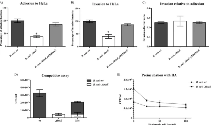
epithelial carcinoma cell line) was compared with that of the wild-type strain. Cells were seeded in multiwell plates and inoculated with the different strains for 1 h. To determine the total number of cell-associated bacteria, cells were lysed with a nonionic detergent and CFU were determined by plating serial dilutions of cell lysates. To determine the number of intracellular viable bacteria, a stan-dard gentamicin protection assay was performed. The number of adherent bacteria was calculated as the difference between total cell-associated and intracellular bacteria. B. suis⌬btaE showed a significant reduction (by 63%⫾ 7%) in the ability to adhere to HeLa cells compared to the wild-type strain (Fig. 3A) as well as a decrease (by 70%⫾ 14%) in the ability to invade these cells (Fig. 3B). Both adhesion to and invasion of HeLa cells were stored by pBBRbtaE, confirming that the absence of btaE was re-sponsible for the observed in vivo phenotypes. In order to deter-mine if the diminished invasion was a consequence of the reduction in adherence or, alternatively, was due to a reduced invasion aptitude, the invasive cell/adherent cell ratio was calcu-lated. As shown inFig. 3C, the wild-type and mutant strains showed similar efficiencies of invasion, suggesting that BtaE is involved in the initial attachment of B. suis to HeLa cells rather than in host cell internalization. To further investigate the ability of the B. suis⌬btaE mutant to attach to HeLa cells, a competitive infection assay was carried out by coinoculating equal amounts of
B. suis wild-type and⌬btaE bacteria into the HeLa cell monolayer.
FIG 3 Adhesion to and invasion of HeLa cells. Approximately 5⫻ 106bacteria were used to challenge 5⫻ 104HeLa cells (MOI, 100:1). Total numbers of
adherent bacteria (A) and intracellular or invasive bacteria (B) were determined, and the invasive cell/adherent cell ratio was calculated (C). Total numbers of adherent and intracellular bacteria are expressed relative to wild-type B. suis 1330, defined as 100%. (D) A competitive assay was carried out in which HeLa cells were coinoculated with wild-type and B. suis⌬btaE bacteria. The number of adherent bacteria was expressed as CFU/ml. (E) Inhibition of bacterial binding to HeLa cells by preincubation with hyaluronic acid (HA); data are expressed as total CFU/ml. Values represent the means⫾ SD of the results of an experiment representative of three independent assays done in triplicate or quadruplicate. Data were analyzed by one-way ANOVA followed by a Tukey a posteriori test. *, significantly different from the wild type (P⬍ 0.05).
on September 2, 2015 by BIBLIOTHEQUE UNIVERSITAIRE
The BtaE-defective mutant was strongly outcompeted by the wild-type strain for adhesion to HeLa (Fig. 3D), thus reinforcing the idea that BtaE participates in the binding of B. suis to these cells.
We also investigated whether hyaluronic acid is able to com-pete the binding of B. suis to HeLa cells. We found that preincu-bation of wild-type bacteria with 25g/ml hyaluronic acid inhib-ited the binding to these cells by about 60%. In contrast, the binding of the btaE mutant was inhibited only slightly by hyal-uronic acid (Fig. 3E). This observation provides further evidence that BtaE is involved in the binding of Brucella to hyaluronic acid. To further investigate the in vivo adhesive phenotype of the
btaE mutant, the A549 epithelial cell line from human lung was
used as an alternative cellular model in the binding assay (39). B.
suis⌬btaE showed a significant reduction in the ability to adhere
to (by 50%⫾ 8%) and invade (by 51% ⫾ 13%) A549 cells com-pared with the wild type (considered 100%) (Fig. 4AandB). As predicted, the pBBRbtaE plasmid restored adhesion and invasion capacity to wild-type levels in the B. suis⌬btaE mutant. Similar to the phenotypes in HeLa cells, our data indicate that the decrease in the invasion capacity of B. suis⌬btaE was a consequence of a lower adhesion to A549 cells (Fig. 4C).
To test the possibility that BtaE could participate in later steps of host cell infection, such as intracellular survival, a classical in-tracellular proliferation assay was carried out, using murine J774 macrophages and HeLa cells. In both cell models, the wild-type and mutant strains showed the same kinetics of intracellular rep-lication (data not shown), indicating that the absence of BtaE does not affect later stages of cellular infection.
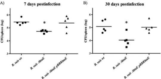
To evaluate the contribution of BtaE to the virulence of B. suis, we used the mouse infection model. Since it is possible that BtaE is involved in the initial stages of infection, we decided to inoculate mice through the intragastric route (40) and evaluate splenic in-fection at different times postinin-fection (p.i.). It was previously reported that the bacterial load in spleens and other organs of mice infected with B. abortus through the digestive track significantly increases at 7 days p.i. and stabilizes at later times (41). Therefore, we analyzed the spleen load after 7 and 30 days p.i. The B. suis ⌬btaE mutant and the complemented (B. suis ⌬btaE pBBRbtaE)
and parental strains were administered intragastrically to mice, and CFU counts in spleens were determined. At 7 and 30 days p.i., the splenic infection by the B. suis⌬btaE mutant was significantly reduced by 0.76 log and 1.24 log, respectively, compared with infection by the parental wild-type strain (Fig. 5AandB). As pre-dicted, the complemented strain harboring the pBBRbtaE plasmid showed a splenic CFU count similar to that of the wild type. These results show that BtaE is required for full virulence of B. suis in mice infected through the oral route.
BtaE has a surface-exposed unipolar localization. While the
passenger domains of monomeric autotransporters may be cleaved after secretion, the passenger domains of characterized TAs remain intact after secretion (33). A recombinant peptide corresponding to a portion of the passenger domain (seeFig. 1, underlined region) was cloned, expressed in E. coli, and purified to generate polyclonal anti-BtaE antibodies in mice. To evaluate the membrane association of BtaE, Western blot analysis of whole membranes from the B. suis wild-type or⌬btaE mutant strain obtained from exponential-phase cells was performed. Similar to other TAs (42,43), we noted that BtaE is difficult to denature. It was suggested that the C-terminal domains of TAs form heat- and so-dium dodecyl sulfate (SDS)-resistant trimers. Hence, the samples were submitted to a strong denaturing process in the presence of formic acid and resolved by SDS-PAGE. Western blot analysis using the anti-BtaE antibodies showed the presence of an⬃75-kDa protein band corresponding to monomeric BtaE, which was absent in the membrane fraction of the B. suis⌬btaE strain (Fig. 6A).
To assess the surface presentation of BtaE, late-exponential-phase cultures of the B. suis wild-type or⌬btaE strain labeled with green fluorescent protein (GFP) were refreshed for 1 h in TSB medium and then fixed without permeabilization, incubated with anti-BtaE antibodies, and observed by confocal microscopy. Un-der the conditions assayed, we found that BtaE was detected in 1% to 3% of bacteria examined, but in all cases (n⫽ 50), the red signal corresponding to BtaE exhibited unipolar localization (Fig. 6B). As expected, no red signal was observed on the surface of the B.
suis ⌬btaE mutant but the unipolar signal was restored in the
complemented strain harboring the pBBRbtaE plasmid (data not
FIG 4 Adhesion to and invasion of A549 lung epithelial cells. Approximately 5⫻ 106bacteria were used to challenge 5⫻ 104A549 cells (MOI, 100:1). Total
numbers of adherent bacteria (A) and intracellular bacteria (B) were determined, and the invasive cell/adherent cell ratio was calculated (C). Total numbers of adherent and intracellular bacteria are expressed relative to the adhesion and invasion values of the wild type, defined as 100%. Values represent the means⫾ SD of the results of an experiment representative of three independent assays done in triplicate. Data were analyzed by one-way ANOVA and a Tukey a posteriori test. *, significantly different from the wild type (P⬍ 0.05).
on September 2, 2015 by BIBLIOTHEQUE UNIVERSITAIRE
http://iai.asm.org/
shown). To our knowledge, this is the first description of a TA with unipolar localization.
We also evaluated the BtaE localization in the E. coli heterolo-gous host, carrying the pBBRbtaE plasmid. Western blot analysis indicated the presence of the 75-kDa protein corresponding to BtaE in the total membrane fraction of E. coli pBBRbtaE, which was absent in the control strain (Fig. 6C). Interestingly, immuno-fluorescence analysis of GFP-tagged E. coli carrying pBBRbtaE showed BtaE foci at one bacterial pole in 5% of bacteria examined (Fig. 6D), while no signal was observed in the control strain (data not shown). This observation suggests that the mechanism in-volved in polar targeting of the BtaE trimeric autotransporter is conserved in E. coli.
Brucella’s new pole is functionally differentiated for
adhe-sion. Brucellae are proposed to be polarized bacteria, exhibiting
asymmetric division (44) and unipolar growth, as seen with
sev-eral other Rhizobiales (45). Moreover, proteins specifically re-cruited to the old pole or to the new pole have been reported. The PdhS histidine kinase is recruited to the old pole (19,20), and the putative DNA repair protein AidB is recruited to the new pole (18). Since both the BmaC and BtaE adhesins showed unipolar localization, the question arises as whether these adhesins are lo-calized at a specific pole and, in that case, if they are lolo-calized at the same pole. Therefore, to assess the relative localizations of BmaC and BtaE, we used the PdhS and AidB proteins as markers of the old and new poles, respectively.
Since the PdhS and AidB pole markers were identified in B.
abortus, we first evaluated the localization of the BmaC
homo-logue of B. abortus, which is 99.6% identical to BmaC from B. suis. By immunofluorescence, it was confirmed that in B. abortus the BmaC homologue consistently displays unipolar localization (data not shown). Next, we used the B. abortus strains carrying the
FIG 5 Role of BtaE in the virulence of B. suis in BALB/c mice. BALB/c mice (10 mice per group) were inoculated by intragastric delivery of the B. suis wild type,
B. suis⌬btaE, or the complemented strains. Five mice from each group were sacrificed at 7 (A) and 30 (B) days postinfection, and then spleens were removed.
Dilutions of spleen homogenates were plated in duplicate, and CFU were counted and expressed as the log10value per spleen. The CFU data were normalized by
log transformation and evaluated by one-way ANOVA followed by Dunnett’s a posteriori test. The experiment was repeated twice with similar results. *, significantly different from the wild type (P⬍ 0.05).
FIG 6 BtaE localization. (A and C) Western blot analysis of whole-membrane fractions from B. suis (A) and E. coli pBBRbtaE (C) was carried out. Membrane
samples were submitted to a strong denaturing process. Samples were electrophoresed by SDS-PAGE and transferred to a PDVF membrane. Blots were incubated with anti-BtaE antisera and with anti-mouse HRP-conjugated secondary antibody and finally revealed using ECL Plus. On the right, a Coomassie blue pattern is shown as a loading control (ctrl). (B and D) Detection of BtaE (in red) on the B. suis surface (B) and on the E. coli pBBRbtaE surface (D) by immunofluorescence of GFP-tagged bacteria. Cultures of GFP-labeled strains were fixed, incubated with anti-BtaE antibodies, and then probed with a CY3-conjugated donkey anti-mouse antibody preparation. Samples were observed with a Plan-Aprochromat 100⫻/1.4 oil DIC objective on a Zeiss LSM 5 Pascal confocal microscope.
on September 2, 2015 by BIBLIOTHEQUE UNIVERSITAIRE
fusion proteins encoded by either pdhS-egfp (19) or aidB-yfp (18) to evaluate by immunofluorescence the relative localization of BmaC. DAPI staining and phase contrast were used to distinguish the bacterial shapes. Analysis by confocal microscopy showed that every time BmaC was detected (red signal) on the surface of B.
abortus carrying PdhS-eGFP protein (green), it was at the pole
opposite PdhS-eGFP (29 occurrences) (Fig. 7A). It was never ob-served that BmaC and PdhS-eGFP colocalized. Conversely, every time BmaC was detected in the strain carrying the AidB-YFP pro-tein (yellow), it was either at the same pole as the AidB-YFP marker (31 occurrences) or in cells with diffuse markers, but never at the opposite pole (Fig. 7B). As expected, a bmaC mutant of B.
abortus did not show any red signal (data not shown).
The pdhS-egfp and aidB-yfp fusions were introduced into the wild-type strain of B. suis and also into the isogenic B. suis⌬bmaC mutant as a negative control. The same analysis described above was carried out with these strains, leading to the same conclusion; i.e., BmaC is localized at the new pole of B. suis (data not shown). We next analyzed the relative localization of BtaE in B. suis. We found that every time BtaE was detected on bacteria expressing the old pole marker (PdhS-eGFP), it was at the opposite pole (30 occurrences) (Fig. 7C), but it was never observed at the same pole as PdhS-eGFP. Instead, in all the cases where BtaE was detected in bacteria carrying the aidB-yfp fusion, it was observed either at the same pole as the AidB-YFP fusion (17 occurrences) or in bacteria in which this marker was diffuse, but on no occasion was the BtaE fluorescent focus at the opposite pole (Fig. 7D). Taken together, our results show that under the conditions tested here both the BmaC and BtaE adhesins are localized at the new pole, suggesting that this pole is functionally differentiated for adhesion.
DISCUSSION
In microbial pathogens, adhesion to host cells is a critical step in the infection process. The importance of adhesion is reflected by the finding that several bacterial pathogens display multiple hesins, which can act synergistically, thereby enhancing their ad-hesive capacity. Despite the relevance of adhesion in the interac-tion with the host, the impact of this step in Brucella pathogenesis was little explored. We presented evidence indicating that the mo-nomeric BmaC autotransporter is required for an efficient adhe-sion of B. suis to host cells (15). In this work, we aimed to explore the role of BtaE, a protein that is predicted to belong to the TA family, in the interaction of B. suis with the host. Since it is ex-pected that pathogens harbor several adhesins, probably exhibit-ing overlappexhibit-ing functions, we initially assessed the function of BtaE using a heterologous approach and found that this protein was able to increase the affinity of a “nonadherent” E. coli strain to immobilized hyaluronic acid and fibronectin. Analysis of the knockout btaE mutant confirmed the role of BtaE in the binding of B. suis to one of these ECM components, the hyaluronic acid glycosaminoglycan. The possibility exists that a bind-ing-defective phenotype can be masked by other fibronectin-binding proteins, such as BmaC (15). The ability of BtaE to medi-ate the binding of Brucella to hyaluronic acid may be relevant at different stages of the infection. For example, it could facilitate initial bacterial adhesion to epithelial cells. It was previously de-scribed that hyaluronic acid is involved in adhesion of other pathogens such as Mycobacterium tuberculosis to the human lung epithelial cell line A549 (46), a cell type to which Brucella also adheres (47). Indeed, binding assays demonstrated that BtaE
con-tributes to the adhesion of B. suis to both A549 and HeLa cells. Interestingly, addition of hyaluronic acid was able to compete the binding of the wild-type strain to HeLa cells, while adhesion of the
btaE mutant was inhibited only slightly by hyaluronic acid. This
observation provides further evidence that BtaE mediates the binding of Brucella to hyaluronic acid. Furthermore, taken to-gether, our observations are consistent with the possibility that BtaE mediates adhesion to cells, at least in part, through hyal-uronic acid. As hyalhyal-uronic acid is a component of the ECM of several tissues, BtaE could also be involved in Brucella dissemina-tion to different target tissues such as cartilage, heart, and bone, which may result in serious complications of brucellosis.
The role of BtaE in the virulence of B. suis was studied by inoculating the wild type, the btaE deletion mutant, and the com-plementing strain in mice through the oral route. At 7 and 30 days p.i., splenic infection by B. suis⌬btaE was significantly reduced, compared to infection by the wild type, while the complemented strain showed a restored phenotype. This result strongly suggests that the BtaE adhesin might be considered a virulence factor and highlights the importance of bacterial adhesion to host compo-nents during the Brucella infection process.
Immunofluorescence analysis showed that BtaE exhibits uni-polar localization. To the best of our knowledge, this is the first report of unipolar localization for a TA. Yet several monomeric (type I) autotransporters from rod-shaped pathogens, including the BmaC adhesin of B. suis (15), the IcsA protein from Shigella
flexneri (48), and the AIDA-I monomeric autotransporter that mediates adhesion to human cells by pathogenic diffusely adher-ent E. coli (DAEC) strains (49), were shown to localize at one bacterial pole. It was reported that IcsA localizes to the pole at which actin assembly occurs, which is the old pole (48). Remark-ably, some of the paradigmatic type I autotransporters, including AIDA-I and IcsA, are polar in the cytoplasm prior to secretion, suggesting that secretion occurs at the pole (49,50). Intriguingly, it was shown that NalP, a monomeric autotransporter of spheri-cally shaped Neisseria meningitidis, contains the molecular infor-mation to localize to the pole of E. coli. In fact, the BtaE TA also showed unipolar localization in the E. coli heterologous host, sup-porting the notion that the polar targeting mechanism of proteins from the autotransporter families is conserved in several bacteria. The observation that both BtaE and BmaC show unipolar lo-calization prompted us to explore whether they are associated with a specific (new or old) pole and, if this is the case, whether they are at the same or a different one. Evidence presented by others and us indicates that the individual Brucella bacterium in-teracts with the host cell surface through one of its poles (15,51). Furthermore, it seems that the pole where BmaC is located on single bacteria is the one that interacts with the cell surface (15). Therefore, it was also interesting to address the issue as to which could be the “adhesive” pole. To determine the pole where the adhesins localize, we used the PdhS-eGFP fusion as a marker of the old pole (19), and the AidB-YFP fusion as a marker of the new pole (18). We observed that every time BmaC or BtaE was detected, it localized to the pole opposite the pole where PdhS-eGFP was lo-cated. In contrast, on every occasion that the BmaC or BtaE fluo-rescent foci were observed in bacteria expressing the new pole fusion, they were found at the same pole as AidB-YFP. We con-clude that under the conditions tested here, both BmaC and BtaE localize to the new pole generated after cell division. Accordingly,
on September 2, 2015 by BIBLIOTHEQUE UNIVERSITAIRE
http://iai.asm.org/
FIG 7 BtaE and BmaC are localized to the new bacterial pole. (A and B) Immunofluorescence microscopy using anti-BmaC antibodies was carried out with fixed
cultures of B. abortus strains expressing PdhS-eGFP (A) and AidB-YFP (B) as old and new pole markers, respectively. DAPI staining and phase contrast (PC) were used to visualize the bacterial DNA content and shape, respectively. Samples were observed with a confocal LSM 510 Meta microscope using a Plan-Apochromat 60⫻/1.4 oil DIC objective. Representative images are shown. BmaC, PdhS-eGFP, and AidB-YFP are indicated with red, green, and yellow arrows, respectively. A schematic representation is also displayed. An intensity profile of the channels (constructed through the cyan dotted arrow) is presented, expressed in arbitrary units. (C and D) The same analysis was carried out for BtaE in B. suis with both PdhS-eGFP (C) and AidB-YFP (D).
on September 2, 2015 by BIBLIOTHEQUE UNIVERSITAIRE
we propose that the new pole is functionally differentiated for adhesion.
BtaE was detected in a low proportion of cells. A similar obser-vation was previously made for the unipolar BmaC adhesin (15). At present, we do not know the reason for these observations. One possibility is that the level of btaE or bmaC gene transcription in
vitro is not high enough to allow detection of fluorescent foci in
most cells. If this is the case, an increase in btaE or bmaC expres-sion is predicted to occur within the host environment before bacterial internalization. Since the anti-BtaE and anti-BmaC an-tibodies used in the immunofluorescence assays recognize a pep-tide of the passenger domains, another nonexclusive explanation is that these peptides are not very accessible to the antibodies in the native-roll structure exposed on the bacterial surface (17). Fi-nally, another interesting hypothesis is that BtaE and BmaC are expressed only in a bacterial subpopulation. This possibility is supported by the finding that several cell types generated by asym-metric division and differentiation coexist in a culture of B.
abor-tus (19,20,44). Furthermore, relatively low expression of btaE or
bmaC in a small subpopulation of bacteria might be sufficient for
bacterial internalization and a successful (but limited) infection. Further studies are required to explore these possibilities.
Several surface virulence factors (including adhesive struc-tures) were found to exhibit polar localization in other alphapro-teobacteria closely related to Brucella. It was reported that all
Agro-bacterium tumefaciens VirB proteins are polarly localized (52). Furthermore, A. tumefaciens attaches efficiently to plant and abi-otic surfaces through one of its poles in a process mediated by several structures, including a unipolar polysaccharide (UPP) (53). Very interestingly, evidence was presented indicating that cells of A. tumefaciens preferentially attach to surfaces via the old cell pole (45). Evidence presented in this work and other previ-ously reported evidence (15) support the hypothesis that the un-ipolar BmaC and BtaE adhesins mediate Brucella binding to host cells at its new pole. It is possible that the characteristics of the polar asymmetry depend on the bacterial life style. In the case of A.
tumefaciens, a sessile or attached mode of life would require that
adhesion to abiotic or biotic surfaces is mediated by adhesins lo-calized at the relatively stable old pole (53); in Brucella, initial binding of single bacteria to host cells prior to cellular internaliza-tion would be mediated by adhesins localized at the new pole of a subpopulation of infective bacteria.
In addition to BmaC and BtaE, other putative adhesins were shown to participate in the interaction of Brucella with the host. Recently, it was reported that a pathogenicity island of four genes contributes to the efficiency of attachment of B. abortus to both HeLa and J774 macrophage cells. One of the proteins encoded by the gene cluster was a hypothetical protein (Bab1_2009) that har-bors an immunoglobulin-like BID_1 adhesion domain, found in adhesins such as the E. coli intimin (13). In addition, it was previ-ously reported that a predicted adhesin from the monomeric au-totransporter family from B. suis (OmaA) would be important during the chronic phase of infection (54). It will be interesting to determine whether Bab1_2009, OmaA, BtaE, and BmaC play re-dundant, synergistic, or complementary roles in the interaction of
Brucella with different cell types and/or host tissues. Furthermore,
different adhesins could exert their roles at different infection stages. Studies on gene regulation could give a hint regarding these possibilities. Through a transcriptomic analysis, it was previously shown that the expression of the btaE orthologue from B.
meliten-sis (BMEI1873) is reduced in a mutant defective in the LuxR
ho-mologue VjbR regulator (55), which modulates the expression of transcripts required for intracellular survival (56). Besides, by mi-croarray analysis it was found that transcription of the bmaC or-thologue of B. abortus was downregulated by the BvrR/BvrS two-component system. This system plays a role in the adjustment of
Brucella physiology to the shift expected to occur during the
tran-sition from the extracellular to the intracellular niche (57). Thus, one plausible hypothesis is that btaE expression and bmaC expres-sion are induced under different stimulus conditions. Future studies will focus on the regulatory elements that control the ex-pression of the autoransporter adhesins during the onset of cell infection.
Not only surface proteins were shown to participate in the adhesive properties of Brucella. Overexpression of an acyl homo-serine-lactone (AHL) acylase (that degrades AHLs) in B. melitensis induced clumping and increased adhesion to both polystyrene and HeLa cells, probably due to the production of both some polymeric substances and outer membrane vesicles (16). It was argued that adhesion of the quorum-sensing-defective strain was somehow reminiscent of the adherence of localized bacterial col-onies of B. abortus to epithelial cells (12). Thus, it is possible that initial and polar attachment of single bacteria to the host cell is mediated by proteinaceous adhesins, such as BtaE or BmaC and that, eventually, subsequent formation of bacterial microcolonies would be promoted by the production of other polymeric sub-stances induced in contact with the cell surface.
Finally, host tropism is a very important feature of the Brucella genus, for which the concept of species is controversial, due to the high degree of similarity between the Brucella genomes (58). For example, comparison of the B. melitensis 16 M and B. suis 1330 genomes revealed extensive gene similarity and synteny, with the majority (⬎90%) of genes sharing 98% to 100% identity (32,59). It is striking that most of the genes that were more variable were genes encoding surface-exposed proteins, leading to the hypoth-esis that they could contribute to differences in host preference and disease manifestation (32). In addition to B. suis and B.
melitensis, we also found BtaE orthologues in B. abortus 2308
(BAB1_0069), B. canis ATCC 23365 (BCAN_A0073), B. ovis 25810 (BOV_0071), and B. microti CCM4915 (BMI_I75). Inter-estingly, in silico analysis indicated that there are significant differ-ences either in protein lengths or in the number of repetitive mo-tifs between the different ortologues. Therefore, variations found in BtaE and other adhesins such as BmaC (15) may well contribute to host preference or tissue tropism. It will be interesting, in the future, to assess this hypothesis.
ACKNOWLEDGMENTS
This work was supported by a grant from University of Buenos Aires (UBACyT, X-240) to A.Z. and by Concerted Research Action 08/13-015, FRFC (grants 2.4541.08 and 2.4510.12 from FNRS-FRS), Interuniversity Attraction Pole (P7/27, Belgian Science Policy Office), and the University of Namur to X.D.B. V.R.-R., D.M.P., G.M.A., and F.A.M. were supported by fellowships from CONICET, Argentina. A.Z., P.L.A., and R.S. are re-search fellows of CONICET. C.V.D.H. was FRIA-FNRS-FRS fellow.
We acknowledge Martín Rumbo and Dolores González Maciel for providing the A549 cell line, Susana Raffo and Marta Bravo for DNA sequencing and other technical support, and Máximiliano Neme for help with confocal microscopy. We also thank our colleagues at the laboratory for helpful discussions.
on September 2, 2015 by BIBLIOTHEQUE UNIVERSITAIRE
http://iai.asm.org/
Survival of the fittest: how Brucella strains adapt to their intracellular niche in the host. Med. Microbiol. Immunol. 198:221–238.
7. Pizarro-Cerda J, Meresse S, Parton RG, van der Goot G, Sola-Landa A,
Lopez-Goni I, Moreno E, Gorvel JP. 1998. Brucella abortus transits
through the autophagic pathway and replicates in the endoplasmic retic-ulum of nonprofessional phagocytes. Infect. Immun. 66:5711–5724. 8. Martirosyan A, Moreno E, Gorvel JP. 2011. An evolutionary strategy for
a stealthy intracellular Brucella pathogen. Immunol. Rev. 240:211–234. 9. Gorvel JP. 2008. Brucella: a Mr “Hide” converted into Dr Jekyll. Microbes
Infect. 10:1010 –1013.
10. Pizarro-Cerda J, Cossart P. 2006. Bacterial adhesion and entry into host cells. Cell 124:715–727.
11. Kline KA, Falker S, Dahlberg S, Normark S, Henriques-Normark B. 2009. Bacterial adhesins in host-microbe interactions. Cell Host Microbe
5:580 –592.
12. Castaneda-Roldan EI, Avelino-Flores F, Dall’Agnol M, Freer E, Cedillo
L, Dornand J, Giron JA. 2004. Adherence of Brucella to human epithelial
cells and macrophages is mediated by sialic acid residues. Cell. Microbiol.
6:435– 445.
13. Czibener C, Ugalde JE. 2012. Identification of a unique gene cluster of
Brucella spp. that mediates adhesion to host cells. Microbes Infect.
14:79 – 85.
14. Hernandez-Castro R, Verdugo-Rodriguez A, Puente JL, Suarez-Guemes
F. 2008. The BMEI0216 gene of Brucella melitensis is required for
inter-nalization in HeLa cells. Microb. Pathog. 44:28 –33.
15. Posadas DM, Ruiz-Ranwez V, Bonomi HR, Martin FA, Zorreguieta A. 2012. BmaC, a novel autotransporter of Brucella suis, is involved in bacte-rial adhesion to host cells. Cell. Microbiol. 14:965–982.
16. Godefroid M, Svensson MV, Cambier P, Uzureau S, Mirabella A, De
Bolle X, Van Cutsem P, Widmalm G, Letesson JJ. 2010. Brucella
meliten-sis 16M produces a mannan and other extracellular matrix components
typical of a biofilm. FEMS Immunol. Med. Microbiol. 59:364 –377. 17. Dautin N, Bernstein HD. 2007. Protein secretion in gram-negative
bac-teria via the autotransporter pathway. Annu. Rev. Microbiol. 61:89 –112. 18. Dotreppe D, Mullier C, Letesson JJ, De Bolle X. 2011. The alkylation response protein AidB is localized at the new poles and constriction sites in
Brucella abortus. BMC Microbiol. 11:257.
19. Hallez R, Mignolet J, Van Mullem V, Wery M, Vandenhaute J, Letesson
JJ, Jacobs-Wagner C, De Bolle X. 2007. The asymmetric distribution of
the essential histidine kinase PdhS indicates a differentiation event in
Bru-cella abortus. EMBO J. 26:1444 –1455.
20. Van der Henst C, Beaufay F, Mignolet J, Didembourg C, Colinet J,
Hallet B, Letesson JJ, De Bolle X. 2012. The histidine kinase PdhS
controls cell cycle progression of the pathogenic alphaproteobacterium
Brucella abortus. J. Bacteriol. 194:5305–5314.
21. Schafer A, Tauch A, Jager W, Kalinowski J, Thierbach G, Puhler A. 1994. Small mobilizable multi-purpose cloning vectors derived from the
Escherichia coli plasmids pK18 and pK19: selection of defined deletions in
the chromosome of Corynebacterium glutamicum. Gene 145:69 –73. 22. Kovach ME, Elzer PH, Hill DS, Robertson GT, Farris MA, Roop RM, II,
Peterson KM. 1995. Four new derivatives of the broad-host-range cloning
vector pBBR1MCS, carrying different antibiotic-resistance cassettes. Gene
166:175–176.
23. Punta M, Coggill PC, Eberhardt RY, Mistry J, Tate J, Boursnell C, Pang
N, Forslund K, Ceric G, Clements J, Heger A, Holm L, Sonnhammer EL, Eddy SR, Bateman A, Finn RD. 2012. The Pfam protein families
database. Nucleic Acids Res. 40:D290 –D301.
24. Altschul SF, Madden TL, Schaffer AA, Zhang J, Zhang Z, Miller W,
lus influenzae biogroup aegyptius, which promotes entry into host cells.
Cell. Microbiol. 11:1044 –1063.
29. Klingman KL, Murphy TF. 1994. Purification and characterization of a high-molecular-weight outer membrane protein of Moraxella
(Branha-mella) catarrhalis. Infect. Immun. 62:1150 –1155.
30. Kovach ME, Phillips RW, Elzer PH, Roop RM, II, Peterson KM. 1994. pBBR1MCS: a broad-host-range cloning vector. Biotechniques 16:800 – 802.
31. Lambertsen L, Sternberg C, Molin S. 2004. Mini-Tn7 transposons for site-specific tagging of bacteria with fluorescent proteins. Environ. Micro-biol. 6:726 –732.
32. Paulsen IT, Seshadri R, Nelson KE, Eisen JA, Heidelberg JF, Read TD,
Dodson RJ, Umayam L, Brinkac LM, Beanan MJ, Daugherty SC, Deboy RT, Durkin AS, Kolonay JF, Madupu R, Nelson WC, Ayodeji B, Kraul M, Shetty J, Malek J, Van Aken SE, Riedmuller S, Tettelin H, Gill SR, White O, Salzberg SL, Hoover DL, Lindler LE, Halling SM, Boyle SM, Fraser CM. 2002. The Brucella suis genome reveals fundamental
similar-ities between animal and plant pathogens and symbionts. Proc. Natl. Acad. Sci. U. S. A. 99:13148 –13153.
33. Cotter SE, Surana NK, St Geme JW, III. 2005. Trimeric autotransport-ers: a distinct subfamily of autotransporter proteins. Trends Microbiol.
13:199 –205.
34. Lyskowski A, Leo JC, Goldman A. 2011. Structure and biology of trim-eric autotransporter adhesins. Adv. Exp. Med. Biol. 715:143–158. 35. Linke D, Riess T, Autenrieth IB, Lupas A, Kempf VA. 2006. Trimeric
autotransporter adhesins: variable structure, common function. Trends Microbiol. 14:264 –270.
36. El Tahir Y, Skurnik M. 2001. YadA, the multifaceted Yersinia adhesin. Int. J. Med. Microbiol. 291:209 –218.
37. Cope LD, Lafontaine ER, Slaughter CA, Hasemann CA, Jr, Aebi C,
Henderson FW, McCracken GH, Jr, Hansen EJ. 1999. Characterization
of the Moraxella catarrhalis uspA1 and uspA2 genes and their encoded products. J. Bacteriol. 181:4026 – 4034.
38. Hoiczyk E, Roggenkamp A, Reichenbecher M, Lupas A, Heesemann J. 2000. Structure and sequence analysis of Yersinia YadA and Moraxella UspAs reveal a novel class of adhesins. EMBO J. 19:5989 –5999. 39. Ferrero MC, Fossati CA, Baldi PC. 2009. Smooth Brucella strains invade
and replicate in human lung epithelial cells without inducing cell death. Microbes Infect. 11:476 – 483.
40. Delpino MV, Marchesini MI, Estein SM, Comerci DJ, Cassataro J,
Fossati CA, Baldi PC. 2007. A bile salt hydrolase of Brucella abortus
contributes to the establishment of a successful infection through the oral route in mice. Infect. Immun. 75:299 –305.
41. Paixao TA, Roux CM, den Hartigh AB, Sankaran-Walters S, Dandekar
S, Santos RL, Tsolis RM. 2009. Establishment of systemic Brucella
melitensis infection through the digestive tract requires urease, the type IV
secretion system, and lipopolysaccharide O antigen. Infect. Immun. 77: 4197– 4208.
42. Roggenkamp A, Ackermann N, Jacobi CA, Truelzsch K, Hoffmann H,
Heesemann J. 2003. Molecular analysis of transport and oligomerization
of the Yersinia enterocolitica adhesin YadA. J. Bacteriol. 185:3735–3744. 43. Surana NK, Cutter D, Barenkamp SJ, St Geme JW, III. 2004. The
Haemophilus influenzae Hia autotransporter contains an unusually short
trimeric translocator domain. J. Biol. Chem. 279:14679 –14685. 44. Hallez R, Bellefontaine AF, Letesson JJ, De Bolle X. 2004.
Morpholog-ical and functional asymmetry in alpha-proteobacteria. Trends Microbiol.
12:361–365.
45. Brown PJ, de Pedro MA, Kysela DT, Van der Henst C, Kim J, De Bolle
on September 2, 2015 by BIBLIOTHEQUE UNIVERSITAIRE
X, Fuqua C, Brun YV. 2012. Polar growth in the Alphaproteobacterial
order Rhizobiales. Proc. Natl. Acad. Sci. U. S. A. 109:1697–1701. 46. Aoki K, Matsumoto S, Hirayama Y, Wada T, Ozeki Y, Niki M,
Dome-nech P, Umemori K, Yamamoto S, Mineda A, Matsumoto M, Ko-bayashi K. 2004. Extracellular mycobacterial DNA-binding protein 1
par-ticipates in Mycobacterium-lung epithelial cell interaction through hyaluronic acid. J. Biol. Chem. 279:39798 –39806.
47. Ferrero MC, Fossati CA, Baldi PC. 2010. Direct and monocyte-induced innate immune response of human lung epithelial cells to Brucella abortus infection. Microbes Infect. 12:736 –747.
48. Goldberg MB, Barzu O, Parsot C, Sansonetti PJ. 1993. Unipolar local-ization and ATPase activity of IcsA, a Shigella flexneri protein involved in intracellular movement. J. Bacteriol. 175:2189 –2196.
49. Jain S, van Ulsen P, Benz I, Schmidt MA, Fernandez R, Tommassen J,
Goldberg MB. 2006. Polar localization of the autotransporter family of
large bacterial virulence proteins. J. Bacteriol. 188:4841– 4850.
50. Steinhauer J, Agha R, Pham T, Varga AW, Goldberg MB. 1999. The unipolar Shigella surface protein IcsA is targeted directly to the bacterial old pole: IcsP cleavage of IcsA occurs over the entire bacterial surface. Mol. Microbiol. 32:367–377.
51. Pizarro-Cerdá J, Moreno E, Gorvel JP. 1999. Brucella abortus: invasion and survival within professional and non-professional phagocytes, vol 6. Jai Press Inc., Greenwich, CT.
52. Judd PK, Kumar RB, Das A. 2005. The type IV secretion apparatus protein VirB6 of Agrobacterium tumefaciens localizes to a cell pole. Mol. Microbiol. 55:115–124.
53. Tomlinson AD, Fuqua C. 2009. Mechanisms and regulation of polar surface attachment in Agrobacterium tumefaciens. Curr. Opin. Microbiol.
12:708 –714.
54. Bandara AB, Sriranganathan N, Schurig GG, Boyle SM. 2005. Putative outer membrane autotransporter protein influences survival of Brucella
suis in BALB/c mice. Vet. Microbiol. 109:95–104.
55. Weeks JN, Galindo CL, Drake KL, Adams GL, Garner HR, Ficht TA. 2010. Brucella melitensis VjbR and C12-HSL regulons: contributions of the N-dodecanoyl homoserine lactone signaling molecule and LuxR homo-logue VjbR to gene expression. BMC Microbiol. 10:167.
56. Delrue RM, Deschamps C, Leonard S, Nijskens C, Danese I, Schaus JM,
Bonnot S, Ferooz J, Tibor A, De Bolle X, Letesson JJ. 2005. A
quorum-sensing regulator controls expression of both the type IV secretion system and the flagellar apparatus of Brucella melitensis. Cell. Microbiol. 7:1151– 1161.
57. Viadas C, Rodriguez MC, Sangari FJ, Gorvel JP, Garcia-Lobo JM,
Lopez-Goni I. 2010. Transcriptome analysis of the Brucella abortus BvrR/
BvrS two-component regulatory system. PLoS One 5:e10216.
58. Moreno E, Cloeckaert A, Moriyon I. 2002. Brucella evolution and tax-onomy. Vet. Microbiol. 90:209 –227.
59. DelVecchio VG, Kapatral V, Redkar RJ, Patra G, Mujer C, Los T,
Ivanova N, Anderson I, Bhattacharyya A, Lykidis A, Reznik G, Jablon-ski L, Larsen N, D’Souza M, Bernal A, Mazur M, Goltsman E, Selkov E, Elzer PH, Hagius S, O’Callaghan D, Letesson JJ, Haselkorn R, Kyrpides N, Overbeek R. 2002. The genome sequence of the facultative
intracellu-lar pathogen Brucella melitensis. Proc. Natl. Acad. Sci. U. S. A. 99:443– 448.