HAL Id: tel-01628207
https://tel.archives-ouvertes.fr/tel-01628207
Submitted on 3 Nov 2017HAL is a multi-disciplinary open access
archive for the deposit and dissemination of sci-entific research documents, whether they are pub-lished or not. The documents may come from teaching and research institutions in France or abroad, or from public or private research centers.
L’archive ouverte pluridisciplinaire HAL, est destinée au dépôt et à la diffusion de documents scientifiques de niveau recherche, publiés ou non, émanant des établissements d’enseignement et de recherche français ou étrangers, des laboratoires publics ou privés.
Application of Alpha-Repeat Proteins as Antiviral
Molecules Against HIV-1 Targeting Viral Assembly or
Maturation
Sudarat Hadpech
To cite this version:
Sudarat Hadpech. Application of Alpha-Repeat Proteins as Antiviral Molecules Against HIV-1 Tar-geting Viral Assembly or Maturation. Virology. Université de Lyon; Mahāwitthayālai Chīang Mai, 2017. English. �NNT : 2017LYSE1139�. �tel-01628207�
N°d’ordre NNT : 2017LYSE1139
THESE de DOCTORAT DE L’UNIVERSITE DE LYON
opérée au sein de
l’Université Claude Bernard Lyon 1
Ecole Doctorale ED341-E2M2
Evolution Ecosystèmes - Microbiologie Modélisation et
University of Chiang Mai
Faculty of Associated Medical Sciences
Diplôme de Doctorat
Soutenue publiquement le 18 Juillet 2017 par :
Mlle Sudarat HADPECH
Nouveaux agents antiviraux dérivés de protéines cellulaires à motifs répétés et ciblant l’assemblage du VIH
Devant le jury composé de :
M. le Professeur ANDRE Patrice Examineur
Mme. la Professeure CHAICAMPA Wanpen Rapporteure
M. le Professeur MINARD Philippe Rapporteur
Dr. KITIDEE Kuntida Rapporteure
Dr. YASAMUT Umpa Examinatrice
Dr. NANGOLA Sawitree Co-Directrice de Thèse
Dr. HONG Saw-See Directrice de Thèse
1
UNIVERSITE CLAUDE BERNARD - LYON 1
Président de l’Université M. le Professeur Frédéric FLEURY
Président du Conseil Académique M. le Professeur Hamda BEN HADID Vice-président du Conseil
d’Administration
M. le Professeur Didier REVEL Vice-président du Conseil Formation et
Vie Universitaire
M. le Professeur Philippe CHEVALIER Vice-président de la Commission
Recherche M. Fabrice VALLÉE
Directrice Générale des Services Mme Dominique MARCHAND COMPOSANTES SANTE
Faculté de Médecine Lyon Est – Claude Bernard
Directeur : M. le Professeur G.RODE Faculté de Médecine et de Maïeutique
Lyon Sud – Charles Mérieux
Directeur : Mme la Professeure C. BURILLON
Faculté d’Odontologie Directeur : M. le Professeur D. BOURGEOIS
Institut des Sciences Pharmaceutiques et Biologiques
Directeur : Mme la Professeure C. VINCIGUERRA
Institut des Sciences et Techniques de la Réadaptation
Directeur : M. X. PERROT Département de formation et Centre de
2
COMPOSANTES ET DEPARTEMENTS DE SCIENCES ET TECHNOLOGIE
Faculté des Sciences et Technologies Directeur : M. F. DE MARCHI Département Biologie Directeur : M. le Professeur F.
THEVENARD
Département Chimie Biochimie Directeur : Mme C. FELIX
Département GEP Directeur : M. Hassan HAMMOURI
Département Informatique Directeur : M. le Professeur S. AKKOUCHE
Département Mathématiques Directeur : M. le Professeur G. TOMANOV
Département Mécanique Directeur : M. le Professeur H. BEN HADID
Département Physique Directeur : M. le Professeur J-C PLENET
UFR Sciences et Techniques des
Activités Physiques et Sportives Directeur : M. Y.VANPOULLE Observatoire des Sciences de l’Univers
de Lyon Directeur : M. B. GUIDERDONI
Polytech Lyon Directeur : M. le Professeur E.PERRIN Ecole Supérieure de Chimie Physique
Electronique
Directeur : M. G. PIGNAULT
Institut Universitaire de Technologie de Lyon 1
Directeur : M. le Professeur C. VITON
Ecole Supérieure du Professorat et de
l’Education Directeur : M. le Professeur A. MOUGNIOTTE Institut de Science Financière et
3
Résumé de la thèse en Français
Au cours de notre programme de thèse, nous avons isolé et caractérisé des molécules protéiques à activité antivirale intracellulaire dirigée contre le VIH-1. Ces protéines, appelées αRep, ont été obtenues par criblage d'une banque de protéines artificielles de nouvelle génération, construites de façon combinatoire à partir de protéines naturelles constituées de motifs alpha-hélicoidaux répétés. La cible virale (ou "appât") utilisée pour ce criblage est une région de la polyprotéine Gag du VIH-1 identifiée comme une cible privilégiée de nouvelles thérapeutiques antivirales, car essentielle à l'assemblage viral, l'empaquetage du génome viral et le clivage de maturation de Gag aboutissant à la formation de virions infectieux. Deux molécules d'αRep à forte affinité pour la cible virale, l'αRep4E3 (32 kDa; 7 motifs répétés) et l'αRep9A8 (28 kDa; 6 motifs répétés) ont ainsi été isolées, clonées et caractérisées. L'étude de l'activité anti-VIH intracellulaire de ces αRep a été réalisée dans différents systèmes d'expression cellulaire, nécessitant la construction de lignées stables de cellules d'insecte et de cellules épithéliales humaines, et leur infection par différents types de vecteurs viraux recombinants, baculovirus ou lentivirus, porteurs du gène rapporteur luciférase. Mais surtout, notre étude a été menée sur des cellules lymphocytaires-T (SupT1), cibles naturelles du virus, exprimant ces αRep et infectées par du VIH-1 naturel infectieux. Nos résultats ont montré que l'αRep4E3 et l'αRep9A8 ont toutes deux un effet négatif significatif sur le cycle réplicatif du VIH-1, mais ciblent des fonctions virales différentes. L'αRep4E3 bloque l'empaquetage du génome viral, tandis que l'αRep9A8 inhibe la maturation et diminue l'infectivité virale. De plus, l'αRep9A8, exprimée de façon constitutive dans les cellules SupT1, leur confère une résistance au VIH: une lignée de SupT1 chroniquement infectée par le VIH-1 a pu être ainsi isolée et maintenue en culture pendant plusieurs semaines, sans effet cytopathique viro-induit apparent. Ces nouvelles données auront des implications non-négligeables dans le choix et la conduite de futures stratégies de thérapie cellulaire anti-VIH.
Mots-clés: VIH-1; antivirals; designed molecular scaffolds; HEAT-like repeat; αRep protéine
4
ABSTRACT
Human immunodeficiency virus type 1 (HIV-1) infection is a long-term disease which required a long-life treatment. Besides the standard highly active retroviral therapy (HAART) regiment, HIV-1 gene therapy is one of a promising alternative strategy which give rise to hope for better HIV-1 treatments. In addition, protein-based therapeutics represent another promising approach which show the high impact results in curing various types of diseases. Nowadays, it has already become a significant part of the current medical treatments. In this study, we focused on αRep proteins, the non-immunoglobulin scaffold proteins which were designed to target nucleocapsid (NC) domain of HIV-1 Pr55Gag polyprotein precursor and investigated their possible roles as intracellular therapeutic agents. Phage display technology was used for the specific isolation of αRep against a critical C-terminal region of the HIV-1 Pr55Gag (CA21
-SP1-NC) from a large and diverse αRep phage library. Several strong αRep binders were thus isolated, but only two binders, referred to as αRep4E3 and αRep9A8, were further characterized. The αRep4E3 contains 7 internal repeat motifs (32 kDa), whereas αRep9A8 has 6 repeat motifs (28 kDa). Both αRep proteins were expressed at high level in a conventional bacterial expression system, and were recovered at high yields in soluble form. Their potential antiviral activity was investigated using different cell systems. The baculovirus system was used for determining the antiviral effects on virus-like particle (VLP) production of both αRep scaffold molecules. Sf9 cells stably expressing N-myristoylated αRep4E3 and αRep9A8 fused to the GFP reporter protein at their C-termini were infected with a recombinant baculovirus carrying the gene encoding the HIV-1 Pr55Gag polyprotein (AcMNPVgag), and the antiviral effects were evaluated
by quantitative and qualitative methods. The results showed that N-myristoylated αRep4E3 and αRep9A8 qualitatively altered the particle formation and were coencapsidated with Pr55Gag into the VLPs released into the culture supernatant. The different patterns of particle morphology were observed by electron microscopy. The αRep4E3-expressing cells mainly produce aberrant VPLs, while αRep9A8 expression induces the accumulation of Pr55Gag at the plasma membrane of
5
AcMNPVgag-infected Sf9 cells. In the human T cell line system, αRep4E3 and αRep9A8
proteins were expressed intracellularly by transducing SupT1 cell with SIN CGW lentiviral vector carrying the αRep genes fused to the EGFP coding sequence at their 3'-end. These stable SupT1 cell lines were then challenged with HIV-1NL4-3.The results
indicated that both αRep4E3 and αRep9A8 displayed antiviral effects, which occurred at the late steps of the HIV-1 life cycle, with no effect on proviral DNA integration. Reduction and delay in the viral progeny production were found in infected SupT1 expressing αRep4E3-EGFP and αRep9A8-EGFP. Difference in the antiviral mechanism was observed between these two αRep proteins: αRep4E3-EGFP mainly interferes with the packaging of the viral genomic RNA, while αRep9A8-EGFP interferes with the proteolytic processing of Pr55Gag polyprotein, and performs as a protease inhibitor to prevent the HIV-1 protease cleavage which is required for the production of newly infectious mature virions. Interestingly, SupT1 expressing αRep9A8-EGFP is able to survive to chronical HIV-1 infection up to 38 days after infection, with a low level of noninfectious HIV-1 particle production. Taken together, our results suggested that αRep, a new type of scaffold protein, could serve as a promising alternative antiviral agent which would influence the future strategies and candidates of antiviral molecules to be used in anti-HIV-1 cell therapy.
Keywords: HIV-1; antivirals; designed molecular scaffolds; HEAT-like repeat; αRep proteins
6
บทคัดย่อ
การติดเชือไวรัสเอชไอวี 1 ก่อให้เกิดโรคติดเชือเรือรังทีต้องรักษาตลอดชีวิต นอกเหนือไปจาก
การรักษาด้วยยาต้านไวรัสซึงเป็นการรักษามาตรฐานในปัจจุบันแล้ว การรักษาด้วยยีนถือเป็นอีกหนึง
แนวทางใหม่ทีช่วยเพิมความหวังในการรักษาให้ดีขึน โดยทําการออกแบบยีนทีกําหนดการสร้าง
โมเลกุลทีมีประสิทธิภาพในการยับยังการแบ่งตัวของเชือไวรัส ในการศึกษานีเป็นครังแรกทีใช้ยีน
สําหรับสร้างโปรตีนแอลฟารีพีทซึงเป็นโปรตีนโครงสร้างพิเศษทีแตกต่างไปจากแอนติบอดี
มาทดสอบการยับยังวงจรชีวิตของไวรัสเอชไอวี 1
โดย
ทําการคัดเลือกโปรตีนแอลฟารีพีททีสามารถ
จับได้กับบริเวณนิวคลีโอแคพซิด ซึงเป็นส่วนหนึงของโปรตีนโครงสร้างของเชือไวรัสโดยเทคนิค
ฟาจดิสเพลย์ พบว่าสามารถคัดเลือกโปรตีนแอลฟารีพีทโคลนทีจับอย่างจําเพาะได้ 2 โคลนคือ
สีอีสาม (αRep4E3) และเก้าเอแปด (αRep4E3) ซึงโปรตีนแอลฟารีพีทดังกล่าวนีประกอบไปด้วย
จํานวนรีพีท 7 รีพีท มีขนาดโมเลกุล 32 กิโลดาลตัน และ 6 รีพีท มีขนาดโมเลกุล 28 กิโลดาลตัน
ตามลําดับ โปรตีนทังสองโมเลกุลมีระดับการแสดงออกทีสูงและมีความคงตัวสูงในเซลล์หลายชนิด
ทังในแบคทีเรีย เซลล์แมลง และเซลล์มนุษย์ จากนันนํายีนของโปรตีนทังสองโคลนนีเข้าสู่เซลล์เพือ
ทดสอบฤทธิในการยับยังวงจรชีวิตของเชือไวรัสภายในเซลล์ ทังในเซลล์แมลงชนิดเอสเอฟเก้า (Sf9)
และในเซลล์เม็ดเลือดขาวของมนุษย์ชนิดทีเซลล์ (SupT1)ซึงการทดสอบผลของแอลฟารีพีททังสอง
ในการรบกวนการผลิตอนุภาคเสมือนไวรัส (VLP) โดยใช้เซลล์แมลงนัน ได้ทําการออกแบบให้
โปรตีนแอลฟารีพีทมีการแสดงออกในรูปแบบทีมีไมริสติกแอซิดติดอยู่ด้านทางปลายอะมิโนและ
เชือมต่อโปรตีนเรืองแสงสีเขียว (GFP) เข้าไปทางด้านปลายคาร์บอกซิลิค จากนันจึงติดเชือเซลล์
ดังกล่าวด้วยแบคคูโลวัสไวรัส AcMNPV
gagทีทําหน้าทีในการนําส่งยีนกําหนดการสร้างโปรตีนแกก
(Gag) ของไวรัสเอชไอวี 1 และติดตามผลการรบกวนการสร้าง VLP ในเซลล์ดังกล่าว จากผลการ
ทดลองพบว่าโปรตีนแอลฟารีพีททังสองมีผลทําให้การกระกอบอนุภาคของไวรัสแปรเปลียนไปใน
เชิงคุณภาพ พบรูปแบบทีแตกต่างกันทางสัณฐานวิทยาของ VLP ซึงเป็นผลมาจากฤทธิของโปรตีน
แอลฟารีพีทเมือศึกษาโดยใช้กล้องจุลทรรศน์อิเล็กตรอน โดยสังเกตพบว่า αRep4E3 มีฤทธิส่งเสริม
ให้มีการผลิต VLP ทีมีรูปร่างผิดปกติ ในขณะที αRep9A8 มีผลทําให้เกิดการสะสมของโปรตีน Gag
ทีผนังเซลล์ Sf9 และพบอีกว่าโปรตีนแอลฟารีพีททังสองถูกบรรจุเข้าไปในอนุภาค VLP จากนันจึง
ทําการศึกษาฤทธิของโปรตีนแอลฟารีพีทต่อในเซลล์เม็ดเลือดขาวของมนุษย์ชนิด SupT1 โดยได้
ออกแบบโปรตีนแอลฟารีพีททังสองให้มีการแสดงออกภายในเซลล์ได้อย่างต่อเนืองโดยได้เชือม
7
EGFP ไว้ทางด้านปลายปลายคาร์บอกซิลิคของโปรตีน จากนันทําการติดเชือเซลล์ทีมีการแสดงออก
ของโปรตีนแอลฟารีพีททังสองชนิดด้วยไวรัสเอชไอวีที 1 สายพันธุ์ NL4-3 ที 1 MOI ติดตามผลการ
ทดลองพบว่าโปรตีนแอลฟารีทังสองตัวมีความสามารถในการต้านการเพิมจํานวนของไวรัสและพบ
อีกว่าโปรตีนแอลฟารีพีททังสองออกฤทธิในช่วงท้ายของวงจรชีวิตของเอชไอวีโดยทีไม่มีผลในการ
ยับยังการฝังตัวของดีเอ็นของไวรัสในโครโมโซมของเซลล์ติดเชือ นอกจากนันพบว่าไวรัสตัวใหม่
สร้างได้น้อยลงและช้าลงในเซลล์ทีมีการแสดงออกของโปรตีนแอลฟารีพีททังสอง และยังพบอีกว่า
กลไกในการยับยังไวรัสนันแตกต่างกันระหว่างโปรตีนแอลฟารีพีททังสองตัว โดย αRep4E3 มีฤทธิ
ในการรบกวนการบรรจุอาร์เอ็นเอจีโนมเข้าไปในอนุภาคไวรัส ในขณะที αRep9A8 พบว่ามีการ
รบกวนกระบวนการสร้างไวรัสทีสมบูรณ์ (maturation)โดยทําหน้าทีเสมือนเป็นตัวขัดขวางเอ็นไซม์
โปรติเอสไม่ให้สามารถเข้าตัดโปรตีน Gag ได้ ซึงขันตอนดังกล่าวนีมีความจําเป็นสําหรับการผลิต
ไวรัสตัวเต็มวัยตัวใหม่ อีกทังยังพบประเด็นทีน่าสนใจว่าเซลล์ SupT1 ทีมีการแสดงออกของ
αRep9A8 สามารถรอดชีวิตนานถึง 38 วันในสภาวะทีเป็นการติดเชือเรือรัง พบการสร้างไวรัสตัวไม่
เต็มวัยและไม่มีความสามารถในการติดเชือออกมาในนําเพาะเลียงในระดับตํา จากการศึกษาทังหมดนี
สามารถกล่าวได้ว่าโปรตีนแอลฟารีพีทเป็นโปรตีนชนิดใหม่ซึงถือเป็นทางเลือกทีน่าสนใจสําหรับการ
นํามาใช้ในการพัฒนาเป็นโมเลกุลต้านไวรัสและสามารถนํามาต่อยอดเพือการพัฒนาเซลล์ต้านไวรัส
เอชไอวีเพือใช้ในการรักษาได้
8
ACKNOWLEDGEMENTS
This thesis is the story which has been arisen for several years in my Ph.D. study. I would say that it is such a great and wonderful memories. However, I will never forget experiencing many important lessons, hard examinations, late night experiments and the stressful I have when I wrote this thesis. During my study program, I have had a very great honor to work with a large number of amazing people. It would not have been possible to finish this thesis without their warm help, support, guidance, and friendship. It also would not have been possible to finish this thesis without financial support throughout my education. I would like to take this opportunity to express that this thesis is the result of many experiences I have encountered from dozens of remarkable individuals who I wish to acknowledge.
First and foremost I wish to express my deepest gratitude and sincere appreciation to my thesis supervisor, Prof. Dr. Chatchai Tayapiwatana, for giving me a great opportunity to be a Ph.D. student under his supervision. It has been a great honor to be his student, not only because of his tremendous academic support but also because he is one of the great teachers and researchers I have ever met in my life. His guidance helped me in all the time of studying and writing of this thesis. He has always made himself available to clarify my doubts despite his busy schedules. I could not have imagined having a better advisor and mentor for my Ph.D. study. He is the type of person called "polymath" which means "a man can do all things if he would like to". I really wish that someday I could be as enthusiastic and energetic as Prof. Chatchai and to someday be able to command an audience as well as he can.
I am extremely grateful to express my gratitude and appreciation to Dr. Saw-See Hong and Prof. Pierre Boulanger, my thesis advisors from UMR754, UCBL, and my parents in Lyon for their support, guidance, and taking care of me. They are the beginning of alpha repeat proteins project, the beginning of this thesis work. Dr. Saw-See is the one that changed my perspective on life. I see things more in a positive way after I know her. I have learned to be more kind, sweet, and be a positive thinking person than I was.
9
Prof. Pierre is a rare type of man I know, I mean it in a good way. He is like a big mobile library with a large storage capacity. He always makes me surprise that how can one man packs all that knowledge inside the brain, not only about sciences but also many others; history, archeology, architecture, and politic, and everything. I must thank you for all of your kind, supportive, encouragement, insightful comments and hard questions during thesis writing and manuscript preparation. Finally, I would like to thank you for taking care of me like a family, without you two, living in France for two years could not be this happy.
I take this opportunity to sincerely acknowledge Prof. Dr. Watchara Kasinrerk for valuable advice, kindly providing monoclonal antibodies, and support laboratory equipment. I also thanks to Dr. Supansa Pata, and other members of Prof. Dr. Watchara Kasinrerk’s laboratory for their technical support and kind help.
I must express my grateful thanks to Dr. Sawitree Nangola from Division of Clinical Immunology and Transfusion Sciences, School of Allied Health Sciences, the University of Phayao. I remember well that she is my first teacher who teaches me how to work in the laboratory since I started working as research assistance 6 years ago before register to graduate school. Since that she is still and will always be my teacher. I would like to thank for her valuable advice, suggestion, support, encouragement, and kindness. This thesis work would not have been completed without her help even in the last experiment that I thought it was impossible, but she makes it possible.
I additional need to express my appreciation to Dr. Kuntida Kitidee from Center of Development and Technology Transfer, Faculty of Medical Technology, Mahidol University for her advice, suggestion, supportive, understanding and personal attention which have provided good and smooth basis for my Ph.D. tenure.
I would like to express my appreciation to Dr. Supachai Sakkhachornphop at Research Institute for Health Sciences (RIHES), Chiang Mai University for cell culture, viral propagation, and flow cytometer facility. I am also thankful Miss Chansunee Panto for technical assistance and her friendship
10
I would like to extend an individual special acknowledge to many people. All of fellow labmates in CT lab, Mr. Somphot Saoin, Miss Wannisa Khamaikawin, Miss Kannaporn Intachai, Mr. Warachai Praditwongwan, Miss Tanchanok Wisitponchai, Miss Supattra Suwanpairoj, Miss Wannarat Jinathep, Miss Kanokwan Samerjai, Mr. Koolawat Chupradit and Mr. Supirat Moonmuang for their warm friendship and taking care me as brother as sister. I would express my special thanks to Miss Weeraya Thongkum for her supportive, encouragement, and taking care of me. I also would like to thank to Mrs. Kongkham patumwan for her kindness. I would like to special thank Mrs. Tuanjai Ponsung for the best services in official document preparations.
I would like to express my grateful to Madame Sylvie Farget for her constant secretarial aid and her kindness and friendly, Madame Marie-Pierre Confort for her skillful technical assistance and friendship, Madame Elisabeth Errazuriz and Madame Christel Cassin (Centre d’Imagerie Quantitative de Lyon-Est) for their valuable assistance in electron microscopy. Moreover, I must extend an individual special acknowledge to all of my friends, Mr. Wilhelm Furnon, Miss Najate Ftaich, Miss Maryline Gomes, Miss Claire Ciancia, Mr. Franck Touret, Miss Margot Enguehard, Miss Emma Reungoat, Mr Nicolas Baillet, and Miss Cyrielle Vituret for their warm welcome and friendship.
The work in Thailand was supported by the Centre of Biomolecular Therapy and Diagnostics (CBTD), the Ph.D. Franco-Thai scholarship program (2013) of the French Government, the 50th CMU Anniversary Ph.D. Program, and the Faculty of Pharmaceutical Sciences, Burapha University. The work in Lyon was financed by the Ministery of Foreigh Affaires, and the Cystic Fibrosis French Association.
Last but not the least, I would like to thank my family: my parents Mr. Sumrit Hadpech, Mrs. Nongyao Hadpech and to my brothers Mr. Warayut Hadpech for supporting me spiritually throughout writing this thesis and my life in general. Thank you for always believing in me and support my decision. I would like to thank my beloved cats Cookie and Candy for their love.
11
CONTENTS
Page Abstract in French 3 Abstract in English Abstract in Thai 4 6 Acknowledgements 8 List of Tables 15 List of Figures 16 List of Abbreviations 18Statement of originality in English 23 Chapter 1 Introduction 24
1.1 Literature review 26
1.1.1 Human immunodeficiency virus type 1 (HIV-1) 26 1.) HIV life cycle 26 2.) HIV-1 structural proteins 29
3.) Matrix (MA) 31 4.) Capsid (CA) 31 5.) Nucleocapsid (NC) 32
6.) Spacer peptides 1 and 2 (SP1 and SP2) 32
7.) p6 protein 34
8.) HIV-1 genome 34
9.) HIV-1 Antiviral Therapy 37
10.) Entry inhibitors 37
11.) Nucleoside reverse transcriptase inhibitors 38 12.) Non-nucleoside reverse transcriptase inhibitors 38
12
13.) Integrase strand transfer inhibitors 14.) Protease inhibitors
38 39 15.) Assembly and Maturation inhibitors 39
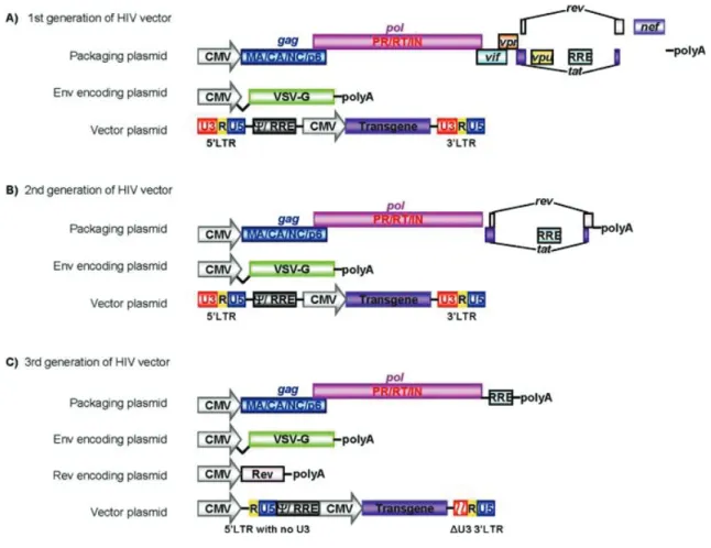
1.1.2 Scaffold proteins 42 1.) αRep proteins 43 1.1.3 Phage-display technology 47 1.1.4 Baculovirus 50 1.1.5 Lentivirus vectors 55 Objectives 59
Chapter 2 Materials and Methods 60
2.1 Chemicals and equipments 60
2.2 E. coli strains 60
2.3 Plasmids and vectors 60
2.4 Cells 61
2.5 Construction of αRep library 61
2.6 Screening of αRep phage library on the viral targets 62
2.7 Production of αRep proteins 63
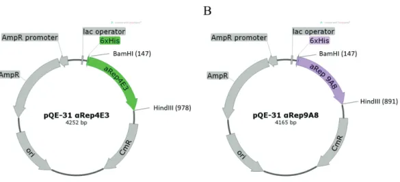
2.7.1 Construction of pQE-31 expression vectors for soluble αRep proteins production
63 2.7.2 Expression and purification of αRep proteins 63
2.8 Western immunoblotting 65
2.9 Construction of Sf9 stable cell lines stably expressing αRep proteins fused to GFP
65 2.9.1 Construction of pIB vectors expressing (Myr+)
αRep4E3-GFP and pIB_(Myr+) αRep9A8-GFP
65 2.9.2 Establishment of Sf9 cells stable expressing
(Myr+) αRep4E3-GFP and pIB_(Myr+) αRep9A8-GFP 66 2.10 Luciferase-based quantitative assay of αReps on HIV-1 VLP assembly 66 2.11 Electron microscopy analysis of HIV-1 Gag assembly into VLPs in
Sf/(Myr+)αRep4E3GFP or Sf/(Myr+)αRep9A8GFP
68
13
2.13 Production of VSV-G-pseudotyped lentiviral vectors 69 2.14 Construction of SupT1 stable cell lines stably expressing αRep proteins 70
2.15 Flow cytometry 72
2.16 Confocal microscopy 72
2.17 HIV-1 stock 73
2.18 Evaluation of antiviral activity of αRep proteins in SupT1 stable cell lines
73 2.18.1 Determination of p24 level by p24 antigen ELISA 74 2.18.2 Quantitation of integrated proviral DNA by
SYBR RT-PCR-based integration assay
74 2.19 Infectivity Assay of HIV-NL4-3 derived from SupT1 cells lines stably
expressing αRep proteins
74 2.20 ELISA based HIV-1 Pr55Gag maturation assay 75
2.21 Generation of HIV-1 target proteins 75
2.22 Protein pull-down assays 77
Chapter 3 Results 78
3.1 A new scaffold protein library and the CA21-SP1-NC domain of
the Gag polyprotein precursor as target
78 3.2 Selection of CA21-SP1-NC binders from a library of phage
displayed αRep proteins
80 3.3 Biochemical and biophysical characterization of CA21-SP1-NC binders 82
3.4 Effects of αRep4E3 and αRep9A8 on HIV-1 virus-like particle assembly
86 3.4.1 Construction of Sf9-derived cell lines stably expressing
αRep proteins
86 3.4.2 Biological effects of αRep4E3 and αRep9A8 on HIV-1
virus-like particle production in heterologous system: quantitative aspects
88
3.4.3 Biological effects of αRep4E3 and αRep9A8 on HIV-1 VLP production in heterologous system: qualitative aspects
14
3.4.4 Co-encapsidation of (Myr+)αRep4E3-GFP and (Myr+)αRep9A8-GFP into VLPs
94 3.5 Expression of αRep4E3-GFP and αRep9A8-GFP proteins in
HeLa cells using a lentiviral vector
96 3.6 Construction of SupT1 cell lines stably expressing αRep proteins 98 3.7 αRep4E3- and αRep9A8-mediated protection of SupT1 cells
against HIV-1 infection
100 3.7.1 Determination of HIV-1 production by CAp24-ELISA 100
3.7.2 HIV-1 proviral DNA detection 103
3.7.3 Status of the extracellular HIV-1 genomic RNA molecules
103 3.7.4 Cell viability and αRep protein expression in HIV-1
infection SupT1 cells
106 3.8 Molecular mechanisms of the antiviral functions of αRep4E3 and
αRep9A8
108 3.8.1 Influence of αRep4E3 and αRep9A8 on HIV-1
infectivity: (i) maturation of the Pr55Gag precursor
108 3.8.2 Influence of αRep4E3 and αRep9A8 on HIV-1
infectivity: (ii) encapsidation of the viral genome
111 3.8.3 Intracellular interaction of αRep and Gag proteins in
HIV-1 infected cells
3.9 Mapping of the αRep binding sites on the viral Gag target Chapter 4 Discussion Chapter 5 Conclusion References List of publications Appendix A Appendix B Appendix C Curriculum Vitae 113 115 118 125 127 138 139 144 145 152
15
LIST OF TABLES
Page
16
LIST OF FIGURES
Page Figure 1.1 Schematic overview of the HIVǦ 1 replication cycle 28 Figure 1.2 Diagram of HIV-1 virus particle structure 30 Figure 1.3 Schematic representation of the HIV-1 genome 36
Figure 1.4 Design of the αRep motif 45
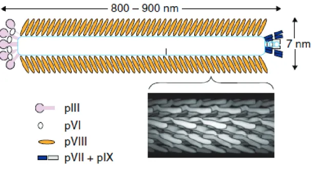
Figure 1.5 Structure of filamentous phage 50
Figure 1.6 Structure of baculovirus virions 53
Figure 1.7 Baculovirus life cycle 54
Figure 1.8 Schematic representation of HIV vectors 58 Figure 2.1 The schematic figure of pQE-31 vectors construction for
expression of 6xHis-tagged αRep proteins in bacterial cells
64
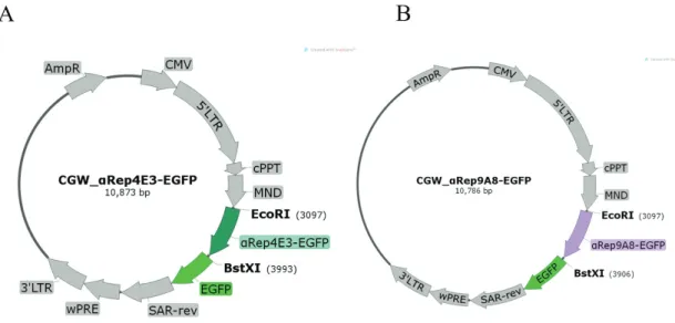
Figure 2.2 Luciferase based VLP assembly assay 67 Figure 2.3 The schematic figure of CGW lentiviral transfer vectors
construction for stable expression of αRep proteins in target cells
69
Figure 2.4 Production of VSV-G pseudotyped lentiviral vector particles 71 Figure 2.5 CA21-SP1-NC full-length bait and deletants 76
Figure 3.1 Amino acid sequences and the NMR structure of the SP1 nucleocapsid (NC)-SP2 domain of HIV-1 Gag polyprotein.
79
Figure 3.2 Screening of αRep clones from the third round of panning 81 Figure 3.3 Target-binding activity of αReps against GST-CA21-SP1-NC 83
Figure 3.4 Amino acid sequence, production and purification of αRep4E3 and αRep9A8
17
Figure 3.5 Construction of Sf9 cells stably expressing N-myristoylated αRep4E3-GFP and N-myristoylated αRep9A8-GFP
87 Figure 3.6 Biological effects of αRep4E3 and αRep9A8 on HIV-1 VLP
assembly
90 Figure 3.7 Morphological analysis of VLPs from αRep4E3-expressing
insect cells
92
Figure 3.8 Morphological analysis of VLPs from αRep9A8-expressing insect cells
93 Figure 3.9 SDS-PAGE &Western blot analysis of gradient fractions 95 Figure 3.10 Confocal microscopy of HeLa cells expressing αReps 97 Figure 3.11 Fluorescence microscopy and flow cytometry analysis of SupT1
cells transduced by a lentiviral vector
99 Figure 3.12 αRep4E3 and αRep9A8-mediated protection of SupT1 cells
against HIV-1 challenge
102 Figure 3.13 Concentration of HIV-1 genome copies in cell culture
supernatants
104 Figure 3.14 Extracellular viral genomes released by HIV-1 infected,
αRep-expressing SupT1 cells
105 Figure 3.15 Total cell number, cell viability and cell morphology after
challenge with HIV-1NL4-3
107 Figure 3.16 Maturation cleavage of Pr55Gag in viral particles released by
different cell types
110 Figure 3.17 Viral infectivity assay based on Jurkat-GFP re-infection. 112 Figure 3.18 Confocal microscopy of HIV-1 infected, αRep-expressing SupT1
cells
114 Figure 3.19 CA21-SP1-NC full-length bait and deletants 116
Figure 3.20 SDS-PAGE Western immunoblotting of protein pull-down assay representation the binding sites of αRep proteins on the viral target
117
Figure 4.1 Schematic representation of the distinct antiviral effects of αRep4E3 and αRep9A8 in HIV-1 infected SupT1 cell
18
LIST OF ABBREVIATIONS
% percent
5’UTR 5′ untranslated region
˚C degree celsius
αRep alpha repeat protein
AcMNPV Autographa californica Multicapsid
Nucleopolyhedrovirus
AIDS acquired immune deficiency syndrome
AR ankyrin
ARM armadillo
ART antiretroviral therapy
BA betulinic acid
BSA bovine serum albumin
BV baculovirus
BVM bevirimat
bp base pair (s)
CA capsid
CCR5 C-C chemokine receptor type 5 CD cluster of differentiation
CMV cytomegalovirus
CRISPR clustered regulatory interplaced short palindromic repeat CRM1 chromosome region maintenance 1
Ct threshold cycle
CTD carboxyl terminal domain
DAPI 4',6-diamidino-2-phenylindole DARPins designed ankyrin repeat proteins
DMEM Dulbecco’s Modified Eagle’s medium
19
EDTA ethylenediaminetetraacetic acid
EF3 elongation factor 3
EGFP enhanced green fluorescent protein ELISA enzyme-linked immunosorbent assay Env envelope
ESCRT endosomal sorting complex required for transport
FBS fetal bovine serum
FI fusion inhibitor
Fwd forward
g gram (s)
GFP green fluorescent protein
GST glutathione S-transferases
h hour (s)
HAART highly active antiretroviral therapy
HBV hepatitis B virus
HEAT huntingtin, elongation factor 3, protein phosphatase 2A, yeast kinase TOR1
HEK293T human embryonic kidney cell
HER2 human epidermal growth factor receptor 2
HGF hepatocyte growth factor
HIV human immunodeficiency virus
HIV-1 human immunodeficiency virus type 1
HRP horseradish peroxidase
Ig immunoglobulin IN integrase
INSTI integrase strand transfer inhibitor IPTG isopropyl β-D-1-thiogalatopyranoside kb kilobases
kDa kiloDaltons LB Luria-Bertani
20
LEDGF lens epithelium-derived growth factor
LLRs leucine-rich repeats
LTR long terminal repeat
Luc luciferase
M molar (s)
MA matrix protein
MFI mean fluorescence intensity
mg milligram (s)
min minute (s)
ml milliliter (s)
mM millimolar (s)
MOI multiplicity of infection
mRNA messenger RNA
MW molecular weight
Myr myristoylation
NaOH sodium hydroxide
NC nucleocapsid Nef negative regulatory factor
Ni-NTA nickel (II)-nitrilotriacetic acid complex NRTI nucleoside reverse transcriptase inhibitor
NTD amino terminal domain
OD optical density (-ies)
ODV occlusion derived virus
ORF open reading frame
PBMC peripheral blood mononuclear cell (s)
PBS phosphate buffered saline
PBS primer binding site
PCR polymerase chain reaction
PDB protein data bank
21
pfu plaque-forming unit
PI protease inhibitor
PIC pre-integration complex
PI(4,5)P2 phosphatidylinositol 4, 5-bisphosphate
PP2A protein phosphatase 2A
PR protease Pr55Gag gag polyprotein precursor
P-TEFb positive transcription elongation factor b RCA rolling circle amplification
RCLs replication competent lentiviruses RLU relative luminescence unit
RPMI Roswell Park Memorial Institute medium
RNA ribonucleic acid
RNAi RNA interference
RNA pol II RNA polymerase II
RSV rous sacroma virus
RT reverse transcriptase
RT reverse transcription
scFv single chain variable fragment
SDS-PAGE sodium dodecyl sulfate-polyacrylamide gel electrophoresis SIN self-inactivating
SL1 stem-loop 1
SL3 stem-loop 3
SP1 spacer peptide 1
SP2 spacer peptide 2
ssDNA single-stranded DNA
scFv single chain variable fragment
TALENs transcription activator-like effector nucleases
TAR trans-activation response
22
TBS Tris buffer saline
TMB 3,3',5,5'-Tetramethylbenzidine
TOR1 yeast kinase TOR1
TPR tetratricopeptide
U unit (s)
VEGF-A vascular endothelial growth factor A VEGF-B vascular endothelial growth factor B
VLP viral-like particle
Vif viral infectivity factor
Vpr viral protein R
Vpu viral protein U
VSV-G vesicular stomatitis virus-G
μg microgram (s)
μl microliter (s)
WCL whole cell lysate
WT wild type
ZFN zinc finger nuclease
23
STATEMENT OF ORIGINALITY
1. This thesis presents a new strategy to combat the HIV-1 infection by applying the αRep protein, a new family of repeat protein scaffold based on HEAT–like repeat for the intracellular interfering and inhibiting the virus replication cycle at the viral genome packaging and the maturation step.
2. This thesis contains a part of the study that used the newly developed method for investigating the ratio of mature per immature virus particle in the sample. This new technique was developed based on the competitive ELISA combined with using the unique anti-MA antibody which recognizes the cleaved C-terminal of MA when processed by HIV-1 protease enzyme.
24
CHAPTER 1
INTRODUCTION
Long-term treatment of Human Immunodeficiency Virus 1 (HIV-1) infection can be readily achieved by combinations of antiretroviral agents, usually referred to as Highly Active Antiretroviral Therapy (HAART). The combination of antiviral drugs can result
in the suppression of plasma viremia to less than the limit of quantification (< 50 copies/ml) with consequential improvement in the level of CD4+ T cell counts.
This was which associated with the resolution of established opportunistic infections although they cannot eradicate the viruses due to latent viral reservoirs. In addition, prolonged treatment tends to result in the occurrence of multi-drug resistant mutants, drug-drug interaction and drug toxic effects. All these drawbacks justify the exploration of alternative therapeutic approaches such as gene therapy and cellular therapy in order to enhance the anti-HIV-1 response (1-8).
In the past 30 years we have witnessed the feasibility of gene and protein therapies for the treatment of diverse human diseases. These approaches apply the powerful genetic engineering which is applicable to the treatment of both genetic and acquired maladies ranging from blood diseases to the treatment of infectious diseases such as HIV infection. Antibody therapy is one of the successful strategy in which a molecule of antibody or antibody-derived protein scaffold is used as the therapeutic agents. Although it is one of the major commercial success of the biotechnology industry to date, antibody molecules have their limitations (9, 10).
In the past decade the development of protein engineering techniques had made it possible to generate novel repeat scaffold protein binders called non-immunoglobulin scaffolds. The absence of disulfide bond in these scaffold molecules is the key factor which makes them suitable for intracellular applications, particularly inside the cytosolic compartment in which the proper folding of bioactive proteins should not be influenced by the reducing conditions. In addition, unlike antibodies, scaffold proteins have less complexity in the
25
production processes. They can be expressed in bacterial cell systems with high production levels, high solubility and high stability. For these reasons, a number of scaffold molecules are now in the preclinical and clinical development stage. DARPins, the designed ankyrin repeat proteins which are genetically engineered antibody mimetic proteins typically exhibiting highly specific and high-affinity target protein binding. These most well-known scaffold molecules are today undergoing clinical trials for targeting various types of tumor antigens for example, DARPin targeting VEGF-A (phase II/III), VEGF-A/PDGF-B (preclinical), and VEGF/HGF (phase I) as well as another type of protein binder, affibodies which specific to HER2 is now in phase I (11).
Recently, it was established that a new type of artificial protein derived from the repeat protein family called Huntingtin, elongation factor 3 (EF3), protein phosphatase 2A (PP2A), and the yeast kinase TOR1 (HEAT repeat protein) has a potential advantage in comparison to other well-known scaffold proteins (12). The HEAT repeat is a tandem repeat protein with a structural motif composed of two alpha helices linked by a short loop. It can form alpha solenoids which are found in a number of cytoplasmic proteins, often involved in intracellular transport and protein-protein interactions (13). Various repeat protein binders can be isolated by powerful bio-panning methods such as phage display or ribosome display. Many repeat proteins have been isolated and characterized, and one good example is GFP/EGFP-binding αReps (14). This repeat protein belonged to a new artificial repeat protein family in which their design is based on the thermostable HEAT-like repeats scaffold. They were able to colocalize with EGFP inside the different cell compartments without forming any aggregates or toxicity to the cells. Therefore, the αRep proteins are molecules of choice for the study of intracellular processes in living cells, by their direct interactions with endogenous protein of interest. In the present study we would like to apply αRep proteins to the development of a novel type of intracellular anti-HIV-1 treatment. The viral protein that we have targeted is a portion of the Pr55Gag polyprotein precursor overlapping the last 21 amino acids of the capsid (CA), the spacer peptide 1 (SP1), and the nucleocapsid domain (NC) which refers to CA21-SP1-NC. Many
evidences showed that the C-terminal domain of CA (CACTD) is critical for the formation
26
SP1 domain has been shown to be involved in the correct particle assembly. Mutation in the key amino acid residues of SP1 especially the first four amino acids can alter the viral morphology and interfere the virus infectiveness. The NC region is essential for the viral genomic RNA packaging and promote Gag-Gag interaction. As mentioned, our target protein CA21-SP1-NC is critical in the process of Pr55Gag assembly and genomic RNA
packaging in HIV-1-infected cells, we therefore assumed that expressing αRep proteins which specifically interact with this viral target CA21-SP1-NC inside the cells might be
possible to inhibit or interfere with the normal HIV-1 replication cycle. 1.1 Literature review
1.1.1. Human immunodeficiency virus (HIV)
The human immunodeficiency virus is a member of the genus Lentivirus, belonging to the Retroviridae family. When the viruses enter the body, they attack the immune system by specifically infecting the CD4+T cells, which help the immune system fight against infections. If improperly treated or left untreated, HIV reduces the number of CD4+T cells in the body, making the infected person susceptible to microbial infections or infection-related cancers. These opportunistic infections or cancers take advantage of the deficient immune system provoked by the viruses, called acquire immune deficiency syndrome (AIDS), the last stage of the HIV infection. There are two types of HIV, HIV-1 is the cause of the majority of HIV infections globally due to the high rate of viral production, while HIV-2, mainly present in Western Africa, is less pathogenic and shows a much lower rate of disease development (15, 16). In this study the literature review will be focused mainly on the HIV-1.
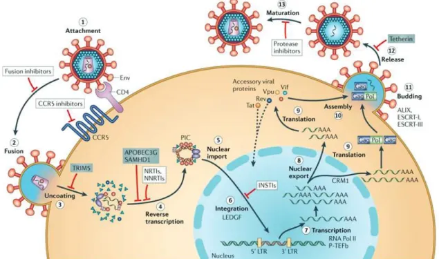
1.) HIV-1 life cycle
HIV-1 begins its life cycle when the mature viruses infect their target cells (Figure 1.1). There are many phases in the HIV-1 replication cycle. It begins with the viral entry (attachment and fusion), uncoating, reverse transcription, integration, particle assembly, budding, and maturation. During the virus entry phase, HIV-1 particle attaches to the membrane outside of a CD4+ T cells. Once attached, the virus can inject its genetic material, the viral
27
genomic RNA and viral enzymes into the cells. After that, one of the viral enzymes, the reverse transcriptase (RT) reverse transcribes the viral RNA into the viral DNA with a high proportion of transcription errors. This promotes a high mutation rate during the replication cycle. In the next step, the viral DNA is integrated into the host chromosome by the action of the integrase enzyme (IN), which transforms the cells into a "factory" to produce large quantities of new HIV-1 virions. After virus integrates its genome into the host chromosome, the viral components which are produced by the infected cells move to the inner leaflet of the cell membrane, where they assemble into new virus particles called the viral progeny. The newly formed virus particles then egress from the cell membrane and the maturation phase occurs when the protease (PR) enzyme of the virus cleaves the immature viral structural proteins inside the particle to transform the particles into mature infectious virions. Since there are many crucial steps in the virus life cycle, antiviral drugs or antiviral strategies were designed to target these steps for example, fusion inhibitors, CCR5 inhibitors, nucleoside reverse transcriptase inhibitors (NRTIs), non-nucleoside reverse transcriptase inhibitors (NNRTIs), integrase strand transfer inhibitors (INSTIs), and protease inhibitors (PIs).
28
Figure 1.1 Schematic overview of the HIV-1 replication cycle. The HIV-1 infection begins when the viruses use envelope (Env) glycoprotein spikes bind to CD4 molecule and the co-receptor CC-chemokine receptor 5 (CCR5) (step 1), leading to fusion of the viral and cellular membranes and enter into the cell (step 2). The CA shell uncoating (step 3) facilitates reverse transcription (step 4), which in turn yields the pre-integration complex (PIC). Following import into the cell nucleus (step 5), PIC-associated integrase orchestrates the formation of the integrated provirus, aided by the lens epithelium-derived growth factor (LEDGF), the host chromatin-binding protein (step 6). Proviral transcription (step 7), mediated by host RNA polymerase II (RNA Pol II ) and positive transcription elongation factor b (P-TEFb), generated the different sizes of viral mRNAs, the larger of which require energy-dependent export to leave the nucleus via host protein CRM1 (step 8). The viral protein production (step 9), and genome-length RNA is encapsidated into viral particles with other viral proteins (step 10). Viral-particle budding (step 11) and release (step 12) from the cell is mediated by ESCRT (endosomal sorting complex required for transport) complexes and ALIX and followed by protease-mediated maturation (step 13) to become an infectious viral particle. Each step in the HIV-1 life cycle is a potential target for antiviral intervention165; the sites of action of clinical inhibitors (white boxes) and cellular restriction factors (blue boxes) are indicated. INSTI,
29
integrase strand transfer inhibitor; LTR, long terminal repeat; NNRTI, non-nucleoside reverse transcriptase inhibitor; NRTI, nucleoside reverse transcriptase inhibitor (17). This figure has been obtained from the site:
http://www.nature.com/nrmicro/journal/v10/n4/full/nrmicro2747.html 2.) HIV-1 structural proteins
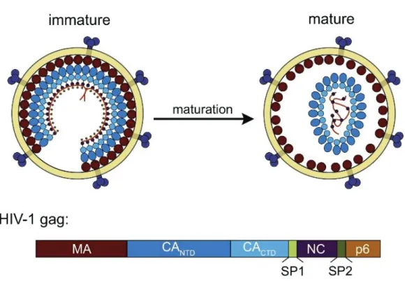
The proteins and gene functions of HIV-1 have been the subject of extensive research since the discovery of the virus in 1980s (18, 19). The formation of HIV-1 particles is driven by the viral structural protein, Gag polyprotein precursor (Pr55Gag) (20). The specific cleavage of Pr55Gag precursor by HIV-1 protease enzyme (PR) generates four major structural components of the mature virions including matrix (MA), capsid (CA), nucleocapsid (NC), and p6 protein and two spacer peptides (SP) which include SP1 and SP2 as shown in (Figure 1.2) (21, 22). Within the mature virions, MA domain is layered underneath membrane envelop meanwhile the shell of the core structure made of CA proteins. Inside the viral core, NC is tightly associated with the 2 copies of viral genomic RNA (23, 24). To be assemble into the new virions, the process begins at the inner leaflet of plasma membrane of the infected cells. When more than 2000 molecules of Pr55Gag and 200 Gag-Pol proteins and viral genomes are incorporated. The viral particles still in their immature form then egress from the infected cell. This form of the virus is non-infectious, and is not able to infect the new target cell as the Pr55Gag protein precursors have to be cleaved by the viral PR to generate a set of mature protein. These newly processed proteins then resemble to form mature particle morphology. MA is associated with the inner viral membrane envelope, CA assemble into conical capsid or viral cone, NC is interacted with the viral genomic RNA and packed inside the capsid core (Figure 1.2) together with viral enzymes, reverse transcriptase (RT), integrase (IN).
30
Figure 1.2 Diagram of HIV-1 virus particle structure. Top panel: Immature HIV-1 virus particle (left) and mature virus particle (right). Bottom panel: The compositions of HIV-1 Gag polyprotein, MA, CA (CANTD and CACTD), SP1, NC, SP2, and p6 respectively
(25). This figure has been obtained from the site:
31 3.) Matrix (MA)
The MA protein is the domain of Pr55Gag which is located on the N-terminal side of the Gag precursor. The MA plays an important role in the trafficking of Pr55Gag to the plasma membrane. This process is mediated by its myristic signal, added to the terminus of Pr55Gag by cytoplasmic N-myristoyl-transferase co-acting with the Gag protein translational machinery. The membrane binding function of Gag is mediated by the insertion of myristoyl group of the myristic acid into the lipid bilayer particularly, the acidic phospholipid, PI(4,5)P2. Moreover, the highly conserved patch of basic residues of MA promotes the Gag membrane interaction. Apart from the membrane targeting function, MA is required for the incorporation of envelope (Env) glycoproteins, gp120 and gp41 into the virus particles. In addition, several studies showed that MA contains a RNA binding site which overlaps the PI(4,5)P2 binding domain. The interaction of MA with nucleic acid has been suggested to enhance the selectivity of MA for PI(4,5)P2-rich cellular membrane and prevent premature intracellular assembly (26-33). 4.) Capsid (CA)
The CA domain of Gag is critical for the viral particle assembly. In mature viral particles, CA forms the conical shape viral core observed in mature virions (23). The viral CA can be divided into two domains which have different functions in the particle morphogenesis. The N-terminal domain of CA (CANTD) comprises of two third of CA (Figure 1.2 bottom). CANTD is
necessary for the mature core formation, but lacks the function necessary for the immature Gag polymerization process (34). The C-terminal domain of CA (CACTD) is critical for both core formation and Gag
oligomerization/polymerization in the particle assembly process (28, 35-37). It has been reported that mutations in CACTD severely impede viral particle
32 5.) Nucleocapsid (NC)
The NC region of Gag, which lies downstream of the CA domain, has been shown to be important for the viral genomic RNA packaging and promoting Gag-Gag interactions (29). The NC domain contains two conserved CCHC zinc finger motifs coordinated with two zinc ions linked by a basic RAPRKKG sequence, and both N-terminal and C-terminal domain are flanked by flexible sequences (38). There is evidence suggesting that this domain is absolutely essential for the virus replication. The function of NC involves its interaction with RNA stem loops, a hundred nucleotide-long portion of the viral RNA which locates at the 5’-end of the untranslated region (UTR) of the viral RNA, and on the 5’-side of the gag initiation codon. This region contains the packaging signal, the so called Psi (ψ), which is required for the efficient encapsidation of the unspliced viral genome. Inside the mature virus particle, NC is found within the CA core where several molecules of NC coat the two copies of viral genetic material (38). Moreover, NC harbors another domain called interacting domain (I domain), which consists of basic residues mediating Gag-Gag interaction. Most of the studies review that primarily ‘tethers’ Gag molecules together via RNA bridges, then additional NC-NC interactions are underwent. Then NC-RNA tether complexes promote the increasing of Gag concentration at the assembly site, resulting in the enhancement of CA-CA interaction. (26, 29, 37-39). More interestingly recent studies show that NC also plays a key role for chaperoning nucleic acid during viral DNA synthesis by HIV-1 reverse transcriptase enzyme and prevents nonspecific self-priming that induced by TAR stem-loop structure at 3’end of minus strand ssDNA (40, 41).
6.) Spacer peptide 1 and spacer peptide 2 (SP1 and SP2)
During the viral maturation step, HIV-1 Gag precursor protein is cleaved by viral PR to obtain the major mature proteins which include MA, CA, NC, and p6 domain. Beside these major domains, there are two spacer peptides, SP1 and SP2, which act as a linker between CA-NC and NC-p6, respectively, and
33
are released after the PR cleavage (21, 42). The SP1 region has been shown to be essential for the assembly of viral like particles (VLP). There are two functional properties of the SP1 domain which have been characterized. Firstly, the importance of the ~6 first residues of SP1 which are involved in the particle formation. Minor changes in the first 4 residues drastically destroy the correct particle assembly in mammalian cells (42, 43). This study suggested that the proper assembly probably requires the basic residues at the N-terminus of SP1. Secondly, several reports indicate that SP1 promotes the strength of Gag-Gag interaction. In summary, SP1 is required for the immature particle assembly and in regulating the formation of the mature HIV capsid essential for virus infectivity (31, 42-45).
SP2 is the domain that separates NC from the p6 domain. Unlike SP1, the function of SP2 in virus morphogenesis and infectivity are not currently well characterized. However, there is a study reporting that the blockage of both cleavage sites between NC and p6 did not affect processing of other Gag cleavage sites upstream, but caused a severe reduction of viral infectivity, close to background levels (46-48). In addition, another study provides evidence that the release of virus particles does not require the presence or the proteolytic processing of SP2. The viral infectivity is almost abolished when both cleavage sites (the "slow" cleavage site between NC and SP2, and the "fast" cleavage site between SP2 and p6) are mutated, and severely reduced when the fast cleavage site SP2-p6 is altered. This correlates with an increased proportion of irregular core structures, although processing of CA is not affected. Mutation of the slow cleavage site NC-SP2, or deletion of most of SP2, had only minor effects on infectivity and did not induce major alterations in mature core morphology. These results suggest that the processing kinetics leading to the separation of NC and p6 is essential for successful maturation of the virions, while SP2 itself is dispensable (49).
34 7.) p6 protein
While MA, CA, and NC are the common proteins found in all retroviral Gag precursors, the p6 domain is a specific feature of HIV-1 and other primate lentiviruses. In the full-length Gag molecule, p6 is located at the C-terminus and separated from the NC by the short spacer peptide SP2. The p6 domain is encoded by a sequence which directs translational frameshifting into the overlapping pol open reading frame. Thus, p6 is the only Gag domain which is absent from the Gag-Pol polyprotein (50). It has been shown that, within the viral particle, p6 is the major phosphoprotein in the mature form, but the function of this phosphorylation remains unclear. By contrast, a major function of p6 has been well characterized: it consists of the release of assembled viruses from the cell surface and/or from each other, which is highly dependent upon p6. In the sequence of p6, the major determinant of this function is the conserved P(T/S)APP motif near the N-terminus of the p6 domain. This region is now known as the late assembly (L) domain. Moreover, the L domain can induce the recruitment of cellular factors to the site of particle assembly (51).
8.) HIV-1 genome
The full HIV-1 genome is encoded in one long strand of RNA. In the free virus particle, there are two copies of single-stranded positive-sense RNA (52) packed inside. Each RNA strand is approximately 9.7 kilobases (kb) in length. The 5' and 3' ends of the RNA genome are flanked by a long terminal repeat (LTR) promoter region (Figure 1.3). Through specific interactions via packaging signal Ψ, the cis-acting elements, the unspliced genomic RNA is packaged into virions (53). The sequences in genomic RNA that is involved in packaging include stem-loop 3 (SL3) (54) and stem-loop 1 (SL1) (55) in the 5’ UTR. Viral RNA once entered into the cytoplasm of the cell, the RT converts the genomic RNA into the viral DNA which is a form ready to integrate into the host chromosome via the action of IN called proviral DNA. Between the two LTR regions, the viral genome contains the genes encoding
35
the three structural polyproteins Gag, Pol, and Env, the genes encoding four accessory proteins Vpu, Vif, Vpr, and Nef, and the genes for the two regulatory proteins Rev and Tat (56, 57).
36 F igure 1.3 Schem atic representat ion of the HIV -1 genom e. The ge nome of HI V-1 HXB 2 (K 03455) wa s used as an ex ample for the HI V-1 ge nome org aniz ation. Ope n re ading fr ames are shown as re ct ang les. The start codon of each g ene is indicated by the small nu mber in the
upper left corner of
each
rectang
le,
while the number
in the lower rig ht records the last positi on
of the stop codo
n. For t he sequenc e encoded for pol, the
start position is the first
T in the se quence TTTTTTAG, which for ms part of the stem
loop that potentiat
es
ribosomal
slippag
e
on the RNA and
a re sulting -1 fr ameshift and the translation of the Ga g-Pol poly protein.
The tat and
re v spliced ex ons are shown as shaded re ctang les. This fig ure has been obtained fr om the site: https://www.hiv.lanl.g ov/content/sequence/HI V/MAP/landmark.html
37 9.) HIV-1 Antiviral Therapy
Antiretroviral therapy (ART) has proven to be highly effective in controlling HIV-1 disease progression to full blown AIDS by reducing the amount of virus in the body, preserving CD4+ T cells and dramatically slowing down the destruction of the immune system (58, 59). Several classes of drugs have been approved as medications and used for interrupting the viral life cycle in patients, e.g. fusion inhibitors (FIs), chemokine receptor antagonists (CCR5 antagonists), nucleoside reverse transcriptase inhibitors (NRTIs), non-nucleoside reverse transcriptase inhibitors (NNRTIs), integrase strand transfer inhibitors (INSTIs), and protease inhibitors (PIs). Each class of drugs acts at the different stages of the HIV life cycle. Typically, the therapeutic use of highly active retroviral therapy (HAART) which is a combination of at least three drugs belonging to two classes of antiretroviral agents.
10.) Entry inhibitors
HIV-1 entry inhibitors also known as fusion inhibitors are the anti-HIV-1 which interferes with the binding, fusion and entry of the virus to the host cell by blocking one of several targets. Maraviroc and enfuvirtide are the two currently available agents in this class. Maraviroc works by targeting CCR5, a chemokine receptor expressed on the surface of human helper T-cell membrane. In rare cases, individuals may have a mutation in the CCR5 delta gene which results in a nonfunctional CCR5 co-receptor and in turn, a means of slow progression of the disease or resistance to the new infection. The most famous case study is Berlin patient. He was diagnosed with HIV in 1995 and started to receive antiretroviral therapy. In 2006, Berlin patient diagnosed with acute myeloid leukemia (AML). His physician therefore, arranged for him to receive a hematopoietic stem cell transplant from a donor with the "delta 32" mutation on the CCR5 receptor results in a mutated CCR5 protein (60, 61). Those individuals who are homozygous for the CCR5 mutation are resistant to HIV and rarely progress to AIDS. The majority of HIV cannot enter a human cell without a functional CCR5 gene. An exception to this is
38
only a small minority of viruses that use alternate receptors, such as CXCR4 or CCR2 (62). To prevent fusion of the virus with the host membrane, enfuvirtide (INN) is one another example of inhibitor in this class. Enfuvirtide is a peptide drug that must be injected into the body and acts by interacting with the N-terminal heptad repeat of gp41 of HIV to inactivate the hetero six-helix bundle formation, therefore preventing infection of host cells (63). 11.) Nucleoside reverse transcriptase inhibitors
HIV-1 is an RNA virus, to be able to integrate its genome into the host chromosome, it must be reverse transcribed into the DNA. Since the conversion of RNA to DNA is not done in the mammalian cell, it is performed by a viral protein which makes it a selective target for inhibition. For mechanism of action, NRTIs are chain terminators such that once incorporated it will prevent other nucleosides from also being incorporated into the DNA chain because of the absence of a 3' OH group. It acts as a competitive substrate inhibitor. The examples of currently used NRTIs include zidovudine, abacavir, lamivudine, emtricitabine, and tenofovir (64). 12.) Non-nucleoside reverse transcriptase inhibitors
Non-Nucleoside reverse transcriptase inhibitor (NNRTI) is a non-competitive inhibitors of reverse transcriptase which inhibit reverse transcriptase by binding to an allosteric site of the enzyme. It can be classified into 2 classes, first generation and second generation NNRTIs. The first generation of NNRTIs are include nevirapine and efavirenz. Meanwhile, the second generation NNRTIs consists of etravirine and rilpivirine (64).
13.) Integrase strand transfer inhibitors
The integrase nuclear strand transfer inhibitors (INSTIs) also known as integrase inhibitors (INs) are the inhibitor that inhibit the function of viral enzyme integrase to prevent the integration of viral DNA into the DNA of the infected cell. There are several integrase inhibitors currently under clinical
39
trial. Raltegravir is the one that became the first to receive FDA approval in 2007. It contains two metal binding groups that compete for substrate with two Mg2+ ions at the metal binding site of integrase. Elvitegravir and
dolutegravir are two other integrase inhibitors which already approved in early 2014 (65).
14.) Protease inhibitors
Protease inhibitors is a type of HIV-1 inhibitor which block the viral protease enzyme and prevent the production of mature virions upon budding from the host membrane. These drugs particulally block the cleavage of gag and gag/pol precursor proteins.[15] Virus particles produced in the presence of protease inhibitors are defective and mostly non-infectious. Examples of HIV protease inhibitors are lopinavir, indinavir, nelfinavir, amprenavir and ritonavir. Darunavir and atazanavir are currently recommended as first line therapy choices (66).
15.) Assembly and maturation inhibitors
The assembly of Gag proteins into immature viral particles followed by proteolytic disassembly of the Gag shell to mature capsids are pivotal steps for the generation of infectious HIV-1 (67). The identification of active peptides as candidates for interfering negatively with the viral assembly step is a promising strategy (27). The HIV-1 CA protein plays essential roles in viral replication and as such represents an attractive new therapeutic target. In 2005, a 12-mer peptide termed capsid assembly inhibitor (CAI), identified by phage display, was reported to disrupt both immature- and mature-like particles in vitro by targeting the C-terminal domain (CTD) of HIV-1 CA (68). However, the CAI peptide itself fails to inhibit HIV-1 assembly in cell culture due to its inability to penetrate cells. A structure-based rational design approach known as hydrocarbon stapling has then been developed to stabilize the alpha-helical structure of CAI and convert it to a cell penetrating peptide. The resulting molecules, named NYAD-1 and NYAD-13, efficiently disrupt HIV-1 assembly in cell cultures. However, because they have a relative low
40
affinity for CA, these peptides are unlikely to progress to the clinic (69). More recently, a third small-molecule binding site on CA was identified via a high-throughput screen for inhibitors of HIV replication. PF-3450074 and related compounds bind between helix 4 and helix 7 of CANTD. Molecules
like the CAP-1, also termed PA-457, and most notably the peptide-based CAI are suitable leader compounds for anti-HIV drug development (67). As the proteolytic processing of Gag is conditioned by the functionality of the viral PR and by the accessibility of its recognition site in Gag, strategies aimed at interfering either with PR enzymatic activity or to reduce access to protein substrate have been considered in the development of anti-HIV-1 drugs. The later concept is represented by a new class of HIV-1 antivirals referred to as maturation inhibitors that have been developed and tested in phase IIb clinical trials. This class of compound is able to inhibit proteolytic maturation of Gag independently of an inhibition of protease activity. The lead drug of this pharmacologic class is betulinic acid (BA), a triterpene compound isolated from the clove-like plant Zygizum cloviflorum. This molecule has been identified as anti-cancer agent. In the last years it has been identified as an anti-HIV-1 molecule. Modification of side chains at position C3 and C28 in the BA scaffold has been used to derive a library of compounds that has been evaluated in vitro for its inhibitory properties. Among these molecules, bevirimat (BVM), potently inhibits HIV-1 replication in tissue culture and is efficacious in HIV-1 infected patients. Biochemical analyses of virus particles grown in the presence of BVM displayed the amount of processed p24 protein decreases in favor of the immature p25 protein reflecting unprocessed CA-SP1 junction. This phenotype is reminiscent of that observed for HIV-1 bearing mutation in CA or at SP1 junction that abolish CA-SP1 processing (27, 70-74).
The combinations of antiretroviral drugs result in the multiple obstacles to HIV-1 to replicate, keep the number of offspring low and reduce the possibility of a superior mutation. In the condition of mutation that conveys resistance to one of the drugs being taken arises, the other drugs will continue to suppress reproduction of that mutation. Combination of antiretroviral drugs