HAL Id: tel-02940144
https://hal.univ-lorraine.fr/tel-02940144
Submitted on 16 Sep 2020HAL is a multi-disciplinary open access archive for the deposit and dissemination of sci-entific research documents, whether they are pub-lished or not. The documents may come from teaching and research institutions in France or abroad, or from public or private research centers.
L’archive ouverte pluridisciplinaire HAL, est destinée au dépôt et à la diffusion de documents scientifiques de niveau recherche, publiés ou non, émanant des établissements d’enseignement et de recherche français ou étrangers, des laboratoires publics ou privés.
Isolation and characterization of Phanerochaete
chrysosporium mutants resistant to antifungal
compounds
Duy Vuong Nguyen
To cite this version:
Duy Vuong Nguyen. Isolation and characterization of Phanerochaete chrysosporium mutants resistant to antifungal compounds. Mycology. Université de Lorraine, 2020. English. �NNT : 2020LORR0045�. �tel-02940144�
AVERTISSEMENT
Ce document est le fruit d'un long travail approuvé par le jury de
soutenance et mis à disposition de l'ensemble de la
communauté universitaire élargie.
Il est soumis à la propriété intellectuelle de l'auteur. Ceci
implique une obligation de citation et de référencement lors de
l’utilisation de ce document.
D'autre part, toute contrefaçon, plagiat, reproduction illicite
encourt une poursuite pénale.
Contact : [email protected]
LIENS
Code de la Propriété Intellectuelle. articles L 122. 4
Code de la Propriété Intellectuelle. articles L 335.2- L 335.10
http://www.cfcopies.com/V2/leg/leg_droi.php
Ecole Doctorale SIReNa
(Sciences et Ingénierie des Ressources Naturelles)
Thèse
Présentée et soutenue publiquement pour l’obtention du titre de
DOCTEUR DE l’UNIVERSITE DE LORRAINE
Mention « Biologie et écologie des forêts et des agrosystèmes »
Par Duy Vuong NGUYEN
Isolation et caractérisation de mutants
de Phanerochaete chrysosporium
résistants à différents composés antifongiques
Soutenance le 3 Juin 2020
Membres du jury :
Rapporteurs :
Mme. Catherine RAFIN MCF, Université du Littoral Côte d’Opale M. Christian MEYER DR, INRAE, Versailles
Examinateurs :
Mme. Harivony RAKOTOARIVONINA MCF, Université de Reims M. Philippe GÉRARDIN Pr, Université de Lorraine
M. Éric GELHAYE DR, INRAE Nancy, Directeur de thèse
M. Rodnay SORMANI MCF, Université de Lorraine, Co-directeur de thèse
Invités :
ACKNOWLEGEMENTS
First of all, I would like to thank the members of the thesis committee, Catherine Rafin, Christian Meyer, Harivony Rakotoarivonina, Nadine Amusant, and Philippe Gérardin for agreeing to read this manuscript and evaluate my thesis work.
The research was carried out at the team Stress Response and Redox Regulation (Redox team), UMR 1136 laboratory at the University of Lorraine where my work was supported by Laboratory of Interactions between Plants and Microorganisms (IAM). This support is warmly acknowledged. I would like to sincerely thank the Vietnam Ministry of Agriculture and Rural Development, Vietnam Ministry of Education and Training, France embassy at Hanoi, and Campus France for the scholarship and supports during my studies in France. I would also like to thank the Lab of Excellence ARBRE for funding to the project ANR-11-LABX-0002-01 that provides materials for my studies.
I am very grateful to my supervisors, Professor Eric Gelhaye and co-supervisor A. Professor Rodnay Sormani, for your valuable guidance and supports during my study and for me a chance and freedom to explore the physiology of filamentous fungi (regulatory and signaling proteins) due to I only studied biochemistry as a branch of physical science in previously. Thank both for reading, providing critical comments, and giving me ideas to improve my thesis manuscript, that is a very big work, and I know that it took so much time of yours. Thank Eric so much for your financial support to my travel to the ECFG14th in Israel, a very big international congress on fungal biology. I consider myself very lucky to work with you as my thesis supervisor. Sincerely, thank Eric and Rodnay for everything.
I warmly thank Professor Jean-Pierre who read carefully my thesis and give me useful comments and suggestions, and also for supporting me English improvement as other PhD students of Redox team during our working time at here. I also wish to thank Professors Nicolas Rouhier and Mélanie Rouhier, leaders of Redox team who keep weekly seminars. I have obtained not only so much knowledge on plant biochemistry and fungal physiology but also slides preparation and presentation skills from those seminars. Thank Nicolas also for reminding all people to organize workspace of the laboratory as cultural space, it is not easy to find a space as here.
Special thanks to Elena, who always motivates me through your bits of help in both work and life. I always remember images we talked on the way coming back home together in many times, they will be importantly memorable of mine.
Special thanks also to Mélanie and Flavian, who started PhD course with me. Both always have paid attention so much to my work, especially during writing the thesis manuscript, which was a very difficult period. Your supportive attitude towards my work is very important. Thanks to your sincere inspirations, I had got more motivation for working.
I would like to thank many colleagues and friends who I have had the pleasure of working during the years for the very open and friendly space in the laboratory. Those include Jérémy, Arnaud, Tiphaine, Benjamin Petre, Benjamin Selles, Jean-Michel, Julien, Muriel, Alexis, Raphael, Fanny, Elodie, Nicolas Valette, Anna, Jonathan, Thomas, Antonio, Delphine, Damien, Loïck, Marion, Kevin, Corrine, and Martine.
I also would like to warmly thank all the administrative staff of the Doctoral Register office, the School of RP2E and SIReNa, for the information necessary for my study activities and also for their aides when I have several issues.
Warm thanks are sent to all my Vietnamese friends for helping me to keep spirits and motivation, also to find a solution for the difficulties and problems I met.
Finally, I feel especially lucky to have such a wonderful family. My parents, my wife Anh Thu, and my daughter Minh Thai always inspire me to have confident thoughts. Thank you all for your love and support. I know that all of the achievements I have obtained here belongs to you.
TABLE OF CONTENTS
ABBREBRIATIONS RÉSUMÉ
INTRODUCTION
I. Fungal adaptation to environment ... 1
I.1 Fungi have to adapt to their environment ... 1
I.2 Fungal sensing of the environment ... 5
I.2.1 Nutrient sensing ... 6
Carbon sensing ... 6
Carbon nutrient sensing and responses in lignocellulose degradation ... 11
Nitrogen sensing ... 13
Target Of Rapamycin (TOR) signaling as a key regulator of growth in the adaptation of filamentous fungi ... 15
I.2.2 Stress sensing and responses for adaptation to environment ... 18
II. Wood and wood degrading fungi ... 22
II.1 Wood components ... 22
II.1.1 Structural compositions ... 25
II.1.1 Cellulose ... 25
II.1.2 Hemicellulose ... 25
II.1.3 Lignin ... 27
II.I.2 Wood extractives ... 27
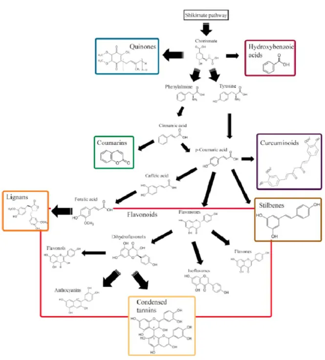
II.I.2.1 Phenolic compounds ... 28
II.I.2.1.1 Flavonoids ... 29 II.I.2.1.2 Tannins ... 31 II.I.2.1.3 Stilbenes ... 32 II.I.2.1.4 Lignans... 34 II.1. 2.1.5 Quinones ... 35 II.1.2.2 Terpenoids ... 35
II.2 Wood decaying fungi ... 37
II.2.1 Brown rot fungi ... 38
II.2.2 White rot fungi ... 38
II.2. 3 General biological characteristics of Phanerochaete chrysosporium ... 39
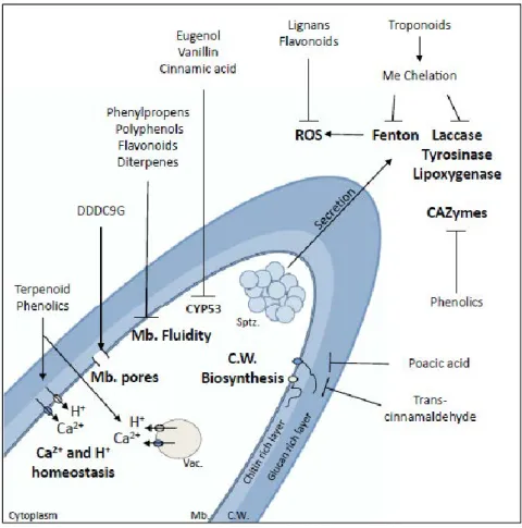
II. 3 Antifungal mechanisms of wood extractives and fungal adaptation ... 41
Objectives ... 47
Article I ... 51 Article II... 75 Supplementary results ... 101 Introduction ... 103 Experiment protocols ... 103 Results ... 105
Discussion and conclusions (Article I & II) ... 115
Article III ... 119
Supplementary results ... 149
Introduction ... 151
Results ... 154
Mutagenesis and screening mutant resistant to CTWE ... 156
Phenotypes of chy mutants ... 158
Identification of the causal mutation(s) leading to CTWE resistance... 158
Involvement of AGR57_10098 in resistance against CTWE... 163
DISCUSSION AND PERSPECTIVES ... 165
Wood extractives as tools to characterize wood decaying fungi ... 168
The possible relationships between identified proteins PcTOR and PcDUF1630 protein and secretion ... 170
The possible relationships between PcTOR and PcNACHT protein ... 172
Understanding the functions of TOR signaling in P. chrysosporium ... 174
Proteomic analysis could be used to characterize deeply the role of TOR in regulating the secretome response of P. chrysosporium ... 174
The possible relationship between TOR and the intracellular detoxification system ... 175
Future perspectives ... 178
General conclusions ... 179
List of Figures
Figure 1. Fungi have to adapt to their environment. ... 2
Figure 2. Wood decaying fungi have to adapt to a specific environment. ... 4
Figure 3. Sugar-sensing membrane proteins of fungal cells. ... 5
Figure 4. Glucose sensing pathways through G-protein in S. cerevisiae. ... 8
Figure 5. Regulation of lignocellulolytic CAZymes in filamentous fungi. ... 10
Figure 6. The molecular mechanism of carbon catabolite repression in S. cerevisiae and filamentous fungi . ... 12
Figure 7. TOR signaling pathway in the model yeasts. ... 16
Figure 8. Stress signaling pathways in S. cerevisiae and C. albicans. ... 19
Figure 9. Structural and chemical composition of ligno-cellulose biomass. ... 22
Figure 10. A chemical chain of cellulose composed of glucose units attached with β-1,4 linkages and organization of cellulose in a microfibril. ... 24
Figure 11. Chemical composition of hemicellulose compounds... 24
Figure 12. Structure of primary lignin monomers and three corresponding lignin units. ... 26
Figure 13. Model structures of lignin. ... 26
Figure 14. Biosynthetic pathways of phenolic compounds based on their carbon skeleton. ... 28
Figure 15. Chemical structure of main groups of flavonoids. ... 29
Figure 16. Structures of taxifolin and quercetin. ... 30
Figure 17. Classification of the tannins. ... 31
Figure 18. Structure of pinosylvin and derivatives. ... 33
Figure 19. Chemical structures of lignans identified in methanol extractives of heartwood of Araucaria araucana (Mol.) K. Koch. ... 34
Figure 20. Classification of quinones. ... 35
Figure 21. Antioxidant activity of ferruginol ... 36
Figure 22. Decay of wood by brown rot fungi. ... 38
Figure 23. Decay of wood by white rot fungi. ... 39
Figure 25. Life cycle of P. chrysosporium. ... 40
Figure 26. Effect of compounds derived from wood extractives at the tip of a fungal hypha. ... 42
Figure 27. Scheme represents the main approach used in the thesis. ... 46
Figure 28. Characterization of P. chrysosporium rap mutants resistant to rapamycin. ... 106
Figure 29. Identified mutations in the P. chrysosporium FKBP12-rapamycin-FRB complexes… ... 109
Figure 30. Inhibitory effects of rapamycin on WT fungus, but not on rap mutants. ... 110
Figure 31. Rapamycin had no significant effects on growth and secretion of mutant rap8... 111
Figure 32. The proposed involvement of signaling pathways in expression of Cyt P450s and extracellular lignin degrading enzyme system (LDS) in P. chrysosporium. ... 117
Figure 33. Effect of wood extractives on germination of P. chrysosporium RP78 conidia determined by nephelometry. ... 152
Figure 34. Effect of dichloromethane cherry tree wood extractives (CTWE) on germination and hyphal growth of P. chrysosporium RP78 conidia. ... 155
Figure 35. Phenotype of chy mutants... 157
Figure 36. Germination phenotype of selected chy mutants in presence of CTWE. ... 159
Figure 37. Genomic survey of the mutations in chy mutants. ... 162
Figure 38. The possible links of PcDUF1630/DENND to TOR signaling and secretory pathway in P. chrysosporium. ... 171
Figure 39. Bagassa wood extractives resistance of bag31 and rap1 mutants in comparison to WT strain. ... 171
Figure 40. Rapamycin resistance of chy14 in comparison to WT strain. ... 173
i
RÉSUMÉ
Isolation et caractérisation de mutants de Phanerochaete chrysosporium
résistants à différents composés antifongiques
Contexte de mes études
Les champignons lignivores, responsables de la dégradation du bois et de la litière, suscitent un intérêt croissant ces dernières années, principalement en raison (i) de leur utilisation potentielle dans la valorisation de la biomasse pour la production de biocarburants, (ii) de leur fonction importante dans le cycle global du carbone et (iii) des dommages qu’ils peuvent causer au matériau bois.
Ces champignons, et en particulier les basidiomycètes, sont des organismes capables de dégrader et d’utiliser la cellulose, les hémicelluloses et la lignine comme sources de carbone et d'énergie. Cependant, les processus oxydatifs utilisés par ces champignons pour décomposer le bois génèrent une myriade de molécules potentiellement toxiques et augmentent la biodisponibilité de molécules de faibles masses moléculaires non liées de façon covalente aux polymères : les substances extractibles. Les extractibles sont des composants non structurels du bois qui peuvent être éliminés avec un solvant neutre à polaire. La composition de ces substances extractibles peut varier en fonction du tissu, des conditions de culture et est spécifique à l'espèce (Kebbi-Benkeder et al., 2015). Les extractibles sont responsables des propriétés du bois telles que sa couleur et sa durabilité, car elles font partie du système de défense de l'arbre.
Pour s'adapter à cet environnement toxique, les champignons responsables de la décomposition du bois ont développé diverses stratégies de détoxication (Morel et al., 2013). Les approches de génomique comparative ont permis de montrer la présence d’extensions de familles de gènes codant pour les systèmes de détoxication chez les champignons décomposeurs du bois par rapport à d'autres champignons (principalement les cytochromes P450 monooxygénases et les glutathion transférases (GST)). L’analyse du transcriptome de Phanerochaete chrysosporium cultivé en présence d’extractibles de chêne a révélé une induction de l'expression de certains de ces gènes impliqués dans la détoxication (Thuillier et al., 2014). Enfin, au niveau fonctionnel, des résultats intéressants concernant la caractérisation biochimique de GST et leur interaction avec les extractibles ont été obtenus (Mathieu et al., 2012; Perrot et al., 2018). Cependant, bien que très informatives, ces données ne sont pas suffisantes pour déterminer le rôle physiologique de ces protéines dans les cellules fongiques lors de la dégradation du bois. Une approche sans a priori est donc essentielle pour identifier les acteurs impliqués dans le processus de détoxication.
ii
Le manque de données physiologiques concernant les voies de détoxication chez les champignons est principalement dû à l'absence d'outils génétiques disponibles pour ces organismes. Pour contourner ce problème, nous avons développé une stratégie de génétique directe chez P.
chrysosporium, champignon dégradeur de bois, choisi par la communauté comme modèle pour
ces études. La première partie du projet que j’ai développé visait à prouver la faisabilité et la pertinence d'une telle approche. Deux études prouvant le concept ont été réalisées, la première utilisant un antifongique appelé itraconazole, la deuxième utilisant la rapamycine.
Description de la stratégie de génétique directe mise en œuvre:
1. Identification d’une molécule toxique.2. Production de mutants de P. chrysosporium.
3. Identification parmi ces mutants de ceux capables de résister à la molécule toxique. 4. Identification des mutations chez les mutants résistants.
5. Comprendre comment la mutation permet la résistance.
Deux études: preuves de concept
Avant de nous lancer dans l’étude de la réponse des champignons aux molécules toxiques contenus dans le bois (les extractibles), nous avons choisi de tester la faisabilité de cette approche en travaillant avec des molécules dont les mécanismes d’actions sont connus au moins chez d’autres organismes.
Preuve de concept 1, l’itraconazole
L'itraconazole est un antifongique appartenant à la famille des triazoles qui inhibe la biosynthèse de l'ergostérol en agissant sur la lanostérol 14α déméthylase (CYP51A) (Bowyer and Denning, 2014). Il y a une décennie, des résistances aux triazoles ont été rapportées dans des isolats d'Aspergillus fumigatus prélevés chez des patients atteints d'aspergillose invasive. La propagation de la résistance a été étudiée en analysant les isolats d'A. fumigatus provenant de patients de 7 pays différents. Les mutations du gène CYP51A se sont avérées être le mécanisme de résistance dominant (Snelders et al., 2008). L’homologue de CYP51A chez P. chrysosporium se trouve sur le
scaffold 16 entre les positions 46319 et 47911
(http://genome.jgi.doe.gov/programs/fungi/index.jsf) du génome de ce champignon. La séquence protéique correspondante présente une identité de séquence de 54,1% avec celle d’A. fumigatus (Warrilow et al., 2008).
iii
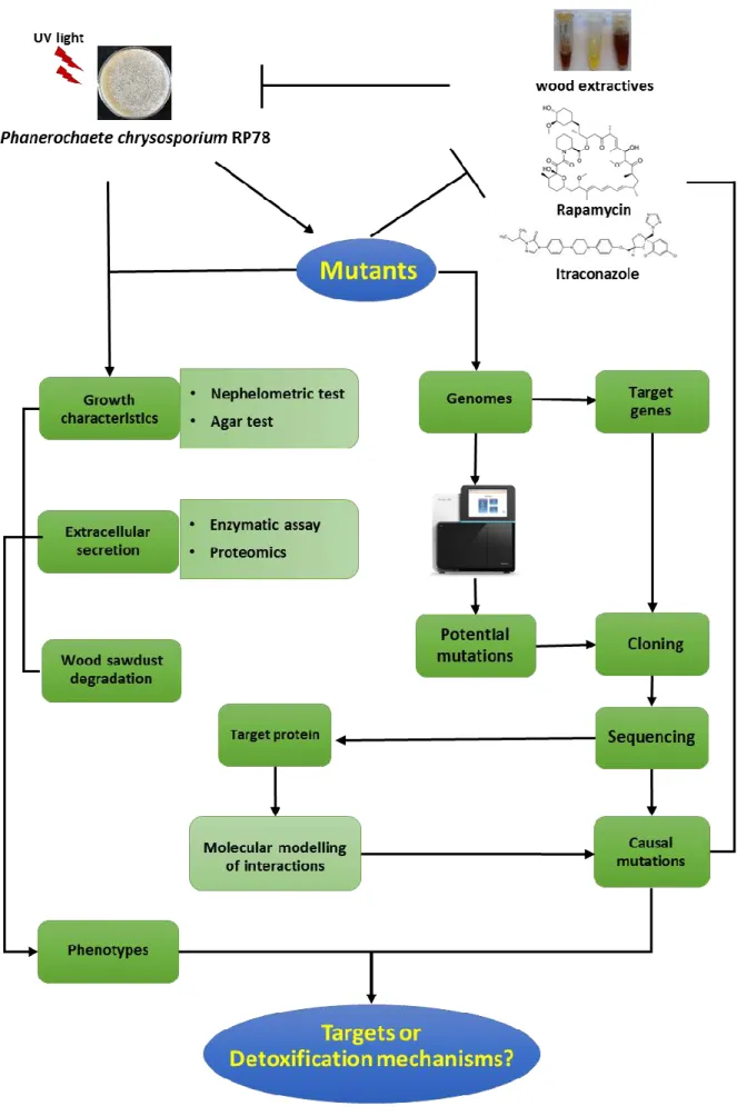
Lors de notre étude, les conidies de P. chrysosporium ont été soumises à U.V. afin de produire des mutations de façon aléatoire dans le génome de ces cellules. Un crible de résistance génétique a été réalisé à l’aide d’une dose létale d’itraconazole. Quarante mutants ont été isolés et appelés rit pour résistant à l’itraconazole. Des lignées de descendance ont été produites pour chacun des 40 mutants identifiés. Tous ces mutants sont résistants à l'itraconazole (Fig. 1).
Figure 1. Effet de l'itraconazole sur la croissance de P. chrysosporium.
Panneau supérieur WT et panneau inférieur rit1 mutant.
8 µg d'itraconazole (itra) ou de DMSO (ctrl) ont été déposés sur le disque en papier. Barre d'échelle : 1 cm.
Les 40 mutants rit obtenus lors de notre expérience portent tous des mutations dans le gène
PcCYP51A. Parmi ces 40 mutants, seuls deux allèles ont été trouvés et ils conduisent à la même
substitution d'acide aminé : l'alanine 290 est remplacée par une valine dans la protéine. Dans 34 séquences, nous avons trouvé GTT à la place de GCT et dans les autres cas GTC à la place de GTT. Pour comprendre le phénotype de résistance observé, une modélisation structurelle du PcCYP51 a été effectuée (Fig. 2).
WT 0h 6h 12h 18h 24h 30h 36h 42h
rit1
ctrl itra.
iv
Figure 2. Prédiction des sites de liaison du lanostérol et de l'itraconazole par modélisation de la 14-α-déméthylase de
P. chrysosporium (PcCYP51). Zoom sur les
sites actifs PcCYP51 et PcCYP51 A290V en présence de lanostérol (respectivement en A et B) ou d'itraconazole (en C et D respectivement). Dans chaque cas, les molécules de l'hème, du lanostérol et de l'itraconazole sont colorées respectivement en noir, violet et jaune. La position Ala290 (Ala ou Val) est également indiquée en rouge.
La structure cristalline du CYP51 (identité de 54,1%) du champignon filamenteux pathogène A. fumigatus (entrée pdb 4UYL) a été utilisée comme matrice pour le modèle structural de la protéine PcCYP51A et les sites de liaison du lanostérol et de l'itraconazole ont été obtenus par modélisation à partir de la structure cristalline obtenue en rayons X de la 14-alpha déméthylase de Saccharomyces cerevisiae lanostérol (entrées pdb 4LXJ et 5EQB, respectivement). À partir de cette analyse, la substitution de Ala290 par des acides aminés plus importants (tels que la valine dans notre cas) ne devrait pas gêner l'activité enzymatique du lanostérol (Fig. 2A et 2B), mais entraverait probablement le positionnement de l'itraconazole en raison d'un voisinage trop proche (Fig. 2C & 2D). Ces résultats expliquent le phénotype de résistance à l'itraconazole sans aucun défaut de croissance des mutants rit (Fig. 1).
Nous avons donc produit et criblé des mutants résistants à l'itraconazole pour lesquels le gène cible a bien été décrit et nous avons confirmé que le phénotype observé était lié à des mutations dans ce gène cible (Sormani et al. à paraitre). Ces mutants sont actuellement utilisés dans le cadre du projet ANR woodwaste (Porteur Pr. Mélanie Rouhier). En effet, les bois de construction et d’ameublement sont traités à l’aide de molécule de la classe des triazoles (classe à laquelle appartient l’itraconazole) pour assurer leur imputrescibilité. Le retraitement des déchets bois traités est un problème écologique que nous tentons d’étudier en utilisant les mutants rit insensibles à l’itraconazole et par nature, dégradeurs de bois.
Preuve de concept 2, la rapamycine
La voie de signalisation cible de la rapamycine (Taget Of Rapamycin, TOR) est très conservée chez les eucaryotes et régule les processus cellulaires essentiels, notamment la synthèse des protéines, la biogenèse des ribosomes, l'autophagie et l'organisation du cytosquelette. Chez les champignons, la voie de TOR est impliquée dans la réponse à la disponibilité des ressources en
v
éléments nutritifs, en particulier le sucre et l'azote. TOR intervient également dans les réponses aux stress, tels que les stress osmotiques et oxydatifs. Mais cette voie restait inconnu chez les basidiomycètes et en particulier, chez P. chrysosporium.
Parmi les basidiomycètes, les champignons responsables de la pourriture blanche sont des modèles écologiques très intéressants en raison de leur capacité à se développer sur du bois mort et à le minéraliser. Le bois est considéré comme une niche écologique très spécifique : en effet, il contient des sources de carbone récalcitrantes à la dégradation, une faible teneur en azote et un environnement hautement toxique en raison de la présence de substances extractibles du bois (Valette et al. 2017). En conséquence, étudier la voie de TOR chez P. chrysosporium en utilisant la rapamycine et notre stratégie de génétique directe nous est apparu pertinent.
Nous avons testé l'effet de la rapamycine sur la croissance et la composition en sécrétomes de P. chrysosporium. En combinant l’analyse génomique à une approche de modélisation des structures tridimensionnelles des domaines protéiques impliqués, nous avons identifié dans cette étude la voie de signalisation TOR de P. chrysosporium et mis en évidence son rôle dans la régulation du sécrétome (Nguyen et al., 2020).
Suivant la même stratégie que précédemment, les conidies de P. chrysosporium ont été soumises aux U.V. afin de générer aléatoirement des mutations dans leur génome. Un crible de résistance génétique a été réalisé à l’aide d’une dose létale de rapamycine. Dix mutants ont été isolés et appelés rap pour résistant à la rapamycine. Les descendances ont été obtenues pour chacun des mutants identifiés. Toutes ces lignées mutantes sont résistantes à la rapamycine comparés au sauvage (Fig. 3).
Figure 3. Effet de la rapamycine sur la croissance de P. chrysosporium sauvage et mutant rap1. A droite, en l’absence de rapamycine, les deux champignons se développent. A gauche en présence de rapamycine, seul rap1 se développe.
La collection de mutants rap est composée de 10 mutants capables de croître et de produire des conidies sur des milieux contenant de la rapamycine. Dans cette collection, des mutations ont été identifiées dans 3 gènes TOR1, TOR2 et FKBP12. Chez d'autres espèces, le complexe TOR-rapamcyine-FKBP12 a déjà été caractérisé. La rapamycine est prise en étau entre le domaine FRB de TOR (domaine de liaison FK506-Rapamycine) et FKBP12 (Fig. 4). À partir de la modélisation, il est facile de comprendre comment les mutations trouvées dans les mutants rap peuvent affecter
+Rapa. -Rapa.
WT rap1
vi
l’interaction avec la rapamycine et conduire au phénotype de résistance (Nguyen et al., manuscrit en préparation).
Figure 4. Mutations identifiées dans les complexes FKBP12-rapamycine-FRB de P.
chrysosporium. Modèles 3D du domaine
prédit FKBP12-rapamycine-FRB1 de P.
chrysosporium de TOR1 (A) et du
domaine FKBP12-rapamycine-FRB2 de TOR2 (B). FKBP12, FRB1, FRB2 et la rapamycine sont colorés en bleu, vert, rouge et blanc, respectivement. Les structures secondaires sont représentées sous forme de ruban et la rapamycine sous forme de bâtons. Les mutations ponctuelles survenant sur FRB1 et FRB2 en fonction de la souche fongique sont mises en évidence par des bâtons jaunes. Le codon d'arrêt trouvé dans FKBP12 (rap8) est également pointé.
Production de mutants résistants aux extractibles (rex) de bois
La première étape à consisté en l’identification de composés toxiques issus de bois. Pour rappel, le bois contient un certain nombre de composés toxiques dénommés extractibles permettant, entre autre, sa durabilité. Une première série d’extractibles a été fournie grâce à une collaboration avec le LERMAB (EA 4370). Cette collection d’extractibles a été obtenue à partir des bois provenant d’arbres présents dans les forêts de l’est de la France (Kebbi-Benkeder et al., 2015). Sur 95 extraits différents fournis, les extraits acétoniques de cerisier ont été les plus efficaces pour retarder la croissance de P. chrysosporium (Fig. 5).
Figure 5. Cinétique de croissance de P.
chrysosporium déterminée par néphélométrie dans des conditions standard ou en présence de substances extractibles du bois de chêne, de substances extractives du bois de cerisier à une concentration finale de 1 µg / ml (n = 3).
vii
Ensuite, une collection d’extractibles provenant de la forêt tropicale de Guyane française a été produite (en collaboration avec l’UMR EcoFoG). L’effet de ces extractibles sur la croissance des champignons et leur capacité à interagir avec des protéines impliquées dans la détoxication chez les champignons de pourriture du bois ont été testés (Perrot et al., 2018). Ces études nous ont conduit à choisir les extraits de deux essences de bois : la bagasse et le saint martin rouge pour leur toxicité vis-à-vis du champignon modèle pour nos études, P. chrysosporium.
La seconde étape a consisté en la production de collection de mutants résistants aux extractibles (rex) de bois. Suivant la même stratégie que décrite dans les études préliminaires, les conidies de P. chrysosporium ont été soumises aux U.V. afin de générer aléatoirement des mutations dans leur génome. Un crible de résistance génétique a été réalisé à l’aide d’une dose létale des différents extractibles. Trois collections d’une quarantaine de mutants ont été isolées et les mutants sont appelés chy pour résistant aux extrait de bois de cerisier (cherry tree), bag pour résistant aux extrait de bois de bagasse, et sam pour résistant aux extrait de bois de saint Martin rouge.
Perspectives
À partir de nos études précédentes, nous avons généré plus d’une centaine de lignées mutantes résistantes aux composés toxiques de trois différentes essences de bois. Le séquençage du génome a été effectué pour les collections de mutants chy, bag et sam, et un séquençage de génome individuel a été effectué pour 2 mutants de bag et un mutant chy. La suite de ce travail sera de réaliser la caractérisation de ces mutants et d’identifier la mutation causale responsable des phénotypes de résistances observés. Ceci devrait nous permettre :
i/ d’identifier les cibles des molécules antifongiques contenues dans les extractibles de bois . ii/ de mieux comprendre les mécanismes de détoxication mis en œuvre par les champignons dégradeurs de bois.
Références
Bowyer, P., Denning, D.W., 2014. Environmental fungicides and triazole resistance in Aspergillus. Pest Management Science 70, 173–178. https://doi.org/10.1002/ps.3567
Kebbi-Benkeder, Z., Colin, F., Dumarçay, S., Gérardin, P., 2015. Quantification and characterization of knotwood extractives of 12 European softwood and hardwood species. Annals of Forest Science 72, 277–284. https://doi.org/10.1007/s13595-014-0428-7
Mathieu, Y., Prosper, P., Buée, M., Dumarçay, S., Favier, F., Gelhaye, E., Gérardin, P., Harvengt, L., Jacquot, J.-P., Lamant, T., Meux, E., Mathiot, S., Didierjean, C., Morel, M., 2012. Characterization of a Phanerochaete chrysosporium Glutathione Transferase Reveals a Novel Structural and
viii
Functional Class with Ligandin Properties. J. Biol. Chem. 287, 39001–39011. https://doi.org/10.1074/jbc.M112.402776
Morel, M., Meux, E., Mathieu, Y., Thuillier, A., Chibani, K., Harvengt, L., Jacquot, J.-P., Gelhaye, E., 2013. Xenomic networks variability and adaptation traits in wood decaying fungi. Microbial Biotechnology 6, 248–263. https://doi.org/10.1111/1751-7915.12015
Nguyen, D.V., Roret, T., Fernandez-Gonzalez, A., Kohler, A., Morel-Rouhier, M., Gelhaye, E., Sormani, R., 2020. Target Of Rapamycin pathway in the white-rot fungus Phanerochaete chrysosporium. PLOS ONE 15, e0224776. https://doi.org/10.1371/journal.pone.0224776
Nguyen Duy V, Fernandez-Gonzalez A, Roret T, Morel-Rouhier M, Gelhaye E, Sormani R. Generation and characterization of mutants resistant to rapamycin in Target Of Rapamycin pathway in the white_rot fungus Phanerochaete chrysosporium. En préparation.
Perrot, T., Schwartz, M., Saiag, F., Salzet, G., Dumarçay, S., Favier, F., Gérardin, P., Girardet, J.-M., Sormani, R., Morel-Rouhier, M., Amusant, N., Didierjean, C., Gelhaye, E., 2018. Fungal Glutathione Transferases as Tools to Explore the Chemical Diversity of Amazonian Wood Extractives. ACS Sustainable Chemistry & Engineering. https://doi.org/10.1021/acssuschemeng.8b02636
Snelders, E., Lee, H.A.L. van der, Kuijpers, J., Rijs, A.J.M.M., Varga, J., Samson, R.A., Mellado, E., Donders, A.R.T., Melchers, W.J.G., Verweij, P.E., 2008. Emergence of Azole Resistance in Aspergillus fumigatus and Spread of a Single Resistance Mechanism. PLOS Medicine 5, e219. https://doi.org/10.1371/journal.pmed.0050219
Thuillier, A., Chibani, K., Belli, G., Herrero, E., Dumarçay, S., Gérardin, P., Kohler, A., Deroy, A., Dhalleine, T., Bchini, R., Jacquot, J.-P., Gelhaye, E., Morel-Rouhier, M., 2014. Transcriptomic Responses of Phanerochaete chrysosporium to Oak Acetonic Extracts: Focus on a New Glutathione Transferase. Appl. Environ. Microbiol. 80, 6316–6327. https://doi.org/10.1128/AEM.02103-14 Warrilow, A., Ugochukwu, C., Lamb, D., Kelly, D., Kelly, S., 2008. Expression and Characterization of CYP51, the Ancient Sterol 14-demethylase Activity for Cytochromes P450 (CYP), in the White-Rot Fungus Phanerochaete chrysosporium. Lipids 43, 1143. https://doi.org/10.1007/s11745-008-3239-5
1
I. Fungal adaptation to environment
As other organisms, fungi are always confronted with many chemicals in their natural habitats. These chemicals include nutritional and signaling molecules, which could influence fungal growth and development. Detection of these signals and appropriate coordinated responses are then essential for the survival of fungi. This process can be separated into three main steps: signal perception, signal transduction, and response (Bahn et al., 2007; van der Does and Rep, 2017; Martín et al., 2019). Molecular mechanisms involved in those three steps are often designated as response signaling pathways. Advances in molecular techniques have led to a better understanding of the involved cellular mechanisms. As multicellular eukaryotic organisms characterized by a rapid growth, fungi are ideal models for studies of environmental sensing and cellular responses. In the first part of this manuscript, molecular mechanisms allowing fungi to sense and respond to environmental cues including nutrients and stress, are discussed.
I.1 Fungi have to adapt to their environment
Fungi are known to be the largest group of eukaryotic organisms on the planet, with an estimate of 3.5 to 5.1 million species, most of them being unknown (O’Brien et al., 2005). The most basic feature of fungal growth and development is the production of spores, which can be distributed in the air or mobilized to all parts of the planet by water (Bayram and Braus, 2012). Therefore, the ability to adapt to new ecological niches is the most important dynamic for their evolution.
Yeasts are fungi that exist mainly as unicellular organisms. They account for approximately 1% of the described fungal species. They have been evolved into several taxonomic groups (Kurtzman and Piškur, 2015), as shown in Fig. 1A. The fundamental feature of the yeast’s developmental program is the transition from the round single-cell yeast form to the filamentous growth mode. The switch between these two modes is known as fungal dimorphism and depends on environmental conditions. The dimorphism can generate pseudohyphae and true hyphae (Fig. 1A). Pseudohyphae are the form with interconnected cellular units produced from adhesion of elongated cells. They are typical forms of diploid budding yeasts.
2 Figure 1.Fungi have to adapt to their environment.
A-C. Cellular forms and life cycle of the filamentous model fungus Aspergillus nidulans. A. Yeast form:
unicellular fungal growth mode; pseudohyphae: filamentous growth form with individual cells; true hyphae: filaments, often separated by permeable septae; conidiophore: composed asexual structure; cleistothecium: spherical closed sexual fruiting body (common in Aspergillus species). S, stalk; V, vesicle; M, metulae; P, phialides; C, conidia; HC, Hülle cells. B. Life cycle from vegetative growth to asexual or sexual alternatives of development (Bayram and Braus, 2012). C. Environmental factors in soil and at the surface. A conidiophore and a cleistothecium of A. nidulans are produced on the surface and in the substrate, respectively. ROS represents for reactive oxygen species (Rodríguez-Romero et al., 2010).
A
B
3
The specific fungal growth mode is the formation of multicellular hyphae, as described in Fig. 1A. Hyphae have a tube-like structures that are formed from the germination of a fungal spore. They are the basic growth units of most filamentous fungi and expand at the apex of the tip cell. Polar tip growth is due to plasma membrane expansion combined with the biosynthesis of cell wall components (Steinberg, 2007).
Filamentous fungi comprise fungi without dimorphic forms. The fundamental feature of their developmental program is the formation of vegetative hyphae before moving on to other development programs. Vegetative growth starts from spore germination with the production of a fungal hypha. The differentiation capability of the latter is determined by its susceptibility to environmental signals, as shown in Fig. 1B. The range of time from spore germination to the fungal hypha, depends on the considered fungal species and is called the competent time, which is linked to the growth rate. For example, the competent time of Aspergillus nidulans is from 12 to 20 hours (Fig. 1B).
For filamentous fungi, developmental programs include the transition to asexual spore formation and to sexual fruiting bodies, as described in Fig. 1B. The transition from asexual to sexual development programs depends on environmental interactions and signals sensing, including nutrients, fungal pheromones, stressors, surface, oxygen, or light (Bayram and Braus, 2012). These interactions also modify the production of secondary metabolites that could play a role in fungal protection against competitive interactions in their ecosystems (Rohlfs et al., 2007).
To adapt to their environment, fungi have to sense their chemical environment and react appropriately. Saprophytic fungi use organic matters as nutritional resources. A. nidulans, a soil-living organism, has for instance to adapt to the low oxygen concentration in underground conditions. Such anoxic environment can induce the generation of cleistothecia resulting from the sexual reproductive system (Fig. 1C). At the soil surface or above the ground, environmental conditions change rapidly and drastically. For instance, A. nidulans can be confronted to a large range of temperature, humidity, and light in a short period of daytime (night-day cycle for instance). In these conditions, the fungal mycelium may desiccate quickly leading to osmotic stress. Besides, light exposure through ultraviolet irradiation could induce damages to fungal DNA or production of harmful reactive oxygen species (ROS) (Fig. 1C). Light affects the fungal metabolism and possibly induce the production of secondary metabolites either potentially toxic for human or animals or industrially valuable (Rodríguez-Romero et al., 2010).
In the case of pathogenic fungi, these latter have to sense the specific physicochemical environment of their host. They have to deal in particular with oxidative stress, chemokines,
4
immune mediators, serum factors, hormones, and host-microbiota. The oxidative environment could be due to reactive oxygen species production by the host in response to infection (Braunsdorf et al., 2016).
Figure 2. Wood decaying fungi have to adapt to a specific environment.
Picture of Phanerochaete carnosa growing on wood bark (copyright creative commons CC3). Fungal growth leads to wood degradation, and wood degradation releases nutrients which sustain fungal growth. Wood degradation also releases toxic compounds such as lignin degradation products and plant cell secondary metabolites found in wood known as extractives. Those compounds inhibit fungal growth and fungi have to detoxify those compounds to develop on wood.
In a similar way, wood decaying fungi are also adapted to wood or lignocellulosic materials. Those materials are specific: rich in carbon and poor in nitrogen. In addition, fungi have to sense and detoxify factors that could be harmful or toxic for fungi including metabolites from lignin transformations, reactive oxygen species (ROS), and wood extractives (Fig. 2). ROS can be produced during the fungal degradation process. Therefore, sensing of the environment will be discussed in this thesis with a focus on carbon, nitrogen, and stress sensing.
5
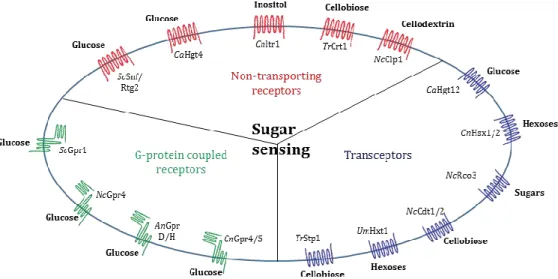
Figure 3. Sugar-sensing membrane proteins of fungal cells.
Sugar GPCRs include the Saccharomyces cerevisiae (Sc) glucose sensors Gpr1, the Neurospora crassa (Nc) glucose sensor Gpr4, the Aspergillus nidulans (An) glucose sensors GprD/H, and the Cryptococcus
neoformans (Cn) glucose sensors Gpr4/5. Nontransporting sugar receptors include the Sc glucose sensors
Snf3/Rgt2, the Candida albicans (Ca) glucose sensor Hgt4, the Cryptococcus neoformans (Cn) inositol sensor Itr1, the Trichoderma reesei (Tr) cellobiose sensor Crt1, and the Cn cellodextrin sensor Clp1. Sugar transceptors include the Ca glucose sensor Hgt12, the Cn hexose sensors Hxs1/2, the Nc sugar sensor Rco3, the Nc cellobiose sensors Cdt1/2, the Ustilago maydis (Um) hexose sensor Hxt1, and the Tc cellobiose sensor Stp1. From Dijck et al, with modifications (Dijck et al., 2017).
I.2 Fungal sensing of the environment
The knowledge of nutrient sensing and responses in fungi is mostly due to studies using the model yeast Saccharomyces cerevisiae. The identified regulatory components are often conserved in filamentous fungi, however, their functions and regulation are poorly known (Martín et al., 2019). Nutrient sensing and response of filamentous fungi, especially lignocellulose-decomposing fungi will be then discussed in comparison with S. cerevisiae in the following chapters.
Nutrient sensing is usually mediated by membrane proteins that activate downstream signaling pathways (Dijck et al., 2017). They are clustered into three classes: non-transporting receptors, transceptors, and G–protein-coupled receptors. Non-transporting receptors are proteins that have a similar structure to that of transport proteins but without transport capacity. They could function as receptors. Transceptors function as both transporters and receptors. All types of receptors are involved in the regulation of transporters involved in nutrient uptake (Dijck et al., 2017). Among those, G–protein-coupled receptors (GPCRs) are the best-studied receptors. They sense extracellular signals and transmit them to intracellular G-proteins allowing the activation of signaling pathways for the coordination of appropriate responses (Dijck et al.,
6
2017). G-proteins are cytosolic proteins that could be associated with cell membrane receptors as well as receptors found in intracellular membranes such as the endoplasmic reticulum, Golgi apparatus, vesicle membranes, and late endosomes. These proteins possess GTPase activity and require GTP in their active state (Martín et al., 2019; Ramanujam et al., 2013).
I.2.1 Nutrient sensing
Carbon sensing
Carbon nutrients include simple sugars and cleaved polysaccharides from the external biomass that can be imported through the cellular membrane to provide energy and matters for fungal biosynthesis and catabolism. They also have regulatory roles in fungi. Glucose is considered the most abundant nutrient for both yeast and filamentous fungi (Bahn et al., 2007; Brown et al., 2014). Besides, other sugars derived from hemicelluloses such as xylose, also play an important role in the growth of filamentous fungi. Lignocellulose mineralization requires the ability to utilize many carbon nutrients, leading to complex and flexible detection mechanisms. However, these sensing mechanisms in filamentous fungi remain largely to be unraveled.
Non-transporting receptors:
In yeast S. cerevisiae, glucose uptake is done by hexose transporters (HXT family) whose expression depends on the availability of extracellular glucose. Glucose uptake is also controlled by sensing the internal/external ratio of glucose via the regulation of the receptors ScSnf3 and ScRgt2 (Fig. 3). ScSnf3 and ScRgt2 are members of HXT family without transport activity. They regulate the expression of other HXT genes through the Hxt suppressor ScRgt1 (Polish et al., 2005).
Due to the diversity of sugars and saccharides derived from the biomass during the degradation process, in lignocellulolytic filamentous fungi, the sensing mechanisms are distinct from those of yeast S. cerevisiae. For example, in the early stage of biomass degradation, the release of saccharidic inducers such as cellobiose induces the secretion of CAZymes by the fungus Trichoderma reesei (Brown et al., 2014). Additionally, few non-transporting receptors have been reported in filamentous fungi, and none of them is homologous to ScSnf3 and ScRgt2 found in S. cerevisiae. Crt1 in A. nidulans and in Neurospora crassa (Zhang et al., 2013) and the cellodextrin transporter-like protein NcClp1 in N. crassa (Cai et al., 2015) have been functionally identified as non-transporting receptors. These receptors have no role in the transport of cellulose, cellobiose, and cellodextrin. They function as inducers or repressors of the cellulolytic expression machinery (Cai et al., 2015; Zhang et al., 2013).
7 Transceptors:
In S. cerevisiae, hexose transporters functioning as plasma membrane transceptors implicated in activating the downstream signaling pathways are not present. In contrast, they were identified in Candida albicans and Cryptococcus neoformans but poorly characterized (Dijck et al., 2017).
In filamentous fungi, transceptors have been identified and functionally characterized in several lignocellulolytic fungi such as N. crassa, T. reesei, and also in phytopathogenic fungi Ustilago maydis (Fig. 3). In these fungi, they could play roles in the detection and uptake of polysaccharides, and then affect the interactions of those fungi with their environment (Dijck et al., 2017). For example, in N. crassa, NcCdt1/2 are cellodextrins (cellulose breakdown products) transporters, and they were proposed to sense cellulose leading to the induction of cellulases in response to crystalline cellulose (Znameroski et al., 2014). In T. reesei, TrStp1 was identified as a cellobiose transporter. It is essential in sensing and transmitting the cellulose signal (Zhang et al., 2013).
G–protein-coupled receptors (GPCRs)
GPCRs activate the signaling pathways for responses through heterotrimeric G-proteins. Heterotrimeric G-proteins comprises three units: α, β, and γ, that are associated with the plasma membrane. In the heterotrimeric state, a GDP molecule binds to the Gα-subunit and G-proteins are inactive. They become active when Gα-subunit dissociated from Gβ-Gγ dimer by guanine nucleotide exchange as a result of binding between extracellular ligands and GPCRs. These Gα-subunit and a Gβ-Gγ dimer interact with downstream proteins to transmit the stimulus signal (Li et al., 2007b; Martín et al., 2019).
GPCRs are classified into five main categories including pheromone, carbon, nitrogen, cAMP, and microbial opsin receptors (Li et al., 2007b). Despite the high conservation of the GPCR-signaling mechanism in eukaryotes, there are significant differences between yeast and filamentous fungi (Dijck et al., 2017).
In yeast S. cerevisiae, extracellular glucose is sensed by GPCR named ScGpr1, leading to the activation of the Gα-protein ScGpa2 which, in turn, activates the adenylate cyclase ScCyr1 (Fig. 4). Besides, ScCyr1 activation is also mediated through ScRas1/2 proteins which are stimulated by intracellular glucose (Fig. 4). This activation results in a cAMP increase, which directly causes PKA activation by docking to the binding pockets of the PKA regulatory subunits releasing the catalytic domain (Yun et al., 1998). These active subunits migrate to the nucleus where they phosphorylate a wide spectrum of target proteins associated with rapid cell growth. PKA is the protein kinase A
8
mediating a large range of cAMP-depending processes in fungi including nutrient sensing and stress response (Casado et al., 2011; Thevelein et al., 2005). Nutrient control of PKA pathway in S. cerevisiae has been considered to be an outstanding model for studying nutrient sensing mechanisms.
Filamentous fungi are able to utilize diverse sugars derived from the biomass including hexoses, pentoses, and complex saccharides. This ability may be reached through the presence of additional Gα-proteins. Indeed, most characterized filamentous fungi contain three Gα-proteins, whereas yeasts only possess two (Li et al., 2007b).
Figure 4. Glucose sensing pathways through G-protein in S. cerevisiae.
Induction of ribosome biogenesis via protein kinase A (PKA), which regulates glucose signaling mediated by G-protein Gpa2. Dashed lines represent regulatory interactions, which may not be direct and in some cases are only surmised (Zaman et al., 2008).
9
In filamentous fungi, glucose sensed by GPCRs also activates protein effectors involved in fungal growth and development via stimulation of the cAMP/PKA signaling pathway. For example, in N. crassa, NcGpr4 (Fig .3) interacts with NcGna1 as an orthologue of Gpa2 in S. cerevisiae (Fig. 4) to trigger cAMP production and asexual development in response to the carbon nutrient composition (Li and Borkovich, 2006). The deletion of NcGpr4 causes a reduction in growth on glycerol, mannitol, and arabinose (Li and Borkovich, 2006). Similarly, in A. nidulans, the putative protein AnGprD, affects hyphal growth, sexual development, metabolic pathways, and stress responses (Souza et al., 2013). When growing on a 1% glucose containing medium, a deletion mutant for GprD exhibits a critical reduction in PKA activity (Souza et al., 2013). Recently, AnGprH, a GPCR highly expressed in A. nidulans under carbon starvation, has been shown to function as an upstream sensor of the cAMP-PKA pathway affecting primary metabolism and hyphal growth and repressing sexual development (Brown et al., 2015). In summary, the cAMP/PKA signaling pathway is a major pathway responding to glucose which controls the growth of filamentous fungi.
10
Figure 5. Regulation of lignocellulolytic CAZymes in filamentous fungi.
Upstream nutrient sensing pathways: The transcription factor Cre1/CreA, regulated by VIB-1/XprG,
controls the utilization of preferred carbon sources. Under activation, it represses transcription of both direct lignocellulolytic regulators and plant cell wall degrading enzymes. Besides, the cAMP-dependent protein kinase A (PKA) have a role in nutrient sensing in the manner depending on species. Inducers derived from the carbohydrates of biomass cell walls activate lignocellulolytic regulators. Direct
lignocellulolytic regulators: XYR1/XLnR activates transcription of both cellulases and hemicelluloses in
T. reesei, A. niger, and P.oxalicum, but only hemicellulases in N. crassa and A. nidulans. In N. crassa, CLR-1
activates transcription of genes required for cellulose utilization and CLR-2 known as the main transcriptional activator of cellulase transcription. The CLR-1 homolog, ClrA has been found to have a small role in the regulation of cellulase transcription in A. nidulans and A. niger, but not control ClrB, the CLR-2 homolog which is involved in cellulase transcription in A. nidulans, A. niger, and P. oxalicum. Secretory
pathway feedback: Under secretion stress, IRE1/IRE-1/IreA activates HAC1/HAC-1/HacA via direct
cleavage a non-canonical intron from the transcription factor. In turn, HAC1/HAC-1/HacA activates the unfolded protein response, which may downregulate the transcription of plant cell wall degrading enzymes. (A.nr. = A. niger, A.ns. = A. nidulans, N.C. = N. crassa, P.o. = Penicillium oxalicum, T.r. = T. reesei). From Huberman et al. (Huberman et al., 2016).
11
Carbon nutrient sensing and responses in lignocellulose degradation
The lignocellulose degrading process is mainly mediated by extracellular enzymes involved in the cleavage of polysaccharides. Nutrient sensing pathways, especially pathways enabling the use of preferred carbon sources are very important for the survival strategy used of the fungi. In filamentous fungi, those pathways function as the upstream of the direct activation of genes encoding CAZymes (Fig. 5). This activation results in the inhibition of the energy-consuming production of lignocellulose degrading enzymes (Huberman et al., 2016). Up to date, the best-studied pathway is carbon catabolite repression (CCR).
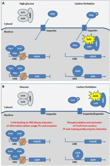
The utilization of a diverse array of carbon sources derived from lignocellulose degradation requires the coordination of the cellular metabolism and the preferential consumption of glucose prior to other carbon sources, a phenomenon known as carbon catabolite repression (CCR). CCR mechanism represses transcription of secreted and intracellular metabolic enzymes and has been described in both yeasts and filamentous fungi (Brown et al., 2014). The transcription of these genes is tightly regulated by a Cys2-His2 type DNA-binding zinc finger factor, named Mig1 in S. cerevisiae, and CreA/Cre1 in filamentous fungi (Fig. 5, 6).
In S. cerevisiae, the repressor protein ScMig1 is imported into the nucleus when glucose is added to derepressed cells (Fig. 6A). In the nucleus, ScMig1 binds to upstream regulatory elements (URE) of genes coding for transcription factors. This leads to inhibit the expression of those transcription factors which transcriptionally regulate alternative carbon usage. In addition to ScMig1, CCR is also regulated by nuclear chromatin structure which can be linked to intrinsic cellular program and environmental factors (Brosch et al., 2008). In yeast, ScMig1 reacts with the corepressors ScTup1 and ScSsn6 (Fig. 6A). The obtained complex binds to promoters of alternative carbon usage genes leading to their repression through the modulation of nucleosome positioning (Li et al., 2007a; Treitel and Carlson, 1995).
In filamentous fungi, the carbon nutrient-dependent nuclear localization of CreA/Cre1 (Fig. 6B) is similar to ScMig1 in S. cerevisiae. However, this repressor is not only regulated by glucose but also by other sugars resulting from carbon nutrient sources (Brown et al., 2013; Sun and Glass, 2011). The role of CreA nuclear localization in regulation of CCR has been confirmed in A. nidulans, and N. crassa by use of GFP fusions (Brown et al., 2013; Sun and Glass, 2011).
Orthologues of the S. cerevisiae corepressors ScTup1 and ScSsn6 have also been widely identified in filamentous fungi (García et al., 2008). In A. nidulans, the homolog of ScTup1 is AnRcoA (Fig. 6B). This corepressor is dispensable for CCR and affects the repressed chromatin structures in the alcR promoter during repression (García et al., 2008). alcR is a trans-acting gene containing a
12
specific site allowing the binding of the repressor CreA (Kulmburg et al., 1993). In comparison with S. cerevisiae, the regulatory role of the CreA/Cre1 complex to CCR is less well-known in detail in filamentous fungi. The role of CreA in nucleosome positioning has been only shown in T. reesei. In particular, TrCreA is essential for correct nucleosome positioning in the cellulase promoter cbh2 under repressing and inducing conditions (Zeilinger et al., 2003).
Figure 6. The molecular mechanism of carbon catabolite repression in S. cerevisiae (A), and filamentous fungi (B).
In presence of glucose, S. cerevisiae and filamentous fungi repress the expression of alternative carbon utilization genes via binding of a repressor protein to upstream regulatory elements (URE), which inhibit the expression of the transcription factors (TF) and metabolic enzymes necessary for alternative carbon utilization. Besides, chromatin structure hampers transcription. In the condition of limited carbon nutrient, Snf1/SnfA mediates phosphorylation and relocalisation of the repressor protein leading to depression, while local modifications to chromatin structure enable transcription. (Grey Snf1/SnfA complex = unactive; yellow star = activate Snf1/SnfA complex; P = phosphorylation; orange circle = nucleosome). From Brown et al (Brown et al., 2014).
13
In addition to CreA/Cre1 considered as a crucial regulator in CCR, the protein kinase Sucrose non-fermenting complex (Snf1/SnfA) (Fig. 6A, B) and the protein kinase A (PKA) play also a regulatory role for CCR and for the expression of lignocellulolytic enzymes (Fig. 5). In S. cerevisiae, ScSnf1 phosphorylates the repressor ScMig1 under carbon limitation and promotes the expression of glucose-repressed genes (Fig. 6A) (García‐Salcedo et al., 2014). ScSnf1, therefore, releases the CCR in glucose depleted conditions. When present at high levels, glucose is imported into the cell and the intracellular production of phosphorylated glucose lead to the inactivation of ScSnf1, which in turn inhibits ScMig1 phosphorylation. As a result, ScMig1 is localized in the nucleus (Fig. 6A). In filamentous fungi as A. nidulans, a similar function for SnfA (Fig. 6B) to phosphorylate CreA was confirmed (Brown et al., 2013; Ries et al., 2016).
Besides the cAMP/PKA pathway, the TOR (Target of Rapamycin) pathway has also been proposed possibly having an important role in regulating the expression of cell wall degrading enzymes in response to lignocellulosic material (Brown et al., 2013; Xiong et al., 2014). The TOR signaling pathway is a highly conserved nutrient sensing pathway in eukaryotes, and is found to work in parallel with the cAMP/PKA pathway to regulate common targets relating to growth in S. cerevisiae (Zurita-Martinez and Cardenas, 2005). The roles of TOR signaling in filamentous fungi will be separately discussed in detail in one following part.
In summary, there are several carbon sensing pathways having a role in regulation of growth of filamentous fungi in response to the lignocellulosic material. Among those, CCR and cAMP/PKA appear to be the major pathways. However, the regulatory role of other nutrient sensing pathways, for instance, the TOR pathway, remains to be unraveled and may provide novel solutions to enhance the extracellular enzyme productivity.
Nitrogen sensing
Fungi are able to use many nitrogen sources such as amino acids, ammonium ions, and other sources such as nucleotidic bases (Bahn et al., 2007; Dijck et al., 2017). Sensing and uptake of these nutrients are essential for the synthesis of many cellular components. However, the number of described nitrogen sensing receptors is smaller than carbon sensing receptors. To date, there are no non-transporting receptors for ammonium sensing described in yeasts. That observation is also true for amino acid sensing with the absence of described transceptors that mediate amino acid sensing in filamentous fungi. For ammonium sensing, transceptors have been found only in the ectomycorrhizal fungus Hebeloma cylindrosporum (Javelle et al., 2003) and in the phytopathogenic fungi Fusarium fujikuroi and Colletotrichum gloeosporioides (Shnaiderman et al., 2013; Teichert et al., 2008).
14
Non-transporting receptor Ssy1 for amino acid sensing in S. cerevisiae
In S. cerevisiae, extracellular amino acids are detected by a multiprotein complex composed of ScSsy1-ScPtr3-ScSsy5 (SPS). This detection induces the transcriptional expression of amino acid transporters genes and other genes encoding nitrogen-metabolizing enzymes (Kodama et al., 2002). ScSsy1 is an amino acid permease (AAP) and functions as a receptor in the SPS complex (Klasson et al., 1999). ScPtr3 is a linker between ScSsy1 and ScSsy5. ScSsy5 contains an inhibitory domain that dissociates in response to extracellular amino acids, freeing a catalytic domain to activate the transcription factor ScStp1p (Pfirrmann et al., 2010).
The receptor induction by binding of amino acids to ScSsy1 results in activating the endoproteolytic cleavage of transcription factors Stp1/2 in a process called receptor-activated proteolysis (RAP) in which Stp1/2 functions as latent cytoplasmic precursors (Pfirrmann et al., 2010). Stp1/2, in turn, induces the expression of genes encoding amino acid–metabolizing enzymes and amino acid permeases. In S. cerevisiae, Stp1 is regulated by the TOR signaling pathway (Fig. 4). Indeed, in presence of amino acids, activated Stp1 localizes in nucleus and induces expression of its target genes. In contrast, in absence of amino acids or in presence of rapamycin at a concentration at which TOR kinase activity is inhibited (TOR pathway will be discussed later), Stp1 is degraded in a manner dependent on the activity of a protein phosphatase 2A (PP2A)-like phosphatase, and Sit4, which have been known as effectors of TOR. This degradation results in the disappearance of Sit4 from the nucleus (Shin et al., 2009).
Transceptors in S. cerevisiae
In the model yeast S. cerevisiae, all identified transceptors belong to the AAP family. The best-studied receptor for amino acid sensing is Gap1, which transports all L-amino acids, several D-amino acids, and polyamines (Uemura et al., 2005). The role as an D-amino acid receptor of Gap1 was found when studying the amino acid induced rapid activation of the PKA pathway, which has been known to be activated in a cAMP dependent reaction (Fig. 4). In this pathway, Gap1 is involved in the downstream relay of the signal according to an unknown mechanism (Donaton et al., 2003).
In S. cerevisiae, three genes encoding for ammonium transporters have been identified, but only one is a transceptor named ScMep2. ScMep2 acts as a regulator of pseudohyphal growth and PKA activation when ammonium is added to nitrogen-starved cells (Boeckstaens et al., 2007). ScMep2 in controlled by the ScNpr1, a kinase that is the target of ScTORC1 (Fig. 4). Under nitrogen starvation or ScTOR inactivation, ScNpr1 is activated by dephosphorylation. In presence of sufficient ammonium, ScNpr1 is phosphorylated and inactive (Boeckstaens et al., 2007).
15
G – protein-coupled receptors (GPCRs) in nitrogen sensing of yeast and filamentous fungi
There are no GPCRs established as nitrogen receptors in S. cerevisiae while orthologues of ScGpr1 known as glucose sensor (Fig. 4) were reported to sense methionine in pathogenic C. albicans and C. neoformans (Maidan et al., 2005; Xue et al., 2005). Methionine sensing is important for the hyphal transition of C. albicans on agar medium, and it can induce a transient cAMP production, as well as mating hyphal elongation in C. neoformans (Maidan et al., 2005; Xue et al., 2005).
In filamentous fungi, amino acids appear to be sensed by the cAMP receptor-like GPCR named GprH which is the glucose sensor in A. nidulans (Fig. 3) (Li et al., 2007b). Orthologues of GprH have been found in other filamentous fungi such as NcGpr4 in N. crassa, and AfGprC/D in Aspergillus flavus (Fig. 3) (Affeldt et al., 2014; Li and Borkovich, 2006). Additionally, the function of cAMP receptor-like GPCRs in the regulation of sexual development is conserved in fungi. GPCRs therefore may have a dual role in sensing both sugars and amino acids.
There is very little literature dealing with GPCRs requirement in ammonium sensing of filamentous fungi. To date, ammonium sensing via GPCRs has been only shown in Aspergillus flavus (Affeldt et al., 2014). In this fungus, mutations in GprC/D receptors impair growth on ammonium chloride and proline (Affeldt et al., 2014).
In summary, in addition to cAMP/PKA signaling pathways, the TOR signaling pathway may play an important role in regulating a wide range of functions in response to both carbon and nitrogen nutrient sources in the filamentous fungi. In the next part, TOR signaling will be described and discussed in these fungi.
Target Of Rapamycin (TOR) signaling as a key regulator of growth in the adaptation
of filamentous fungi
All living organisms sense and respond to nutrient signals by regulating growth and developmental programs for survival. In eukaryotic organisms, a role for the TOR signaling pathway has emerged as a global regulator of cell growth. The central component of this signaling pathway is Target Of Rapamycin (TOR), a conserved serine/threonine kinase which belongs to the family of phosphatidylinositol kinase-related kinases (PIKK) (Wullschleger et al., 2006).
TOR was first found in S. cerevisiae via the characterization of strains resistant to rapamycin. Rapamycin is a metabolite produced by Streptomyces hygroscopicus (Heitman et al., 1991). Rapamycin binds to the intracellular receptor FKBP12 (FK506 binding protein 12) to form a complex which then inhibits TOR kinase activity resulting in cellular toxicity (Wullschleger
16
et al., 2006). In S. cerevisiae, two genes encoding two proteins named TOR1 and TOR2 are present. The resulting proteins are the main components of two complexes named TORC1 and TORC2. Up to date, TOR characterization in fungi has been mainly carried out in S. cerevisiae wherein TOR has been demonstrated as the central controller of growth, proliferation, and survival in response to nutrients and stress (Loewith and Hall, 2011). Up to date, TOR has been identified in all tested eukaryotes, controlling cellular processes via regulating networks named TOR signaling pathways.
Figure 7. TOR signaling pathway in the model yeasts.
The TOR pathway components in S. cerevisiae (A) and S. pombe (B). The functional homologs between the two species are shown in the same shape and color (Shertz et al., 2010).
17
In the fungal kingdom, TOR signaling pathways have been best-characterized in model yeasts S. cerevisiae and S. pombe as presented in Fig. 7 (Shertz et al., 2010). In S. cerevisiae, only ScTORC1 is sensitive to rapamycin. It comprises ScTOR1 or ScTOR2, ScKog1, ScTco89, and ScLst8 (Fig. 7A). ScTORC1 controls protein synthesis, mRNA synthesis and degradation, ribosome biogenesis and autophagy. ScTORC2 is composed of ScTOR2, ScLst8, ScAvo1, ScAvo2, ScAvo3 (Fig. 7A), it is insensitive to rapamycin and is involved in the control of actin polarization and cell wall integrity (Loewith and Hall, 2011). The downstream effectors of ScTORC1 are the ScSit4, one PP2A-like phosphatase, and the AGC kinase ScSch9. ScSch9 has been deeply studied due to its involvement in stress response. Under stress conditions including nutrient starvation, high salt, redox stress, temperature, ScSch9 phosphorylation is strongly reduced (Urban et al., 2007). Due to this property, ScSch9 has been used to quantify ScTORC1 activity through its phosphorylation in various nutrient and stress conditions (González et al., 2015). In comparison to S. cerevisiae, there is an extension or expansion in upstream components of the TOR pathway in S. pombe, with the presence of ScTsc1 and ScTsc2 (Fig. 7B). In this fungus, the TOR pathway has functional roles in nutrient and stress signaling, cell growth and differentiation, and sexual development (Alvarez and Moreno, 2006; Otsubo and Yamamoto, 2010; Weisman and Choder, 2001).
In addition to S. cerevisiae and S. pombe, TOR signaling has been also well-studied in human pathogenic yeasts such as C. albicans and C. neoformans (Rutherford et al., 2019). C. albicans is the first fungus found to be inhibited by rapamycin in 1975 which opened the story of TOR signaling researches (Sehgal et al., 1975). In contrast to yeasts, TOR signaling pathways have been less investigated in filamentous fungi. In these organisms, TOR and TOR signaling pathways have been only identified and partially functionally characterized (focusing on genetics, development, and pathogenicity) in several fungal models such as A. nidulans (Fitzgibbon et al., 2005), N. crassa (Park et al., 2011), Podospora anserina (Pinan-Lucarré et al., 2006), A. fumigatus (Castro et al., 2016), C. neoformans (Lee et al., 2012), Fusarium fujikuroi (Teichert et al., 2006), Fusarium graminearum (Yu et al., 2014), Fusarium oxysporum (López-Berges et al., 2010), Magnaporthe oryzae (Qian et al., 2018), and Verticillium dahliae (Li et al., 2019). In all these fungi, TOR signaling is functionally conserved and involved in the regulation of cellular growth, metabolism, virulence and vegetative development. In addition to the TOR identification, the components of the TOR signaling pathway have also been identified in the focus on downstream effectors of TOR. Indeed, homologs of ScSit4 (AnSit4) and ScSch9 (AnSchA and NcSch9) were identified in A. nidulans (Fitzgibbon et al., 2005) and in N. crassa (Park et al., 2011). Functional characterization of these components showed similar roles in response to stress and in autophagy regulation under rapamycin treatment or nutrient-starvation. For these fungi, to date, only one upstream component of TOR signaling pathways has been identified in N. crassa: the protein VTA for