HAL Id: tel-02079405
https://tel.archives-ouvertes.fr/tel-02079405
Submitted on 26 Mar 2019HAL is a multi-disciplinary open access archive for the deposit and dissemination of sci-entific research documents, whether they are pub-lished or not. The documents may come from teaching and research institutions in France or abroad, or from public or private research centers.
L’archive ouverte pluridisciplinaire HAL, est destinée au dépôt et à la diffusion de documents scientifiques de niveau recherche, publiés ou non, émanant des établissements d’enseignement et de recherche français ou étrangers, des laboratoires publics ou privés.
Vivek Thakare Sudam
To cite this version:
Vivek Thakare Sudam. Multifunctional platforms for cancer theranosis. Radiochemistry. Université Bourgogne Franche-Comté, 2018. English. �NNT : 2018UBFCK022�. �tel-02079405�
Thesis
Presented toTHE UNIVERSITY OF BOURGOGNE FRANCHE-COMTE
to obtain the title ofDOCTOR OF
THE UNIVERSITY OF BOURGOGNE-FRANCHE-COMTE
Discipline: Chemical Sciencesby
VIVEK SUDAM THAKARE
M. S (Pharm.)Multifunctional platforms for cancer theranosis
Defended on July, 19th 2018 in front of the committee
Éva JAKAB TÓTH Directrice de Recherche, Centre de Biophysique Moléculaire, CNRS, Orléans
Rapporteur Fabienne GAUFFRE Directrice de Recherche, Université de Rennes 1, CNRS, Rennes Rapporteur Stéphane ROUX Professeur à l’Université de Bourgogne Franche-Comte Examinateur
François LUX Maître de Conférences, Université
Lyon1 Examinateur
Frédéric BOSCHETTI Chief Executive Officer, Chematech Encadrant de thèse Franck DENAT Professeur à Université de
Multifunctional platforms for cancer theranosis 3
Acknowledgements
This thesis work was carried out within the company CheMatech and also at the Institute of Molecular Chemistry of the University of Burgundy (ICMUB) in the team Polyamines, Porphyrins Developments and Applications (P2DA).
I express my gratitude to Dr. Frédéric BOSCHETTI, (PhD, University of Bourgogne) and CEO of the company CheMatech for having given me the opportunity to perform my doctoral research work at Chematech. My sincere thanks to him for guiding me in my research and for providing constant support throughout my doctoral training.
I would especially like to thank Professor Franck DENAT, Director of ICMUB, for welcoming me and having me in his excellent team at ICMUB. I thank him for having offered me his valuable supervision and timely motivation. I also acknowledge his pleasant nature and attention for details.
A big thank you to my colleague and friend, Mr. Guillaume PAULIN, Manager of the GMP laboratory at CheMatech, for always being available for interesting discussion and his precious help and advice in the chemical synthesis. The great thing was, all our communications were in French, which was fun and learning experience.
I would also like to thank my colleague Mr. Stéphane MARTEL, Manager of the R & D and Analysis laboratory at CheMatech, for providing me guidance in addressing routine chemistry and technical problems. I also thank him for his work in the laboratory as well as for the maintenance of many devices.
I would also like to express my gratitude to Dr. Mathieu MOREAU and Dr. Claire BERNHARD for interesting scientific discussions and their valuable advice, particularly while planning radiolabelling and animal studies at CGFL. This thesis work would not have been possible without their timely support, encouragement and friendship.
I equally thank all wonderful colleagues from CGFL, Dijon who I got chance to interact with and have directly or indirectly contributed to my knowledge.
I would like to thank Prof. Olivier TILLEMENT, Dr. Lux FRANÇOIS and Vulong TRAN for their collaboration while working on AGuIX nanoparticles and welcoming me to work in their laboratory during the secondment.
I would like to extend my sincere gratitude to Dr. Victor GONÇALVES and Coline CANOVAS, for collaborating in the development of PSMA based theranostics.
I thank Prof. Anthony ROMIEU for his valuable time, support and scientific advice on many interesting topics including fluorescent dyes and their characterization.
I also extend my gratitude to Prof. Stephané ROUX and his team in Besançon for excellent collaboration in developing multifunctional gold nanoparticles. I hope this collaboration will go a long way in developing diverse theranostics in future.
I express my sincere gratitude to Prof. Kevin PRISE and Dr. Karl BUTTERWORTH for welcoming me at their laboratory in Centre for Cancer Research and Cell Biology, Queens University Belfast, Belfast, UK during the secondment.
I would like to thank Professor Sandrine LACOMBE, Co-ordinator of ARGENT, Prof. Nigel MASON, The Open University, Milton Keynes, UK and all other supervisors/members of the ARGENT project consortium who have timely trained, motivated me whist assessing my progress throughout the project.
I also thank Marie-José PENOUILH, Fanny CHAUX and Myriam HEYDEL for their help and advice on mass spectrometry, NMR and other analyses. I also thank Marcel SOUSTELLE for the elementary analysis
I would also like to thank the IT team consisting of Dr. Alain TABARD (deceased), Dr. Christine STERN, Anne COMBET and Thierry BELLOIR of P2DA group whose constant support has been crucial in realising and facilitating my work and stay in Chematech and ICMUB.
I do not forget to extend my thanks to all the permanent and non-permanent members of the P2DA team and ICMUB, who have been great and accommodating. All these people have added a great deal of value to my experience, learning and stay in ICMUB.
Lastly, I would like to warmly thank my colleagues and friends met during these three years of thesis, with whom I spent very good moments; Adrien DUBOIS, Sophie POTY, Mylène BONNAUD, Coline CANOVAS, Jacques PLIQUETT, Léo BUCHER, Clement MICHELIN, Yann BERNHARD, Damien LHENRY, Bertrand BRIZET, Valentine QUESNEAU, Sylvain DEBIEU, Vivian LIORET, Ibai VALVERDE, Marc PIRROTTA, Victor GONCALVES and Richard DECREAU.
Finally, I dedicate this work to my parents who have always been there for me with their unconditional support and to my wife Debarati for her unwavering support and love, without whom nothing would have been possible.
Multifunctional platforms for cancer theranosis 5
Table of Contents:
Chapter I . Introduction ... 11
I-1 Preface ... 12
I-1.1 Overview of ARGENT project: ... 13
I-2 Cancer theranosis: Approaches and avenues ... 15
I-2.1 Cancer therapy: ... 16
I-2.2 Cancer Diagnosis: ... 22
I-3 Monomolecular Multimodal Platform in cancer theranosis: ... 30
I-3.1 Chelators used in multimodal platforms: ... 31
I-3.2 Fluorescent probes for NIR based optical imaging: ... 35
I-3.3 Targeting ligands for cancer: ... 38
I-3.4 Conjugation Site and Chemistry: ... 43
I-4 Objectives of the research thesis: ... 48
Chapter II . Multifunctional polysiloxane (AGuIX) nanoparticles for cancer theranosis ... 51
II-1 AGuIX Nanoparticles in cancer theranosis: ... 52
II-1.1 Synthesis and structure of AGuIX: ... 53
II-2 Functionalization of AGuIX: ... 56
II-2.1 Nanoparticle synthesis and functionalization with silane chelators: ... 57
II-2.2 Physico-chemical properties of functionalized nanoparticles: ... 62
II-2.3 Radiolabelling and study of the stability of radiolabelled nanoparticles: ... 68
II-3 Animal imaging in TSA tumor model: ... 70
II-4 Conclusions: ... 72
Chapter III . Multifunctional gold nanoparticles for cancer theranosis ... 73
III-1 Gold Nanoparticles for cancer theranosis: ... 74
III-2 Development of DOTAGA based amine functionalized gold nanoparticle: ... 77
III-2.1 Synthesis of DOTAGA-Lys-TA-NH2 ... 77
III-2.2 Amine functionalized DOTAGA gold nanoparticle (Au@DOTAGA-Lys-TA-NH2): ... 79
III-3 Development of gold nanoparticles for PET-MRI: ... 80
III-3.1 Synthesis of DOTAGA-Lys-TA-NODAGA ligand ... 80
III-3.2 Gold nanoparticles based on DOTAGA-Lys-TA-NODAGA for PET-MRI: ... 84
III-4 Radiolabelling and stability of the radiolabelled nanoparticles for PET-MRI: ... 86
III-5 Animal imaging and biodistribution studies in TSA tumor model: ... 88
III-6 Development of PSMA targeted gold nanoparticles: ... 90
III-6.1 Synthesis of the DOTAGA-Lys-TA-Glu-PSMA ... 90
III-6.2 Determination of the PSMA binding activity using the enzymatic assay: ... 92
III-6.3 Gold nanoparticle based on DOTAGA-Lys-TA-Glu-PSMA: ... 93
6
III-8 Development of a PET-Optical ligand for gold nanoparticles: ... 95
III-8.1 Synthesis of a PET-Optical ligand for gold nanoparticles ... 95
III-8.2 Photo-physical characterization of the PET-Optical probe (24): ... 99
III-9 Development of amine functionalized NODAGA ligands for gold nanoparticle synthesis: ... 100
III-10 Conclusions: ... 101
Chapter IV PSMA targeted multimodal imaging ... 103
IV-1 Introduction: ... 104
IV-1.1 PSMA as a biomarker in prostate cancer: ... 105
IV-2 PSMA inhibitors in prostate cancer theranosis: ... 106
IV-2.1 Anti-PSMA monoclonal antibodies in prostate cancer theranosis: ... 106
IV-2.2 Small molecule based PSMA inhibitors in prostate cancer theranosis: ... 107
IV-3 Synthesis of PSMA targeted monomolecular PET-Optical imaging probe: ... 114
IV-3.1 Synthesis of chelator-linker moiety: ... 114
IV-3.2 Synthesis of PSMA ligand and its derivative: ... 115
IV-3.3 Synthesis of chelator-linker-PSMA ligand: ... 116
IV-3.4 Synthesis of bioconjugatable IR-783: ... 119
IV-3.5 Synthesis of chelator-linker-PSMA ligand-IR783: ... 121
IV-4 Characterization of the PET-Optical probe ... 123
IV-4.1 Photophysical characterization of the PET-Optical probe: ... 123
IV-4.2 Radiolabelling and stability of the PET-Optical probe: ... 125
IV-4.3 Binding affinity measurements using NAALDase assay: ... 127
IV-5 Conclusions: ... 129
Chapter V . Bimodal imaging probes for bioconjugation to antibody fragments and nanoparticle functionalization ... 131
V-1 Multimodal imaging probes for bioconjugation: ... 132
V-2 Synthesis of trifunctional probe for PET-Optical imaging: ... 134
V-2.1 Synthesis of bifunctional chelating agent: ... 134
V-2.2 Synthesis of the linker system bearing chelator and conjugation site: ... 134
V-2.3 Synthesis of a trifunctional probe using bioconjugatable IR-783: ... 136
V-3 Photo-physical characterization of the trifunctional probe: ... 139
V-4 Chemical biology of the antibody drug conjugates: ... 141
V-4.1 Non-specific Conjugation through Native Residues ... 141
V-4.2 Conjugation through genetically engineered sites ... 143
V-4.3 Enzymatic Bioconjugation: ... 143
V-4.4 Labelling strategies in antibody conjugation: ... 145
V-4.5 Difference between conjugates of mAb fragmentsand full scale mAb? ... 146
Multifunctional platforms for cancer theranosis 7
V-6 Development of HER-2 targeted bimodal probe based on antibody fragment: ... 151
V-6.1 Synthesis of F(ab’)2 of trastuzumab: ... 152
V-6.2 Reduction of F(ab’)2 fragments: ... 153
V-6.3 Conjugation of the bimodal probe to Fab’ fragment: ... 154
V-6.4 Characterization of the bimodal conjugate: ... 154
V-7 Animal imaging in breast cancer model: ... 157
V-8 Development of the multifunctional nanoparticles: ... 159
V-9 Chemical functionalization of AGuIX using bimodal probe: ... 160
V-9.1 Thiolation of the AGuIX: ... 160
V-9.2 Conjugation of the bimodal probe to AGuIX: ... 161
V-10 Characterization of the functionalized nanoparticles: ... 162
V-10.1 Physico-chemical characterization of nanoparticle properties: ... 162
V-10.2 Photophysical characteristics of the functionalized nanoparticles: ... 164
V-11 Radiolabelling and stability of the functionalized nanoparticles: ... 165
V-12 Animal imaging in breast cancer model: ... 167
V-13 Conclusions: ... 169
Chapter VI . Experimental Section ... 171
Chapter VII . References ... 219
Multifunctional platforms for cancer theranosis 9
Glossary of abbreviations
ADC Antibody drug conjugates
AGuIX Activation et Guidage de l’Irradiation X
ARGENT Advanced Radiotherapy Generated by Exploiting Nanoprocesses and Technologies
APTES (3-Aminopropyl) triethoxysilane
BFC Bifunctional chelator
Boc tertiary butyloxycarbonyl
BODIPY 4,4-difluoro-4-borata-3a-azonia-4a-aza-s-indacene cGMP current Good Manufacturing Practices
CT Computed tomography
DIPEA N,N-diisopropyl ethyl amine 4-dimethylaminopyridine
DLS Dynamic light scattering
DMF Dimethyl formamide
DOTA 1,4,7,10-tetraazacyclododecane-1,4,7,10-tetraacetic acid
DOTAGA 1,4,7,10-tetraazacyclododecan-1-glutaric acid-4,7,10-triacetic acid
DTPA Diethylene triamine penta acetic acid
EDC 1-ethyl-3-(3-dimethylaminopropyl) carbodiimide
EDTA Ethylene diamine tetraacetic acid
FDG fluorodeoxyglucose
Gln Glutamine
Glu Glutamic acid
HBED N, N'-bis-(2-HydroxyBenzyl)ethylenediamine-N, N'-diacetic acid
HBTU 2-(1H-benzotriazol-1-yl) -1,1,3,3-tetramethyluronium hexafluorophosphate
HEPES 4-(2-HydroxyEthyl)-1-piperazineethane sulfonic acid
HER-2 Human epidermal growth factor receptor 2
HOBt 4-hydroxybenzotriazole
10
HRMS High resolution mass spectrometry
IC50 Inhibitory Concentration at 50%
ITLC Instant thin layer chromatography
Lys lysine
mAb monoclonal Antibody
MALDI-TOF Matrix Assisted Laser Desorption Ionisation- Time of Flight MRI Magnetic Resonance Imaging
NCS: Isothiocyanate
NHS N-Hydroxy-Succinimidyl
NIR Near infrared
NMR Nuclear Magnetic Resonance
NOTA 1,4,7-triazacyclononane-1,4,7-triacetic acid
NODAGA 1,4,7-triazacyclononane-1-glutaric acid-4,7-diacetic acid PBS Phosphate Buffer Saline
PEG Polyethylene glycol
PET Positron Emission Tomography
PSMA Prostate-Specific Membrane Antigen
SDS Sodium dodecyl sulphate
SPECT Single Photon Emission Computed Tomography
TACN 1,4,7-Triazacyclononane
t-Bu tert butyl
TCEP tris(2-carboxyethyl)phosphine
TFA Trifluoroacetic acid
Chapter 1. Introduction
Multifunctional platforms for cancer theranosis 11
12 I-1 Preface
Cancer is one the deadliest maladies afflicting mankind amounting to 1 in 6 of all global death totalling to about 8.8 million deaths in 2015, as estimated by WHO. Cancer is the second leading cause of death globally, and the number of new cases is expected to rise by about 70% over the next 2 decades. Approximately 70% of deaths from cancer occur in low and middle-income countries. The financial burden of cancer management has been tremendous, with the estimated total annual economic cost of cancer in 2010 to be around US$ 1.16 trillion (www.who.int). A significant quantum of resources has been invested to address this medical need at the academic as well as industry level that has led to improvements in our understanding of the disease.
However, this comprehension of the cancer biology has not been proportionately translated into corresponding improvements in cancer care. One of the important reasons that precluded this is the lack of selective delivery of anti-cancer compounds to cancerous tissue. Owing to lack of selectivity, a high systemic exposure to anti-cancer agents more often leads to a dose-limiting toxicity. As a result, many efforts are being directed towards selectively delivering the toxic payloads to cancer, making targeted delivery an important approach in overcoming the current limitations of cancer therapy. Recent developments in immunology, gene/cell therapy and nanotechnology are expected to significantly improve the therapy and diagnosis of cancer, thereby increasing efficacy with which the disease is being treated currently.
Nanotechnology has been one of the major breakthroughs in research that has found its value in life sciences amongst several other applications and cancer is no exception to this. Several nanotechnology driven approaches have borne fruits as evident from the growing list of the nanomedicines approved by health authorities across the globe. In 2011, the European Commission published a recommendation on the definition of nanomaterial predisposing size as the critical factor (1-100 nm) with the acknowledgment that the upper limit of 100nm not justified across whole range of nanomaterials. Several research programs have been funded by European Union (EU) focussing on leveraging the science at nanoscale and ARGENT has been one of them. The FP7 European Multi-ITN (Marie Curie Actions Initial Training Network) project “Advanced Radiotherapy, Generated by Exploiting Nanoprocesses and Technologies (ITN ARGENT)” started in March 2014. The prime objective of this inter-sectorial and multidisciplinary ITN is to create a new generation of researchers and experts able to develop and propose to the society new tools and concepts for the improvement of cancer therapy and diagnosis.
Chapter 1. Introduction
Multifunctional platforms for cancer theranosis 13
I-1.1 Overview of ARGENT project:
50% of the patients receive radiotherapy as part of their cancer treatment. The main limitation of this treatment is the lack of tumor selectivity, which causes severe side effects, and radioresistance. The most promising developments to improve the performances of radiation-based therapies is the use of fast ion beam radiation (carbon therapy and proton therapy) and nanoparticles-enhanced therapies. ARGENT brings together world-leading researchers of different disciplines, physicists and medical physicists, chemists, biologists, medical doctors and SMEs with the aim of understanding and exploiting the nanoscale processes that drive these phenomena (http://itn-argent.eu/). This European effort should lead to the development and optimization of new nanodrugs together with advanced radiation protocols. This will open a new era for radiotherapy with subsequent economic and ‘quality of life’ benefits for the EU population. In order for Europe to fully exploit its world-lead, a new generation of supra-disciplinary researchers familiar with following domains must work in an orchestrated manner: i) Physics and medical physics: explaining the physical interactions of radiation; ii) Chemistry: describing the chemical processes and methodologies for tailoring nano-agents; iii) Biology: elucidating the effects in vitro and in vivo.
Figure I-1: The partners within the ARGENT consortium spearheading the collaborative interdisciplinary research on cancer.
This project is strongly supported by the medical community. As an end point, this inter-sectoral and multi-disciplinary programme will form young researchers and experts able to
14
create a platform on which next generation cancer therapy will be built. The consortium aims to train a cohort of 13 PhDs (Early Stage Researchers – ESRs) to subsequently act as leaders and ambassadors in the field. The ITN ARGENT strategy relies on i) improving our understanding of the processes and mechanisms underlying radiation damage on a nanoscopic level, ii) applying the improved know-how in the production and development of functionalised nanodrugs to amplify the effects of medical beams and iii) developing of concepts and codes for clinical applications, taking into account the new information.
The ITN-ARGENT consortium is represented by 6 academic, 3 industrial and 6 associated partners that complement and leverage the competencies and expertise developed since their establishment (Figure I-1). The ARGENT research program is based on three work packages; nanodosimetry, theranostic nanoagents and preclinical evaluation. Each work package is with group of ESRs based on their respective projects and scientific/technical competencies. The Preclinical Evaluation team combines their efforts to understand better how nanoscale processes initiated by the interaction of radiation with living matter affects biological responses, establishing a link between nanoscale interaction and clinical effects. Combining advanced experimental, theoretical, and modelling tools, the team investigates nanoscale interactions for preclinical testing in cell-based models and exploring their clinical applicability. The major goal of this team in ARGENT is to evaluate the use of the new methods and tools developed in the project for better patient outcome. The scientific work package on theranostic nanoagents is comprised of experimentalists from chemistry, biology, and medical physics. The goal of this work package is to design, synthesize, and characterize the next generation of technologies as radiosensitizers and diagnostic tools. The group also studies their fate inside the cellular environment and the cell damage caused by the combination of nanoparticles and different radiation beams. The Nanodosimetry team combines experiments and simulations to answer the most fundamental questions regarding the mechanisms present in radiation induced damage in cells. They study the interactions between biomolecules, nanoagents, and ion and photon radiation with both experiment and simulation. Being a part of the ARGENT team as one of the ESRs gave me an opportunity to perform my doctoral research at Chematech, one of the industrial partners in the consortium along with its affiliate Institute of Molecular Chemistry (UMR CNRS 6302), Université de Bourgogne-Franche Comté, Dijon.
Chematech is one the leading European research based enterprise developing chemical tools based on chelation chemistry. These chemistries find applications in antibody conjugation, nanoparticle development and other technologies useful in therapy and diagnosis of diseases. Chematech is led by its CEO, Dr. Frédéric Boschetti and represents one of the pillars of the
Chapter 1. Introduction
Multifunctional platforms for cancer theranosis 15
ARGENT team owing to its unique capability in the development of the macrocyclic compounds used in imaging and nuclear medicine. This is further strengthened by the scientific and advisory support received from Institute of Molecular Chemistry, Université de Bourgogne-Franche Comté, Dijon which is spearheaded by Prof. Franck Denat. Apparently, this thesis has received the unique mentorship of both Dr. Boschetti and Prof. Denat who are experts in the field of macrocyclic and conjugation chemistry from industry and academia. In the context of the aforementioned objectives, the work package designated towards ‘Nanoagents for cancer theranosis’ forms the important aspect of the current thesis and steers in the direction that aims to develop novel tools for cancer theranosis.
I-2 Cancer theranosis: Approaches and avenues
Figure I-2: Key drivers of cancer therapy, diagnosis and theranosis.
The term “theranostics” signifies any material that permits the combined diagnosis and treatment along with the potential to follow up of a disease and has been coined by the US consultant John Funkhouser, in August 1998 [1]. Theranostics implies the development of the two modalities in an integrated manner that serves the function of a diagnostic test and a therapeutic agent in an interdependent and concerted manner so as to cater towards comprehensive care of a specific disease. The ultimate aim of developing a theranostic is to simultaneously image and monitor the diseased tissue, drug pharmacokinetics, and pharmacodynamics with the long-term objective of fine tuning the therapy and dose with a control unattainable hitherto.
16
I-2.1 Cancer therapy:
As implied in the Figure I-2, the cancer therapy relies on four major treatment modalities namely; surgery, radiotherapy, chemotherapy and immunotherapy. These approaches could be used as standalone or in combination with each other depending on the tumor type and its stage. Majority of the benign cancers can be treated by surgical resection, whereas the malignant ones need a concomitant approach. Radiotherapy involves exposing the tumor lesions to high energy ionising radiations (e.g: X-rays) as an external beam radiotherapy. On the other hand, ‘brachytherapy’ or internal radiation therapy involves locally exposing the cancerous tissue to radiation by implanting the seeds of radioactive substance at the affected site. Alternatively, the radiotherapy can also be provided by delivering the sequestered radioisotope (e.g: 177Lu, 90Y) at the cancerous lesions in conjunction with a targeting ligand. This typically involves conjugating the chelator molecule to the targeting ligand that targets specific receptors on the cancer cells. The chelator molecule serves the purpose of sequestering the radioisotope which otherwise would be difficult to target. The ligand can be a peptide as in the case of recently (January 2018) FDA approved Lutathera(Lutetium dotatate) which is referred to as Peptide Receptor Radionuclide Therapy (PRRT) or it can be a monoclonal antibody where it is referred to as ‘Radioimmunotherapy’. Zevalin(90Y ibritumomab tiuxetan) and Bexxar (131I- tositumomab) are the approved products in this category (www.fda.gov). Chemotherapy represents one of the most traditional approaches in the cancer therapy that involves administration of a chemotherapeutic (synthetic or semi-synthetic drugs) to the cancer patients. These drugs are cytotoxic as they usually interfere with the key metabolic processes that are important for the cell survival or cell growth. Since the action of the cytotoxic drugs is not selective to cancer cells they also affect the normal healthy cells resulting in serious side effects. Many research efforts are being directed to address this issue by modifying the existing drugs or developing the new drugs that are more selective to cancerous cells. Much of the nanoparticle based research focussing on drug delivery encompasses the delivery of such anti-cancer drugs so as to target them only to the tumor site minimising the deleterious effects on the healthy tissue [2]. Immunotherapy represents the most recent and advanced therapeutic modalities for cancer therapy based on sound understanding of the molecular immune mechanisms underlying the pathophysiology. Monoclonal antibodies (mAbs) form the mainstay of the immunotherapy and were first produced by Milstein and Köhler (Cambridge University), who were awarded the Nobel Prize in Physiology or Medicine in 1984. mAbs possesses exquisite specificity owing to their greater surface area binding, which results in decreased ‘off-target’ effects/toxic effects as
Chapter 1. Introduction
Multifunctional platforms for cancer theranosis 17
compared with most small molecule drugs. With the advancement and the synergy of immunology, molecular biology and protein engineering, it has been possible to developed engineered proteins including monoclonal antibodies that are highly efficacious, stable and cater to different type of diseases/cancers. Various mechanisms have been thought to play roles in mediating the tumoricidal effects of mAb. These include signalling promoted by cross-linking of surface antigen that leads to cell death, blockade of an activation signal that is essential for continued cell growth, antibody-dependent cellular cytotoxicity (ADCC), complement mediated cytotoxicity (CMC) and the ability of mAb to alter the cytokine milieu or augment development of an active anti-tumor immune response. Of the various monoclonal antibodies approved for diverse disease categories 27 have been only approved for cancer highlighting the importance these magic bullets in cancer therapy [3, 4]. Bolstering the concept of the magic bullets, monoclonal antibodies have also been utilized as a cancer targeting cargo to deliver the highly potent cytotoxic molecules giving rise to new class of drugs; antibody drug conjugates referred to as ADCs. With only four approved ADCs currently in market, this is a very recent addition to the armamentarium against cancer, yet with lot of promise as witnessed by the prolific pipeline (> 50) that is at moment under clinical investigation (www.adcreview.com).
I-2.1.1 Nanoparticles in cancer therapy:
One of the hallmarks of the cancerous lesion is its altered physiological and anatomical state that makes it more vulnerable to the action of nanoparticles by virtue of an effect termed as ‘Enhanced Permeability and Retention effect’. The EPR effect is a combined result of complex biological processes viz; angiogenesis, vascular permeability, lymph-angiogenesis, heterogeneous tumor genetic profile and microenvironment and poor lymphatic clearance. Any macromolecular system including nanoparticle that is injected into the biological subject, has propensity to get lodged in the milieu of the cancerous tissue owing to its leaky vasculature and poor lymphatic drainage. This enables the dislodged cargo to exert its action locally at the site endogenously (drug release from nanoparticle) or under the influence of an external stimuli (radiosensitization by nanoparticles upon radiation). Many approaches based on the macromolecular/nanoparticulate delivery have been developed for the cancer therapy and diagnosis as exemplified in the Figure I-3. Categorically, they have been classified as lipid based carriers, polymeric carriers, inorganic nanoparticles, conjugates and viral nanoparticles.
18
Figure I-3: Schematic illustration of different nanotherapeutic platforms. Adapted from [5]. Lipid nanocarriers include liposomes, solid lipid nanoparticles, SMEDDS etc. that are made from fatty acid derivatives. Several liposomal formulations like Doxil, Myocet, Mepact, Marqibo, DaunoXome etc. have been appoved in US/EU/Asia for cancer therapy. Polymeric nanoparticles are composed of the matrix based nanoparticles fabricated using chemically synthesized polymers (e.g: PLGA), polymeric micelles which are made up of amphiphilic copolymers (e.g: Polycaprolactone-polyglutamic acid) and polymer encapsulated/coated systems (e.g: Abraxane – Albumin bound paclitaxel nanoparticles). Conjugates include more often covalently conjugated polymer drug systems or antibody drug conjugates which have been one of the hot topics in the targeted drug delivery with the clinical pipeline abuzz with several lead candidates. Virus like particles (VLP) are viruses devoid of genetic materials making them non-infectious. VLP are made up of the viral surface proteins/capsids that can house drug/tracer/gene/protein which acts as a cargo and can be useful in therapeutic, imaging and vaccine applications. Inorganic nanoparticles typically encompass silica nanoparticles, gold nanoparticles, iron oxide nanoparticles and other metallic nanoparticles (e.g: quantum dots etc). Inorganic nanoparticles are usually non-biodegradable and can pose toxicity issues and hence need a due consideration with respect to their physico-chemical characteristics so as to address any potential biocompatibility/bio persistence issues. Nonetheless, despite these challenges several inorganic nanoparticles have made it to clinic (ferumoxtran, ferucarbotran etc.) and many are on their way to do so (e.g: AGuIX – Phase I and Hafnium oxide NPs – Phase I/II) as can be seen from the clinical trial database (www.clinicaltrials.gov).
Chapter 1. Introduction
Multifunctional platforms for cancer theranosis 19
I-2.1.1.1 What is the difference between passive and active targeting of nanoparticles? By virtue of the anatomic and physiological peculiarities of the cancerous lesions, the nanotechnology based research has been able to drive the results in its strides and is one of the main reasons that justify the application of nanomedicines in oncology compared to other disease areas. Passive targeting of the nanoparticles harnesses the EPR effect and involves use of the nanoparticles without any specific surface chemistry manipulations but within a certain size range. Active targeting involves modification of the nanoparticle surface with the ligands that have higher affinity to the receptors expressed mainly on the surface of the cancerous cells.
Although the approved nanomedicines justify the promise offered by the nanoparticles, there are multitude of factors that pathophysiologically differentiate patients from each other depending on the tumor type and its extent. Moreover, the distribution and accumulation of NPs in tumors could be highly variable and is likely influenced by the biological and physicochemical properties of each material. As a result of this, an EPR effect alone might not be effective in eliciting the adequate response giving rise to molecularly specific approaches in the context of personalized medicine. Active targeting of the nanoparticles with small molecule ligands/peptides/antibody or its fragments that target the specific receptors on the cancer cells presents one such avenue to bolster the performance of nanoparticles [6].
I-2.1.2 Nanoparticles in radiotherapy:
The line of treatment for the cancer has improved in recent years owing to development of the drugs with targeted mechanism of actions. However, the elements associated with diet, environment, lifestyle and the ageing population still increase the likelihood of cancer incidence. Amongst the population benefitted by the increase in the survival post treatment, it has been observed that 49% are cured by surgery, 40% by radiotherapy alone or combined with other modalities, and 11% by chemotherapy alone or combined with other modalities. Radiotherapy represents a very cost-effective component (5% of total) of cancer care particularly for palliation and symptom control in patients with advanced-stage or recurrent cancer [7].
Radiotherapy has witnessed significant advances resulting in sophisticated modalities such as image-guided radiotherapy, stereotactic radiotherapy, intensity-modulated radio- therapy, and proton therapy. These techniques have enabled the application of the higher doses to cancerous tissue with higher accuracy and precision whilst sparing or minimising the radiation exposure to the surrounding healthy tissue making radiation therapy more effective. Recently, the nanoparticles based on heavy atoms (Z >50) have shown the promise in improving the radiotherapeutic outcomes by acting as radiosensitizers. Continued research in
20
this field; understanding the biological matter and radiation interactions, developing new chemistries for nanoparticles and preclinical and clinical studies of the promising nanoparticulate candidates is helping understand their potential to address and explore this avenue.
I-2.1.2.1 How do nanoparticles enhance the radiotherapy?
Upon exposure to the ionising radiations, elements with high atomic number like; gadolinium (Gd, Z = 64), gold (Au, Z = 79), iodine (I, Z = 53) and platinum (Pt, Z = 78), exhibit elevated photoelectric absorption of IR energy in comparison to surrounding soft tissue eliciting a radiosensitizing effect. The effect of ionizing radiations on biological systems can be broadly classified into physical, chemical, and biological phases. The phase in which high energy particles (photons, electrons, protons, or heavy ions) travel through their biological medium and bring about ionization and/or excitation of the molecules is referred to as physical phase, which leads to breakage of chemical bonds and generation of free radicals [8].
The physical phase is followed by the chemical phase that entails; scavenging and fixation reactions involving highly reactive free radicals that interact rapidly with molecules. Scavenging indicates the inactivation of free radicals by reducing agents such as thiol containing molecules (e.g: glutathione), whereas fixation causes an irreparable damage to biological components by molecules with high electron affinity, such as oxygen. Damage to the key components of the cellular machinery causes impairment of the cell function [9]. In the context of radiobiology, the biological (molecular, cellular and tissue) phase/effect is exhibited as the 5 Rs: repair, reoxygenation, redistribution, repopulation, and intrinsic radiosensitivity. Radiosensitizers enhance the effects of radiotherapy via multiple mechanisms through a cascade of events entailing three phases that ultimately results in the amplification of damage caused to the targeted tissue [10].
The inorganic nanoparticles have a key role in augmenting the radiation therapy as most of these nanoparticles are composed of the elements with high atomic mass number making them ideal for radiosensitizing aplications. Within the purview of the ARGENT program, gadolinium based silica nanoparticles and gold nanoparticles have been identified as key theranostics considering several years of research experience of consortium members dedicated to the development of these systems.
Gold nanoparticles represent one of the widely researched inorganic nanoparticles for their radiosensitizing potential. The chelate functionalized gold nanoparticles is one of the main areas of research within the frame of collaboration between Chematech andICMUB, Université de Bourgogne-Franche Comté (from Dijon)and UTINAM, Université de Bourgogne-Franche Comté (Besançon). The group in Besançon is led by Prof. Stephane Roux
Chapter 1. Introduction
Multifunctional platforms for cancer theranosis 21
who has been spearheading the activities relevant to development of these nanoparticles since last few years. The nanoparticles developed by the team of Prof. Roux have also been extensively studied by researchers within the consortium of ARGENT.
AGuIX nanoparticles are a novel class of Gadolinium based nanoparticles that have shown promise as radiotherapeutic sensititizers and MR contrast agents. With the proof of concept demonstrated at the pre-clinical stages, these nanoparticles are under clinical development (Phase I) for indications relevant to brain metastases. These nanoparticles are being developed under the concerted efforts of Nano-H and Theraguix (co-founded by Prof. Olivier Tillement) along with other academic partners.
These nanoparticles will be exploited in this thesis by developing advanced synthetic ligands that will be used as building blocks for the fabrication of the next generation of multifunctional nanoparticles.
22
I-2.2 Cancer Diagnosis:
Cancer diagnosis forms one of the integral part of the cancer management even before the commencement of the therapy. It entails understanding the spatial and temporal distribution of the cancer lesions and also involves identifying the molecular signatures associated with cancer form. Imaging plays a crucial role in cancer diagnosis with different modalities offering different levels of information and can be broadly categorized as nuclear and non-nuclear; nuclear imaging typically involves use of a radioisotope/ionising radiation whereas a non-nuclear imaging involves use of electromagnetic radiation or energy forms least likely to cause any radiation induced damage (Figure I-4 and
Table I-1). Although, diagnosis based on imaging is crucial in terms of the body level assessment of the malignancy, this has to be supported by the histopathological and immunochemical examination of the cancerous tissue to identify and get molecular insights into nature of the cancerous lesions. This can also help and subsequently refine the selection of the tracer and imaging tools to keep track on the disease prognosis.
I-2.2.1 Imaging in cancer diagnosis:
Figure I-4: Key nuclear and non-nuclear molecular imaging tools used in pre-clinical and clinical set ups. Adapted from [11].
I-2.2.1.1 PET and SPECT imaging:
PET and SPECT are nuclear/radioactivity based molecular imaging tools that can ascertain the biochemical changes and extent of molecular targets with a seamless depth of penetration and high sensitivity. Imaging of the molecular target using PET/SPECT entails identification and synthesis of a radiolabelled imaging agent that is specific and selective for the target of
Chapter 1. Introduction
Multifunctional platforms for cancer theranosis 23
interest.
Table I-1: Molecular imaging tools and their features. Adapted from [12].
Imaging Technique Source of energy Spatial Resolution (mm) Acquisition Time Amount of tracer Detection Sensitivity Depth of
penetration Safety Profile Animal Clinical (time units) (ng) (mol/L) (mm)
SPECT Gamma rays 5-12 1-4 min-h 1-1000 10-10-10-11 limitless Ionising Radiation
PET Photon
annihilation 3-8 1-3 min-h 1-100 10
-11-10-12 limitless Ionising Radiation
MRI Radiofrequency
0.01-0.1 0.5-1.5 min-h 10
3- 106 10-5-10-6 limitless Non-Ionising
Radiation
CT X-rays 50m 1-2mm min - 10-3 limitless Ionising Radiation
Optical Visible to
infrared waves 1-5 <5 s-min 10
3- 106 10-9-10-12 1-20
Non-Ionising Radiation, safety linked probe dose
The small quantity of this radiolabelled agent in the magnitude of nanomolar quantities is administered intravenously to the subject following which, the radioactivity is then traced through the body and its distribution determined from scans obtained with a PET/SPECT camera or detector.
Figure I-5: Scheme illustrating the principles imaging based on PET and SPECT[13]. In SPECT imaging, the radioisotope emits γ-rays that is detected by a 360° rotating gamma camera consisting of photon detector array. The detection of gamma rays enables reconstruction of an image that identifies the location of the radioisotopes which in turns helps in diagnosis. On the other hand, PET is based on positron emission decay, where the injected radioisotope emits a positron that traverses through the tissue (∼ <2 mm) and gets decelerated by loss of its kinetic energy till it ensues collision with an electron. The emission
24
of a positron and its annihilation with electron results in an emission of two 511 keV photons at 180° to each other; these photons are detected as a coincident event, making it possible to localize more precisely their source and reconstruct an image (Figure I-5).
I-2.2.1.2 Radioisotopes used in nuclear medicine:
Radioactive isotopes have been widely used in diagnosis as well as for therapy in oncology in addition to their use in drug development research. These isotopes can be used after integration into a pertinent chemical structure (e.g: 18F, 11C) or they can be sequestered/chelated using chelators (ligands) to have them in a form that is stable under physiological conditions. A vast number of radiometals are being routinely produced, with a broad variety of half-lives and emission profiles. A wide range of radioisotopes at disposal makes it feasible to select specific nuclear properties that are necessary for requisite applications. Table I-2 summarizes the list of the radioisotopes with their properties and applications that are currently in use in nuclear medicine and research.
Table I-2:Different radioisotopes used in imaging and therapy [14, 15].
*EC- Electron Capture; IT-Isomeric Transition. I-2.2.1.3 Magnetic Resonance Imaging:
MRI is a widely used imaging modality that is analogous to nuclear magnetic resonance (NMR) in principle and allows imaging of atomic nuclei within the body. MRI involves using
Radioisotopes Atomic Number Half-Life Decay Mode (%) Production Application
11C 6 20.4 min + (100) Cyclotron PET Imaging 13N 7 9.96 min + (100) Cyclotron PET Imaging 15O 8 2.03 min + (100) Cyclotron PET Imaging 18F 9 109.8 min + (97) Cyclotron PET Imaging 62Cu 29 9.76min + (100), EC (3) Cyclotron PET Imaging 64Cu 29 12.8 h + or -, EC Cyclotron PET Imaging 67Ga 31 3.3 days EC (100) Cyclotron SPECT Imaging
68Ga 31 68 min + (89), EC (11) Generator PET Imaging 82Rb 37 1.25 min + (95), EC (5) Generator PET Imaging 94mTc 43 52 min + (72), EC (28) Cyclotron PET Imaging 99mTc 43 6 hr IT (100) Generator SPECT Imaging
111In 49 2.8 days EC (100) Cyclotron SPECT Imaging 123I 53 13.2 hr EC (100) Cyclotron PET Imaging
124I 53 4.2 days + (23), EC (77) Cyclotron PET Imaging 125I 53 60 days EC (100) Reactor SPECT Imaging,
Brachytherapy
44Sc 21 3.9 hr + (94), EC (6) Generator PET Imaging 47Sc 21 80.2 hr - (100) Generator Radiotherapy 90Y 39 64.1 hr - (100) 90Zr (n, p)90Y Radiotherapy 89Zr 40 78.5 hr + (23), EC (77) Cyclotron PET Imaging 177Lu 71 159.4 hr - (100) 176Lu(n,)177Lu Radiotherapy
Chapter 1. Introduction
Multifunctional platforms for cancer theranosis 25
high power magnet and radiofrequency (RF) energy so as to visualize the internal structure and soft tissue morphology of the body.
MRI involves use of a strong magnetic field that aligns the magnetic moments of protons in a sample producing an equilibrium magnetization along the z-axis (Mz) with a magnitude of M0. When a radio frequency (RF) pulse is applied to the sample, at a resonance frequency capable of transferring energy to protons, magnetic moments of the protons rotate away from the z-axis by an angle called the flip angle. The flip angle depends on the imaging sequence applied, but it is generally the transverse plane (xy-plane), resulting in a net magnetization of Mxy. When the RF is removed, the magnetic moments of the protons relax to equilibrium. The time needed for the magnetic moments to relax to its equilibrium state is termed as the relaxation time and is dependent on the tissue under investigation [16, 17]. By varying the parameters of the pulse sequence, different contrasts may be generated between tissues based on the relaxation properties of the hydrogen atoms therein. In principle, MRI is based on the study of Nuclear Magnetic Resonance (NMR) of protons (water) present in the tissues and the contrast of an MRI image depends in particular on the density of the water and the relaxation times of the proton spins, which vary according to the physiological properties of tissue/site [18, 19].
The contrast in soft tissue with MRI arises due to differences in the proton density, spin-lattice or longitudinal relaxation time (T1) and spin-spin or transverse relaxation time (T2) of the protons. T1 is the time constant of the exponential recovery process of M0 along the z-axis after an RF pulse. Protons that relax quickly (short T1) are able to recover full magnetization along the z-axis and can produce high signal intensities. For protons that relax more slowly (long T1), full magnetization is not recovered before subsequent RF pulses, as a result they produce less signal and result in saturation effect. T1 weighted images illustrate anatomical information and are envisaged when a clear image of the structure is necessary. On the other hand, T2 is the time constant of the exponential decay of the transverse magnetization (Mxy) after an RF pulse. T2 reflects the amount of time taken by the magnetic moments of protons to become randomly aligned in the xy-plane after an RF pulse, resulting in a net magnetic moment of zero in the xy-plane. T2 weighted images are generated by eliminating the dephasing effects caused by extrinsic magnetic field inhomogeneities and taking into account only the molecular interactions. Thus, T2 weighted images can generate good pathological information wherein accumulation of the abnormal fluid appears bright against the normal tissue background [16, 20].
Although, water in the biological milieu acts as an endogenous contrast for the MRI the intrinsic variations of T1 and T2 are small. As a result, it has become obvious that in many
26
clinical situations an exogenous contrast agent is administered which can greatly improve the diagnostic value of MR, just as the case for X-ray and CT [21]. Ideally, a MR contrast agent must induce a strong local effect on the T1 or T2 relaxation times of water, have suitable pharmacokinetic properties and be nontoxic at the administered doses. One of the common approaches to altering the relaxivity of water is to introduce a high spin paramagnetic metal such as Fe or Gd into its vicinity. Water molecules bound to the high-spin metals relax in orders of magnitude faster than free water, leading to a dramatic change in T1 that can be observed by MR. As the paramagnetic ions cannot be administered directly, owing to their inherent toxicity resulting from the interference of the physiological process, it is imperative to administer these ions in a form that is innocuous to cellular machinery. This can be typically achieved by sequestering the metal using an organic chelate or administering the metal in non-ionisable atomic form [22, 23].
At present, there are two main classes of MRI contrast agents: the first is gadolinium-containing small molecule complexes (with chelate) and the second is superparamagnetic iron oxide nanoparticles (SPIONs). Whereas, gadolinium shortens the T1 relaxation time of protons inside tissues, the iron present in SPIONs possesses a large magnetic moment that reduces the MRI signal intensities resulting in a negative contrast enhancement in T2 weighted images. In the context of the targeted MRI agents, the nanoparticles based on gadolinium and iron oxide have been widely studied and reported [23, 24].
MRI has a number of important advantages compared with other imaging modalities including 1) no need for ionizing radiation. 2) unlimited depth of penetration. 3) high spatial resolution (
Table I-1).4) concurrent collection of physiological or metabolic data with high-resolution anatomical images. 5) High soft tissue contrast superior to that attainable with CT.6) molecular information/imaging when used in conjunction with targeted MRI compatible imaging agents; and 7) Excellent clinical utility and value. Although MRI is an extremely useful imaging technique, it suffers from few drawbacks like; poor sensitivity compared to other imaging modalities, high cost and use of contrast agents (potential dose limiting toxicity concerns). Nonetheless, the high spatial resolution of MRI has hugely benefitted medical diagnostics and basic research [11].
I-2.2.1.4 Optical imaging:
Optical imaging is advantageous compared to radiological imaging as it is rapid, inexpensive and sensitive. Optical imaging obviates the need for exposure to harmful radiation by employing non-ionizing radiation like visible, ultraviolet, and/or infrared light. This electromagnetic radiation spectrum generates images by exciting electrons which upon
Chapter 1. Introduction
Multifunctional platforms for cancer theranosis 27
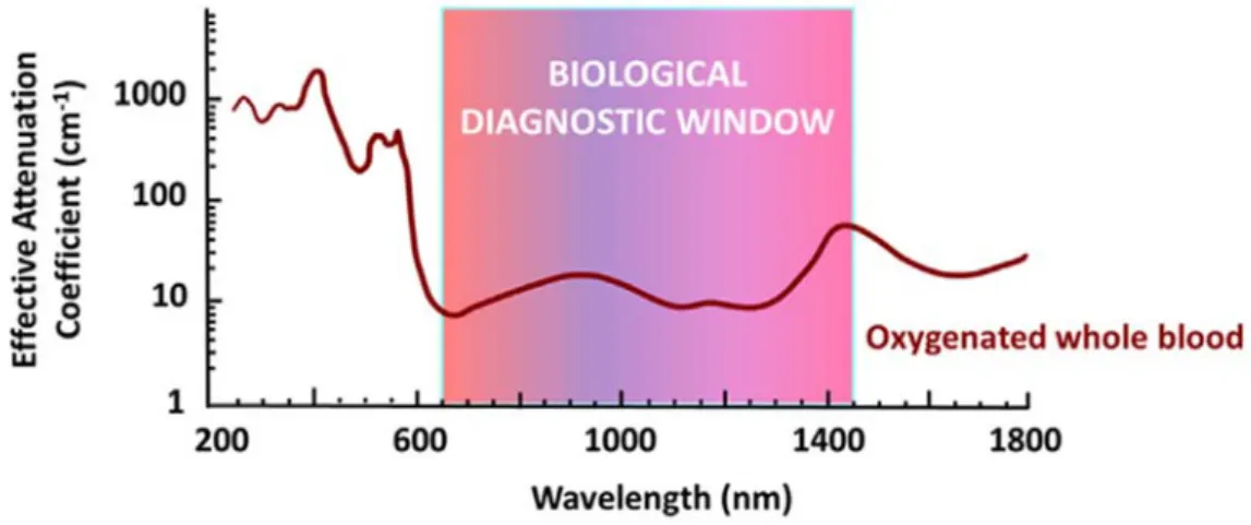
relaxation to ground state release energy that is transduced into signals revealing the information associated with the tissue, cell or organelles in the form of an image. Unlike the whole-body imaging techniques such as MRI and PET, optical imaging can offer real-time images with high resolution. As, the subcellular processes can be captured by optical microscopy, optical imaging has been used to understand the behaviour of various nanomaterials at the cellular levels in living subjects [25]. Currently, numerous fluorescent dyes are being widely used as optical imaging probes (discussed in detail in following sections). However, in vivo optical imaging suffers from a major drawback of limited penetration depth in tissue and background signals caused by light scattering. As result, many efforts in advancing optical imaging techniques and probes are being made to maximize penetration depth and minimize background signals. Advanced techniques stemming from optical imaging include; Optical Coherence Tomography, Photoacoustic Imaging, and Raman Spectroscopy to list few. Employing NIR radiation and NIR based fluorophores have been proven to overcome the tissue auto-fluorescence. On the other hand, the photo-bleaching and the limited absorption coefficient of fluorescent dyes can be to certain extent overcome by developing nanoparticle-based optical probes [26].
28
I-2.2.2 Multimodal Imaging:
Despite development of various imaging techniques, no single imaging modality offers a comprehensive functional or anatomic information. Every technique has its own unique shortcomings on spatial or temporal resolution, depth of tissue penetration detection sensitivity and cost [27]. MRI and CT can provide high resolution images with detailed information of anatomy but lacks target sensitivity (mM to μM range). In contrast, PET is high sensitivity imaging modality, but is limited by the range of positron in tissue, resulting in low spatial resolution (4–6 mm for human whole-body PET scanners, 1 mm in small animal imaging and preclinical PET scanners).This causes partial volume effects and the anatomic information offered is inconclusive [28]. On the other hand, optical imaging techniques suffer from low tissue penetration, despite the high detection sensitivity; while ultrasound offers good spatial resolution, but is limited by its poor penetration and sensitivity. Thus, the above nuclear and non-nuclear imaging techniques represent the strengths of molecular imaging each with their own share of advantages and disadvantages. Synergistic combination of the above modalities offers hope to overcome clinical diagnostic challenges by offering complementary and comprehensive information. Multimodality aims to combine the techniques that provide structural/anatomical information (MRI, CT or US) and the one that offers high sensitivity with functional or molecular information (PET, SPECT or optical). This has resulted in different dual modal technologies like PET/CT, PET/MRI, SPECT/MRI, and MRI/optical [27, 29]. Combination of PET or SPECT with optical imaging also holds potential as a technology that helps combine whole body imaging with image guided surgery giving rise to PET/optical and SPECT/optical imaging [30].
Although multimodality offers potential to improve diagnostic capabilities at clinic, it has several challenges at different levels. Hybrid imaging necessitates development of the multimodal scanners that present design and engineering complexities. This has to be supported by the image processing advancements to combine the results obtained from the two measurements without any loss/masking of the information [31]. Multimodal imaging also poses the significant challenge to chemists to develop the multimodal probes or contrast agents that can help maximise and refine the hybrid imaging outcomes. Administering a single agent that combines two modalities is a desirable feature in terms of the patient convenience and compliance. Emergence of the targeted or personalized therapy based on molecular signatures of the cancer has further added a new dimension to this area of hybrid imaging. Small molecule/peptide, antibody and nanoparticles based multimodal contrast agents play a very crucial role in multimodal imaging. Research outlined in this thesis aims to address this concern and contribute to the new strides taken in this direction.
Chapter 1. Introduction
Multifunctional platforms for cancer theranosis 29
I-2.2.2.1 Nanoparticles in multimodal imaging:
Although there are lot of preclinical studies reported in literature [32], very limited number of nanoparticles are used in the clinic [24]. Amongst the ones used in clinic, majority of them are for the cancer therapy as most of them cater to the drug delivery challenges of the anti-cancer drugs. Nanoparticles for imaging or theranosis is relatively a new concept and present challenges and opportunities different from the nanoparticles for drug delivery. As an unmet clinical need, the detection of early tumor development at primary and metastatic sites can be improved by use of nanoparticles owing to their potential of targeting accuracy to tumors. The design of imaging methodologies based on nanoprocesses and technologies that could detect tumors earlier would significantly improve patient outcomes. The properties of the nanoparticles that are desirable from the viewpoint of imaging are slightly different from those expected for drug delivery and this difference stems mainly from the pharmacokinetic requirements. A nanoparticle for imaging is expected to quickly reach the tumor environment and get cleared from the system as early as possible whereas the one for drug delivery needs to remain in the circulation for relatively longer time so as to deliver adequate dose at the site of cancerous lesion. As a result of this, nanoparticles in the size range of 5-10nm are generally favourable for imaging application. Size in this range also ensures that the nanoparticles are quickly cleared from the circulation mostly via renal clearance [33]. Other factors that can influence the biological behaviour of nanoparticles is the surface chemistry, surface potential/charge and particle shape. Nanoparticles present a versatile platform to combine different modalities in order to be used as bi/multimodal tracer in imaging and/or theranosis. Presence of the high specific surface area (surface area per unit weight or volume) enables appending of the high sensitivity probes to the nanoparticles surface whereas the bulk/core of the nanoparticles can possess the properties needed for other imaging tracers/therapy.
30 I-3 Monomolecular Multimodal Platform in cancer theranosis:
Figure I-6: Multimodal platform for the development of diverse theranostics.
Broadly, imaging contrast agents include small molecules, peptides, proteins, and nanoparticles. Majority of the scans use small molecules based tracers that are below 2,000 kDa and measure approximately 1 nm (e.g:18F-FDG for PET, iodinated small molecules for CT, and chelated gadolinium for MRI). Peptide based imaging agents are gaining momentum owing to their specificity and ease of synthesis relative to larger proteins. This has led to the approval of such agents towards specific class of cancer (e.g: NETSPOTTM/68 Ga-DOTATATEfor neuroendocrine tumors)(www.adacap.com).Protein imaging agents, such as radiolabelled monoclonal antibodies, are less common but offer precise molecular information and are a growing area of research. Nanoparticles offer an exciting class of imaging agents that can be used for both anatomic and molecular imaging. Developing a platform that caters to multiple such avenues forms the basis of the current thesis. Monomolecular multimodal platforms have been one of the thrust areas of research at Chematech and ICMUB. Monomolecular multimodal platform (MOMIP) aims to develop the systems (conjugates/nanoparticles) that are easy to develop and characterize and yet retain their multimodality. For instance, modification of an antibody for PET-Optical probe development using independent stepwise approach i.e conjugation first with PET probe and then optical probe appears very complex in terms of the developing optimal process of conjugation as well as the precise characterization of the probe conjugated in this manner.
Chapter 1. Introduction
Multifunctional platforms for cancer theranosis 31
Whereas, conjugation with MOMIP is one step process along with precise characterization of the resulting bioconjugate. This is furthermore important and valuable in development/functionalization of the nanoparticles where these systems are inherently complex to characterize. Thus, the purpose of this platform is to provide researchers and the clinicians, the tools that encompass diverse modalities in a single integrated system towards improved cancer/disease management. In this context, the current thesis aims to harness this approach in diverse possible manner and generate multimodal tools for cancer therapy. This platform can be applied for the development of the small molecule based multimodal imaging ligands or for the ligands that can be in turn used for nanoparticle synthesis or antibody bioconjugation that can have substantial theranostic value. Figure I-6 highlights this approach in a pictorial/infographic format representing the general and versatile nature of the multimodal platform that can be easily customized or adapted based on the product and the feasibility of the chemistry desired.
I-3.1 Chelators used in multimodal platforms:
The increasing number of radioisotopes in nuclear medicine has necessitated the corresponding increase in the development of the novel chelators to suit the requirement of the complex’s stability in terms of thermodynamic stability and kinetic inertness. Each radiometal ion has different physical and chemical properties like; ligand donor atom preferences (e.g. N, O, S), size, oxidation state, coordination number and coordination geometry. As result of this, a correct choice of the chelator suiting the attributes of the chosen radioisotope has to be made so that the resulting complex exhibits optimal characteristics suitable for the in vivo stability. Broadly, the chelators have been classified as linear or acyclic and macrocyclic. In the context of this doctoral thesis, the key radiometals/metals that are of interest for theranostic applications include Gadolinium/Gd (for MRI and radiosensitization), Copper 64/64Cu (for PET) and Indium-111/111In (for SPECT). As a result, only those chelators useful for the complexation of above radiometals will be discussed. A detailed account of the chelators used in radiochemistry has been outlined in an excellent review by Price and Orvig [14].
32
Table I-3: Properties of the chelators used in this thesis for theranostic applications.
Chelator Radiometal/ metal
Labelling/ radiolabelling
conditions logKML
DOTA (1,4,7,10-tetraazacyclododecane- 1,4,7,10-tetraacetic acid)
Coordination Number: maximum 8 Donor Site: N4O4
Bifunctional Derivative used in this thesis: DOTAGA 64Cu+2 25–90°C, 30–60 min, pH 5.5–6.5 22.2, 22.7 111In+3 37–100°C, 15–60 min, pH 4.0–6.0 23.9 (pM 17.8–18.8) Gd+3 37–100°C, 15min– 6 h, pH 5.0–6.0 24.7 NOTA (1,4,7-triazacyclononane-1,4,7- triacetic acid) Coordination Number = 6 Donor Site: N3O3
Bifunctional Derivative used in this thesis: NODAGA 64Cu+2 25 °C, 30–60 min, pH 5.5–6.5 21.6 111In+3 60–95 °C, 20–30 min, pH 4.0–5.0 26.2 (pM 21.6) DTPA, diethylenetriaminepentaacetic acid Coordination Number = 8 Donor Site: N3O5
Bifunctional Derivative: DTDTPA
64Cu+2 40 °C, 60 min, pH 6.5 21.4 111In+3 25 °C, 5–10 min, pH 4.5–5.5 29.0 (pM 24.9)
The table has been adapted from [14, 34, 35]. Green-Best match; Orange-moderate match; Red-poor match. DTPA is one of the oldest and most widely used acyclic chelator in radiochemistry and can be radiolabelled with many radiometal ions at room temperature within few minutes. However, the complexes of DTPA suffer from potential stability issues in vivo and are not as stable as
Chapter 1. Introduction
Multifunctional platforms for cancer theranosis 33
the ones formed with macrocyclic chelators. As a result of this, there is a decrease in its use which is gradually being replaced by chelators like DOTA and NOTA derivatives [36].
Nonetheless, DTPA has been successfully used in the FDA approved SPECT agent OctreoScanTM(111In-DTPA-octreotide), a somatostatin-targeting peptide-conjugate used for imaging neuroendocrine tumors [37]. DTPA (Gd complexed) based contrast agents for MRI have been approved and marketed under different brand names (Table I-4). Also, the first-generation gold nanoparticles from our group were based on the bifunctional chelator (BFC); DTDTPA (thiolated DTDTPA) and are currently being upgraded to the advanced form by use of the BFC based on DOTA and NOTA.
Table I-4: Gadolinium based contrast agents marketed in US and Europe [34].
Chemical Name Generic Name Trade/Product Name Acyclic Chelators
Gd-DTPA Gadopentetate Dimeglumine Magnevist®
Gd-DTPA-BMA Gadodiamide Omniscan®
Gd-DTPA-BMEA Gadoversetamide Optimark®
Gd-BOPTA Gadobenate Disodium Multihance®
Gd-EOB-DTPA Gadoxetate Disodium Primovist®
MS-325 Gadofosveset Trisodium Vasovist®
Macrocyclic Chelators
Gd-DOTA Gadoterate Meglumine Dotarem®
Gd-HP-DO3A Gadoteridol Prohance®
Gd-DO3A-Butrol Gadobutrol Gadovist®
DOTA is the most versatile and widely used chelator and is of significant value for MRI as several marketed products are based on the gadolinium complex of the DOTA in its different forms as can be seen from the Table I-4. Due to relatively lower stability, the DTPA based contrast agents are more likely to release the Gd+3 in vivo. This can have implications in potential kidney toxicity that can result in Nephrogenic Systemic Fibrosis (NSF) and hence are being widely replaced by DOTA based complexes [38]. DOTA has been gold standard for some of the radioisotopes and forms stable complexes with 111In, 177Lu, 86/90Y, 225Ac, and 44/47Sc. Owing to the lack of geometric fit, DOTA does not form very stable complexes with PET isotopes like 64Cu and 68Ga, which can be addressed by deployment of NOTA. NOTA is a hexadentate N3O3chelator and has been successfully used as chelator of choice for67/68Ga and 64Cu. NOTA is now considered to be the ‘‘gold standard’’ for 64Cu+2 and Ga3+ chelation, with facile and favourable radiolabelling conditions (RT, 30–60 minutes) and excellent in
34
vivo stability [39, 40].
Table I-1 summarizes the properties of the key chelators that are relevant to the research work described in this thesis.
I-3.1.1 Influence of the chelator on the properties and stability of the theranostics:
Selection of the correct combination of the chelator and metal is key to the successful development of theranostics. In vivo kinetic inertness of the metal–chelate complex is of utmost consideration in identifying the right match between the chelator and metal. Although, thermodynamic stability constants (KML= [ML]/[M][L]) can be a useful gauge for the initial level comparisons, but they do not necessarily predict the in vivo stability. Experiments like acid dissociation and competitive radiolabelling have been proposed but they are not representative and do not reflect the environment encountered at physiological conditions. Challenge studies can be performed by incubating the chelate-metal complex with ions like Na+, K+, Ca2+, Mg2+, Cu2+ or Fe3+ to identify the trans-chelation mediated instabilities. Alternatively, the complex can be subjected to EDTA challenge assays to mimic the potential endogenous chelators. Ultimately, the correct match can only be justified by performing the in vitro stability assessment in serum and performing in vivo studies focussing on biodistribution and pharmacokinetics.
Despite the stability and inertness with a given radiometal (e.g. NOTA vs. DOTA for 68Ga), it may not be the optimal match for a certain application (e.g. a specific peptide vector). For instance, NOTA forms a more stable complex with 68Ga than does DOTA, but owing to differences in charge and physical properties (e.g. neutral vs. charged complex), DOTA may provide superior in vivo properties with certain vectors [41]. This exemplifies the complex set of variables that need due consideration when constructing radiometal-based radiopharmaceuticals. Another interesting feature related to the influence of chelator, is that they can modulate the binding affinity of the peptide based radiopharmaceuticals. Findings by Maecke et al., claim that ‘The chelate makes difference’ in the context of peptide based imaging agents. It has been demonstrated that the combination of the chelate and metal can have a substantial effect on the binding affinity as well as the tumor localization in animal models [42].
In the view of the multimodality, it is necessary to use the right form of the bifunctional chelators so as to conjugate it to the multimodal assembly. This necessitates the use of the bifunctional chelator so that properties of the base chelators are unaffected. One of the strategies in offering the bifunctionality to the chelator (e.g: DOTA and NOTA) is to append a side arm containing two carboxylate groups on the macrocyclic ring. This can be done for instance, by employing the glutaric arm during the macrocycle synthesis so that the original

![Figure I-5: Scheme illustrating the principles imaging based on PET and SPECT[13].](https://thumb-eu.123doks.com/thumbv2/123doknet/14670055.741484/24.892.201.689.614.909/figure-scheme-illustrating-principles-imaging-based-pet-spect.webp)