Accuracy of Cerebral Monitoring in Detecting Cerebral
Ischemia during Carotid Endarterectomy
A Comparison of Transcranial Doppler Sonography, Near-infrared
Spectroscopy, Stump Pressure, and Somatosensory Evoked Potentials
Stefan Moritz, M.D.,* Piotr Kasprzak, Ph.D.,† Matthias Arlt, M.D.,* Kai Taeger, Ph.D.,‡ Christoph Metz, Ph.D.§Background: This study compares the accuracy of cerebral monitoring systems in detecting cerebral ischemia during ca-rotid endarterectomy.
Methods:The authors compared transcranial Doppler sonog-raphy (TCD), near-infrared spectroscopy (NIRS), stump pres-sure (SP) meapres-surement, and somatosensory evoked potentials (SEP) in 48 patients undergoing carotid surgery during regional anesthesia. Cerebral ischemia was assumed when neurologic deterioration occurred. During clamping, the minimum mean middle cerebral artery velocity (TCDmin), its percentage change
(TCD%), the minimum regional saturation of oxygen (NIRSmin),
its percentage change (NIRS%), the mean SP, and the changes of
SEP amplitude were recorded. To analyze the corresponding sensitivity and specificity of each parameter, the authors per-formed receiver operating characteristic analysis.
Results:Neurologic deterioration occurred in 12 patients. SP and NIRS were successfully performed in all patients. TCD mon-itoring was not possible in 10 (21%); SEP was not possible in 2 patients (4%). All parameters provided the ability to distinguish between ischemic and nonischemic patients. TCD%and NIRS%
showed significantly better discrimination than TCDmin and
NIRSmin(P < 0.05). The highest area under the curve (AUC) was
found for TCD% (AUCⴝ 0.973), but there was no significant
difference compared with NIRS%(AUCⴝ 0.905) and SP (AUC ⴝ
0.925). The lowest AUC was found for SEP (AUCⴝ 0.749), which was significantly lower than that for TCD%, NIRS%, and SP.
Conclusions:TCD%, NIRS%, and SP measurement provide
simi-lar accuracy for the detection of cerebral ischemia during carotid surgery. Lower accuracy was found for SEP monitoring. Because of the high rate of technical difficulties (21%), TCD monitoring was the least practical of the investigated monitoring devices.
THE beneficial effects of carotid endarterectomy in symptomatic and asymptomatic patients with high-grade carotid stenosis have been published in detail in previ-ous studies. Unfortunately, the incidence of periopera-tive stroke due to clamping-induced cerebral ischemia or embolization is significant (3–5%).1– 4
Currently, selective shunting during carotid endarter-ectomy is widespread, although there is no evidence that
it is better than routine shunting or nonshunting.5 Al-though shunting may prevent cerebral hypoperfusion due to cross clamping, the routine use of an intraluminal shunt in all patients increases the risk of perioperative stroke resulting from embolic events.6,7Therefore, many authors advocate selective shunting guided by signs and measurements of cerebral hypoperfusion during clamping. Currently, there are three different kinds of monitoring devices: monitoring of cerebral hemodynamics (i.e., transcranial Doppler sonography [TCD], carotid artery stump pressure [SP]), monitoring of cerebral oxygen metabolism (i.e., jugular bulb monitoring, near-infrared spectroscopy [NIRS]), and monitoring of cerebral func-tional state (i.e., electroencephalography, evoked poten-tials). Despite of an enormous number of publications, no single method has been shown to be superior or to provide better outcome after surgery.8
Awake patient monitoring during local or regional an-esthesia seems the most reliable method of predicting the need for a shunt after carotid clamping and can be regarded as the accepted standard for the evaluation of intraoperative monitoring.9,10
In the current study, TCD, NIRS, somatosensory evoked potentials (SEP), and carotid artery SP measurement were simultaneously performed in patients undergoing carotid surgery during regional anesthesia. The overall objective of the current investigation was to compare the accuracy of the different monitoring systems and to determine which of them allows the best discrimination of patients with and without cerebral ischemia.
Materials and Methods Patient Characteristics
After approval of the ethics committee of the University of Regensburg Medical Center (Regensburg, Germany) and written consent were obtained, 48 patients undergoing elective carotid endarterectomy were enrolled in the study. The group included 34 men and 14 women with a mean age of 69 yr (51– 89 yr). The indications for operation included 80 –99% internal carotid artery stenoses for asymptomatic patients (n ⫽ 24) and greater than 70% stenoses for symptomatic patients, i.e., hemispheric tran-sient ischemic attacks and amaurosis fugax (n ⫽ 11), nonhemispheric symptoms (n⫽ 8), reversible ischemic neurologic deficits (n⫽ 1), and previous stroke (n ⫽ 4). The degree of contralateral stenosis was mild in 23, * Staff Anesthesiologist, ‡ Professor, Director, and Chair, Department of
Anesthesiology, † Head of Department, Department of Vascular Surgery, Univer-sity of Regensburg. § Head of Department, Department of Anesthesiology, Klinikum Freising, Freising, Germany.
Received from the Department of Anesthesiology, University of Regensburg, Regensburg, Germany. Submitted for publication November 27, 2006. Accepted for publication June 11, 2007. Support was provided solely from institutional and/or departmental sources. Presented in part at the Annual Meeting of the American Society of Anesthesiologists, Chicago, Illinois, October 14 –18, 2006.
Address correspondence to Dr. Moritz: University of Regensburg, Medical Center, 93053 Regensburg, Germany. stefan.moritz@klinik.uni-regensburg.de. Information on purchasing reprints may be found at www.anesthesiology.org or on the masthead page at the beginning of this issue. ANESTHESIOLOGY’s articles are made freely accessible to all readers, for personal use only, 6 months from the cover date of the issue.
moderate in 17, high grade in 6, and occlusion in 2 patients.
Anesthesia
All patients received standard premedication; antihyper-tensive drugs were continued on the day of surgery, with the exception of angiotensin-converting enzyme inhibitors. Regional anesthesia consisted of single-shot superficial and deep cervical plexus blocks performed with 40 ml prilocaine, 1%. After accomplishing the block, we installed the neuromonitoring devices, which took us 30 – 45 min. Thus, a complete spread of the cervical plexus blocks was assured until the recording of the baseline values (45– 60 min after injection of the local anesthetic) Patients received supplemental facemask oxygen during the whole proce-dure. Sedation was avoided to maintain the patients’ re-sponsiveness to verbal stimuli and to prevent respiratory depression. Invasive blood pressure, peripheral oxygen sat-uration, and ST-segment analysis by five-lead electrocardio-gram were recorded continuously.
The arterial carbon dioxide partial pressure (PaCO2) and oxygen content (CaO2) were monitored intermit-tently at the time points described in the Data Analysis and Statistical Methods section.
The patients were brought to a semirecumbent posi-tion for the operaposi-tion.
Neuromonitoring
During the procedure, one anesthetist was exclusively responsible for the neurologic surveillance of the pa-tient. Before carotid clamping, the patient was told to keep his eyes continuously open for the whole clamping period and to stay in contact with the anesthetist. The patients’ eyes were continuously observed by the anes-thetist. At 30-s intervals, the patient was asked for his behavior, his age, the actual time, and the location. For a semiquantitative assessment of the motor function, the patient was requested to squeeze the hand of the anes-thetist. When the patient closed his eyes, he was again asked to open them, to answer the questions, and to squeeze the anesthetist’s hand. Cerebral ischemia was assumed when any new neurologic deficit such as speech abnormalities, motor weakness, or impaired con-sciousness occurred. These symptoms were also used as criteria for shunt insertion.
For the detection of new postoperative deficits, all patients underwent preoperative and postoperative (24 h) cranial computed tomography scanning. In addition, the patients underwent preoperative and postoperative (second postoperative day) neurologic examination. The performing neurologist was blinded to the intraoperative course of the procedure.
Transcranial Doppler
A 2-MHz pulsed Doppler ultrasound device (Medason-ics Inc., Fremont, CA) was used to access the ipsilateral
middle cerebral artery (MCA) at a depth of 45–50 mm. A specially designed headband fixed the probe to the pa-tient’s head with provisions for adjusting the probe po-sition and angle and thereby ensured constant in-sonation angle and depth. Mean MCA velocity was recorded at the time points described in the Data Analysis and Statistical Methods section. The minimum blood flow velocity during clamping (TCDmin) and its percentage change compared with baseline (TCD%) were calculated.
Stump Pressure
Stump pressure monitoring was conducted according to the recommendations of Spencer et al.11with a zero reference level of the pressure measurement at the com-mon carotid artery bifurcation and was readjusted just before insertion of the needle into the artery. Mean SP was measured 5–10 min after cross clamping.
Near-infrared Spectroscopy
The INVOS 3100 cerebral oximeter (Somanetics, Troy, MI) measures regional intracerebral oxygen saturation (rSO2) continuously and noninvasively by spectroscopy of reflected near-infrared light. Technical details of this device were described elsewhere.12,13The probe was positioned over the ipsilateral parietotemporal area of the brain, just above the temporal muscle. To place and fix the probe properly, it was necessary in some patients to shave the scalp in this area. Informed consent was obtained for each patient. For data analysis, rSO2 values were continuously recorded on a disk. The minimum rSO2value during clamp-ing (NIRSmin) and its relative change compared with base-line (NIRS%) were determined.
Somatosensory Evoked Potentials
Somatosensory evoked potentials were continuously monitored and recorded with the Axon Sentinel-4 evoked potential system (AXON Systems, Inc., Hauppauge, NY). Steel EEG needles were placed at the scalp at C3= and C4=. The reference electrode was placed over the forehead (Fpz). Impedance was kept below 10 k⍀. The median nerve at the wrist contralateral to the operated side was stimulated with a frequency of 4.3 Hz and a duration of 0.2 ms. The stimulus intensity was set at 10 mA. The high-bandpass and low-high-bandpass filters were set at 30 and 300 Hz. Final traces used for data analysis were the average of 250 sweeps. Evoked potentials were recorded above the ipsilateral somatosensory region (C3= and C4=). The peak-to-peak amplitudes of the primary cortical response N20/ P25 complex were measured online. Values of the ampli-tude are given as percent of baseline ampliampli-tude.
Data Analysis and Statistical Methods
For data analysis, patients were assigned to one of two groups: those with clinical signs of cerebral ischemia (ischemic patients) and those without any signs of neu-rologic change (nonischemic patients).
All data were recorded exactly at the following time points: 2 min before carotid cross clamping, 2 and 10 min after clamping, and immediately before declamping. In ischemic patients, we in addition recorded values at the onset of neurologic deterioration and 2 min after shunt insertion. Only data recorded at these time points were used for data analysis.
After assuring stable signal quality conditions, the base-lines for TCD, NIRS, and SEP were determined 2 min before carotid clamping. During clamping we selected the mini-mum rSO2 (NIRSmin), the percentage change of NIRSmin compared with baseline (NIRS%), the minimum mean MCA velocity (TCDmin), the percentage change of TCDmin com-pared with baseline (TCD%), the absolute mean SP values, and the minimum amplitude of the N20/P25 complex given as percent of baseline. Parameters recorded during shunt use were excluded for this analysis.
For statistical analysis, the Kolmogorov–Smirnov mod-ification of the Lilliefor test was used to test for normal distribution. The Mann–Whitney U test was used to com-pare data between the two groups.
For the assessment of the accuracy of each parameter in discriminating ischemic from nonischemic patients, we performed receiver operating characteristic (ROC) analysis. A ROC curve is the plot of a test’s true-positive rate (sen-sitivity) versus its false-positive rate (1-specificity). It is constructed by calculating the sensitivity and specificity of a test for each possible test result. Thus, the ROC curve describes the trade-off between the sensitivity and specific-ity of the test as the criterion for defining “negative” and “positive” result changes. The area under the ROC curve (AUC) is a quantitative measure of the selectivity (1.0 ⫽ best selectivity; 0.5⫽ worst selectivity). It is a function of both sensitivity and specificity of a test and takes into account the entire range of error rates. The value for the area under the ROC curve can be interpreted as follows: An area of 0.75, for example, means that a randomly selected individual from the positive group has a test value larger than that for a randomly chosen individual from the nega-tive group 75% of the time.14 When the variable under study cannot distinguish between the two groups, i.e., where there is no difference between the two distribu-tions, the area will be equal to 0.5 (the ROC curve will coincide with the diagonal). When there is a perfect sepa-ration of the values of the two groups, i.e., there is no overlapping of the distributions, the area under the ROC curve equals 1 (the ROC curve will reach the upper left corner of the plot).
The 95% confidence interval for the area can be used to test the hypothesis that the theoretical area is 0.5. If the confidence interval does not include the 0.5 value, there is evidence that the test does have an ability to distinguish between the two groups.14,15
Three different cutoff points for each parameter were determined: the value correlating 100% sensitivity with the highest specificity, the value correlating 100% specificity
with the highest sensitivity, and a “best fit’” cutoff point. The latter is the value corresponding with the highest accuracy (minimum false-negative and false-positive re-sults). For each cutoff point, sensitivity, specificity, and positive and negative likelihood ratios are reported.
For statistical analysis, we used MedCalc version 8.1.1.0 (MedCalc Software, Mariakerke, Belgium).
Results
Cervical plexus blocks provided sufficient anesthesia in all patients. None of them showed neurologic changes after accomplishing the blocks. NIRS and SP measurements were successfully performed in all 48 patients. Twelve of these patients showed neurologic deterioration. TCD mon-itoring was successfully performed in 38 patients, 9 with and 29 without clinical signs of cerebral ischemia. In the remaining 10 patients (21%), insufficient signal quality was obtained or the Doppler probe dislocated intraoperatively. The latter were excluded because a constant insonation angle was no longer guaranteed. SEP monitoring was not possible because of technical problems in 2 patients; 1 of them showed signs of cerebral ischemia.
The kinds of neurologic disorders ranged from hemi-paresis (n ⫽ 1) and dysarthria (n ⫽ 2) to impaired consciousness (n⫽ 9). Neurologic changes disappeared after insertion of an intravascular shunt in 8 of these patients. In the remaining 4 patients, the surgeon de-cided to complete the operation without the use of a shunt, because the operation was near completion and flow could be restored quickly. None of the patients showed postoperative neurologic deficit.
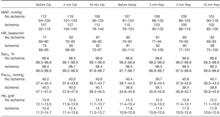
Values for mean arterial blood pressure, heart rate, PaCO2, arterial oxygen saturation (SaO2), and hemoglobin concentration are shown in table 1; they did not differ significantly between the two groups during the whole study period.
At baseline, there were no significant differences be-tween the two groups in any parameter. Figure 1 shows the ROC plots, the AUCs, and the 95% confidence intervals of all investigated parameters. For each parameter, the AUC and the corresponding confidence interval was greater than 0.5. For TCD as well as for NIRS, the AUCs of percent-age changes (i.e., TCD% and NIRS%) were significantly higher than the AUCs of the minimum values (i.e., TCDmin and NIRSmin) (P⬍ 0.05). The highest AUC was found for TCD%, but there was no significant difference compared with NIRS%(P⫽ 0.278) and SP (P ⫽ 0.753).
Compared with SEP monitoring, we found a signifi-cantly better discrimination for SP measurement, TCD%, and NIRS%(P⬍ 0.05).
Table 2 shows the three cutoff points and the corre-sponding sensitivity, specificity, and positive and nega-tive likelihood ratios for each monitoring parameter.
NIRS%and the kinds of neurologic deterioration of the eight patients with shunt are shown. The SEP amplitude was diminished in six of the eight patients at the onset of cerebral ischemia. In these patients, the SEP signal re-covered with the insertion of an intraluminal shunt. The remaining two patients lost consciousness 3 and 10 min after clamping of the carotid artery, and both recovered immediately after shunt insertion. In these two patients,
SEP signals remained unchanged during the whole time. All patients with sufficient signal quality on TCD showed a profound decrease of TCD flow even before the ap-pearance of neurologic deterioration. With shunt inser-tion, TCD flow increased in all patients. NIRS decreased in all patients continuously until the onset of neuro-logic deterioration and increased immediately after shunt insertion.
Table 1. Cardiac and Respiratory Data of Patients with and without Signs of Cerebral Ischemia
Before Clp 2 min Clp 10 min Clp Before Declp 2 min Rep 5 min Rep 10 min Rep
MAP, mmHg No ischemia 112 119 109 107 108 109 103 64–126 101–133 95–122 91–124 88–125 86–125 90–118 Ischemia 107 127 112 110 123 100 102 92–118 104–140 78–140 76–123 95–132 88–119 82–126 HR, beats/min No ischemia 77 82 81 80 81 80 82 66–90 70–90 69–92 70–92 71–94 70–95 68–93 Ischemia 75 80 82 81 92 80 88 68–90 68–92 73–97 65–114 74–105 71–101 72–100 SaO2, % No ischemia 98.6 98.5 98.6 98.6 98.6 98.6 98.6 98.3–98.8 98.1–98.9 98.1–98.9 98.2–98.9 98.2–98.9 98.2–98.9 98.3–98.8 Ischemia 98.4 98.7 98.4 98.5 98.5 98.5 98.3 98.0–98.9 98.2–98.9 97.8–98.7 97.7–98.7 96.9–98.7 97.5–98.8 98.0–98.8 PaCO2, mmHg No ischemia 40.2 40.5 40.6 40.4 39.7 40.8 40.1 37.4–42.4 37.6–43.6 38.7–43.7 38.7–44.0 37.6–43.4 37.6–43.6 38.0–43.5 Ischemia 40.3 40.0 40.1 38.6 39.1 38.4 38.8 37.1–47.0 37.8–47.6 38.4–45.5 34.8–45.9 35.9–42.9 36.8–42.7 36.8–43.6 Hb, g/dl No ischemia 12.9 12.6 12.5 12.3 12.3 12.0 12.2 12.1–13.5 11.9–13.9 11.7–13.7 11.4–13.4 11.3–13.2 11.4–13.1 11.1–13.0 Ischemia 13.0 12.4 12.7 11.8 11.4 11.5 11.9 11.2–14.1 11.4–13.6 11.3–13.7 10.9–12.9 10.8–12.6 10.5–12.6 10.6–12.6
Values are given as median and 25th–75th percentiles. There was no significant difference between the two groups.
Clp⫽ clamping; Declp ⫽ declamping; Hb ⫽ hemoglobin concentration; HR ⫽ heart rate; MAP ⫽ mean arterial pressure; PaCO2⫽ arterial partial pressure of carbon dioxide; Rep⫽ reperfusion; SaO2⫽ arterial oxygen saturation.
Fig. 1. Receiver operating characteristic curves of the investigated monitoring methods. % ⴝ relative reduction com-pared with baseline; AUC ⴝ area under the curve; CIⴝ 95% confidence interval; minⴝ minimum during clamping; NIRS
ⴝ near-infrared spectroscopy; SEP ⴝ
so-matosensory evoked potentials; TCD ⴝ transcranial Doppler sonography.
Discussion
The current study was designed to compare the accuracy of different neuromonitoring methods in detecting cerebral ischemia during carotid endarterectomy. Clinical signs of cerebral hypoperfusion developed during 12 operations and were related to occlusion of internal carotid artery. In all patients, neurologic deterioration could be reversed by shunt insertion or declamping. There was no new postop-erative neurologic deficit in any of these patients, demon-strating that a short period of cerebral ischemia is tolerated without permanent sequelae.
In the current study, all AUCs of the investigated pa-rameters differ significantly from 0.5. Therefore, all mon-itoring systems have the ability to distinguish between ischemic and nonischemic patients. In contrast, none of the parameters reached an AUC of 1.0, indicating that no monitoring is equal to awake patient monitoring.
The best results were found for TCD%, but there was
no significant difference compared with SP and NIRS%.
The worst results were found for SEP monitoring. For TCD as well as for NIRS, the current study shows that relative changes are more accurate for the diagnosis of cerebral ischemia than absolute values.
We found a 50% reduction in MCA velocity to provide 100% sensitivity and 86% specificity. To avoid false-pos-itive results (100% specificity), the corresponding reduc-tion in MCA velocity was 70%. However, sensitivity de-clines to 78%. These findings agree with earlier studies.7,16For TCDmin, we found a cutoff point of 25 cm/s to provide 100% sensitivity and a specificity of 69%. The proposed cutoff values from other authors vary widely from 10 to 24 cm/s.16,17The high variance of the proposed cutoff values is obvious because these values highly depend on individual vessel diameter and the insonation angle, both of which cannot be determined. Our data show that the accuracy of TCD%is equal to
Table 2. Cutoff Values
100% Sensitivity 100% Specificity Best Fit
Cutoff Specificity PLR Cutoff Sensitivity NLR Cutoff Sensitivity Specificity PLR NLR
TCDmin 25 cm/s 69% 3.22 6 cm/s 22% 0.78 25 cm/s 100% 69% 3.22 0.00 TCD% 48% 86% 7.25 70% 78% 0.22 48% 100% 86% 7.25 0.00 NIRSmin 59 47% 1.89 36 17% 0.83 59 100% 47% 1.89 0.00 NIRS% 13% 64% 2.77 34% 8% 0.22 20% 83% 83% 5.00 0.20 Stump pressure 40 mmHg 75% 4.0 33 mmHg 58% 0.42 40 mmHg 100% 75% 4.00 0.00 SEP 100% 0% 1.0 25% 18% 0.82 50% 82% 57% 1.91 0.32
Cutoff values and the corresponding sensitivity, specificity, negative likelihood ratio (NLR), and positive likelihood ratio (PLR) of the investigated monitoring parameters.
%⫽ relative reduction compared with baseline; min ⫽ minimum during clamping; NIRS ⫽ near-infrared spectroscopy; SEP ⫽ somatosensory evoked potentials; TCD⫽ transcranial Doppler sonography.
Fig. 2. Individual courses of eight shunted patients. For each patient, somatosen-sory evoked potentials (SEP), transcra-nial Doppler sonography (TCD%), and
near-infrared spectroscopy (NIRS%) are
shown at the following time points: base-line (2 min before clamping), before is-chemia (the last regular time point dur-ing clampdur-ing before the onset of cerebral ischemia, i.e., 2 or 10 min clamping), ischemia (at the onset of cerebral isch-emia), shunt (2 min after shunt inser-tion). Line style reflects the kind of neu-rologic deterioration: motor weakness (dashed line), impaired consciousness (solid line), dysarthria (dashed– dotted
that of NIRS%and SP monitoring. However, because of a missing acoustic temporal window, TCD sonography cannot be successfully performed in all patients.16,18,19 In addition, the relative proximity of the operation field implies a high risk of intraoperative probe dislocation. For these reasons, TCD monitoring was not successfully in 21% of our investigated patients. Three of 12 patients with clinical signs of cerebral ischemia were missed because of technical problems with TCD monitoring. Including these missed patients, the sensitivity of TCD is reduced to 75%. Therefore, TCD monitoring is less prac-tical compared with NIRS and SP, both easy-to-accom-plish monitors.
Stump pressure measurement is a simple and inexpen-sive operative monitoring technique that requires no addi-tional equipment or personnel. Despite numerous investi-gations and validations by different authors, there is still no consensus on the appropriate cutoff value indicating cere-bral ischemia. For mean SP measurement, we found the highest accuracy for a cutoff point of 40 mmHg. Previous studies found similar accuracy for a threshold value of 50 mmHg in awake patients.7,16,20 Two principal differences must be considered when comparing our findings with their results. First, the studies did not report the zero reference level for SP measurement. Years ago, Spencer et
al.11 stated that only SP measurement with the common carotid artery as the zero reference is reliable for the cor-rect diagnosis. Because SP is recorded in absolute values, the reference zero level is crucial.11In our patients, there was at least a difference of 20 cm between heart level and carotid bifurcation level. Second, some authors used sys-tolic SP for analysis, whereas we recorded mean SP.
Cerebral NIRS as described by Jobsis21is a noninvasive technique to monitor cerebral oxygenation. This moni-toring device is easily and rapidly to apply. Sufficient signal quality was obtained in all patients within a few seconds. For NIRS, we found rSO2 values below 59 to provide 100% sensitivity at 47% specificity. To take in-tersubject variability into account, Samra et al.22 pro-posed using percentage decrease from baseline as crite-ria for cerebral ischemia. We confirmed their proposed cutoff value of 20% and found 83% sensitivity and 83% specificity for this cutoff value.22However, this measure-ment modality is controversial. The regional nature, the high intersubject variability and the unclear contribution of nonbrain sources are only some of the discussed limitations. All of these are potential reasons for its im-perfect sensitivity. A detailed summary of its limitations has been recently published by Davies and Janelle.23
For SEP, previous studies reported 89 –100% sensitivity and 93–100% specificity, which we could not con-firm.24,25In our investigation, two patients did not show any change of N20/P25 waveform while experiencing neurologic deterioration. In both patients, loss of con-sciousness occurred within a short period after carotid artery clamping, and both patients recovered
immedi-ately after shunt insertion. Because of this close relation and because other monitoring devices showed signifi-cant changes (fig. 2), reasons other than cerebral isch-emia seem unlikely for the loss of consciousness. Previ-ous publications also reported some cases of unchanged SEP with the occurrence of new neurologic deficits.25,26 For a decrease of SEP amplitude to 50% of baseline, we found a sensitivity of 81% and a specificity of 57%, indicating that every second patient is falsely classified as ischemic. Some limitations must be considered for the interpretation of the SEP results. Because we used re-gional anesthesia of the cervical plexus, the relatively poor results of SEP may be related to an inadvertent block of ascending pathways. However, none of the patients showed neurologic changes after application of the local anesthetic. As mentioned above, the time delay from accomplishing the blocks to the record of the baseline values was at least 45– 60 min. The spread of the local anesthetic should be completed at this time point. At this time, a sufficient signal was obtained in all in-cluded patients.
The used SEP monitor shows moving averages (250 recordings) for the analysis of SEP amplitudes. Conse-quently, there is a time delay until measured changes of the SEP amplitude result in changed amplitudes on the screen. This may lead to an underestimation of the sen-sitivity. Conversely, we did not record evoked potentials above the cervical spine for artifact identification. There-fore, we cannot completely exclude that the low speci-ficity is influenced in part by artifacts. However, in all but one nonischemic patient, SEP amplitudes recovered with clamp release. Therefore, a peripheral artifact seems unlikely in most of the cases.
None of the monitoring systems under investigation provided perfect sensitivity and specificity. The regional (e.g., NIRS) and global nature (e.g., SP) of the monitoring devices are likely to play a major role in this context. In addition, the site of measurement needs to be consid-ered. All used monitoring devices record changes in the vascular region of the middle cerebral artery, which is at the highest risk to suffer from cerebral ischemia.27 How-ever, changes in the vascular region of the anterior and posterior cerebral artery are missed by the used moni-toring devices.
There are some limitations of our study. First, the current study was conducted in awake patients, and the results are therefore primarily only applicable for pa-tients undergoing carotid endarterectomy during re-gional anesthesia. Whether the determined cutoff values are also applicable for patients in general anesthesia remains unclear. Short of an accepted standard of mon-itoring in general anesthesia, it would be necessary to demonstrate a reduction in postoperative neurologic deficits by using a specific monitoring device. But such a study would require a sample size of many thousands of patients.
Second, the accepted standard, neurologic impair-ment, was measured using consciousness, speech, and hemiparesis. Because these measurements are relatively crude and intermittent measures, this implies the risk of labeling some monitoring modalities as excessively non-specific. We cannot finally exclude that we missed some minor neurologic changes during clamping, but we found no new postoperative neurologic deficit by neu-rologic examination or by computed tomography scan-ning. Therefore, in the current study, the performed neurologic surveillance provided sufficient sensitivity to prevent persisting neurologic sequels.
Third, the current study was performed in only 48 patients. In a post hoc power analysis, we calculated 90% power to detect significant differences from an AUC of 0.5. In contrast, because of the similar results for SP, NIRS%, and TCD%, a sample size of approxi-mately 200 patients would have been necessary to detect significant differences with sufficient power (i.e., 80%). Therefore, the lack of significant differ-ences between the three mentioned monitoring types may be due to the insufficient power of the current study to detect these.
The observed rate of 25% of cerebral symptoms after cross clamping in the current study is a high percentage compared with previous studies and even with our own clinical experience. In this context, the following as-pects need discussion. First, both surgeon and anesthe-siologist were not blinded to the applied monitoring systems, and therefore, we cannot definitely exclude its influence on the diagnosis of neurologic deterioration. For example, in patients with speech abnormalities, it is important to differentiate between ischemia-induced dysarthria and slurred speech due to dry mouth. In these patients, the knowledge of the monitoring results may influence an objective differentiation. Second, cerebral symptoms were observed in 25% of the patients, while the shunting rate was 16.6%, which is most commonly reported in other studies. Third, in the current study, nearly 50% of the investigated patients showed signifi-cant contralateral stenosis, and this is known to increase the risk of intraoperative cerebral ischemia.
In conclusion, no single monitoring method provides 100% sensitivity and 100% specificity compared with awake patient monitoring. Following our data, the use of TCD%, NIRS%, and SP monitoring for selective shunting provide equal sensitivity and specificity. Lower accuracy was found for SEP monitoring. However, in the clinical setting, it is often difficult to obtain sufficient signal quality with TCD monitoring and to prevent probe dis-lodgement. Because TCD did not show improved accu-racy compared with SP or NIRS, which are both easy to apply, the use of these monitoring systems might be superior in clinical practice.
References
1. Executive Committee for the Asymptomatic Carotid Atherosclerosis Study: Endarterectomy for asymptomatic carotid artery stenosis. JAMA 1995; 273:1421–8
2. European Carotid Surgery Trialists’ Collaborative Group: Randomised trial of endarterectomy for recently symptomatic carotid stenosis: Final results of the MRC European Carotid Surgery Trial (ECST). Lancet 1998; 351:1379–87
3. Barnett HJ, Taylor DW, Eliasziw M, Fox AJ, Ferguson GG, Haynes RB, Rankin RN, Clagett GP, Hachinski VC, Sackett DL, Thorpe KE, Meldrum HE, Spence JD: Benefit of carotid endarterectomy in patients with symptomatic moderate or severe stenosis. North American Symptomatic Carotid Endarterectomy Trial Collaborators. N Engl J Med 1998; 339:1415–25
4. Rothwell PM, Eliasziw M, Gutnikov SA, Fox AJ, Taylor DW, Mayberg MR, Warlow CP, Barnett HJ: Analysis of pooled data from the randomised controlled trials of endarterectomy for symptomatic carotid stenosis. Lancet 2003; 361:107–16
5. Bond R, Rerkasem K, Rothwell PM: Routine or selective carotid artery shunting for carotid endarterectomy (and different methods of monitoring in selective shunting). Stroke 2003; 34:824–5
6. Halsey JH Jr: Risks and benefits of shunting in carotid endarterectomy. The International Transcranial Doppler Collaborators. Stroke 1992; 23:1583–7
7. Cao P, Giordano G, Zannetti S, De Rango P, Maghini M, Parente B, Simoncini F, Moggi L: Transcranial Doppler monitoring during carotid endarterectomy: Is it appropriate for selecting patients in need of a shunt? J Vasc Surg 1997;26:973–9 8. Bond R, Rerkasem K, Counsell C, Salinas R, Naylor R, Warlow CP, Rothwell PM: Routine or selective carotid artery shunting for carotid endarterectomy (and different methods of monitoring in selective shunting). Cochrane Database Syst Rev 2002:CD000190
9. Benjamin ME, Silva MB Jr, Watt C, McCaffrey MT, Burford-Foggs A, Flinn WR: Awake patient monitoring to determine the need for shunting during carotid endarterectomy. Surgery 1993; 114:673–9
10. Hafner CD, Evans WE: Carotid endarterectomy with local anesthesia: Results and advantages. J Vasc Surg 1988; 7:232–9
11. Spencer MP, Thomas GI, Moehring MA: Relation between middle cerebral artery blood flow velocity and stump pressure during carotid endarterectomy. Stroke 1992; 23:1439–45
12. Samra SK, Dorje P, Zelenock GB, Stanley JC: Cerebral oximetry in patients undergoing carotid endarterectomy under regional anesthesia. Stroke 1996; 27: 49–55
13. Williams IM, Vohra R, Farrell A, Picton AJ, Mortimer AJ, McCollum CN: Cerebral oxygen saturation, transcranial Doppler ultrasonography and stump pressure in carotid surgery. Br J Surg 1994; 81:960–4
14. Zweig MH, Campbell G: Receiver-operating characteristic (ROC) plots: A fundamental evaluation tool in clinical medicine. Clin Chem 1993; 39:561–77
15. Hanley JA, McNeil BJ: The meaning and use of the area under a receiver operating characteristic (ROC) curve. Radiology 1982; 143:29–36
16. Belardi P, Lucertini G, Ermirio D: Stump pressure and transcranial Doppler for predicting shunting in carotid endarterectomy. Eur J Vasc Endovasc Surg 2003; 25:164–7
17. Giannoni MF, Sbarigia E, Panico MA, Speziale F, Antonini M, Maraglino C, Fiorani P: Intraoperative transcranial Doppler sonography monitoring during carotid surgery under locoregional anaesthesia. Eur J Vasc Endovasc Surg 1996; 12:407–11 18. Finocchi C, Gandolfo C, Carissimi T, Del Sette M, Bertoglio C: Role of transcranial Doppler and stump pressure during carotid endarterectomy. Stroke 1997; 28:2448–52
19. McCarthy RJ, McCabe AE, Walker R, Horrocks M: The value of transcranial Doppler in predicting cerebral ischaemia during carotid endarterectomy. Eur J Vasc Endovasc Surg 2001; 21:408–12
20. Calligaro KD, Dougherty MJ: Correlation of carotid artery stump pressure and neurologic changes during 474 carotid endarterectomies performed in awake patients. J Vasc Surg 2005; 42:684–9
21. Jobsis FF: Noninvasive, infrared monitoring of cerebral and myocardial oxygen sufficiency and circulatory parameters. Science 1977; 198:1264–7
22. Samra SK, Dy EA, Welch K, Dorje P, Zelenock GB, Stanley JC: Evaluation of a cerebral oximeter as a monitor of cerebral ischemia during carotid endar-terectomy. ANESTHESIOLOGY2000; 93:964–70
23. Davies LK, Janelle GM: Con: All cardiac surgical patients should not have intraoperative cerebral oxygenation monitoring. J Cardiothorac Vasc Anesth 2006; 20:450–5
24. Markand ON, Dilley RS, Moorthy SS, Warren C Jr: Monitoring of somato-sensory evoked responses during carotid endarterectomy. Arch Neurol 1984; 41:375–8
25. Sbarigia E, Schioppa A, Misuraca M, Panico MA, Battocchio C, Maraglino C, Speziale F, Fiorani P: Somatosensory evoked potentials versus locoregional an-aesthesia in the monitoring of cerebral function during carotid artery surgery: Preliminary results of a prospective study. Eur J Vasc Endovasc Surg 2001; 21:413–6
26. De Vleeschauwer P, Horsch S, Matamoros R: Monitoring of somatosensory evoked potentials in carotid surgery: Results, usefulness and limitations of the method. Ann Vasc Surg 1988; 2:63–8
27. Algotsson L, Ryding E, Rehncrona S, Messeter K: Cerebral blood flow during carotid endarterectomy determined by three dimensional SPECT measure-ment: Relation to preoperative risk assessment. Eur J Vasc Surg 1993; 7:46–53