RESEARCH OUTPUTS / RÉSULTATS DE RECHERCHE
Author(s) - Auteur(s) :
Publication date - Date de publication :
Permanent link - Permalien :
Rights / License - Licence de droit d’auteur :
Institutional Repository - Research Portal
Dépôt Institutionnel - Portail de la Recherche
researchportal.unamur.be
University of Namur
Members of the EGF receptor family in normal and pathological epidermis
Poumay, Yves; Mitev, Vanio
Published in:
Folia Medica
Publication date:
2009
Document Version
Publisher's PDF, also known as Version of record Link to publication
Citation for pulished version (HARVARD):
Poumay, Y & Mitev, V 2009, 'Members of the EGF receptor family in normal and pathological epidermis', Folia
Medica, vol. 51, no. 3, pp. 5-17.
General rights
Copyright and moral rights for the publications made accessible in the public portal are retained by the authors and/or other copyright owners and it is a condition of accessing publications that users recognise and abide by the legal requirements associated with these rights. • Users may download and print one copy of any publication from the public portal for the purpose of private study or research. • You may not further distribute the material or use it for any profit-making activity or commercial gain
• You may freely distribute the URL identifying the publication in the public portal ? Take down policy
If you believe that this document breaches copyright please contact us providing details, and we will remove access to the work immediately and investigate your claim.
MeMbers of the eGf receptor faMily in norMal and
patholoGical epiderMis
Yves Poumay, Vanio Mitev
1Cell and Tissue Laboratory, URPHYM, University of Namur (FUNDP), Belgium, 1Department of
Biochem-istry, Medical University, Sofia, Bulgaria
abstract
epidermal growth factor (eGf) and analogs bind to transmembrane receptors that exhibit tyrosine kinase activity in their cytoplasmic domain and which belong to the eGf receptor family. These growth factors and relevant receptors were named after initial identification of the functions of their founding members in the epidermis. however, since the eGf re-ceptor was recognized as an oncogene, it has been mainly analyzed in cancer; the members of the receptor’s family were also found and characterized initially in tumors and can-cer cells. the present article reviews mainly the expression and function of eGf receptor family members in normal epidermis, together with the expression and function of their respective ligands, and we extend the review to potential involvement of these systems in epidermal disease.
Key words: epidermal growth factor, heregulin, c-erbB, neu, skin
introdUction
Deregulation of growth factors and protooncogenes expression and activation have been suspected to be potentially critical agents in the pathogenesis of complex skin diseases, including psoriasis and cancer, in which the program of normal epidermal proliferation and differentiation is altered.1-3 Mul-tiple in vitro and in vivo experiments, including also the production of transgenic mice, have now demonstrated that growth factors and oncogene overexpression in keratinocytes result indeed in variable anomalies in epidermal cell proliferation and differentiation.4-7 More specifically, a large proportion of the reports addressing the role of growth factors in skin and published during the last fifteen years dealt with the epidermal growth factor (EGF), with its activity-related ligands [transforming growth factor-α (TGF-α), amphiregulin, heparin-binding EGF, epiregulin], and with the so-called EGF receptor, the receptor originally identified by its specific affinity for EGF.8,9
The number of characterized EGF-related growth factors increased rapidly and the EGF receptor was found to be the first member of a family of growth factor receptors composed of four members (Fig. 1) and named the type 1 family of growth factor receptors.10 For years, except for the EGF receptor itself, the involvement of the other recep-tors of this family has received very little
atten-tion by skin biologists. Most likely, this lack of interest was the result of a longstanding absence of identified ligands showing specificity for those receptors. The identification of such ligands in 1992 led skin biology researchers to pay more attention to other members of the EGF receptor family as receptors involved in skin in general, and in the epidermis in particular.11,12 Data from our labs, but also from several other labs, have motivated the present short review in which we summarize the knowledge about EGF receptor family members that are of special interest for epidermal biology and pathology.
the eGf receptor (c-ERBB-1/her-1) is an iMportant epiderMal reGUlator
The EGF receptor, the founding member of the type 1 family of growth factor10, is a transmembrane glycoprotein (170 kDa) exhibiting extracellular cysteine-rich regions with high affinity binding capac-ity for EGF, a cytoplasmic domain with characteristic tyrosine kinase activity, and a carboxy-terminal tail with potent autophosphorylation docking sites for signal transduction proteins (phospholipase C-γ, phosphoinositol-3-kinase, Shc or Grb2). Also called c-erbB-1, due to the similarity of its gene with the v-erb gene of avian erythroblastosis virus, the EGF receptor is named in human tissues the Human EGF Receptor-1 (HER1). As a result of EGF binding to
HER1, receptor dimerization occurs and phosphory-lation of at least five cytoplasmic tyrosine residues can be detected, thereby revealing activation of the kinase and production of downstream intracellular signaling.9,10,13,14 Receptor dimerization (Fig. 2) is a process largely utilized by transmembrane growth factor receptors which exhibit inducible cytoplasmic tyrosine kinase activity to transduce signal from the environment into the cell.15 TGF-α, amphiregulin, betacellulin, and several other proteins, in addition to EGF, bind to HER1, promoting dimerization and tyrosine kinase activation.8,14 Most of these EGF-like ligands are normally expressed in skin and their expressions have interestingly been found particularly elevated in psoriatic skin.8
The signaling pathway induced by the EGF re-ceptor and its ligands is of particular relevance in normal and pathological skin. The multiple effects produced by activation of the EGF receptor on the physiology of normal epidermal keratinocytes, as well as evidence for roles in human skin diseases, have already been widely illustrated before.1,8,9,13,16 We emphasize here only a few data from cell
cul-ture, transgenic animals and pathological samples to illustrate cutaneous effects. in human keratinocyte cultures, the autocrine cell growth, i.e. cell growth in a chemically-defined medium free of any peptide17, requires EGF receptor occupancy18. These cultures when confluent initiate terminal differentiation, but a treatment of growth-arrested keratinocytes with EGF inhibits the expression of early differentiation markers, although EGF does not impede the loss of cell clonogenicity.19 These finding provide evidence for the crucial roles of the EGF receptor and the binding of its ligands in controlling the cellular physiology of keratinocytes. With the production of transgenic mice lacking the EGF receptor, re-searchers have provided deeper insights into the critical role of this signaling pathway for epidermal cells in vivo.20,21 These studies have revealed that, amongst several other organ anomalies, offspring lacking the EGF receptor and surviving for several days after birth exhibited anomalies in the skin, particularly in epidermis and epidermal appendages. interestingly, the lack of EGF receptor expression in mice produced stronger effects than the lack of
figure 1. Members of the Human EGF Receptor family and their ligands. The EGF receptor, also called HER1,
exhibits an extracellular domain which binds several ligands (including EGF, HB-EGF, amphiregulin, epiregulin, β-cellulin and TGF-α), a transmembrane domain, and a cytoplasmic domain containing tyrosine kinase activity responsible for cytoplasmic phosphorylation of associated members of the HER family. HER2 is encoded by the c-erbB-2/neu oncogene, is homologous to HER1 but has no identified ligand. HER3 and HER4 are homologous to other members of the HER family. Their extracellular domains are bound by neuregulins (nRGs). The tyrosine kinase domain of HER3 exhibits deficient activity. HER4 is boxed and faded because this receptor is probably not expressed by epidermal cells, as reported in several independent references.
only one growth factor activating this receptor22, suggesting that each ligand of HER1 may have its own effect on development and homeostasis of cutaneous tissues. indeed, although all those different ligands activate the same receptor, the regulation of its activation is not identical. Firstly, each ligand seems to activate its own set of HER1 tyrosine phosphorylation. Secondly, since the sorting of ligand-receptor complexes in early endosomes depends on potential ligand-receptor dissociation at acidic pH, some ligands allow the dissociated EGF receptor to recycle, while on the other hand, other ligands do not dissociate, leading both the ligand and the EGF receptor to degradation.14,23 indicating also the involvement of the EGF receptor in skin
pathology, this receptor was identified as a proto-oncogene and is suspected of playing some role in epidermal cancers: for instance, it is overexpressed in certain squamous carcinoma-derived cell lines.3 Presently, inhibitors of signaling through the EGF receptor are under trial as anticancer drugs. Vari-able successes have been reported to date, probably due to variable sensitivity of cancer cells to those inhibitors24, but it is also of interest for the scope of this review to note that epidermal side-effects of those drugs have been reported and indicate again that the EGF receptor normally acts in the control of epidermal cells25.
interestingly, ligand-independent activation of the EGF receptor can be achieved in keratinocytes. For
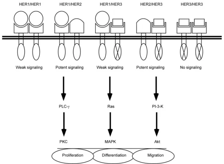
figure 2. Dimerization of HERs upon ligand binding triggers cell signaling cascades which may affect keratinocyte
proliferation, differentiation, or migration. Expression of three members of the HER family in keratinocytes leads to the formation of five homo- and/or hetero-dimers upon ligand binding. HER2 has no identified ligand, thus ho-modimers of HER2 are unlikely to form. Hoho-modimers of HER3 produce no signal. Heterodimers containing HER2 are preferentially formed when this receptor is present. The cell signaling includes the classical Ras-Mek-MApK pathway, pKc, and activation of phosphatidylinositol-3-kinase and Akt. it regulates keratinocyte proliferation, differentiation and also migration.
instance, cholesterol depletion of HaCaT keratino-cytes26 or of normal keratinocytes27 results in EGF receptor activation without the addition of ligand. Such activation is followed by a stimulation of the expression of heparin-binding EGF-like growth factor (HB-EGF)28 and leads to an atypical internalization of the EGF receptor29, both phenomena being under control of the activity of p38 MApK.
the neU/c-ERBB-2/her-2 oncoGene is eX-pressed by epiderMal Keratinocytes The second member of the type 1 family of growth factor receptors is a glycoprotein of 185 kDa closely related to the EGF receptor. This receptor, identi-fied first in rat neuroblastomas chemically induced by the carcinogen ethylnitrosourea, is the product of the neu oncogene whose name was chosen to depict its particular neuronal relevance.30 in this kind of tumor, a transforming potential of the rat glycoprotein was found to result from an oncogenic point mutation. However, in the corresponding gene subsequently identified in humans, no such precise point mutation was ever found. Because of its high homology with the EGF receptor, the product of the human neu gene was further named c-erbB-2, or HER2, although EGF does not bind nor activates this receptor.31-33 The HER2 gene product has generated a considerable interest in cancer researchers as this gene was found overexpressed in some human breast and ovarian cancers with poor prognosis.34
normal expression of HER2 in rat and human skin, particularly in the epidermis, has been reported by several groups studying tissue samples from donors of different ages and from various body sites.35-37 in the human epidermis, HER2 expres-sion is up-regulated with keratinocyte differentiation and this correlates with the relocalization of the protein into the plasma membrane, especially in keratinocytes of the granular layer.36,38 in vitro, we have demonstrated expression of HER2 in cultures of human epidermal keratinocytes and found that the HER2 expression is similarly up-regulated when cultured cells differentiate.39
The significant expression of the HER2 receptor in cutaneous tissues, particularly in the epidermis, implies that some role in the regulation of normal epidermal cell biology is most probably assigned to this molecule. like the EGF receptor, HER2 exhibits an activable tyrosine kinase cytoplasmic domain responsible for signal transduction and subsequent regulation of the cellular physiology.14,40 This property makes normally signaling HER
recep-tors important contriburecep-tors to normal cell control, but also makes them, when deregulated, potentially harmful inducers of anarchic cell proliferation. However, contrary to what has been described for the EGF receptor, no particular overexpression of the c-erbB-2 oncogene can be detected in skin tumors, including squamous cell carcinomas.36,41 Thus, despite there being numerous structural ho-mologies between the EGF receptor and HER2, it is suggested that these molecules may have different implications in cutaneous physiological processes and in malignancies of epidermal origin.
However, during investigations performed on the skin of mice that had been treated with the tumour-promoting phorbol ester TpA, the following observation could be made: tyrosine phosphorylation of HER2 is elevated by this kind of treatment, in good correlation with the formation of HER2:EGF receptor heterodimers.42 These data were completed by other studies performed on cultured mouse and human keratinocytes: heterodimerization between HER2 and the EGF receptor was found after ex-posure to EGF, together with phosphorylation of tyrosine residues.39,42 These results demonstrate that ErbB2/HER2 is able to interact with HER1. indeed, when this receptor is present, ErbB-2/ HER2-containing HER heterodimers are formed preferentially.43 Thus, as in other cell types, the presence of HER2 in variable amount available for heterodimerization in keratinocyte plasma membrane can modulate signal transduction by external ligands binding to members of the HER family. Furthermore, ligand-independent activation through cholesterol depletion of keratinocytes ap-parently leads also to the formation of heterodimers between the EGF receptor and HER2.
Other studies of the epidermal expression of HER2 utilized the production of transgenic mice expressing the HER2 under the control of the basal keratin 5 promoter.44 The animals reveal severe hyperplasia of the follicular and interfol-licular epidermis, as well as dramatic stimulation of keratinocyte proliferation, with appearance of papillomas.44 Thus, in cooperation with the EGF receptor, HER2 can induce signaling and play a previously unexpected role in the control of epi-dermal cell proliferation.
On the other hand, differences in the normal epidermal localization of HER2 and the EGF re-ceptor argue for potentially different roles in the program of epidermal differentiation undergone by keratinocytes committed to tissue maintenance and
renewal.36 Finally, interesting but still poorly un-derstood findings are the associations of the HER2 receptor with adhesion molecules: β-catenin and plakoglobin45, or the integrin α6β4 involved in the basal anchorage through hemidesmosomes.46 As a consequence, we may postulate that the activation of HER2 might indirectly regulate cellular adhesion and/or migration in keratinocytes. in normal cells, this type of regulation could be beneficial during tissue development and/or repair, however, in car-cinomas, such a regulation could be disastrous if it helps cell dissociation and metastasis.
in the present review, we focus on HER family members in the epidermis; however, it must be reminded that epidermis is certainly not the tis-sue where HER2 is mainly involved. Rather, the production of mice carrying a null allele of HER2 has shown the vital importance of this gene in the development of the neural system, as well as in cardiac development.47 indeed, the embryos carrying the null mutation suffer from a markedly affected development of cranial sensory ganglia, but also from a lacking formation of cardiac trabeculae. This absence leads to severe cardiac dysfunctions very likely responsible for the embryos death before day E11.47 Of course and unfortunately, the inter-rupted development of HER2 knock-out mice makes impossible any evaluation, with such an in vivo model, of the role of HER2 in the differentiation of mature epidermis and epidermal appendages, but to date, no particular abnormality has been reported in the developing epidermal tissue.
the c-ERBB-3/her-3 oncoGene is eXpressed by epiderMal Keratinocytes
The third member of the HER family of receptors was identified later than HER2 by reduced stringency hybridization of human genomic dnA.48,49 The so-called c-erbB-3/HER3 gene also encodes a receptor of 180 kDa with typical cytoplasmic tyrosine kinase domain. As for the HER2 receptor, it has been deduced from hybrid receptors constructed to as-sociate the extracellular domain of the EGF receptor with the cytoplasmic domain of HER3, that ligand binding on the extracellular moiety of the receptor triggers the tyrosine kinase activity of HER3.50 However, although other authors have demonstrated that the cytoplasmic domain of the receptor has an impaired tyrosine kinase activity51, HER3 remains an important partner for receptor heterodimerization with its numerous docking sites for transduction proteins such as pi3K, for example48,49.
As early as in the first report on HER3, it was noticed that this gene is expressed by epidermal cell types (keratinocyte and melanocyte). Conversely, dermal fibroblasts lack detectable HER3 mRnA.48 immunohistochemical methods confirmed later the localization of this receptor in the epidermis and in epidermal appendages.52 As we noted about HER2, the expression of HER3 was found also in autocrine cultures of human keratinocytes detected as the HER3 protein or detected as its encoding transcript. An up-regulation of its expression was again concomitant with the differentiation process.39 in vivo, the protein is particularly expressed by the differentiated cells of the spinous and granular lay-ers. These data seriously argue for a regulation of epidermal keratinocyte biology by signaling events transmitted through the HER3 receptor. nonetheless, such a regulation of keratinocytes through HER3 would not affect the development of the epidermis, since it was found that mice with targeted mutations in HER3 show no obvious developmental defect in epithelia.53 in consequence, we must admit that the HER3 receptor may regulate epidermal cells, but only after birth, or that other signaling molecules compensate during development for a defect in this receptor.
interestingly, one initial paper on HER3 also reported overexpression of this gene in several hu-man mammary tumor cell lines48, and analysis of its expression in breast cancer later confirmed that the transcription of the HER3 gene was upregulated in 22% of the cases analyzed in that particular study54. More recently, however, the HER3 gene has been found expressed at a much higher level in normal colon epithelium than in colorectal cancers, suggesting that HER3 was not systemati-cally overexpressed in malignant cell types.55 To the best of our knowledge, unfortunately, no pub-lished study has been specifically devoted so far to the analysis of HER3 expression in cutaneous malignant tissues.
Recently, forced HER3 expression was found able to accelerate the resurfacing of wounds in a pig model56, allowing hypotheses that link this receptor with healing and growth factors57. the foUrth MeMber of the her-faMily (c-ERBB-4/her-4) is absent froM the sKin A few years after the discovery of HER3, plowman and collaborators identified a fourth member of the HER family of receptors which was logically named c-erbB-4 or HER4.58 The HER4 receptor exhibits
also a molecular weight of 180 kDa and denotes highly homologous domains with the three previ-ously identified members, including a cytoplasmic domain with inducible tyrosine kinase activity. The expression of HER4 has been reported to be mostly neural, but the receptor can also be expressed by some breast-derived cancer cell lines. On the other hand, the HER4 receptor was not found in the epi-dermis58, and accordingly, we verified with other groups that no transcript for HER4 was detectable in either human38,39,59 or mouse42,60 cultured epi-dermal keratinocytes.
hereGUlins, the her3 and her4 liGand faMily, indUce the her2 tyrosine Kinase actiVity
Whereas the EGF receptor HER1 had been identi-fied by its ability to bind a previously identiidenti-fied growth factor, EGF itself, the c-erbB/HER2, -3, and -4 were initially characterized as orphan receptors with no identified ligand, yet these receptors exhibit a good homology with the EGF receptor. Thus, the members of the increasing family of growth factor exhibiting capacity to bind and activate the EGF receptor were unable to bind and activate HER2, -3 or -4.
in 1992, two independent groups reported iden-tification of a factor described as an activator of the HER2 receptor as detected by phosphorylation of cytoplasmic tyrosine residues on the HER2 (185 kDa) transmembrane protein. This suggested that the factor could be a ligand of HER2.11,12 This factor has been originally named by one group neu differentiation Factor (ndF) because the isolated 44 kDa rat protein induces differentiation in a breast cancer cell line overexpressing HER2.11,61 The second group named the factor heregulin to refer to its specificity for activation of the HER2 receptor.12 We will use here the term heregulin to designate the ndF/heregulin factor. Heregulin is a glycoprotein produced, like similar growth factors, as a transmembrane precursor exhibiting, from the n-terminal extracellular moiety to the c-terminal extremity, an immunoglobulin-like domain, a gly-cosylated site, an EGF-like domain as well as a juxtamembrane and transmembrane domains, and a cytoplasmic tail. Several isoforms, have been identified and result from alternative splicing of transcription products of a common gene. The isoforms combine the EGF-like domain alpha or beta, one juxtamembranous domain 1, 2, 3 or 4, and a cytoplasmic tail identified as a, b or c.12,40,61
The identity (α or β) of the heregulin EGF-like domain, responsible for the ligand-receptor inter-actions, doesn’t influence the specificity of the ligand but define the binding affinity: the affinity of β isoforms is ten-fold higher than that of the α isoforms.62
Some members of the heregulin family called Glial Growth Factor (GGF) or Acetylcholine Recep-tor inducing Activity (ARiA) have been identified as growth factors for glial cells produced by neu-ronal cells63 or as factors released by presynaptic terminals inducing the expression of acetylcholine receptors by muscle cells at the neuromuscular junction64. Because there is a clear predominance of the expression of heregulin by the nervous system65 and because of the relationship of heregulin with HER2, the product of neu oncogene, the factor is often referred to as neuregulin.40,63
The critical role in the nervous system of these HER2-activating factors has been assessed by the production of mice embryos lacking the heregulin gene.66 interestingly, these embryos die during de-velopment and exhibit abnormalities of the neural and cardiac systems which mimic abnormalities reported in mice lacking the HER2 receptor.47
Recently, three other genes were found related to the heregulin gene and their products were named neuregulin-267,68, neuregulin-369 and neuregulin-470. These new factors signal through HER3 and/or HER4 receptors and are apparently not expressed in cutaneous tissues.
hereGUlins are eXpressed by epiderMal Keratinocytes
Already in the first paper reporting the characteris-tics of the neu differentiation Factor, it was noted that this factor could be expressed in skin tissues.61 Subsequent studies, using in situ hybridization to localize the expression of heregulin in developing mice embryos, noticed also that beside predominant expression in neural tissues there is a significant hybridization of probes over the basal layer of the developing skin.65
The expression of heregulin in normal mature skin, however, has been studied in rabbits using an immunohistochemical approach. in the study, it was found that heregulin expression is predomi-nant in dermal cells, most likely in fibroblasts.71 looking for heregulin expression in samples of superficial normal human skin by RT-pcR detec-tion of transcripts, we have found a small amount of amplified products.39 However, this technique
does not allow to distinguish between epidermal or dermal origin of the amplified material. north-ern blot hybridizations of extracts from cultured mouse keratinocytes revealed first that these cells express small amounts of heregulin transcripts60; however, when we performed a similar study on samples from cultured human dermal fibroblasts and cultured human epidermal keratinocytes, we found a much higher expression of heregulins in keratinocytes, especially in subconfluent proliferating cells, than in fibroblasts39. The level of heregulin transcripts detected in rapidly dividing keratinocytes was so high that it did overcome the level found in MdA-231 cells, the human breast cancer cell line utilized for the initial isolation of heregulin.12 Such high expression is detected in subconfluent cultures of human keratinocytes, but drastically decreases when the cells become confluent and initiate the expression of early markers of epider-mal differentiation.19,39 In vivo, the expression of heregulin is also down-regulated by differentiation. To our knowledge, no such regulation of heregu-lin expression depending on culture conditions of a normal cell type has been previously reported. Therefore, cultures of keratinocytes could be of special interest to study the basic regulation of heregulin expression.
hereGUlin siGnals throUGh receptor heterodiMers.
Relative binding affinities of different growth fac-tors of the EGF family measured on soluble recep-tors72 have demonstrated that we may discriminate between three types of ligands. A few growth fac-tors like EGF, TGF-α and amphiregulin(AR) bind only to HER1. Betacellulin (BC), Heparin binding (HB)-EGF and epiregulin bind to HER1 but also to HER4. Heregulins bind only to HER3 and HER4. HER2 remains an orphan receptor but is thought to play a crucial role as a preferential partner for heterodimerization between HER family members. indeed, despite a first line of evidence suggested that incubations of certain cell lines with heregulins resulted in signal transduction through the HER2 receptor, it was rapidly recognized though that the interactions between heregulins and members of the EGF receptor family could be more complex.73 indeed, more evidence followed, suggesting that co-expression of the HER2 receptor with the HER4 receptor, or alternatively of the HER2 receptor with the HER3 receptor, was required to obtain
activa-tion of the HER2 molecule by heregulins.74,75 The particular importance of this heterodimerization process for signaling through heregulins and HER2 has been recently demonstrated: mice embryos were produced that lacked the HER4 receptor and these embryos died in utero, between E10 and E11. At that time of embryogenesis, HER4-null mice were shown to suffer exactly like HER2-null mice or heregulin-null mice from aberrant neural and cardiac development.47,66,76
in epithelial cells, and particularly in the epi-dermis where keratinocytes do not express HER4, HER3 and its impaired tyrosine kinase activity heterodimerizes with HER2 to constitute the pre-dominant form of heregulin receptor.39,77
hereGUlins with her2 and her3 consti-tUte a potential paracrine systeM in epiderMis.
The variations of the expression of the different HER family members and of heregulin in normal human keratinocytes undergoing normal epidermal differentiation, as described above, suggest that these molecules may constitute an intraepidermal paracrine system in which proliferating cells express amounts of a growth factor which act mainly on differentiating cells (Fig. 3). it seems, therefore, reasonable to speculate potential involvement of heregulins and their complex receptor system in later skin development (i.e. after E11 in mouse), in normal homeostasis of epidermal layers and/or in tissue repair during cutaneous wound healing. Prob-ably, the most straightforward experimentation to evaluate the role of an identified gene is to produce knock-out mice for this particular gene. Unfortu-nately, the role of heregulins and their receptors in mature cutaneous cell signaling in vivo cannot be studied with null mice since these embryos lack-ing heregulin die in utero before birth, due to the cardiac anomalies also observed in embryos lacking HER2 or HER3.47,66,76
interestingly, the expression of HER2 and HER3 is regulated in cultured human keratinocytes in accordance with in vivo data, being minimal in subconfluent rapidly growing cultures but being increased several times when the cells become confluent and initiate terminal differentiation.39 Thus, treatment of cultures of human epidermal keratinocytes with heregulin isoforms induces HER2 activation (tyrosine phosphorylation) in confluent culture conditions only. Accordingly, we have not been able to show any effect of heregulin
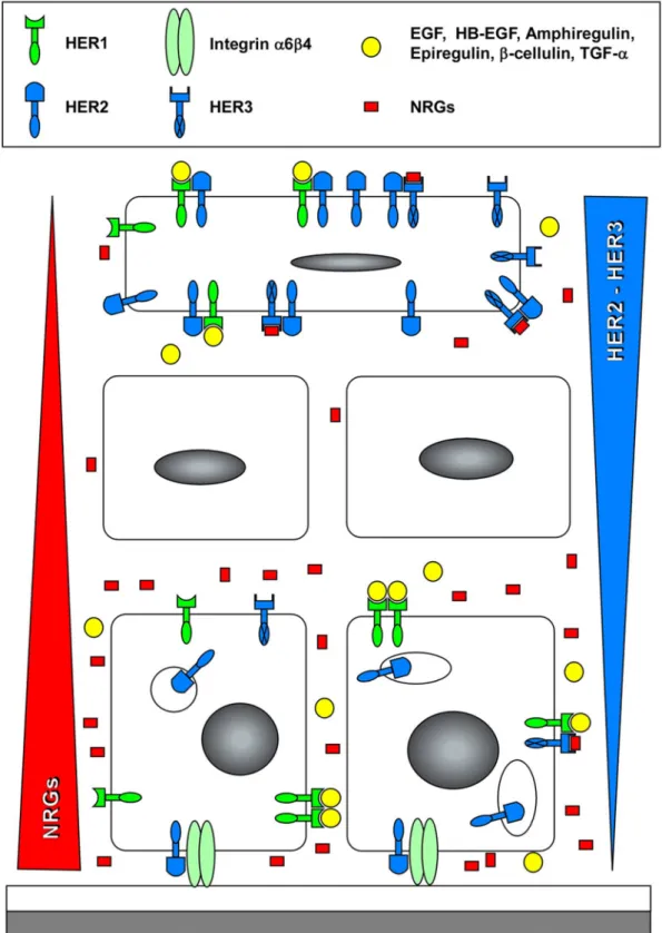
treat-figure 3. localization of HER-dependent signaling in keratinocytes embedded in a normal epidermis.
Keratino-cytes produce ligands for HER1 (TGF-α, Hb-EGF, Amphiregulin), as well as neuregulins (nRGs). neuregulins are mainly produced by basal keratinocytes producing a gradient represented by the red triangle, whereas HER3 is mainly expressed by suprabasal keratinocytes, producing an inverted gradient of the receptors (blue triangle). HER2 is also upregulated in differentiating keratinocytes. Heterodimers including HER2 induce the most potent signaling in suprabasal layers. HER2 produced in proliferating keratinocytes is mostly intracellular and relocated into the plasma membrane of differentiating keratinocytes of the upper spinous and granular cell layers38, while
HER2 expressed in basal keratinocytes is associated with the integrin α6β4, possibly regulating migratory prop-erties of cells46.
ment on subconfluent keratinocytes, whereas a significant suppression of the expression of markers of epidermal differentiation was produced by this treatment.39 This effect of an activation of HER2 in keratinocytes is similar to the effect produced when these cells are incubated with EGF in order to activate HER1.19
According to data available in the literature, such a regulation of HER3 expression in a normal cell type is unique so far to the human keratinocyte culture system. Therefore, culture of keratinocytes could be of value to study the normal control of HER3 expression.
Finally, we have to mention that apparently the responsiveness of mouse keratinocytes to heregu-lins60 differs from the responsiveness of human keratinocytes39, most probably as a consequence of the yet unexplained different regulation of HER3 expression in mouse epidermal cells.
possible inVolVeMent of her2 in cUtane-oUs patholoGies
The HER2 proto-oncogene seems to be critical in certain mammary and ovarian cancers where over-expression of this receptor is detected. Thereby, HER2 is an interesting target for immunotherapy of these cancers.77,78 Since HER2 is not over-expressed in squamous cell carcinomas, no unusual signaling through this receptor during this kind of tumoral development can be hypothesized. On the other hand, our findings about heregulin production by epidermal keratinocytes and the requirement for HER2 and HER3 heterodimerization in order to signal in these cells39,60 indicate future investiga-tions that should identify whether this system of ligands and receptors is involved in the etiology of epidermal tumors. nevertheless, HER2 has been shown to partially induce a malignant phenotype in papilloma-derived keratinocytes, but the effect pro-duced through HER2 was reported as less potent than the induction obtained with the ras oncogene.79
in some human dermal naevi and cutaneous mela-nomas, the HER2 receptor is apparently expressed, but at a level lower than in normal keratinocytes.80,81 Also, when the expression of HER2 was measured at mRnA level in psoriatic epidermis and com-pared with the same expression in normal tissue, no significant difference was found.82 On the other hand, over-expression has been interestingly reported in human radiation-induced skin ulcers83 and the involvement of HER2 could thereby be suspected
in the cancer transformation and poor healing of these particular radiation-induced skin lesions. More recent results obtained with transgenic mice further support this hypothesis.44 in these mice, HER2, under the control of the bovine keratin 5 promoter, is overexpressed in keratinocytes of the epidermal basal layer resulting in a significant epidermis hyperplasia, a dramatic increase in keratinocyte proliferation and the development of a squamous cell carcinoma-like appearance.
Finally, due to the epidermal localization of the abnormal cells observed in mammary Paget’s disease, the studies reporting the expression of HER2 in such cases have to be mentioned here.83,84 particularly, the localization of the HER2 receptor at tumor cell membranes was noticed in Paget’s disease.85,86 in order to explain the pathogenesis of this disease, an interesting hypothesis, proposing that epidermal keratinocytes may release chemot-actic factors attracting in the epidermis carcinoma cells over-expressing HER2, has been tested. Such a putative motility factor, which probably acts as a ligand with specificity for HER2, has indeed been detected during in vitro investigations.86 Recently, it was demonstrated that HRGα could be a potent chemoattractant protein eventually implicated in the Paget's disease.87
perspectiVes
in this review, we have focused our attention on the existence of an intraepidermal system in which heregulin is expressed by proliferating keratinocyte and may activate, by a paracrine loop, the differenti-ated keratinocytes of the suprabasal layers (Fig. 3). in this model, heregulin may also activate, by an autocrine loop, the weakly expressing HER2 and HER3 keratinocytes of the basal layer. We may postulate that this system is implicated in normal homeostasis of epidermal layers and/or in tissue repair during cutaneous wound healing. nonethe-less, studying the role of heregulin-HER receptor system in the control of the keratinocyte physiology remains a real challenge for the future.
acKnowledGeMents
This work received financial support from FRFc grant 2.4.506.01 and FnRS grant 1.5.033.06F to YP.
references
1. pittelkow MR. Keratinocyte abnormalities. in: Roenigk HHJr, Maibach Hi, eds. psoriasis. new york: Marcel dekker; 1991:305-25.
2. pittelkow MR. Growth factors and intracellular control mechanisms of keratinocyte growth and differentiation. in: ohkawara A, McGuire J, eds. The biology of the Epidermis. Amsterdam: Elsevier Science publishers; 1992:109-22.
3. Montano X. The role of oncogenes in skin cancer. in: leigh iM, lane Eb, Watt FM, eds. The keratinocyte handbook. Cambridge: Cambridge University Press; 1994:459-69.
4. boukamp p, breitkreutz d, Hülsen A, et al. in vitro transformation and tumor progression: a multistep model for skin carcinogenesis. in: leigh iM, lane EB, Watt FM, eds. The keratinocyte handbook. Cam-bridge: Cambridge University Press; 1994:485-99. 5. Greenhalgh dA, Rothnagel JA, dominey AM, et al.
Transgenic mouse models for inherited and acquired skin disorders: paradigms for molecular mechanisms and potential therapeutics. in: leigh iM, lane Eb, Watt FM, eds. The keratinocyte handbook. Cam-bridge: Cambridge University Press; 1994:471 84. 6. Greenhalgh dA, Wang XJ, Roop dR. Multistage
epidermal carcinogenesis in transgenic mice: coop-erativity and paradox. J invest dermatol Symposium proc 1996;1:162-76.
7. Cook PW, Piepkorn M, Clegg CH, et al. Trans-genic expression of the human amphiregulin gene induces a psoriasis-like phenotype. J clin invest 1997;100:2286-94.
8. Elder JT. Transforming growth factor alpha and re-lated growth factors. in: luger TA, Schwarz T (eds) Epidermal growth factors and cytokines. new york: Marcel dekker inc; 1994:205-40.
9. Jost M, Kari c, Rodeck U. The EGF receptor - an essential regulator of multiple epidermal functions Eur J dermatol 2000;10:505-10.
10. prigent SA, lemoine nR. The type 1 (EGFR-related) family of growth factor receptors and their ligands. progr Growth Factor Res 1992;4:1-24.
11. peles E, bacus SS, Koski RA, et al. isolation of the neu/HER-2 stimulatory ligand: a 44 kd glycoprotein that induces differentiation of mammary tumor cells. cell 1992;69:205-16.
12. Holmes WE, Sliwkowski MX, Akita RW, et al. identification of heregulin, a specific activator of p185erbb2. Science 1992;256:1205-10.
13. Mitev V, Miteva l. Signal transduction in keratino-cytes. Exp Dermatol 1999;8:96-108.
14. Yarden Y, Sliwkowski MX. Untangling the Erbb signaling network. nat Rev Mol cell biol 2001;2:127-37.
15. Heldin CH. Dimerization of cell surface receptors in signal transduction. cell 1995;80:213-223. 16. King lEJr, Gates RE, Stoscheck cM, et al. The
EGF/TGF alpha receptor in skin. J invest dermatol 1990;94:164S-170S.
17. Cook PW, Pittelkow MR, Shippley GD. Growth factor-independent proliferation of normal neo-natal keratinocytes: production of autocrine- and paracrine-acting mitogenic factors. J Cell Physiol 1991;146:277-89.
18. pittelkow MR, cook pW, Shipley Gd, et al. Au-tonomous growth of human keratinocytes requires epidermal growth factor receptor occupancy. Cell Growth diff 1993;4:513-21.
19. Poumay Y, Pittelkow MR. Cell density and culture factors regulate keratinocyte commitment to dif-ferentiation and expression of suprabasal K1/K10 keratins. J invest dermatol 1995;104:271-6. 20. Sibilia M, Wagner EF. Strain-dependent epithelial
defects in mice lacking the EGF receptor. Science 1995;269:234-8.
21. Threadgill dW, dlugosz AA, Hansen lA, et al. Tar-geted disruption of mouse EGF receptor: effect of genetic background on mutant phenotype. Science 1995;269:230-4.
22. Mann Gb, Fowler KJ, Gabriel A, et al. Mice with a null mutation of the TGF alpha gene have abnormal skin architecture, wavy hair, and curly whiskers and often develop corneal inflammation. Cell 1993;73:249-61.
23. levkowitz G, Waterman H, Zamir E, et al. c-cbl/ Sli-1 regulates endocytic sorting and ubiniquitina-tion of the epidermal growth factor receptor. Genes dev 1998;12:3663-74.
24. bishop pc, Myers T, Robey R, et al. differential sensitivity of cancer cells to inhibitors of the epi-dermal growth factor receptor family. Oncogene 2002;21:119-27.
25. lacouture ME. Mechanisms of cutaneous toxicities to EGFR inhibitors. nat Rev cancer 2006;6:803-12. 26. lambert S, Vind-Kezunovic d, Karvinen S, et al.
li-gand-independent activation of the EGFR by lipid raft disruption. J invest dermatol 2006;126:954-62. 27. Jans R, Atanasova G, Jadot M, et al. cholesterol
depletion upregulates involucrin expression in epi-dermal keratinocytes through activation of p38. J invest dermatol 2004;123:564-73.
28. Mathay c, Giltaire S, Minner F, et al. Heparin-bind-ing EGF-like growth factor is induced by disruption of lipid rafts and oxidative stress in keratinocytes and participates in the epidermal response to cutaneous wounds. J invest dermatol 2008;128:717-27. 29. lambert S, Ameels H, Gniadecki R, et al.
disruption in keratinocytes is delayed and de-pendent on p38 MApK activation. J cell physiol 2008;217:834-45.
30. bargmann ci, Hung Mc, Weinberg RA. Multiple independent activations of the neu oncogene by a point mutation altering the transmembrane domain of p185. Cell 1986;45:649-57.
31. bargmann ci, Hung Mc, Weinberg RA. The neu oncogene encodes an epidermal growth factor receptor-related protein. nature 1986;319:226-30. 32. coussens l, yang-Feng Tl, liao yc, et al. Tyrosine
kinase receptor with extensive homology to EGF receptor shares chromosomal location with neu oncogene. Science 1985;230:1132-9.
33. yamamoto T, ikawa S, Akiyama T, et al. Similar-ity of protein encoded by the human c-erbb-2 gene to epidermal growth factor receptor. nature 1986;319:230-4.
34. Slamon dJ, Godolphin W, Jones lA, et al. Studies of the HER-2/neu proto-oncogene in breast and ovarian cancer. Science 1989;244:707-12.
35. Kokai y, cohen JA, drebin JA, et al. Stage- and tissue-specific expression of the neu oncogene in rat development. proc natl Acad Sci USA 1987;84:8498-501.
36. Maguire HcJr, Jaworsky c, cohen JA, et al. distri-bution of neu (c-erbb-2) protein in human skin. J invest dermatol 1989;89:786-90.
37. Press MF, Cordon-Cardo C, Slamon DJ. Expression of the HER-2/neu proto-oncogene in normal human adult and fetal tissues. oncogene 1990;5:953-62. 38. Stoll SW, Kansra S, peshick S, et al. differential
utilization and localization of ErbB receptor tyrosine kinases in skin compared to normal and malignant keratinocytes. neoplasia 2001;3:339-50.
39. de potter iy, poumay y, Squillace KA, et al. Human EGF receptor (HER) family and heregulin members are differentially expressed in epidermal keratino-cytes and modulate differentiation. Exp Cell Res 2001;271:315-28.
40. peles E, yarden y. neu and its ligands: from an onco-gene to neural factors. bioEssays 1993;15:815-24. 41. Kearsley JH, leonard JH, Walsh Md, et al. A comparison of epidermal growth factor receptor (EGFR) and c-erbb-2 oncogene expression in head and neck squamous cell carcinomas. Pathology 1991;23:189-94.
42. Xian W, Rosenberg Mp, diGiovanni J. Activation of erbb-2 and c-src in phorbol ester-treated mouse epidermis: possible role in mouse skin tumor promo-tion. Oncogene 1997;14:1435-44.
43. Graus-porta d, beerli RR, daly JM, et al. Erbb-2, the preferred heterodimerization partner of all ErbB receptors, is a mediator of lateral signaling. EMBO
J 1997;16:1647-55.
44. bol d, Kiguchi K, beltran l, et al. Severe follicular hyperplasia and spontaneous papilloma formation in transgenic mice expressing the neu oncogene under the control of the bovine keratin 5 promoter. Mol carcinog 1998;21:2-12.
45. ochiai A, Akimoto S, Kanai y, et al. c-erbb-2 gene product associates with catenins in human cancer cells. Biochem Biophys Res Commun 1994;205:73-8.
46. Hintermann E, bilban M, Sharabi A, et al. inhibitory role of alpha 6 beta 4-associated erbb-2 and phos-phoinositide 3-kinase in keratinocyte haptotactic migration dependent on alpha 3 beta 1 integrin. J cell biol 2001;153: 465-78.
47. lee KF, Simon H, chen H, et al. Requirement for neuregulin receptor erbb2 in neural and cardiac development. nature 1995;378:394-8.
48. Kraus MH, issing W, Miki T, et al. isolation and char-acterization of ERbb3, a third member of the ERbb/ epidermal growth factor receptor family: evidence for overexpression in a subset of human mammary tumors. proc natl Acad Sci USA 1989;86:9193-7. 49. plowman Gd, Whitney GS, neubauer MG, et al.
Molecular cloning and expression of an additional epidermal growth factor receptor-related gene. Proc natl Acad Sci USA 1990;87:4905-09.
50. Kraus MH, Fedi, p, Starks, V, et al. demonstra-tion of ligand-dependent signaling by the erbB-3 tyrosine kinase and its constitutive activation in human breast tumor cells. proc natl Acad Sci USA 1993;90:2900-4.
51. Guy pM, platko JV, cantley lc, et al. insect cell-expressed p180erbB3 possesses an impaired tyrosine kinase activity. proc natl Acad Sci USA 1994;91:8132-6.
52. prigent SA, lemoine nR, Hughes cM, et al. Expres-sion of the c-erbB-3 protein in normal human adult and fetal tissues. oncogene 1992;7:1273-8. 53. Riethmacher D, Sonnenberg-Riethmacher E,
Brink-mann V, et al. Severe neuropathies in mice with targeted mutations in the Erbb3 receptor. nature 1997;389:725-30.
54. lemoine nR, barnes dM, Hollywood dp, et al. Expression of the ERBB3 gene product in breast cancer. br J cancer 1992;66:1116-1121.
55. Zhang l, Zhou W, Velculescu VE, et al. Gene ex-pression profiles in normal and cancer cells. Science 1997;276:1268-72.
56. okwueze Mi, cardwell nl, pollins Ac, et al. Modu-lation of porcine wound repair with a transfected Erbb3 gene and relevant EGF-like ligands. J invest dermatol 2007;127:1030-41.
J invest dermatol 2007;127:995-7.
58. Plowman GD, Culouscou JM, Withney GS, et al. ligand-specific activation of HER4/p180erbb4, a fourth member of the epidermal growth fac-tor recepfac-tor family. proc natl Acad Sci USA 1993;90:1746-50.
59. Marqués MM, Martinez n, Rodriguez-Garcia i, et al. EGFR family-mediated signal transduction in the human keratinocyte cell line HaCaT. Exp Cell Res 1999;252:432-8.
60. Marikovsky M, lavi S, pinkas-Kramarski R, et al. ErbB-3 mediates differential mitogenic effects of ndF/heregulin isoforms on mouse keratinocytes. Oncogene 1995;10:1403-11.
61. Wen d, peles E, cupples R, et al. neu differentiation Factor: a transmembrane glycoprotein containing an EGF domain and an immunoglobulin homology unit. cell 1992;69:559-72.
62. Wen d, Suggs SV, Karunagaran d, et al. Structural and functional aspects of the multiplicity of neu differ-entiation factors. Mol Cell Biol 1994;14:1909-19. 63. Marchionni MA, Goodearl AdJ, chen MS, et al.
Glial growth factors are alternatively spliced erbb2 ligands expressed in the nervous system. nature 1993;362:312-18.
64. Falls dl, Rosen KM, corfas, G, et al. ARiA, a protein that stimulates acetylcholine receptor syn-thesis, is a member of the neu ligand family. Cell 1993;72:801-15.
65. orr-Urtreger A, Trakhtenbrot l, ben-levy R, et al. neural expression and chromosomal mapping of neu differentiation factor to 8p12-p21. proc natl Acad Sci USA 1993;90:1867-71.
66. Meyer D, Birchmeier C. Multiple essential func-tions of neuregulin in development. nature 1995;378:386-90.
67. chang H, Riese dJ ii, Gilbert W, et al. ligands for erbB-family receptors encoded by a neuregulin-like gene. nature 1997;387:509-12.
68. carraway Kl iii, Weber Jl, Unger MJ, et al. neu-regulin-2, a new ligand of erbb3/erbb4-receptor tyrosine kinases. nature 1997;387:512-16.
69. Zhang d, Sliwkowski MX, Mark M, et al. neuregu-lin-3 (nRG3): a novel neural tissue-enriched protein that binds and activates erbb4. proc natl Acad Sci USA 1997;94:9562-7.
70. Harari d, Tzahar E, Romano J, et al. neuregulin-4: a novel growth factor that acts through the ErbB-4 re-ceptor tyrosine kinase. oncogene 1999;18:2681-9. 71. danilenko dM, Ring bd, lu JZ, et al. neu dif-ferentiation factor upregulates epidermal migration and integrin expression in excisional wounds. J Clin invest 1995;95:842-51.
72. Jones FE, Jerry dJ, Guarino bc, et al. Heregulin
induces in vivo proliferation and differentiation of mammary epithelium into secretory lobuloalveoli. Cell Growth Diff 1996;7:1031-8.
73. peles E, ben-levy R, Tzahar E, et al. cell-type specific interaction of neu differentiation fac-tor (ndF/heregulin) with neu/HER-2 suggests complex ligand-receptor relationships. EMBO J 1993;12:961-71.
74. Plowman GD, Green JM, Culouscou JM, et al. He-regulin induces tyrosine phosphorylation of HER4/ p180erbb4. nature 1993;366:473-5.
75. Sliwkowski MX, Schaefer G, Akita RW, et al. coex-pression of erbb2 and erbb3 proteins reconstitutes a high affinity receptor for heregulin. J biol chem 1994;269:14661-5.
76. Gassmann M, casagranda F, orioli d, et al. Ab-errant neural and cardiac development in mice lacking the erbb4 neuregulin receptor. nature 1995;378:390-4.
77. park JW, Stagg R, lewis Gd, et al. Anti-p185HER2 monoclonal antibodies: biological properties and potential for immunotherapy. Cancer Treat Res 1992;61: 193-211.
78. pegram Md, Konecny G, Slamon dJ. The molecular and cellular biology of HER2/neu gene amplifica-tion/overexpression and the clinical development of herceptin (trastuzumab) therapy for breast cancer. cancer Treat Res 2000;103: 57-75.
79. Dotto GP, O’Connell J, Patskan G, et al. Malignant progression of papilloma-derived keratinocytes: dif-ferential effects of the ras, neu, and p53 oncogenes. Molecular Carcinogenesis 1988;1:171-9.
80. peris K, cerroni l, chimenti S, et al. proto-oncogene expression in dermal naevi and melanomas. Arch dermatol Res 1991;283:500-5.
81. natali pG, nicotra MR, digiesi G, et al. Expression of gp185HER-2 in human cutaneous melanoma: implications for experimental therapeutics. int J Cancer 1994;56:341-6.
82. Schulz bS, Michel G, Wagner S, et al. increased expression of epidermal il-8 receptor in psoriasis. down-regulation by FK-506 in vitro. J immunol 1993;151:4399-406.
83. Meissner K, Rivière A, Haupt G, et al. Study of neu-protein expression in mammary Paget’s disease with and without underlying breast carcinoma and in extramammary paget’s disease. Am J pathol 1990;137:1305-9.
84. nakamura G, Shikata n, Shoji T, et al. immunohis-tochemical study of mammary and extramammary paget’s disease. Anticancer Res 1995;15:467-70. 85. nishi M, yoshida H, Setoyama M, et al.
immunohis-tochemical study of c-erbb-2 oncoprotein expression in extramammary Paget’s disease. Dermatology
1994;188:100-2.
86. de potter cR, Eeckhout i, Schelfhout AM, et al. Keratinocyte induced chemotaxis in the pathogen-esis of Paget’s disease of the breast. Histopathology 1994;24:349-56.
87. Schelfhout VR, Coene ED, Delaey B, et al.
Pathogen-esis of Paget’s disease: epidermal heregulin-alpha, motility factor, and the HER receptor family. J natl cancer inst 2000;92:622-8. Представители EGF- рецеПторной фамилии в нормальном и Патоло-гическом эПидермисе Ив Пуме, В. Митев резюме Эпидермальный фактор роста (EGF) и его аналоги связываются с трансмембранными рецепторами EGF рецепторной фамилии, обла-дающими тирозин-киназной активностью в своем цитоплазманическом домене. Эти факторы роста и их соответствующие рецепторы получили свои наименования после первоначального установления их функций в эпидермисе. После доказывания действия EGF-рецептора как онкоген его ши-роко исследовали в случаях наличия опухоли. Ус-тановлены и другие члены той же фамилии, которых охарактеризовали и анализировали в доброкачественных и злокачественных опухолевых клетках. В настоящем обзоре преимущественно рассматриваются функции EGF рецепторной семьи в нормальном эпидермисе одновременно с экспрессией и ролью их соответствующих лиганд. Обсуждается и потенциальное участие этих молекул в различных заболеваниях эпидермиса.