HAL Id: hal-02086847
https://hal-amu.archives-ouvertes.fr/hal-02086847
Submitted on 1 Apr 2019
HAL is a multi-disciplinary open access archive for the deposit and dissemination of sci-entific research documents, whether they are pub-lished or not. The documents may come from teaching and research institutions in France or abroad, or from public or private research centers.
L’archive ouverte pluridisciplinaire HAL, est destinée au dépôt et à la diffusion de documents scientifiques de niveau recherche, publiés ou non, émanant des établissements d’enseignement et de recherche français ou étrangers, des laboratoires publics ou privés.
Distributed under a Creative Commons Attribution - NonCommercial - NoDerivatives| 4.0 International License
A point mutation in the Ncr1 signal peptide impairs the
development of innate lymphoid cell subsets.
Francisca F. Almeida, Sara Tognarelli, Antoine Marcais, Andrew J. Kueh,
Miriam E. Friede, Yang Liao, Simon N. Willis, Kylie Luong, Fabrice Faure,
Francois E. Mercier, et al.
To cite this version:
Francisca F. Almeida, Sara Tognarelli, Antoine Marcais, Andrew J. Kueh, Miriam E. Friede, et al.. A point mutation in the Ncr1 signal peptide impairs the development of innate lymphoid cell subsets.. OncoImmunology, Taylor & Francis, 2018, 7 (10), pp.e1475875. �10.1080/2162402X.2018.1475875�. �hal-02086847�
OncoImmunology
ISSN: (Print) 2162-402X (Online) Journal homepage: https://www.tandfonline.com/loi/koni20
A point mutation in the Ncr1 signal peptide
impairs the development of innate lymphoid cell
subsets
Francisca F. Almeida, Sara Tognarelli, Antoine Marçais, Andrew J. Kueh,
Miriam E. Friede, Yang Liao, Simon N. Willis, Kylie Luong, Fabrice Faure,
Francois E. Mercier, Justine Galluso, Matthew Firth, Emilie Narni-Mancinelli,
Bushra Rais, David T. Scadden, Francesco Spallotta, Sandra Weil, Ariane
Giannattasio, Franziska Kalensee, Tobias Zöller, Nicholas D. Huntington,
Ulrike Schleicher, Andreas G. Chiocchetti, Sophie Ugolini, Marco J. Herold,
Wei Shi, Joachim Koch, Alexander Steinle, Eric Vivier, Thierry Walzer,
Gabrielle T. Belz & Evelyn Ullrich
To cite this article: Francisca F. Almeida, Sara Tognarelli, Antoine Marçais, Andrew J. Kueh, Miriam E. Friede, Yang Liao, Simon N. Willis, Kylie Luong, Fabrice Faure, Francois E. Mercier, Justine Galluso, Matthew Firth, Emilie Narni-Mancinelli, Bushra Rais, David T. Scadden, Francesco Spallotta, Sandra Weil, Ariane Giannattasio, Franziska Kalensee, Tobias Zöller, Nicholas D. Huntington, Ulrike Schleicher, Andreas G. Chiocchetti, Sophie Ugolini, Marco J. Herold, Wei Shi, Joachim Koch, Alexander Steinle, Eric Vivier, Thierry Walzer, Gabrielle T. Belz & Evelyn Ullrich (2018) A point mutation in the Ncr1 signal peptide impairs the development of innate lymphoid cell subsets, OncoImmunology, 7:10, e1475875, DOI: 10.1080/2162402X.2018.1475875
To link to this article: https://doi.org/10.1080/2162402X.2018.1475875
© 2018 The Author(s). Published by Taylor & Francis.
View supplementary material
Published online: 15 Aug 2018.
Submit your article to this journal
Article views: 620
Full Terms & Conditions of access and use can be found at
ORIGINAL RESEARCH
A point mutation in the
Ncr1 signal peptide impairs the development of innate
lymphoid cell subsets
Francisca F. Almeida a,b, Sara Tognarellic,d, Antoine Marçaise, Andrew J. Kueha,b, Miriam E. Friedef, Yang Liaoa,b,
Simon N. Willisa,b, Kylie Luonga,b, Fabrice Fauree, Francois E. Mercierg, Justine Gallusoh, Matthew Firtha,b,
Emilie Narni-Mancinellih, Bushra Raisc,d, David T. Scaddeni, Francesco Spallottaj, Sandra Weilk,l, Ariane Giannattasiok,l,
Franziska Kalenseec,d, Tobias Zöllerf, Nicholas D. Huntingtona,b, Ulrike Schleicherm, Andreas G. Chiocchettin,
Sophie Ugolini h, Marco J. Herolda,b, Wei Shia,o, Joachim Kochk,l, Alexander Steinle f, Eric Vivier h,p,q,
Thierry Walzere, Gabrielle T. Belz a,b, and Evelyn Ullrich d,c
aDivision of Molecular Immunology, Walter and Eliza Hall Institute of Medical Research, Melbourne, Victoria, Australia;bDepartment of Medical
Biology, University of Melbourne, Melbourne, Victoria, Australia;cDivision of Stem Cell Transplantation and Immunology, Department for Children
and Adolescents Medicine, Johann Wolfgang Goethe University Hospital, Frankfurt am Main, Germany;dLOEWE Center for Cell and Gene Therapy,
Johann Wolfgang Goethe University, Frankfurt am Main, Germany;eCIRI, Centre International de Recherche en Infectiologie - International Center
for Infectiology Research, Inserm, U1111, CNRS, UMR5308, Ecole Normale Supérieure de Lyon, Université Lyon 1, Lyon, France;fInstitute for
Molecular Medicine, Johann Wolfgang Goethe University, Frankfurt am Main, Germany;gDepartment of Medicine, McGill University, Montreal,
Quebec, Canada;hCNRS, INSERM, CIML, Centre d'Immunologie de Marseille-Luminy, Aix Marseille University, Marseille, France;iHarvard Stem Cell
Institute, Cambridge, MA, USA;jDivision of Cardiovascular Epigenetics, Department of Cardiology, Johann Wolfgang Goethe University, Frankfurt
am Main, Germany;kGeorg Speyer Haus, Institute for Tumor Biology and Experimental Therapy, Frankfurt am Main, Germany;lInstitute of Medical
Microbiology and Hygiene, University of Mainz Medical Center, Mainz, Germany;mMikrobiologisches Institut-Klinische Mikrobiologie, Immunologie
und Hygiene, Friedrich-Alexander-Universität Erlangen-Nürnberg und Universitätsklinikum Erlangen, Erlangen, Germany;nMolecular Genetics
Laboratory, Department for Child and Adolescent Psychiatry, Psychosomatics and Psychotherapy, Johann Wolfgang Goethe University, Frankfurt am Main, Germany;oDepartment of Computing and Information Systems, University of Melbourne, Melbourne, Victoria, Australia;pInnate Pharma,
Marseille, France;qService d’Immunologie, Hôpital de la Timone, Marseille Immunopole, Assistance Publique - Hôpitaux de Marseille, Marseille,
France
ABSTRACT
NKp46 (CD335) is a surface receptor shared by both human and mouse natural killer (NK) cells and innate lymphoid cells (ILCs) that transduces activating signals necessary to eliminate virus-infected cells and tumors. Here, we describe a spontaneous point mutation of cysteine to arginine (C14R) in the signal peptide of the NKp46 protein in congenic Ly5.1 mice and the newly generated NCRB6C14R strain. Ly5.1C14R NK cells expressed similar levels of Ncr1 mRNA as C57BL/6, but showed impaired surface NKp46 and reduced ability to control melanoma tumorsin vivo. Expression of the mutant NKp46C14Rin 293T cells showed that NKp46 protein trafficking to the cell surface was compromised. Although Ly5.1C14Rmice had normal number of NK cells, they showed an increased number of early maturation stage NK cells. CD49a+ILC1s were also increased but these cells lacked the expression of TRAIL. ILC3s that expressed NKp46 were not detectable and were not apparent when examined by T-bet expression. Thus, the C14R mutation reveals that NKp46 is important for NK cell and ILC differentiation, maturation and function.
Significance
Innate lymphoid cells (ILCs) play important roles in immune protection. Various subsets of ILCs express the activating receptor NKp46 which is capable of recognizing pathogen derived and tumor ligands and is necessary for immune protection. Here, we describe a spontaneous point mutation in the signal peptide of the NKp46 protein in congenic Ly5.1 mice which are widely used for tracking cellsin vivo. This Ncr1 C14R mutation impairs NKp46 surface expression resulting in destabilization of Ncr1 and accumula-tion of NKp46 in the endoplasmic reticulum. Loss of stable NKp46 expression impaired the maturaaccumula-tion of NKp46+ILCs and altered the expression of TRAIL and T-bet in ILC1 and ILC3, respectively.
ARTICLE HISTORY
Received 7 May 2018 Accepted 7 May 2018
KEYWORDS
innate lymphoid cells; activation receptors; intracellular trafficking; congenic mice
Introduction
Natural killer (NK) cells are cytolytic and cytokine-producing cells that contribute to eradicate pathogen-infected cells and
cancers thereby mediating frontline defense and
immunosurveillance.1,2The capacity to mediate these functions
depends on the balance between inhibitory and activating sig-nals. Activating receptors include the family of natural
CONTACTGabrielle T. Belz [email protected]; Evelyn Ullrich [email protected] Walter and Eliza Hall Institute of Medical Research, Melbourne, Victoria, 3052, Australia; Division of Stem Cell Transplantation and Immunology, Department for Children and Adolescents Medicine, Johann Wolfgang Goethe University Hospital, Frankfurt am Main, Germany
F.F.A and S.T. contributed equally and G.T.B. and E.U. contributed equally.
Color versions of one or more of the figures in the article can be found online atwww.tandfonline.com/koni. Supplemental data for this article can be accessedhere.
ONCOIMMUNOLOGY
2018, VOL. 7, NO. 10, e1475875 (15 pages)
https://doi.org/10.1080/2162402X.2018.1475875
cytotoxicity receptors (NCRs) NKp46, NKp30 and NKp44.3,4 NKp46 is expressed by NK cells and group 1 and 3 innate
lymphoid cells (ILCs).5-7NKp46 is encoded by Ncr1 and
associ-ates with the ITAM-containing CD3ζ or FcRγ polypeptides.
Several endogenous ligands of NKp46 have been described
including the complement factor P,8 the intracellular
filamen-tous cytoskeletal protein vimentin expressed on the surface of
Mycobacterium tuberculosis–infected monocytes,9 and several
viral proteins such as hemagglutinin and hemagglutinin
neur-aminidases of the influenza,10,11Sendai,12Newcastle disease,13
ectromelia and vaccinia viruses.14 NKp46 was also shown to
recognize PfEMP1 of Plasmodium falciparum,14 an unknown
ligand from Fusobacterium nucleatum,15 adhesins from
Candida glabrata.16 This wide expression supports that the
triggering of NKp46 is essential for effective immune responses. Previous studies have shown that mutations of the Ncr1 gene can cause a reduction in the cell surface expression of the NKp46 protein. Various strains of mice have been generated that either lack Ncr1 or exhibit mutations that have been
chemically induced.12,17,18 These lines (Ncrgfp/gfp, Ncr1Noe/Noe
and Ncr1iCre/iCre) exhibit different phenotypes, possibly as a
consequence of their difference in NKp46 protein expression.
For example, Ncr1Noe/Noeand Ncr1iCre/iCrestrains harbor
mod-est gene changes but preserve the protein while Ncr1gfp/gfp
mice,17 and Ncr1gfp/gfpKlrk1−/- double knockout mice which
have defects in both NKp46 and NKG2D expression,19 lack
exons 5–7 resulting in a complete disruption of the protein.
Narni-Mancinelli et al.18first described the effect of the point
mutation W32R in a colony of C57BL/6J mice carrying
N-ethyl-N-nitrosourea (ENU)–induced mutations (called Noé mice).
These mice had normal numbers of NK cells but showed an impaired expression of NKp46 on the cell surface. Moreover, Noé mice displayed an increased expression of the zinc-finger protein Helios which is involved in the regulation of lymphocyte development. Noé NK cells were hyper responsive to various stimuli in vitro and Noé mice were more resistant to infection
with mouse cytomegalovirus.18Glasner et al.20further
charac-terized the effect of the Noé mutation clarifying that the
NKp46W32Rprotein could reach the surface of NK cells, but in
a slow and unstable manner. This resulted in an accumulation of NKp46 in the endoplasmic reticulum (ER) and limited transport
to the cell surface.20Recently, studies of human NK cells carrying
the same mutation NKp46W32Rled to similar conclusions.21
Here we describe the analysis of two independent colonies of
congenic CD45.1+ (Ly5.1) mice that exhibited a spontaneous
single point mutation (C14R, designated Ly5.1C14Rmice) in the
signal sequence of the Ncr1 gene. This mutation impaired NKp46 surface expression in ILC subsets. The C14R mutation did not alter the overall expression of Ncr1 mRNA in mutant NK cells but impaired the surface expression of NKp46 in ILCs and was associated with alteration in ILC maturation. These results
were also confirmed by a newly generated NCRB6C14Rstrain.
Results
Ly5.1 congenic mouse strains exhibit a reduced surface expression of NKp46
Ly5.1 (B6.SJL-Ptprca Pepcb/BoyJ) congenic mice expressing CD45.1 on the surface of all lymphocytes are classically used
in combination with CD45.2-expressing C57BL/6 mice to establish experimental systems in which donor and host immune cells can be faithfully tracked both at steady-state and during an immune challenge. However, during analyses
of these types of systems, it was noticed that Ly5.1+ mice
consistently exhibited very poor expression of the receptor
NKp46, a key phenotypic marker of NK cells22 and some
subsets of ILCs, in multiple tissues including peripheral
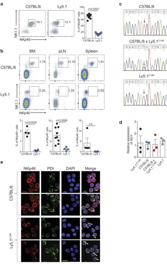
blood (Figure 1A), bone marrow, peripheral lymph nodes
and spleen (Figure 1B). This observation was made in two
mouse colonies completely independent of each other but which were originally derived from the Jackson Laboratory (Jax) imported in 2008 (University Hospital Erlangen) and 2010 (Walter and Eliza Hall Institute, WEHI) and maintained as closed colonies. This was distinct from newly imported mice (2012, University Hospital Erlangen; 2016, WEHI), also from Jax, which exhibited NKp46 expression equivalent to the C57BL/6 lines and F1 mice derived from the originally imported Ly5.1 line (C57BL/6 × Ly5.1WEHI) (SI Appendix, Figure S1A). These findings suggested that this alteration was a recessive trait limited to specific colonies that may have carried the mutation. While this mutation was tightly linked to the Ptprca locus on chromosome 1, Ncr1 is found on chromosome 7 indicating that the alteration was not within Ptprca itself.
A single amino acid change in the Ncr1 gene abrogates stable NKp46 surface expression
To understand the basis of this alteration, we used whole exome sequencing to examine C57BL/6 Ly5.1 (WEHI) and Ly5.1 (WEHI) from the mouse colonies in Melbourne, Australia. Assuming a recessive pattern of inheritance, 1042 SNPs were identified that were homozygous in the affected mice and heterozygous in the parental strain (SI Appendix, Dataset S1). Of these, 670 SNPs were classified as low impact mutations based on variant effector predictor analysis and as such were excluded from further analysis. Based on the observed phenotype, we initially concentrated on the candi-date gene Ncr1. NKp46 is a 46 kDa type 1 transmembrane glycoprotein characterized by a short intracellular tail, a single
transmembrane domain, and two extracellular Ig-like
domains. Sequence analysis identified a single homozygous missense point mutation (T to C) in the signal peptide of Ncr1 in both strains of mice that exhibited low NKp46 protein expression. We confirmed by Sanger sequencing that the mutation was present in Ly5.1 WEHI (hereinafter referred
to as Ly5.1C14R ) mice and C57BL/6 × Ly5.1C14R but not
C57BL/6 mice and resulted in a single amino acid substitution
of cysteine to arginine (C14R; Figure 1C). In contrast, the
RNA level of Ncr1 did not significantly differ in NK cells from
C57BL/6, Ly5.1C14R, WT Ly5.1 and C57BL/6 × Ly5.1C14Rmice
(Figure 1D). It should be noted that subsequent to identifica-tion of this mutaidentifica-tion in the established lines at each institu-tion, new founders were imported to the WEHI (2016) to
establish a new ‘unmutated’ colony. Breedings were kept
entirely separate from the original colonies and progeny from the new breedings were analysed. Of these, 39 of 88 mice tested exhibited a reduction in NKp46 expression by
Figure 1.Ly5.1 congenic mouse strain exhibits reduced surface expression of NKp46 that alters the localization of the NKp46 protein. (A) Dot plot showing the staining and frequency of NK1.1+NKp46+ cells in the peripheral blood lymphocytes of C57BL/6 and Ly5.1 mice. Data show representative plots gated on live lymphocytes CD3−CD19−(n = 12 mice/genotype). (B) Dot plots showing the expression and frequency quantification of NK1.1+NKp46+cells in the bone marrow (BM), peripheral lymph node (pLN) and spleen of C57BL/6 and Ly5.1 mice. (A, B) Data show representative plots gated on live lymphocytes CD3−CD19−pooled from two to five independent experiments (n = 4–12 mice/genotype/tissue). (C) Sanger sequencing analysis of the Ncr1 gene in C57BL/6, C57BL/6 × Ly5.1C14Rand Ly5.1C14Rmice showing the position of the point mutation. (D) Relative levels of NKp46 transcripts in splenic NK cells of C57BL/6, Ly5.1C14R, WT Ly5.1 and C57BL/6 × Ly5.1C14Rmice. Data show the mean ± SEM of 3–4 mice/genotype for one of three similar experiments. P values were calculated using an unpaired two-tailed Student’s t test. (E) NKp46 localization in primary NK cells. Representative images of NK cells isolated from C57BL/6 and mutant Ly5.1C14Rmice stained with anti-NKp46 and anti-PDI primary antibodies, and AlexaFluor488-conjugated anti-goat and AlexaFluor546-conjugated anti-rabbit secondary antibodies (DAPI nuclear stain, blue; anti-NKp46, red; PDI, green). Images were obtained using confocal scanning microscopy. Arrows indicate ER localization. Scale bar, 10μm.
flow cytometry.We further sequenced 29 mice from the new colony; 9 were homozygous wild-type for the Ncr1 gene and were used to reestablish the colony while the remainder were heterozygous. Thus, it appears that the mutation has inadver-tently been retained undetected long term in parent colonies.
The C14R Ncr1 mutation significantly impairs surface NKp46 expression in 293T cells
As the discovery that a mutation in the leader sequence might affect NKp46 cell surface expression or trafficking was unex-pected, we sought to demonstrate that ectopic expression of the
NKp46C14Rmutation itself was responsible for the alterations we
observed in the mutated Ly5.1 mice. To this end, 293T cells were
transfected with wild-type NKp46 or NKp46C14RcDNA,
respec-tively, and the expression of NKp46 was tracked by flow
cyto-metry. The surface expression of the NKp46C14R protein on
293T transfectants was significantly reduced compared with wild-type NKp46 while the total cellular NKp46 expression was at a comparable level (SI Appendix, Figure S1B and C), demon-strating that the impaired surface expression of NKp46 in Ly5.1 mice in fact is due to the C14R mutation in the NKp46 signal peptide. Analyses of freshly isolated naïve NK cells from
wild-type or Ly5.1C14Rmice by confocal laser scanning microscopy
not only confirmed the reduced NKp46C14Rsurface localization,
but also revealed a spotted intracellular distribution of the
mutated NKp46C14R protein. Furthermore, the colocalization
of NKp46 with protein disulfide isomerase (PDI), an enzyme in the endoplasmic reticulum, indicated an accumulation of
NKp46C14R in that cell compartment (Figure 1E and SI
Appendix, S1 D). Thus the C14R mutation in the signal peptide
of NKp46C14Rresulted in disruption of NKp46 surface
expres-sion by failed trafficking within the cell.
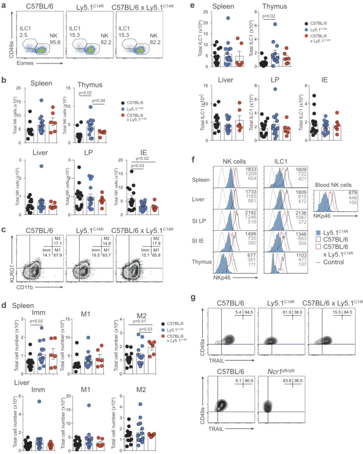
Ly5.1C14Rmice have normal numbers of total NK cells but altered distribution of ILC subsets
We then interrogate if the C14R mutation might impact the subsets of cells that normally express NKp46, i.e. NK cells, ILC1
and NCR+ILC3s. Ly5.1C14Rmice exhibited normal numbers of
NK1.1+CD3−NK cells in the majority of the tissues but these
numbers were significantly increased in the thymus compared
with control mice (Figure 2A and B). We then examined the
maturation status of NK cells by measuring the expression of
KLRG1 and CD11b in C57BL/6 and Ly5.1C14R NK cells
(Figure 2C and D). This revealed an increase in the immature (Imm) NK cell subsets in the spleen but not in the liver of
Ly5.1C14R mice suggesting that steady-state signalling through
NKp46 may act to modulate early NK cell maturation. Similar findings were observed when cells were analysed using CD27 and CD11b (SI Appendix, Figure S2A and B) and in Noé NK cells (SI Appendix, Figure S2C and D), a phenotype that was rescued by the expression of human NKp46.
ILC1 were identified through the expression of the CD49a integrin and of the absence of expression of the transcriptional regulator Eomesodermin (Eomes) which is required for the
devel-opment of NK cells.23This showed that similar to NK cells, ILC1
were also enriched in the thymus (Figure 2E) although the level of
expression of NKp46 in Ly5.1C14Rmice was reduced by ~3-fold
for both populations across all tissues when compared to that
found in C57BL/6 mice and ~1.5-fold for C57BL/6 × Ly5.1C14R
mice (Figure 2F). A similar reduction in the expression of NKp46
was seen in NK cells and ILC1s from tissues analysed in mixed bone marrow chimeras (SI Appendix, Figure S3). This effect was not seen in mice that carried only the six base pair mutation that
encodes CD45.1 (CD45.1STEM) (SI Appendix, Figure S4)
demon-strating that the NKp46 expression defect in Ly5.1C14R mice is
intrinsic and not linked to Ly5.1 itself. In addition, hepatic ILC1
isolated from Ly5.1C14Rmice lacked expression of the TNF-related
apoptosis-inducing ligand, TRAIL (encoded by Tnfsf10) normally
characteristic of this population24(Figure 2G, upper panels). Such
an effect was also seen in the Ncrgfp/gfpstrain but not in Ly5.1STEM
mice (Figure 2G, lower panels and SI Appendix, S4 B and C).
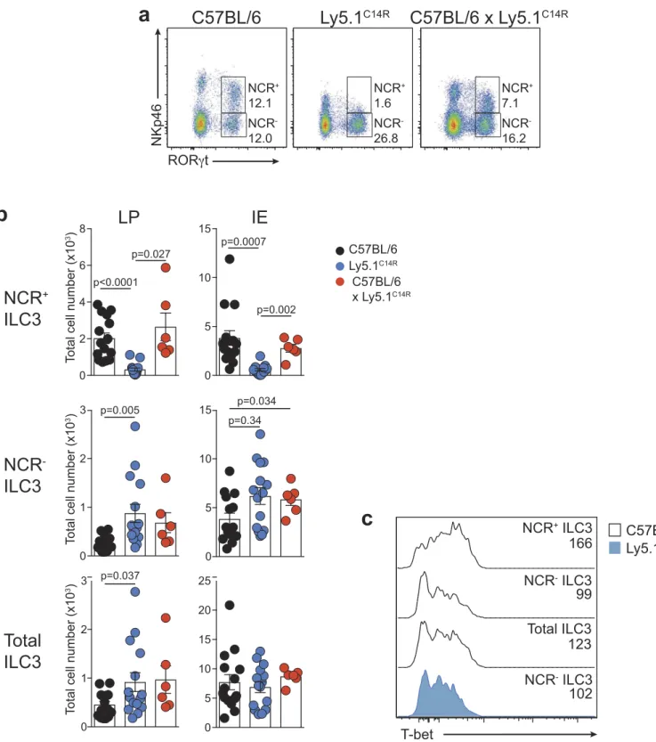
To extend these analyses, we investigated how loss of
NKp46 influenced NCR+ and NCR− ILC3 subsets in the
lamina propria (LP) and intraepithelial (IE) compartments
of the small intestine (Figure 3). As expected, NCR+ ILC3
were not detectable in the Ly5.1C14Rmice (Figure 3A and B)
while ILC2 were present in normal numbers (data not shown). This was accompanied by an accumulation in both
the frequency and number of NCR−ILC3. However, the
over-all number of Rorγt+ILC3 was similar suggesting that loss of
NKp46 did not adversely affect the development of total ILC3 at steady-state. Nevertheless, analyses of T-bet expression
revealed that it was upregulated within the NCR+subset in
C57BL/6 mice as previously reported,25,26 while NCR−ILC3
from Ly5.1C14R mice did not show elevated levels of T-bet
when compared with the NCR−population in C57BL/6 mice
suggesting that NCR+cells fail to develop (Figure 3C).
Ly5.1C14R mice have an altered sensitivity to stimuli in vitro and fail to control tumors in vivo
To gain insight on the functional relevance of the C14R point mutation, the degranulation capacity of total NK cells from
C57BL/6 and Ly5.1C14Rmice was determined by staining for
sur-face CD107a expression following exposure to various stimuli in
vitro. As might be expected, NK cells from NKp46C14Rmice were
significantly less responsive to NKp46 stimulation. When NK cells were cross-linked through NK1.1, they showed a trend towards increased degranulation which was not statistically significant on
the whole NK cell population (Figure 4A), but was statistically
significant when restricted to the immature NK cell subset (SI Appendix, Figure S5), consistent with previous findings on Noé
mice.18Regardless of this point, however, the in vitro anti-tumor
NK cell cytolytic capacity was intact in NK cells from Ly5.1C14R
mice (Figure 4Band SI Appendix, Figure S5). Combined, these
data suggest that NKp46C14RNK cells are functionally competent
and can induce cell lysis through multiple pathways but exhibit defects in vitro when recognition requires engagement of NKp46. These findings suggested that despite the alteration of NKp46
in Ly5.1C14R, NK cells should be competent to protect against in
vivo tumor challenge. To test this point, mice were inoculated with B16F10 melanoma cells which are known to be controlled by NK
cells.27,28While C57BL/6, C57BL/6 × Ly5.1C14Rand Ly5.1 (Jax,
2017) mice were able to largely control tumor growth as measured by the number of primary lung tumors 14 days after tumor
a b ILC1 2.5 ILC1 15.3 NK 95.6 NK 82.2 e d c g C57BL/6 Ly5.1C14R C57BL/6 x Ly5.1C14R ILC1 15.3 NK 82.2 CD49a M2 14.8 M1 63.7 Imm 19.5 M2 17.9 M1 65.8 Imm 15.1 M2 17.1 M1 67.9 Imm 14.1 Eomes C57BL/6 Ly5.1C14R C57BL/6 x Ly5.1C14R KLRG1 CD11b 5.4 94.5 61.9 38.0 15.5 84.5 C57BL/6 Ly5.1C14R C57BL/6 x Ly5.1C14R 0 5 10 15 20 0 1 2 3 0 5 10 15 20 0 5 10 15 Total NK cells (x10 5) Spleen LP IE Thymus Total NK cells (x10 3) Total NK cells (x10 3) Total NK cells (x10 2)
Total cell number (x10
5)
Imm
Imm M1 M2
M1
Total cell number (x10
4)
Total cell number (x10
5)
Total cell number (x10
4)
Total cell number (x10
4) Spleen Total ILC1 (x10 4) Spleen Liver LP IE Thymus Total ILC1 (x10 4 ) Total ILC1 (x10 3) Total ILC1 (x10 3) Total ILC1 (x10 3) M2
Total cell number (x10
5) Liver 0 5 10 15 0 1 2 3 4 0 2 4 6 0 5 10 15 20 0 1 2 3 4 5 0 5 10 15 20 25 0 5 10 15 0 2 4 6 8 0 2 4 6 0 2 4 6 8 p=0.02 p=0.02 p=0.04 p=0.03 p=0.02 p=0.03 p=0.01 p=0.02 0 1 2 3 Liver Total NK cells (x10 5) Spleen Liver SI LP NKp46 SI IE Thymus Ly5.1C14R C57BL/6 f Control NK cells ILC1 654 1833 1209 661 1733 1165 519 2192 1246 390 1499 735 111 677 351 421 1609 722 472 1809 810 372 2136 1087 304 1346 653 107 1103 477 C57BL/6 x Ly5.1C14R C57BL/6 Ly5.1C14R C57BL/6 x Ly5.1C14R C57BL/6 Ly5.1C14R C57BL/6 x Ly5.1C14R Blood NK cells NKp46 156 879 446 9.1 90.9 63.8 36.5 C57BL/6 Ncr1gfp/gfp CD49a TRAIL CD49a TRAIL 0 1 2 3 C57BL/6 Ly5.1C14R C57BL/6 x Ly5.1C14R
Figure 2.Disruption of NK cell homeostasis and maturation of ILC1 and NK cells in Ly5.1C14Rmice. (A) Dot plots showing the frequency of ILC1 and NK cells in the spleen. Data show representative plots gated on live NK1.1+lymphocytes excluding T and B cells in C57BL/6, Ly5.1C14Rand C57BL/6 × Ly5.1C14Rmice. (B) Total number of NK cells in spleen, thymus, liver, small intestine lamina propria (LP) and within the intestinal intraepithelial compartment (IE). Data show the mean ± SEM pooled from three to six independent experiments (n = 6–15 mice/genotype); thymus are pooled from three to five independent experiments (n = 6–12 mice/genotype). P values were calculated using an unpaired two-tailed Student’s t test. (C) FACS plots showing the frequency of immature (Imm, KLRG1−CD11b−), mature 1 (M1, KLRG1−CD11b+) and mature 2 (M2, KLRG1+CD11b+) NK cells in splenic NK1.1+CD3−CD19−NK cells. (D) Total number of Imm, M1 and M2 NK cells in the spleen and liver of C57BL/6, Ly5.1C14Rand C57BL/6 × Ly5.1C14Rmice showing the mean ± SEM pooled from three to six independent experiments (n = 6–15 mice/genotype). P values were calculated using an unpaired two-tailed Student’s t test. (E) Total number of ILC1s in spleen, thymus, liver, small intestine lamina propria (LP) and within the intestinal intraepithelial compartment (IE). Data show the mean ± SEM pooled from three to six independent experiments (n = 6–15 mice/genotype); thymus data are pooled from three to five independent experiments (n = 6–12 mice/genotype). P values were calculated using an unpaired two-tailed Student’s t test. (F) Histograms showing the mean fluorescence intensity of NKp46 in various tissues for both NK cells and ILC1 for wild-type (black solid line), Ly5.1C14R(solid blue) and C57BL/6 × Ly5.1C14R(red solid line). CD3ε+cells were used as a control for NKp46 expression (black dashed line). Data are representative of tissues analyzed in (A–E). (G) Expression of TRAIL on NK1.1+CD49a+CD3−CD19−hepatic ILC1 in C57BL/6, Ly5.1C14Rand C57BL/6 × Ly5.1C14R(results shown in the upper panels) and in C57BL/6 controls compared to Ncr1gfp/gfp(results from experiment shown in the lower panels). Data show representative plots from three to five independent experiments and indicate the the frequency of expression (n = 6–12 mice/genotype).
NKp46C14Rsingle point mutation developed significantly more lung metastases, a condition that was accompanied by respiratory distress. These mice also presented metastases in other organs such as the kidney, liver and bone marrow (SI Appendix, Figure S5 D), a
phenotype which was similar to that observed in Mcl-1fl/flNcr1iCre
mice in which NCR+cells are conditionally ablated29(Figure 4C
andDand SI Appendix, Figure S5D). Thus, although some
func-tion was preserved in the presence of the C14R mutafunc-tion including the ability of NK cells to localize to the lung itself (SI Appendix,
Figure S6), alteration of surface NKp46 in Ly5.1C14Rmice strongly
b
c
Ly5.1C14R C57BL/6 NCR+ ILC3 NCR- ILC3 Total ILC3 NCR- ILC3 166 99 102 123 T-betLP
a
C57BL/6
Ly5.1
C14RC57BL/6 x Ly5.1
C14R NKp46 ROR t NCR+ 12.1 NCR -12.0 NCR+ 1.6 NCR -26.8 NCR+ 7.1 NCR -16.2 8 15IE
Total cell number (x10
3) 0 2 4 6 0 5 10
NCR
+ILC3
p<0.0001 p=0.027 p=0.0007 p=0.002 p=0.034 0 1 2 3 5 10 15 0NCR
-ILC3
Total cell number (x10
3) p=0.005 p=0.34
Total
ILC3
0 1 2 3 0 5 10 15 20 25 Total cell number (x10
3) p=0.037 C57BL/6 Ly5.1C14R C57BL/6 x Ly5.1C14R
Figure 3.Ly5.1C14Rmice have abnormal numbers of ILC3. (A) Dot plots showing the frequency of NCR+and NCR−ILC3 in the LP of the small intestine of C57BL/6, Ly5.1C14Rand C57BL/6 × Ly5.1C14Rmice. Data show representative plots gated on live CD3−CD19−lymphocytes. (B) Enumeration of NCR+, NCR−and total ILC3 for LP and IE in the small intestine. Data showing the mean ± SEM pooled from three to six independent experiments (n = 6–15 mice/genotype). P values were calculated using an unpaired two-tailed Student’s t test. (C) Histograms show the mean fluorescence intensity of intracellular staining for T-bet in ILC3 subsets from the small intestine of C57BL/6 and Ly5.1C14Rmice (n = 6 mice/genotype). P values were calculated using a Student’s t test.
impaired the capacity of NK cells to control tumor development and escape in vivo.
C14R mutation disrupts cellular pathways associated with protein trafficking
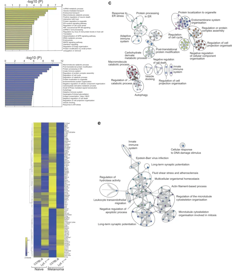
To further dissect the molecular alterations that underpin the changes that occur when the NKp46 expression is altered, we
analysed the transcriptome of naive C57BL/6 and Ly5.1C14R
NK1.1+NK cells by RNA-seq analyses (Figure 5, SI Appendix,
Dataset S2). This revealed that there was a significant enrich-ment in differentially regulated genes involved in intracellular trafficking compartments including the endoplasmic reticu-lum, endosome, ER to Golgi vesicle-mediated transport and
Golgi apparatus in Ly5.1C14Rcells (Figure 5A–C). In addition,
alterations occurred in pathways associated with protein ubi-quitination and transport together with enzymes associated with processing of proteins (eg. GTPase, peptidase activity,
Number of tumors/lungs C57BL/6Ly5.1 C14R 0 100 200 300 >400 Mcl-1 fl/fl NCR iCre/+
c
C57BL/6 Ly5.1C14R Mcl1fl/flNCRiCre/+a
b
+ % CD107a cells % Specific lysis (B16F10) C57BL/6 Ly5.1C14R E:T Ratiod
5:1 10:1 20:1 0 20 40 60 Medium NK1.1 NKp46 YAC1 0 20 40 60 n.s. n.s. n.s. n.s. n.s. n.s. p<0.0001 C57BL/6 Ly5.1C14R C57BL/6 Ly5.1C14R p<0.0001 C57BL/6 x Ly5.1C14R Ly5.1 Jax C57BL/6 x Ly5. 1C14RLy5.1 Jax p<0.0001 p<0.0001Figure 4.Ly5.1C14RNK cells cells show altered sensitivity to stimuli in vitro but fail to control melanoma tumor development in vivo. (A) Degranulation capacity of total NK1.1+NK cells determined by surface CD107a expression in C57BL/6 and Ly5.1C14Rcells. Data show frequencies of CD107a+NK cells ± SEM after coculture with various stimuli. Data shown from 2 independent experiments (n = 5 mice/genotype). (B) Cytolytic activity of C57BL/6 and Ly5.1C14RNK cells sensitized to B16F10 tumor cells. NK cells have been activated overnight with IL-2 (1000 U/ml). Data show the mean lysis ± SEM pooled from three independent experiments (n = 3 mice/ genotype in each experiment). P values were calculated using a Student’s t test. (C) Representative whole mounts of the metastatic burden in the lungs of C57BL/6, Ly5.1C14R, Mcl-1fl/flNcr1iCre, C57BL/6 × Ly5.1C14Rand Ly5.1 (Jax, 2017) mice 14 days after i.v injection of B16F10 melanoma cells. (D) Total tumor burden in the lungs of C57BL/6, Ly5.1C14R, Mcl-1fl/flNcr1iCre, C57BL/6 × Ly5.1C14Rand Ly5.1 (Jax, 2017) mice shown in (C) 14 days after injection of B16F10 melanoma cells. Data show the mean ± SEM of tumor burden pooled from five independent experiments (n = 28–30 mice/genotype). Mcl-1fl/flNcr1iCremice included in a single experiment (n = 5 mice/genotype) while C57BL/6 × Ly5.1C14Rand Ly5.1 (Jax, 2016) mice were included in two experiments (n = 12 mice/genotype). P values were calculated using an unpaired two-tailed Student’s t test.
a
0 1 2 3-log10 (P)4 5 6 7 8ncRNA metabolic process Vesicle-mediated transport Endosomal transport Macromolecule catabolic process Positive regulation of neuron death Interaction with host Protein regulation to organelle ER-nucleus signaling pathway Regulation of cell cycle arrest NIK: noncanonical NF-kB signaling snRNA 3’-end processing
Regulation by virus of viral protein levels in host cell Translation
Desensitization of GPR signaling pathway tRNA metabolic process Endocytosis MAPK signaling pathway Interferon signaling Regulation of Golgi organization Protein modification by small protein conjugation or removal
Macromolecule catabolic process Post-translational protein modification Protein processing in ER Innate Immune system Regulation of protein complex assembly Regulation of cell cycle Regulation of catabolic process Protein localization to organelle Endomembrane system organization Negative regulation of cellular component organization Carbohydrate derivative metabolic process Small GTPase mediated signal transduction Autophagy
Adaptive immune system Asparagine N-linked glycosilation Antigen presentation, class I MHC Negative regulation of cell death Regulation of cell projection organization Vesicle docking Response to ER stress 2 1 0 1 8 6 0 2 4 -log10 (P)
b
Protein localization to organelle
Endomembrane system organisation Regulation or protein complex assembly Regulation of cell projection organisation Negative regulation of cellular component organisation Regulation of cell cycle
c
d
Cellular response to DNA damage stimulus Innateimmune system
Epstein-Barr virus infection
Long-term synaptic potentiation
Regulation of hydrolase activity
Fluid shear stress and atherosclerosis
Actin filament-based process Multicellular organismal homeostasis
Regulation of the microtubule cytoskeleton organisation
Microtubule cytoskeleton organisation involved in mitosis Leukocyte transendothelial
migration
Long-term synaptic potentiation Negative regulation of apoptotic process
e
Protein processing in ER Adaptive immune system Response to ER stress Post-translational protein modification Carbohydrate derivate metabolic process Macromolecule catabolic process Regulation of catabolic process Autophagy Vesicle docking Negative regulation of cell death Regulation of cell projection organisation Innate immune system Naive C57BL/6Ly5.1 C14R C57BL/6Ly5.1 C14R Melanoma S100a6 Itga4 Btg2 H2-Q7 Dbi Pik3ca 1700112E06Rik Cd55 Ccl9 Plk2 Capg Tubb6 Serpine2 Chit1 Hsp90b1 Gpr25 Gm4956 S100a4 Ltbp3 Adgrl4 Rgs2 Ppp1r3b Klrg1 Tnfaip3 Sgk1 Arrdc3 Bhlhe40 Parp3 Entpd1 Lyz2 Rps4l Nfkbiz Rnpep Zfp667 Mlh3 Spp1 Rgs16 Elk3 Atmin Mtmr10 Hip1 Epb41l2 Kcnj8 Ctnna1 Eif3a Chaf1a Nsmf Dcaf4 Map3k5 Wdr62 Dstyk Nfam1 Fes Hic1 Gpc1 Lig1 Avil Cxcr4 Tnni1 Nnt Slc7a5 AA474408 Cd3g B3galt4 Ramp1 Cars 1700030C10Rik Cox6a2 Ctse Wdfy1 Rassf7 Arl4d Zfp672 Tbxa2r Zfp764 Aimp2 Cdk5rap1 Nab2 Csrp1 Trmt61a Steap3 Camk2b COX2 Cxcr3 Rsrp1 AI467606 Kcnk5 Ccdc127 Fam195a Exog -1 Z-Score +1Figure 5.Altered molecular machinery in naïve Ly5.1C14Rmutant NK cells affects antigen processing and protein trafficking pathways. (A, B) Enrichment clusters from genes upregulated and downregulated, respectively, in naïve Ly5.1C14RNK cells compared with C57BL/6 NK cells. (C) Gene ontology (GO) network analysis of significantly reduced gene expression levels in Ly5.1C14RNK cells (shown in B) via Metascape and visualized with Cytoskape (v3.1.2). (D) Heatmap of genes significantly up and downregulated in NK cells responding to B16F10 melanoma tumor cells seven days after challenge presented to show differences in gene expression patterns for C57BL/6 and Ly5.1C14Rmice and the comparative gene expression found in naïve NK cells. (E) Gene ontology (GO) network analysis of significantly reduced gene expression levels in Ly5.1C14RNK cells isolated from day 7 lungs of mice challenged with B16F10 tumor cells. Nodes are coloured by p-value via Metascape and visualized with Cytoskape.
protein kinase). Although changes to NKp46 trafficking may have been expected, consistent with a similar alteration that
occurs in Noé mice,20,21broader changes in protein trafficking
were not anticipated. Exome sequencing has uncovered that a
broad gene set is altered in the Ly5.1C14Rstrain that warrants
further investigation.
To determine how the NKp46C14Rmutation might affect cells
responding to a tumor challenge, we isolated total NK cells from the lungs of mice challenged with B16F10 tumor cells 7 days earlier and subjected them to RNAseq (SI Appendix, Dataset S3). Ninety-one genes were found to be differentially expressed
between C57BL/6 and Ly5.1C14R in NK cells in response to
melanoma (Figure 5D). Genes up regulated in C14R NK cells
were associated with the mitochondrion and cell differentiation, while genes involved in protein processing in the endoplasmic reticulum, transcriptional regulators and cellular organization
were downregulated (Figure 5D and E). However, genes such as
Cxcr3 and Cxcr4, both of which are essential for infiltration and
function of NK cells,30–32and the transcriptional regulator Nab2
were upregulated in Ly5.1C14R. We observed that in Ly5.1C14R
mice, the number of NK cells found in the lung were enriched ~2.1 fold indicating that the failure of tumor control was not driven by the inability to migrate to the lung.
Loss of NKp46 signaling is encoded by the point mutation C14R in Ncr1
It was clear from the analyses above that many more genes than
would be anticipated are altered in Ly5.1C14R mice. Thus, we
could not be completely certain that the effects identified in NKp46 expression in these mice are due solely to the C14R mutation detected in the Ncr1 gene. To determine whether this was indeed the case, we generated mice using the CRISPR/Cas9
technology33 that would carry only the C14R mutation on a
C57BL/6 background. These mice are designated as the NCRB6C14Rstrain and were healthy, viable and bred normally to produce homozygous animals. Analyses of NKp46 surface
expression in C57BL/6 and NCRB6C14R mice showed that the
C14R mutation alone abrogated NKp46 binding phenocopying
the Ly5.1C14R mice for this feature (Figure 6). Similarly, the
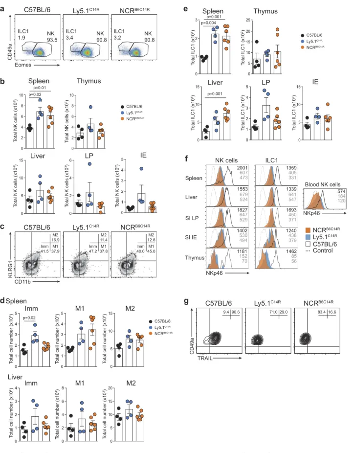
distribution of NK cells in NCRB6C14Rmice were concordant
with the pattern observed in Ly5.1C14Rmice (Figure 6B–D) while
in both NCRB6C14Rand Ly5.1C14RILC1 were enriched in spleen
and liver in four to five week old mice (Figure 6E). We also
observed that NKp46+ILC3 were reduced in NCRB6C14Rsimilar
to that observed in Ly5.1C14Rmice. Notably across all organs
examined, the surface expression of NKp46 was strongly reduced in NK cells and ILC1 and in blood NK cells (Figure 6F). This correlated with a strong reduction in TRAIL expression when C57BL/6 mice expressed the C14R mutation demonstrating that an alteration in NKp46 which affects the stability of the expression at the surface of the cell is sufficient to alter the induction of TRAIL in hepatic ILC1.
Discussion
Spontaneous mutations occur in eukaryotes at a rate of 0.1–
100 per genome per sexual generation. In mice, nearly 5,000 spontaneous and induced mouse mutant alleles with clinically
relevant phenotypes have been described in the Mouse Genome Informatics database and only about one third of
these have characterized phenotypes.34 In many cases,
how-ever, identification of these sequence alterations has served as a rich source of animal models for human genetic diseases.
Although the Ly5.1 (CD45.1) line should differ from its
‘wild-type’ counterpart by just five amino acids within the extracellular
domain,35 it is now clear that the B6.SJL-Ptprca Pepcb/BoyJ
derived from Jax differs in many genes from the C57BL/6 line
spanning ~40Mb and ~300 genes.36 A single amino acid
accounts for the difference between the CD45.1 and CD45.2
congenic markers that define these two strains36and this
differ-ence has formed the basis for their use in extensive tracking experiments. These previously unappreciated alterations in the genome have most likely impacted experimental interpretations. We now add to this list the detection of a spontaneous mutation in the Ncr1 gene that has affected multiple mouse colonies in geographically distinct locations. This mutation may have arisen independently, but seems most likely to have arisen from an individual breeding founder from the Jax. As the C14R mutation of the Ncr1 gene remains undetected in heterozygous animals, this mutation has inadvertently been retained undetected long term in parent colonies. Using whole exome sequencing we identified a spontaneous single autosomal recessive mutation in the Ncr1 gene that significantly altered the expression of the NKp46 surface protein by interfering with its export to the cell surface. Subsequently, we generated C57BL/6 mice in which we introduced the C14R mutation in the Ncr1 gene negating the
effect of other genes altered in the Ly5.1C14Rstrain. This directly
confirmed that the change in NKp46 expression was solely driven by this mutation and will allow this specific mutation to be investigated in detail in future studies.
The C14R mutation in the Ncr1 gene of the mutant Ly5.1 lines occurred just proximal to the previously reported W32R mutation in the signal peptide which can typically affect the synthesis and/or secretion of a
protein.-18,21 We show that expression of Ncr1 mRNA was similar
in NK cells of both wild-type, Ly5.1C14R mouse strains
suggesting that the gene is effectively transcribed but that alterations occur after this stage. The Noé strain also showed poor cell surface expression of NKp46, and like
the NKp46C14R NK cells, displayed no change in mRNA
expression.18 This was supported by analysis of transduced
293T cells where intracellular NKp46C14R and NKp46
expression were similar, but cell surface expression was
strongly impaired in NKp46C14R. Genomic analyses
revealed disruption in intracellular trafficking although a broader group of genes involved in transcriptional regula-tion and protein processing in the ER, endosome, Golgi apparatus and cellular organization were detected suggest-ing other potential defects in this mouse line. Collectively, however, our data suggest that in the mutants, NKp46 could be expressed on the cell surface but is unstable posing the possibility that an altered signaling pathway could be triggered by the constant internalization of NKp46. It also implies that the stability and/or level of NKp46 expression is important in signaling and prompts further dissection of the molecular control of NKp46 that appears to impact on ILC differentiation.
0 1 2 3 4 0 2 4 6 8 0 5 10 15 20 0 1 2 3 4 5 0 1 2 3 4 5 0 5 10 15 0 5 10 15 0 2 4 6 0 1 2 3 4 5 0 2 4 6 8 10 0 2 4 6 8 10 0 5 10 15 0 1 2 3 4 5 0 5 10 15 0 5 10 15 20 25 0 1 2 3 a b ILC1 1.9 ILC1 3.4 NK 93.5 NK 90.8 e d c g C57BL/6 Ly5.1C14R ILC1 3.2 NK90.8 CD49a M2 11.4 M1 37.8 Imm 47.2 M2 12.8 M1 45.0 Imm 40.0 M2 16.9 M1 37.9 Imm 41.5 Eomes C57BL/6 Ly5.1C14R KLRG1 CD11b 9.4 90.6 71.0 29.0 83.4 16.6 C57BL/6 Ly5.1C14R NCRB6C14R CD49a TRAIL T otal NK cells (x10 5) Spleen LP IE Thymus T otal NK cells (x10 3) T otal NK cells (x10 3) T otal NK cells (x10 2) T
otal cell number (x10
5)
Imm
Imm M1 M2
M1
T
otal cell number (x10
4)
T
otal cell number (x10
5)
T
otal cell number (x10
4)
T
otal cell number (x10
3) Spleen T otal ILC1 (x10 4) Spleen Liver LP IE Thymus T otal ILC1 (x10 4) T otal ILC1 (x10 2) T otal ILC1 (x10 3) T otal ILC1 (x10 2) M2 T
otal cell number (x10
4) Liver p=0.001 Liver T otal NK cells (x10 4) Spleen Liver SI LP NKp46 SI IE Thymus Ly5.1C14R C57BL/6 f Control NK cells ILC1 607 2001 473 679 1553 524 647 1827 529 530 1402 494 152 1181 70 405 1359 331 641 1339 547 450 1693 371 438 1240 379 85 1462 56 C57BL/6 Ly5.1C14R Blood NK cells NKp46 184 574 120 NCRB6C14R NCRB6C14R NCRB6C14R NCRB6C14R p=0.004 p=0.001 C57BL/6 Ly5.1C14R NCRB6C14R p=0.01 p=0.02 p=0.02 C57BL/6 Ly5.1C14R NCRB6C14R
Figure 6.Loss of NKp46 surface expression depends on the C14R mutation in the Ncr1 gene. (A) Dot plots showing the frequency of ILC1 and NK cells in the spleen. Data show representative plots gated on live NK1.1+lymphocytes excluding T and B cells. (B) Total number of NK cells in spleen, thymus, liver, small intestine lamina propria (LP) and within the intestinal intraepithelial compartment (IE). Data are pooled from two independent experiments and show the mean ± SEM (n = 4–6 mice/genotype). P values were calculated using an unpaired two-tailed Student’s t test. (C) FACS plots showing the frequency of immature (Imm, KLRG1−CD11b−), mature 1 (M1, KLRG1−CD11b+) and mature 2 (M2, KLRG1+CD11b+) NK cells in splenic NK1.1+CD3−CD19−NK cells. (D) Total number of Imm, M1 and M2 NK cells in the spleen and liver of C57BL/6, Ly5.1C14Rand NCRB6C14R mice showing the mean ± SEM pooled from two experiments (n = 4–6 mice/genotype). P values were calculated using an unpaired two-tailed Student’s t test. (E) Total number of ILC1s in spleen, thymus, liver, small intestine lamina propria (LP) and within the intestinal intraepithelial compartment (IE). Data show the mean ± SEM pooled from two independent experiments (n = 4–6 mice/genotype). P values were calculated using an unpaired two-tailed Student’s t test. (F) Histograms showing the mean fluorescence intensity of NKp46 in various tissues for both NK cells and ILC1 for wild-type (black solid line), Ly5.1C14R(solid blue) and NCRB6C14R(solid orange). CD3ε+cells were used as a control for NKp46 expression (black dashed line). Data are representative of tissues analyzed in (A-E). (G) Expression of TRAIL on NK1.1+CD49a+CD3−CD19−hepatic ILC1 in C57BL/6, Ly5.1C14Rand NCRB6C14R. Data show representative plots from two independent experiments and indicate the the frequency of expression (n = 4–6 mice/genotype).
In the Ly5.1C14R mice, we observed that NK cell develop-ment in terms of cellularity was not impaired in the absence of NKp46. Neverthless, we show that the differentiation of NK
cells and ILC1 were altered in both Ly5.1C14Rand Noé mice.
NK cell subsets were skewed towards an accumulation of less
mature NK cells and NCR− ILC3s. The lack of NKp46
induced in Ncr1gfp/gfp mice has also recently been shown to
be associated with an impaired NK cell maturation in
response to mouse cytomegalovirus infection (MCMV).37
This was associated with changes in CXCR3-driven migration of NK cells to the regional lymph nodes which can affect NK-dendritic cell crosstalk necessary for priming MCMV-specific
CD4+T cells.38In our study, we found that in contrast to this
observation, Cxcr3 mRNA was upregulated, consistent with the higher expression of CXCR3 on the less mature
CD27highM1 NK cells.39,40 Indeed, the number of NK cells
that localized in the lung in response to a B16F10 tumor cells which are likely to express the ligand for NKp46 was not
reduced in Ly5.1C14R mice (SI Appendix, Figure S4) and the
cytolytic machinery was intact. Ncr1gfp/gfp mice appear to
exhibit slightly reduced IFN-γ responses to NK1.1 stimulation
in vitro and murine cytomegalovirus infection.19,37
Stimulation through the NKp46 receptor has been shown to
drive IFN-γ production. This NK cell-driven IFN-γ
produc-tion has recently been shown to induce the extracellular matrix protein fibronectin 1 that could modulate tumor
formation.41 In contrast, diminished NKp46 appears to
reg-ulate phosphorylation of Dok-1 to influence IFN-γ
produc-tion and pathogen protecproduc-tion.42 In our in vitro analyses, a
reduction in IFN-γ was not observed in our mutant mice
although signaling directly through NKp46 was strongly impaired. Thus, it appears that the complete loss of function
of the Ncr1 allele in Ncr1gfp/gfp mice differs from the
disrup-tion induced by the introducdisrup-tion of a point mutadisrup-tion in the gene.
In addition to changes in NK cell differentiation, we observed that diminished NKp46 expression caused alterations in other ILCs. Although loss of NKp46 did not impair ILC1 numbers, we show that the apoptosis-inducing ligand TRAIL, which can be
induced by TGF-β43and through viral binding to NKp4644was
lacking implying that NKp46 may play a role in regulating TRAIL expression. NKp46 is also a key marker of one of the
three subsets of ILC3.18In Ly5.1C14Rmice, as expected NCR+
ILC3 were not detectable via NKp46 expression but we also did not see enrichment of expression of T-bet which is normally
associated with induction of this subset,25,26 although ILC3
development itself was not affected implying that activation of NKp46 is important for the transcriptional program of these cells. This is supported by cell fate mapping experiments that suggest that instability of NKp46 expression in ILC3s found in the intestine reflects a major role of this receptor in tuning the very dynamic environmental signals encountered that drive
ILC3 plasticity.45,46 Loss of Ncr1 has previously been shown
not to affect ILC3 development with Ncr1gfp/gfpRag2−/- mice
showing survival and clinical scores similar to control mice when challenged with the enteric infection Citrobacter
rodentium.47In this setting, NKp46 expression did not appear
to be essential for protection.
In summary, we have identified a commonly used mouse
line, the Ly5.1C14R, that carries a point mutation in the Ncr1
gene together with a large number of previously unknown
genes. The NCRB6C14R strain confirmed that the C14R Ncr1
mutation directly impaired the expression of surface NKp46 and alters differentiation of NK cells with increased accumu-lation of immature stages. In addition, increased numbers of ILC1 were found and these cells lacked TRAIL expression
while NCR+ILC3 were not detected. Thus, although reduced
NKp46 expression did not disrupt either NK cell or ILC development, it exerted an important impact on normal dif-ferentiation, maturation and activation of these lineages.
Materials and methods Mice
C57BL/6 and B6.SJL-Ptprca Pepcb/BoyJ (Ly5.1, originally obtained from The Jackson Laboratory), C57BL/6 × B6.SJL-PtprcaPepcb/BoyJ (C57BL/6 × Ly5.1 and B6.SJL-Ptprca Pepcb/ BoyJAX (imported 2016 and sequence verified) mice were bred and maintained in-house at either the Walter and Eliza Hall Institute of Medical Research (WEHI, Australia) or the University Hospital Erlangen (Germany) under specific pathogen-free conditions. Mice used in Frankfurt were obtained from the Preclinical Experimental Animal Center (PETZ) of the Friedrich Alexander University (FAU)
Erlangen-Nürnberg, Germany. Ncr1gfp/gfp, Noé mice and
Mcl1fl/flNcr1iCre mice have been previously described.17,18,29
CD45.1STEMmice36 were bred at the Massachusetts General
Hospital animal house (Boston). Male and female mice were used between the ages of six to eight weeks and were age
matched unless otherwise stated. NCRB6C14R mice were used
at four to five weeks of age. Animal experiments followed the National Health and Medical Research Council (NHMRC) Code of Practice for the Care and Use of Animals for Scientific Purposes guidelines and were approved by the Animal Ethics Committee of the respective institutions.
Generation of NCRB6C14Rmice
A new C57BL/6 mouse strain that carried the C14R mutation
in the Ncr1 gene (NCRB6C14R) was generated using CRISPR/
Cas9 as previously described.33 Briefly, one sgRNA of the
sequence TAGGGCTATGTCTGAGCCAG (10ng/μl), an
oligo donor of the sequence (gttgaatcaagagcagattggggggag acagcatgccattaaccctgttttctagGGCTACGACTGAGCCAGCGT- ATCAACACTGAAAAGGgtaagtccttccctcgaagtctcagggttgttctt-atgggttca; 10ng/ul) and Cas9 mRNA (5ng/μl) were injected
into the cytosol of C57BL/6J zygotes to generate NCRB6C14R
point mutant mice.
Generation of mixed bone marrow chimeric mice
Mixed bone marrow chimeric mice were generated by
recon-stituting lethally irradiated (2 x 5.5Gy γ-irradiation, 137Cs
source) with a mixture (1:1 ratio; total 10 × 106 bone
mar-row cells/mouse) of Ly5.1C14R (CD45.1+/+) and C57BL/6
× Ly5.1C14R (CD45.2+CD45.1+) bone marrow. Mice were
analysed 6–8 wk after reconstitution.
Isolation of intestinal intraepithelial and lamina propria lymphocytes
Intestinal LPL were isolated from the intestine as previously
described.26,48
Cell isolation and flow cytometric analyses
Single cell suspensions were generated by forcing organs
through 70μm sieves. Lymphocytes were isolated from liver
using a 33% isotonic Percoll (Amersham Pharmacia Biotech) gradient centrifuged at 2000 rpm for 13 min at ambient temperature. Single cell suspensions were blocked with PBS
containing 5μg/ml anti-CD16/CD32 (2.4G2) and stained for
20–30 min on ice with fluorophore-conjugated antibodies,
unless stated otherwise. For analysis of surface molecules, cells were stained with the following antibodies: anti-CD11b
(M1/70) (BD Biosciences, or Miltenyi), anti-CD3ε (145-2C11)
and anti-CD19 (1D3 and 6D5) (BioLegend), anti-KLRG1 (2F1), and NK1.1 (PK136), CD27 (LG.3A11),
anti-CD45.1 (A20), anti-CD45.2 (104), anti-CD49a (HMα1),
anti-B220 (RA3-6B2) (all from BD Biosciences), anti-NKp46 (29A1.4, eBioscience) and anti-CD49b (DX5, generated at The Walter and Eliza Hall Institute hybridoma facility). Live cells were identified by exclusion after staining with a fixable viability dye (BD Biosciences or BioLegend). Intracellular staining was performed using the Transcription Factor Staining Buffer Set (eBioscience) and monoclonal antibodies to EOMES (Dan11mag), GATA3 (TWAJ) (both from
eBioscience) and RORγt (Q31-378, BD Biosciences). For
IFN-γ detection, the monoclonal antibody XMG1.2 (BD
Pharmingen) was used. Flow cytometry was performed using a LSRFortessa X-20 or Canto10C (BD Biosciences) and FlowJo analysis software (Treestar).
Murine melanoma cells and pulmonary metastasis
B16F10 murine melanoma cells (mycoplasma negative) were maintained in complete DMEM (Gibco) with 10% heat-inac-tivated FCS, 2 mM of L-Glutamine, 100 U/mL penicillin, and
100μg/mL streptomycin. 2.5 × 10⁵B16F10 cells were injected
into the tail vein of recipient mice. Fourteen days after injec-tion, the lungs, liver, long bones and kidneys were harvested and fixed in Bouin’s solution. B16F10 metastases were
counted using a dissecting microscope.49
Exome sequencing
To identify the genetic mutation responsible for the reduced
NKp46 expression in the Ly5.1C14R strain we performed
exome sequencing on two mice from this strain and the
C57BL/6 × Ly5.1C14R mice which expressed normal NKp46
protein levels (SI Appendix, Methods).
Sanger sequencing
To further confirm the Ncr1 mutation identified by exome sequencing we performed Sanger sequencing on C57BL/6, C57BL/6 × Ly5.1 and WEHI B6.SJL-Ptprca Pepcb/BoyJ mice (SI Appendix, Methods).
Quantitative PCR
RNA was isolated from sorted NK cells and NKp46 expres-sion was determined as described in SI Appendix, Methods.
Cloning
NKp46 cDNA were generated from RNA isolated from
purified NK cells of either C57BL/6 or Ly5.1C14R mice by
RT-PCR and cloned into the plasmid RSV.5neo-FlagHis which provides C-terminal FLAG- and hexahistidine-tags.
Primers: NKp46 cDNA forward 5ʹ-GACTCCGCG
GGCCACCATGCTGCCAACACTCACTG-3ʹ, NKp46 cDNA reverse 5ʹ-AGTCCTCGAG CAAGGCCCCAGGAGTTGC-3ʹ.
Transfection and flow cytometric analysis
293T cells were cultured in Dulbecco’s Modified Eagle’s
Medium and were transfected with the appropriate
RSV.5neo expression vectors containing NKp46 WT or mutant cDNA with Flag- and hexahistidine-tag using Applifect (AppliChem) (SI Appendix, Methods). Cells were fixed and permeabilized with BD Cytofix/Cytoperm (BD Biosciences) and subsequently stained with appropriate anti-bodies for 20 min at 4°C. Antianti-bodies used: FLAG-tag mAb M2 (Sigma), anti-NKp46-PE mAb 29A1.4 (BioLegend), rat IgG2a-PE isotype control RTK2758 (BioLegend), mouse IgG1 isotype control N1G9, APC-conjugated F(ab)2-frag-ments of goat anti-mouse IgG (Jackson ImmunoResearch). Flow cytometry analysis was performed with a FACS Canto II (BD Biosciences, Heidelberg, Germany) and data analyzed using FlowJo (Tree Star, Ashland, OH).
Immunofluorescence staining
NK cells were isolated from the spleens of Ly5.1C14R and
C57BL/6 mice and centrifuged on lysine coated slides, then fixed and stained with goat anti-mouse NKp46 antibody (R&D Systems, AF2225, 1:100) and rabbit anti-mouse PDI (Cell Signaling Technology, 3501S, 1:100) overnight, followed by AlexaFluor488-conjugated donkey anti-goat IgG (H + L) (ThermoFisher, A-11055, 1:250) and AlexFluor546-conjugated donkey anti-rabbit IgG (H + L) (ThermoFisher, A-10040,
1:200). Images were analyzed using LAS AF (Leica
Application Suite – Advanced Fluorescence) software (SI
Appendix, Methods).
NK cell stimulation
Plates were coated overnight at 4°C with antibodies against
NK1.1 and NKp46 (10μg/ml in PBS). 1.5 × 10⁶ splenocytes
containing 10% heat-inactivated FCS, 2mM of L-Glutamine,
100 U/mL penicillin, and 100μg/mL streptomycin in the
pre-sence of Golgi Stop and soluble CD107a for 4h at 37°C. The
YAC1 stimulation condition was obtained by adding 0.25 106
YAC1 cells to the well. Cells were subsequently stained and analyzed by flow cytometry with an LSR Fortessa and data analyzed using FlowJo (Tree Star, Ashland, OH).
Cytotoxicity assay
Europium release assay was used to assess the lytic capacity of murine NK cells as described in SI Appendix, Methods.
RNA-seq analysis
RNA-seq analyses was performed on NK cells isolated from
the lungs of Ly5.1C14R and C57BL/6 mice seven days after
challenge with B16F10 melanoma tumor cells. These strains were sourced from the sourced from the WEHI colonies. Samples were sequenced on an Illumina NextSeq 500 gener-ating 75 bp paired end reads. Two biological replicates of each cell type were sequenced. RNA-seq Reads were aligned to the GRCm38/mm10 build of the Mus musculus genome using the
Subread aligner.50Only uniquely mapped reads were retained.
Genewise counts were obtained using featureCounts.51Reads
overlapping exons in annotation build 38.1 of NCBI RefSeq database were included. Genes were excluded from down-stream analysis if they failed to achieve a CPM (counts per million mapped reads) value of greater than 0.5 in at least two libraries. Counts were converted to log2 counts per million, quantile normalized and precision weighted with the voom
function of the limma package.52,53A linear model was fitted
to each gene, and empirical Bayes moderated t-statistics were
used to assess differences in expression.54 A false discovery
rate cut-off of 0.15 was applied for calling differentially expressed genes. Gene Ontology and gene enrichment
ana-lyses were performed using Metascape (http://metascape.org).
Raw data files for the RNA sequencing have been deposited in the NCBI Gene Expression Omnibus under accession number GSE113030.
Highlights
● A spontaneous mutation in the signal sequence of the Ncr1 gene arose in the CD45.1 mouse strain
● Ncr1 C14R mutation impairs NKp46 surface expression in NK cells, ILC1 and ILC3
● Destabilization of Ncr1 by the C14R mutation results in accumulation of NKp46 in the endoplasmic reticulum ● Loss of stable NKp46 expression impairs the maturation
of NKp46+ILCs and impairs tumor control
Acknowledgments
We thank M. Camilleri, J. Janssen, S. Cree, C. O’Brien and A. Abendroth and K. Stein for expert technical support and Dr J. Babon for helpful discussions, and Prof. C. Bogdan for providing mice and helpful discus-sion of results. This study was made possible through Victorian State
Government Operational Infrastructure Support and Australian Government NHMRC Independent Research Institute Infrastructure Support Scheme (Walter and Eliza Hall Institute of Medical Research) and the LOEWE Center for Cell and Gene therapy (to ST and EU), Frankfurt, funded by the Hessian Ministry of Higher Education, Germany (III L 4-518/17.004).
Funding
This work was supported by the National Health and Medical Research Council Australia [1054925]; and the National Health and Medical Research Council Australia [1124907]. E.U. lab has been supported by the LOEWE Center for Cell and Gene Therapy Germany [III L 4-518/ 17.004]. S.U. lab is supported by funding form the European Research Council (ERC) under the European Union’s Horizon 2020 research and innovation programme under grant agreement No 648768; from the ANR (No ANR-14-CE14-0009-01) and from the fondation ARC (No PGA120140200817). E.V. lab is supported by funding form the European Research Council (ERC) under the European Union’s Horizon 2020 research and innovation programme (TILC, grant agreement N° 694502); the Agence Nationale de la Recherche; Equipe Labellisée“La Ligue,” Ligue Nationale contre le Cancer, MSDAvenir, Innate Pharma and institutional grants to the CIML (INSERM, CNRS, and Aix-Marseille University) and to Marseille Immunopôle. S.T. received a GO-IN post-doc fellowship at the University Frankfurt Germany [PCOFUND-GA-2011-291776] and by the Madeleine Schickedanz-KinderKrebs-Stiftung; W.S. is supported by a Walter and Eliza Hall Institute Centenary Fellowship sponsored by CSL Limited. S.N.W. was supported by a Walter and Eliza Hall Trust Centenary Fellowship.
Declaration of interest statement
E.V. is cofounder and employee of Innate Pharma.
Authors Contributions
M.E.F., S.W., A.G., T.Z., T.W., J.K., A.S., A.M., F.G., S.U., E.N-M., B.R., F.S., F.F., S.T., M.A.F., J.G., S.N.W., K.L., A.G.C. and F.F.A performed experiments and analysed data; M.H. and A.K. helped create the NCRB6C14Rmice. T.W., U.S., J.K., A.S., N.D.H., F.E.M, D.T.S., E.U., E. V. and G.T.B. provided intellectual input and tools or reagents; A.G., Y.L. and W.S. performed bioinformatics analyses. G.T.B. and E.U. conceived the ideas and interpreted data; G.T.B, E.U., F.F.A. and S.T. wrote the paper and edited it with the assistance of the other authors.
ORCID
Francisca F. Almeida http://orcid.org/0000-0001-6632-7610
Sophie Ugolini http://orcid.org/0000-0003-4041-0103
Alexander Steinle http://orcid.org/0000-0001-5081-8503
Eric Vivier http://orcid.org/0000-0001-7022-8287
Gabrielle T. Belz http://orcid.org/0000-0002-9660-9587
Evelyn Ullrich http://orcid.org/0000-0001-8530-1192
References
1. Lam VC, Lanier LL. NK cells in host responses to viral infections. Curr Opin Immunol.2017;44:43–51. doi:10.1016/j.coi.2016.11.003. 2. Vivier E, Tomasello E, Baratin M, Walzer T, Ugolini S. Functions of natural killer cells. Nat Immunol.2008;9(5):503–510. doi:10.1038/ ni1582.
3. Seidel E, Glasner A, Mandelboim O. Virus-mediated inhibition of natural cytotoxicity receptor recognition. Cell Mol Life Sci.
2012;69(23):3911–3920. doi:10.1007/s00018-012-1001-x.
4. Thielens A, Vivier E, Romagne F. NK cell MHC class I specific receptors (KIR): from biology to clinical intervention. Curr Opin Immunol.2012;24(2):239–245. doi:10.1016/j.coi.2012.01.001.
5. Satoh-Takayama N, et al.. Microbial flora drives interleukin 22 production in intestinal NKp46+ cells that provide innate mucosal immune defense. Immunity. 2008;29(6):958–970. doi:10.1016/j. immuni.2008.11.001.
6. Luci C, et al.. Influence of the transcription factor RORgammat on the development of NKp46+ cell populations in gut and skin. Nat Immunol.2009;10(1):75–82. doi:10.1038/ni.1681.
7. Sanos SL, Bui VL, Mortha A, Oberle K, Heners C, Johner C, Diefenbach A. RORγt and commensal microflora are required for the differentiation of mucosal interleukin IL22-producion NKp46+ cells. Nature Immunology.2009;10(1):83–91. doi:10.1038/ni.1684. 8. Narni-Mancinelli E, et al.. Complement factor P is a ligand for the
natural killer cell-activating receptor NKp46. Sci Immunol.
2017;2:10. doi:10.1126/sciimmunol.aam9628.
9. Vankayalapati R, et al.. The NKp46 receptor contributes to NK cell lysis of mononuclear phagocytes infected with an intracellular bacterium. J Immunol. 2002;168(7):3451–3457. doi:10.4049/ jimmunol.168.7.3451.
10. Mandelboim O, et al.. Recognition of haemagglutinins on virus-infected cells by NKp46 activates lysis by human NK cells. Nature.
2001;409(6823):1055–1060. doi:10.1038/35059110.
11. Draghi M, et al.. NKp46 and NKG2D recognition of infected den-dritic cells is necessary for NK cell activation in the human response to influenza infection. J Immunol. 2007;178(5):2688–2698. doi:10.4049/jimmunol.178.5.2688.
12. Arnon TI, et al.. Recognition of viral hemagglutinins by NKp44 but not by NKp30. Eur J Immunol.2001;31(9):2680–2689. doi:10.1002/ 1521-4141(200109)31:9<2680::AID-IMMU2680>3.0.CO;2-A. 13. Jarahian M, et al.. Activation of natural killer cells by newcastle
disease virus hemagglutinin-neuraminidase. J Virol. 2009;83 (16):8108–8121. doi:10.1128/JVI.00211-09.
14. Jarahian M, et al.. Modulation of NKp30- and NKp46-mediated natural killer cell responses by poxviral hemagglutinin. PLoS Pathog.2011;7(8):e1002195. doi:10.1371/journal.ppat.1002195. 15. Chaushu S, et al.. Direct recognition of Fusobacterium nucleatum
by the NK cell natural cytotoxicity receptor NKp46 aggravates periodontal disease. PLoS Pathog. 2012;8(3):e1002601. doi:10.1371/journal.ppat.1002601.
16. Vitenshtein A, et al.. NK cell recognition of candida glabrata through binding of NKp46 and NCR1 to fungal ligands Epa1, Epa6, and Epa7. Cell Host Microbe. 2016;20(4):527–534. doi:10.1016/j.chom.2016.09.008.
17. Gazit R, et al.. Lethal influenza infection in the absence of the natural killer cell receptor gene Ncr1. Nat Immunol. 2006;7 (5):517–523. doi:10.1038/ni1322.
18. Narni-Mancinelli E, et al.. Tuning of natural killer cell reactivity by NKp46 and Helios calibrates T cell responses. Science.
2012;335(6066):344–348. doi:10.1126/science.1215621.
19. Sheppard S, et al.. Characterization of a novel NKG2D and NKp46 double-mutant mouse reveals subtle variations in the NK cell repertoire. Blood. 2013;121(25):5025–5033. doi: 10.1182/blood-2012-12-471607.
20. Glasner A, et al.. Expression, function, and molecular properties of the killer receptor Ncr1-Noe. J Immunol.2015;195(8):3959–3969. doi:10.4049/jimmunol.1501234.
21. Glasner A, Isaacson B, Mandelboim O. Expression and function of NKp46 W32R: the human homologous protein of mouse NKp46 W32R (Noe). Sci Rep.2017;7:40944. doi:10.1038/srep40944. 22. Walzer T, et al.. Identification, activation, and selective in vivo
ablation of mouse NK cells via NKp46. Proc Natl Acad Sci U S A.
2007;104(9):3384–3389. doi:10.1073/pnas.0609692104.
23. Gordon SM, et al.. The transcription factors T-bet and Eomes control key checkpoints of natural killer cell maturation. Immunity.2012;36(1):55–67. doi:10.1016/j.immuni.2011.11.016. 24. Seillet C, et al.. Differential requirement for Nfil3 during NK cell
development. J Immunol. 2014;192(6):2667–2676. doi:10.4049/ jimmunol.1302605.
25. Klose CS, et al.. A T-bet gradient controls the fate and function of CCR6-RORgammat+ innate lymphoid cells. Nature. 2013;494 (7436):261–265. doi:10.1038/nature11813.
26. Rankin LC, et al.. The transcription factor T-bet is essential for the development of NKp46(+) innate lymphocytes via the Notch pathway. Nat Immunol. 2013;14(4):389–395. doi:10.1038/ ni.2545.
27. Grundy MA, Zhang T, Sentman CL. NK cells rapidly remove B16F10 tumor cells in a perforin and interferon-gamma indepen-dent manner in vivo. Cancer Immunol Immunother. 2007;56 (8):1153–1161. doi:10.1007/s00262-006-0264-1.
28. Smyth MJ, et al.. Differential tumor surveillance by natural killer (NK) and NKT cells. J Exp Med.2000;191(4):661–668. doi:10.1084/ jem.191.4.661.
29. Sathe P, et al.. Innate immunodeficiency following genetic abla-tion of Mcl1 in natural killer cells. Nat Commun. 2014;5:4539. doi:10.1038/ncomms5539.
30. Noda M, et al.. CXCL12-CXCR4 chemokine signaling is essential for NK-cell development in adult mice. Blood. 2011;117(2):451–458. doi:10.1182/blood-2010-04-277897.
31. Bernardini G, Antonangeli F, Bonanni V, Santoni A. Dysregulation of chemokine/chemokine receptor axes and NK cell tissue localization during diseases. Front Immunol.
2016;7:402. doi:10.3389/fimmu.2016.00402.
32. Pak-Wittel MA, Yang L, Sojka DK, Rivenbark JG, Yokoyama WM. Interferon-gamma mediates chemokine-dependent recruitment of natural killer cells during viral infection. Proc Natl Acad Sci U S A. 2013;110(1):E50–59. doi:10.1073/ pnas.1220456110.
33. Wang H, et al.. One-step generation of mice carrying mutations in multiple genes by CRISPR/Cas-mediated genome engineering. Cell.2013;153(4):910–918. doi:10.1016/j.cell.2013.04.025. 34. Blake JA, et al.. The Mouse Genome Database (MGD): premier
model organism resource for mammalian genomics and genetics. Nucleic Acids Res. 2011;39(Database issue):D842–848. doi:10.1093/nar/gkq1008.
35. Zebedee SL, Barritt DS, Raschke WC. Comparison of mouse Ly5a and Ly5b leucocyte common antigen alleles. Dev Immunol.1991;1 (4):243–254. doi:10.1155/1991/52686.
36. Mercier FE, Sykes DB, Scadden DT. Single targeted exon muta-tion creates a true congenic mouse for competitive hematopoietic stem cell transplantation: the C57BL/6-CD45.1(STEM) mouse. Stem Cell Reports. 2016;6(6):985–992. doi:10.1016/j. stemcr.2016.04.010.
37. Miletic A, et al.. NCR1-deficiency diminishes the generation of pro-tective murine cytomegalovirus antibodies by limiting follicular helper T-cell maturation. Eur J Immunol.2017. doi:10.1002/eji.201646763. 38. Mandaric S, et al.. IL-10 suppression of NK/DC crosstalk leads to
poor priming of MCMV-specific CD4 T cells and prolonged MCMV persistence. PLoS Pathog. 2012;8(8):e1002846. doi:10.1371/journal.ppat.1002846.
39. Hayakawa Y, Smyth MJ. CD27 dissects mature NK cells into two subsets with distinct responsiveness and migratory capacity. J Immunol. 2006;176(3):1517–1524. doi:10.4049/ jimmunol.176.3.1517.
40. Meinhardt K, et al.. Identification and characterization of the specific murine NK cell subset supporting
graft-versus-leuke-mia- and reducing graft-versus-host-effects.
Oncoimmunology. 2015;4(1):e981483. doi:10.4161/ 2162402X.2014.981483.
41. Glasner A, et al.. NKp46 receptor-mediated interferon-g produc-tion by natural killer cells increases fibronectin 1 to alter tumor architecture and control metastasis. Immunity. 2018;48:1–13. doi:10.1016/j.immuni.2017.12.007.
42. Jang Y, et al.. Cutting edge: check your mice-A point muta-tion in the Ncr1 locus identified in CD45.1 congenic mice with consequences in mouse susceptibility to infection. J