HAL Id: tel-03209463
https://tel.archives-ouvertes.fr/tel-03209463
Submitted on 27 Apr 2021
HAL is a multi-disciplinary open access
archive for the deposit and dissemination of sci-entific research documents, whether they are pub-lished or not. The documents may come from teaching and research institutions in France or abroad, or from public or private research centers.
L’archive ouverte pluridisciplinaire HAL, est destinée au dépôt et à la diffusion de documents scientifiques de niveau recherche, publiés ou non, émanant des établissements d’enseignement et de recherche français ou étrangers, des laboratoires publics ou privés.
La spectroscopie de résonance magnétique nucléaire à
l’état solide : un outil pour la caractérisation des
systèmes poreux de délivrance de médicaments
Marianna Porcino
To cite this version:
Marianna Porcino. La spectroscopie de résonance magnétique nucléaire à l’état solide : un outil pour la caractérisation des systèmes poreux de délivrance de médicaments. Autre. Université d’Orléans, 2020. Français. �NNT : 2020ORLE3054�. �tel-03209463�
UNIVERSITÉ D’ORLÉANS
ÉCOLE DOCTORALE
ENERGIE, MATERIAUX, SCIENCE DE LA TERRE ET DE L’UNIVERS
Conditions Extrêmes et Matériaux : Haute Température et Irradiation CEMHTI UPR 3079 CNRS
THÈSE
présentée par :Marianna PORCINO
soutenue le : 22 Octobre 2020
pour obtenir le grade de : Docteur de l’Université d’Orléans Discipline/ Spécialité : Chimie
Solid state nuclear magnetic resonance
spectroscopy: a tool for the characterization of
porous drug delivery systems
THÈSE dirigée par :
M. FLORIAN Pierre Ingénieur de Recherche-HDR, CEMHTI-CNRS, Orléans
Mme MARTINEAU-CORCOS Charlotte Maître de Conférences-HDR, UVSQ, Versailles
RAPPORTEURS :
Mme LAURENCIN Danielle Chargée de Recherche-HDR, ICGM-CNRS, Montpellier
M. AZAïS Thierry Maître de Conférences-HDR, Université Pierre et Marie Curie, Paris
JURY :
M. DESCHAMPS Michaël Président du jury, Professeur, Université d’Orléans, Orléans
M. AZAïS Thierry Maître de Conférences-HDR, Université Pierre et Marie Curie, Paris
Mme LAURENCIN Danielle Chargée de Recherche-HDR, ICGM-CNRS, Montpellier
M. ALONSO Bruno Directeur de Recherche, ICGM-CNRS, Montpellier
M. GIRAUD Nicolas Professeur, Université Paris Descartes, Paris
Mme GREF Ruxandra Directrice de Recherche, ISMO-CNRS, Orsay
M. FLORIAN Pierre Ingénieur de Recherche-HDR, CEMHTI-CNRS, Orléans
Mme MARTINEAU-CORCOS Charlotte Maître de Conférences-HDR, UVSQ, Versailles
INVITÉ :
1
Table of contents
Abbreviations ... 5Résumé en Français
Références ... 20General Introduction
References ... 28Chapter 1- Drug Delivery and Drug Delivery Systems
1. Drug Delivery... 312. Drug Delivery Systems ... 33
2.1 Liposomes ... 35
2.2 Polymer micelles ... 36
2.3 Microsphere and microcapsules ... 36
2.4 Red blood cells ... 37
2.5 Polymer Nanoparticles ... 37
2.6 Mesoporous Silica Nanoparticles ... 37
2.7 Periodic Mesoporous Organosilica Nanoparticles ... 38
2.8 Metal-Organic Frameworks ... 38
2.8.1 Synthesis and structural characteristics ... 39
2.8.2 Applications ... 41
2.8.3 General techniques for the structural characterization of MOFs ... 41
2.8.4 MOFs as Drug Delivery Systems ... 43
2.8.5 MOFs used in this thesis ... 45
2.8.5.1 Cyclodextrin-based Metal-Organic Frameworks ... 45
2.8.5.2 MIL-100 ... 48
3. Conclusions ... 52
References ... 54
Chapter 2- Solid State Nuclear Magnetic Resonance
1. The Basics of Nuclear Magnetic Resonance spectroscopy3,4 ... 611.1 The principal interactions in NMR spectroscopy ... 64
1.1.1 Shielding interaction ... 65
1.1.2 Indirect spin-spin (J) coupling interaction ... 67
1.1.3 Direct dipole-dipole (D) coupling interaction ... 68
1.1.4 Quadrupolar coupling interaction... 69
1.2 Magic-Angle Spinning (MAS) ... 72
2
2.1 One-pulse ... 74
2.2 Cross Polarization for spin-1/2 ... 76
2.3 Triple resonance Cross-Polarization ... 78
2.4 Spin Echo ... 78
2.5 MQMAS ... 79
2.6 Exchange experiments ... 80
2.7 Double Quantum-Single Quantum (DQ-SQ) correlation experiments ... 82
2.7.1 INADEQUATE ... 82
2.7.2 Double-quantum recoupling experiment ... 83
2.8 D-HMQC ... 84
2.9 S-RESPDOR ... 86
2.10 Measurement of relaxation times ... 88
2.10.1 T1 ... 88
2.10.2 T2 ... 89
References ... 90
Chapter 3- Solid-state NMR spectroscopy of drug delivery systems
1. Introduction ... 95 2. 1D 1H solid-state NMR spectroscopy ... 97 3. 1H-1H 2D NMR ... 100 4. 1H-X NMR ... 104 5. Conclusions ... 114 References ... 115Chapter 4- γ-CD-MOFs as drug carriers for Lansoprazole
1. Introduction ... 1192. The system: LPZ@-CD-MOF ... 119
3. Solution-state NMR spectroscopy ... 123 4. Solid-state NMR spectroscopy ... 125 4.1 -CD-MOF ... 125 4.2 LPZ@-CD-MOFs ... 128 5. Conclusions ... 136 6. Experimental ssNMR section ... 137 References ... 139
Chapter 5- Surface modified nanoMIL-100(Al) as drug carrier for phosphated drugs
1. Introduction ... 1433
3. CD-P coated nanoMIL-100(Al) ... 151
4. ATP loaded nanoMIL-100(Al) ... 157
5. CD-P coated ATP loaded nanoMIL-100(Al) ... 165
6. Conclusions ... 170
7. Experimental ssNMR section ... 175
References ... 177
Chapter 6- Surface modified nanoMIL-100(Al) as drug carrier for Doxorubicin
1. Introduction ... 1812. CD-CO coating on nanoMIL-100(Al) ... 185
3. DOX loaded in nanoMIL-100(Al) ... 193
4. DOX loaded in the CD-CO polymer ... 197
5. DOX loaded CD-13CO coated nanoMIL-100(Al) ... 200
6. Conclusions ... 204
7. Experimental ssNMR section ... 205
References ... 208
Conclusions and Perspectives
References ... 216Publications
5
Abbreviations
1D: One dimension 2D: Two dimension
API: Active Pharmaceutical Ingredient
ATP: Adenosine triphosphate
AZT-TP: Azidothymidine 5’-triphosphate BABA: Back to Back
BET: Brunauer-Emmett-Teller BTC: Benzene 1,3,5-tricarboxylate CD: Cyclodextrin CD-P: Cyclodextrin Phosphate CO: Citrate CP: Cross polarization
CTAB: CetylTrimethyl Ammonium Bromide CUR: Curcumin
CUS: Coordinatively Unsaturated metal Sites
CW: Continuous Waves DDS: Drug Delivery System
D-HMQC: Dipolar Heteronuclear Multi-Quantum Correlation
DOX: Doxorubicin DQ: Double Quantum
ED: Effective Dose
EXSY: Exchange spectroscopy GI: Gastro Intestinal
HETCOR: Heteronuclear correlation Ibu: Ibuprofen
6 INADEQUATE: INcredible Natural Abundance DoublE QUAntum Transfer Experiment
LPZ: Lansoprazole
LPZS: Lansoprazole sulfide MAS: Magic Angle spinning MCM: Mobil Crystalline Materials MIL: Materials of Lavoisier Institute MOF: Metal Organic Framework
MQMAS: Multi-Quantum Magic Angle spinning
MSNs: Mesoporous silica nanoparticles multiCP: multiple Cross Polarization NMR: Nuclear Magnetic Resonance NPs: Nanoparticles
PBS: Phosphate Buffer Saline PEG: Polyethylene glycole
PLA: PolyLactic Acid
PLGA: Polylactic Acid-Glycolic Acid ppm: part per million
PVP: PolyVynilPyrrolidone PXRD: powder X-Ray Diffraction
REDOR: Rotational Echo DOuble Resonance
RFDR: Radio-frequency driven dipolar recoupling s: second
SBA: Santa Barbara Amorphous type material SEM: Scanning Electron Microscopy
SPINAL-64: Small Phase INcrementation Alteration with 64 steps SQ: Single Quantum
7 ssNMR: solid state Nuclear Magnetic Resonance
TCP: Triple resonance Cross Polarization
TD: Toxic Dose
TEM: Transmission Electron Microscopy TGA: Thermogravimetric Analysis TI: Therapeutic Index
9
Résumé en Français
11 Le processus d'administration d'un médicament consiste à injecter un produit pharmaceutique
dans l'organisme, avec un transport et une distribution contrôlés vers les cellules ou organes cibles.
Diverses voies d'administration anatomiques peuvent être utilisées, telles que la voie orale,
pulmonaire, transdermique, parentérale, transmuqueuse, colorectal. Le choix de la voie dépend de la
maladie, de l'effet souhaité et des caractéristiques du produit. Des matériaux pour l'administration du
médicament peuvent être utilisés pour mieux contrôler la dose ou permettre une libération directe
du médicament sur la cible. Le matériau idéal pour l'administration d'un médicament doit être
biocompatible, non toxique et non immunogène. Il doit avoir des propriétés structurales précises et
reproductibles, avec une bio-élimination adéquate, et une surface modifiable. Sa synthèse doit être
facile, ne pas employer de solvants toxiques et pouvoir être produite à grande échelle. Les matériaux
répondant à ces critères sont appelés systèmes d'administration de médicaments (Drug Delivery
Systems (DDSs) en anglais). Les DDSs sont des formulations ou des dispositifs utilisés pour améliorer
l'administration de médicaments peu efficaces et peu sûrs. De plus, ils peuvent aider le patient à se
sentir plus à l'aise et à se conformer aux prescriptions. D'autres points importants sont la réduction
du coût de développement des médicaments, du risque d'échec dans le développement de nouveaux
produits et la prolongation de la durée de vie du produit1. Il n'existe malheureusement pas de
nanovecteurs de médicaments universels, et pour chaque cible médicale, un nouveau vecteur de
médicament doit être correctement conçu. Le DDS idéal doit avoir une grande stabilité colloïdale, une
capacité de charge élevée du médicament, être préparé sans avoir besoin de solvant toxique, être
inerte, biocompatible, mécaniquement fort, biodégradable, confortable pour le patient, sans risque
de libération accidentelle, simple à administrer et enfin facile et rentable à fabriquer et à stériliser. La
plupart des DDSs sont des systèmes complexes coeur-coquille, composés d'un noyau biodégradable
dans lequel le médicament est chargé et d'un enrobage, qui peut être fonctionnalisé avec un ligand
spécifique. Le coeur du système accueille le médicament, le protège et le stabilise. Il doit être
12 but d'augmenter la stabilité colloïdale des nanoparticules, de les rendre furtives pour le système
immunitaire et d'assurer une libération spécifique vers la cible biologique.
Figure 1: Illustration de la composition d'un système d'administration de médicaments. Le médicament est chargé dans le coeur biodégradable qui doit le protéger et le stabiliser. La couronne est composée d'un revêtement fonctionnel qui
peut interagir avec des ligands spécifiques pour assurer l'administration du médicament à la cible.
Il existe différents types de DDSs, tels que les liposomes, les microsphères et microcapsules,
les micelles et nanoparticules de polymère, les globules rouges, les nanoparticules de silice
mésoporeuse et les composés organique-inorganique appelés ‘metal-organique frameworks’ (MOF).
Les MOFs sont une classe de polymères de coordination comprenant des ligands organiques dans
lesquels l'interaction métal-ligand conduit à des structures de réseau cristallin à 2 ou 3 dimensions2.
La taille, forme et dimensionnalité des pores peuvent être contrôlés par en choisissant judicieusement
les briques de construction (le centre métallique et le ligand organique) ainsi que la façon dont ils sont
connectés. De plus, il possible de fonctionnaliser la partie organique, ce qui permet d’ajuster les
propriétés des matériaux pour des applications spécifiques3. La synthèse biologique, la charge
médicamenteuse élevée dans les pores et les modifications de surface permettant une bonne stabilité
et un ciblage sélectif ne sont que quelques-uns des avantages des MOFs utilisés comme vecteurs de
13 L'analyse de la structure d'un DDS est importante pour comprendre sa stabilité, son mécanisme
d’action, son interaction avec le médicament et avec l'organisme et pour effectuer des modifications
susceptibles d'augmenter son potentiel. Les techniques analytiques, telles que l'analyse thermique,
l'adsorption d'azote, la diffraction des rayons X, fournissent des informations sur la structure
cristalline, la morphologie, la porosité, la charge et la libération du médicament.
La résonance magnétique nucléaire à l'état solide (ssNMR) est devenue au cours des dernières
décennies un outil physique très puissant pour étudier la structure de différents types de matériaux.
Elle donne la possibilité d'observer sélectivement la signature de plusieurs noyaux et d'obtenir des
informations structurelles très détaillées. Elle est donc très utile non seulement comme outil de
diagnostic dans les processus de synthèse, mais elle offre également la possibilité d'étudier la
structure locale, de comprendre le niveau moléculaire et la dynamique des cristaux et des solides
amorphes et d'étudier l'interaction entre les molécules6,7. La RMN a divers avantages, notamment la
possibilité de fournir des informations structurelles détaillées et d'examiner les défauts et les troubles
locaux.
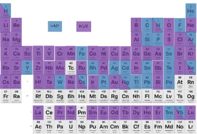
Dans le contexte des DDS, comme de nombreux noyaux RMN actifs sont présents dans le coeur
(tels que 1H, 2H, 13C, 17O 19F and 27Al), la couronne (1H, 13C, 17O, 27Al, 29Si) et à la surface (13C, 17O, 29Si, 31P) des nanoparticules, ils peuvent fournir des informations sur la localisation, l'état et les interactions
du médicament dans le système et sur les interactions cœur/couronne et sur la dégradation de la
matrice et donc, la délivrance/libération du médicament. Ces informations peuvent contribuer de
manière significative à orienter la conception et le développement de nouveaux DDSs. De plus, cette
technique est non destructive, c'est-à-dire que le DDS peut être étudié intact sans avoir besoin d'être
dissous, comme cela serait nécessaire pour la RMN liquide, ou d'être modifié chimiquement comme
cela serait nécessaire pour la spectroscopie de fluorescence.
Les expériences de RMN 1D 1H en rotation à l’angle magique (MAS) sont faciles à réaliser, donnant
14 entre les composants du système. Toutefois, en raison de la complexité de certains systèmes, même
en utilisant des hauts champs magnétiques et la rotation MAS très élevée, La RMN du 1H ne permet
pas toujours d'obtenir des spectres résolus et, par conséquent, d'avoir des informations sur le
système. Dans ce cas, des expériences de RMN 2D peuvent être réalisées. En plus de la réalisation des
spectres 2D 1H-1H, mais une solution alternative peut être les expériences 1H-X (X = noyau actif en
RMN autre que le 1H) pour essayer de tirer profit des hétéronoyaux (X) qui sont présents dans le
médicament ou le nanovecteur. Si elles sont informatives dans de nombreux DDS, les expériences 1
H-X ont également leurs limites dans le cas de systèmes trop complexes. Dans ces cas, des expériences
X-Y 2D peuvent être réalisées. Ces types d'expériences constituent une nouvelle approche dans la
caractérisation structurelle des DDS qui permet de contourner la faible résolution de la RMN du 1H.
Dans cette thèse, en étroite collaboration avec le groupe du Dr Ruxandra Gref (DR CNRS ISMO
Orsay), nous avons étudié les DDS basés sur les MOFs comme vecteur de médicament en utilisant la
spectroscopie RMN à rotation à angle magique. Sur la base de récentes expériences de RMN à base
de protons dans des MOF diamagnétiques et paramagnétiques sur des MOF purs, nous voulions
initialement aborder ces DDS en utilisant la RMN 1H en raison de sa grande sensibilité. Cependant,
nous avons rapidement réalisé que si l'utilisation d'un instrument RMN de pointe (aimants à champ
magnétique élevé, fréquence de rotation à angle magique ultra-rapide) et de séquences d'impulsions,
permettant d'obtenir une très bonne résolution sur des MOF purs, la situation était beaucoup plus
difficile sur les systèmes supramoléculaires complexes que sont les DDSs. En outre, les DDSs basés sur
les MOFs les plus populaires sont constitués de cations Fe3+ non toxiques (par exemple, MIL-100(Fe),
MIL-88(Fe)). La présence de ces centres paramagnétiques puissants rend les spectres RMN 1H MAS
encore plus complexes.
Nous avons donc choisi d'étudier les analogues diamagnétiques de MIL-100(Fe) à base d'Al, à savoir
MIL-100(Al), en considérant comme hypothèse qu'ils se comporteraient de manière similaire. Nous
15 étude sur les hétéronoyaux (tels que 19F, 27Al, 31P, 13C, 2H, 17O, etc.) présents de manière endogène ou
ajoutés volontairement dans le médicament ou dans le nanovecteur. Cela s'est fait au prix d'une
grande perte de sensibilité (car les hétéronoyaux sont présents en concentration inférieure d'au moins
un ordre de grandeur à celle des protons), mais au bénéfice d'une plus grande sélectivité (puisque
nous pouvons choisir quel et où chaque hérétonoyau peut être placé, c'est-à-dire sur la surface ou
dans le cœur de la nanoparticule, sur le médicament, etc.). Grâce à cette approche, nous avons pu
analyser en détail des DDSs supramoléculaires très complexes. En relation constante avec les
chimistes, ces informations ont été utilisées pour modifier à leur tour la conception des DDSs, ce qui
a permis d'améliorer les performances.
Le premier DDS étudié consistait en un promédicament antituberculeux, le Lansoprazole (LPZ),
chargé dans un MOF à base de cyclodextrine- (-CD), -CD-MOF. Le LPZ est une molécule fragile qui
se dégrade facilement dans des conditions acides 8–10 et basiques, est instable sur une étagère8,11,12
(même en condition de stockage à 4°C 13) et a une forte tendance à cristalliser. Le stockage et
l'administration de ce médicament sont donc des challenges et le chargement du médicament dans
un DDS apparaît comme une solution intéressante pour résoudre ces problèmes.
-CD-MOF a été choisi comme vecteur du LPZ en raison de sa capacité de chargement élevée pour ce médicament (23 ± 2 % en poids)14. Les -CD-MOFs sont des MOFs biocompatibles résultant de la
coordination de cations potassium à des unités de D-glucopyranosil alternées à liaison -1,4 sur les
faces primaire et secondaire des CDs. Six unités CDs sont tenues par la coordination de quatre cations
K+ aux groupes C
6OH et aux atomes d'oxygène du cycle glycosidique sur les faces primaires des -CD,
tandis que les cubes sont attachés les uns aux autres par la coordination de quatre ions K+ aux
carbones des groupes OH des faces secondaires de la cyclodextrine. Les -CD-MOFs ont une structure
poreuse ouverte, avec un grand pore sphérique de 1,7 nm de diamètre situé au centre de chaque cube
16 sont définies par le -CD, qui adopte la face du cube. De plus, la structure du canal (avec une ouverture
de 0,42 nm) relie les deux types de pores entre eux 15–17.
Un tensioactif est utilisé dans la synthèse du -CD-MOF pour obtenir des particules de MOF de
taille homogène. En utilisant des mesures RMN 1H-1H, nous avons localisé ce tensioactif et montré
qu’il restait présent dans les particules finales, un problème potentiel pour l’encapsulation des
médicaments. Toutefois, par RMN 1H-1H, 1H-13C et 1H-19F-13C, nous avons montré que la compétition
tensioactif/LPZ était favorable au LPZ, qui présente une affinité meilleure avec les CD. L’ensemble des
mesures, ainsi que des mesures 15N en abondance naturelle sur ce système, nous ont permis de
localiser le médicament dans le MOF, ainsi que son état de protonation, critère important pour la
libération.
Le second système à l'étude était un DDS basé sur le nanoMIL-100(Al), choisi comme analogue du
nanoMIL-100(Fe) très étudié in vivo (préclinique).
Dans une première étape, des groupes fonctionnels de surface et des médicaments contenant des
atomes de phosphore ont été sélectionnés parce qu'il a été démontré qu'ils avaient une grande
affinité avec l'analogue du MOF à base de Fe, et les liaisons chimiques Al-O-P sont connues pour être
énergétiquement favorables. En utilisant les expériences 27Al {31P} 2D D-HMQC, nous avons confirmé
la grande affinité attendue entre le revêtement phosphaté (phosphate -CD) et le médicament
(adénosine triphosphate) avec les cations aluminium de surface et/ou du cœur des particules de
MOF18. Grâce à cette méthodologie, la faible résolution de la spectroscopie RMN 1H MAS a été
contournée et il a été possible de montrer pour la première fois une signature RMN 27Al à température
ambiante des espèces d'aluminium présentes à la surface ou sous la surface des nanoparticules et leur
interaction avec le revêtement CD-P. La formation d'une forte liaison covalente P-O-Al entre le CD et
le MOF pourrait être la clé pour assurer une grande stabilité des nanoparticules cœur-couronne in
vivo. Les expériences 27Al {31P} 2D D-HMQC ont ensuite été utilisées pour analyser la dégradation du
17 d'administration de médicaments, était obtenue, probablement en raison de la formation d'une
couche de type aluminophosphate sur la surface externe du MOF.
C'est pourquoi un autre revêtement de surface plus couvrant a été envisagé : les oligomères de
-CD-citrate (CD-CO). Ce revêtement a été choisi parce qu'on s'attendait à ce que le -CD présente une
plus grande affinité pour le médicament que le -CD (qui est plus petit), ce qui pourrait entraîner une
charge utile plus importante. Ici, le médicament choisi est la doxorubicine (DOX) en raison de son
affinité connue avec les CD-CO. Dans ce système, aucun atome autre que 1H et 13C n'étaient présent.
Par conséquent, la stratégie adoptée a consisté à synthétiser le polymère CD-CO en utilisant le citrate
marqué au 13C comme précurseur. De cette façon, en utilisant27Al {13C} 2D D-HMQC, 13C {27Al} RESPDOR
et 1H {13C} D-HMQC la bonne interaction du nouveau revêtement avec la surface du nanoMIL-100(Al)
a été confirmée par la formation d'une nouvelle résonance sur les spectres RMN 13C appartenant à la
liaison entre le groupe carboxylique du citrate et l'aluminium du MIL-100. La charge du DOX à la fois
dans les pores des MOFs et dans le revêtement a été validée par la même approche. Nous avons
remarqué que l'interaction de la DOX avec le revêtement réduisait l'interaction du citrate avec
l'aluminium de la surface, mais n'affectait pas la stabilité des nanoparticules. L'étude de libération du
médicament est toujours en cours.
L'ensemble des données rapportées dans cette thèse a montré que l'utilisation d'hétéroatomes
pour étudier des DDSs au niveau moléculaire était une bonne alternative à la spectroscopie RMN 1H,
au prix d'une moindre sensibilité mais avec l'avantage d'une plus grande sélectivité. Cette
méthodologie est poursuivie par M. V. D. Le dans sa thèse. En particulier, l'effet de la longueur de la
chaîne de phosphate du médicament (mono- vs tri-phosphate) sur son affinité et la délivrance du
médicament est en cours d'évaluation. Si seuls quelques noyaux ont été étudiés dans ce travail (19F,
27Al, 31P, 13C, 15N), d'autres pourraient être envisagés.
Le deutérium est un nouveau noyau "à la mode" dans le développement des médicaments. La
18 destruction du médicament dans l'organisme, d'où une biodisponibilité accrue et une administration
plus faible du médicament. De plus, le remplacement de H par D dans une molécule connue réduit
considérablement la durée de développement du médicament. Jusqu'à présent, un seul médicament
deutéré a été approuvé par la FDA (Austedo® de Teva), mais une douzaine d'autres sont à l'étude à
différents stades (I à III)19. D’un point de vue de la spectroscopie RMN, le deutérium dilue les protons,
ce qui simplifie les spectres RMN 1H MAS et permet de modifier les liaisons hydrogène. Le D
2O peut
donc être utilisé pour jouer sur les interactions médicament/porteur. Par exemple, le médicament
Vancomycine (VCM) a été chargé à l'intérieur de nanoparticules de PLGA, dans lesquelles le PVA est
mélangé à la surface du PLGA. En raison du fort chevauchement des résonances des protons et de la
présence de molécules d'eau, une stratégie de deutération a été utilisée pour tenter de simplifier les
spectres RMN 1H. Nous avons comparé l'effet du pH sur le NP chargé avec le médicament avec
l’échantillon synthétisé en D2O. Si aucune différence n'est perceptible dans les spectres de carbone
effectués dans les échantillons à différents pH, dans les spectres de protons, il est possible de
remarquer une augmentation de l'intensité des résonances appartenant aux protons de la
vancomycine (OH, NH). Ceci peut être mieux remarqué dans l'échantillon synthétisé en D2O. On peut
remarquer que les résonances appartenant aux OH et NH sont plus intenses dans l'échantillon deutéré
et dans celui à pH=7,5. À pH=4, il y a des interactions, créées par les liaisons H, entre le médicament
et les polymères. Une fois que nous changeons le pH, en passant à un pH neutre (pH=7,5), le
médicament se cristallise à l'intérieur du polymère, et en perdant ces interactions, les pics semblent
plus étroits. Les mêmes lignes étroites peuvent être observées dans le spectre de l'échantillon
deutéré, car la stratégie de deutération "change" la liaison H et donc les interactions entre le
médicament et le polymère.
Enfin, un autre atome très présent dans la DDS est l'oxygène. Le seul isotope de l'oxygène actif en
RMN est le 17O, qui est un spin 5/2 et qui est présent en abondance à 0,037 %. Cette faible abondance
naturelle nécessite un enrichissement isotopique (souvent) coûteux. Récemment, de nouvelles
19 broyage planétaire, ii) par simple imprégnation dans de l'eau marquée en 17O. Cette dernière a été
appliquée avec succès pour les zéolithes20,21. Les MOFs présentant des similarités chimiques avec les
zéolithes, cette stratégie a également été tentée sur des MOFs. En outre, un MOF à base de scandium,
NOTT-400(Sc), qui pu être sélectivement marqué en 17O sur la position pontante Sc-O-Sc
(correspondant au site d’oxygène le plus actif dans le MOF) soit i) en introduisant dans la dernière
étape de la synthèse, ii) soit par simple imprégnation du MOF pendant 3 semaines dans une solution
H217O. Cela montre (ou confirme) que les fonctions oxo des MOFs sont très labiles et peuvent être
facilement échangées. Ceci est très important car ces fonctions oxo sont généralement les sites les
plus réactifs des MOFs, que ce soit pour la catalyse, l'adsorption de gaz ou l'adsorption de
médicaments.
En conclusion, la spectroscopie RMN est un outil analytique important pour comprendre les DDSs
au niveau atomique, et représente un guide important pour la conception de DDS avec des
20
Références
(1) Drug Delivery Systems; Jain, K. K., Ed.; Humana Press: Totowa, NJ, 2008.
(2) Seth, S.; Matzger, A. J. Metal–Organic Frameworks: Examples, Counterexamples, and an Actionable Definition. Cryst. Growth Des. 2017, 17 (8), 4043–4048.
(3) Corma, A.; García, H.; Llabrés i Xamena, F. X. Engineering Metal Organic Frameworks for Heterogeneous Catalysis. Chem. Rev. 2010, 110 (8), 4606–4655.
(4) Chen, W.; Wu, C. Synthesis, Functionalization, and Applications of Metal–Organic Frameworks in Biomedicine. Dalton Trans. 2018, 47 (7), 2114–2133.
(5) McKinlay, A. C.; Morris, R. E.; Horcajada, P.; Férey, G.; Gref, R.; Couvreur, P.; Serre, C. BioMOFs: Metal-Organic Frameworks for Biological and Medical Applications. Angew. Chem. Int. Ed.
2010, 49 (36), 6260–6266.
(6) Gossert, A. D.; Jahnke, W. NMR in Drug Discovery: A Practical Guide to Identification and Validation of Ligands Interacting with Biological Macromolecules. Prog. Nucl. Magn. Reson.
Spectrosc. 2016, 97, 82–125.
(7) Martineau-Corcos, C. NMR Crystallography: A Tool for the Characterization of Microporous Hybrid Solids. Curr. Opin. Colloid Interface Sci. 2018, 33, 35–43.
(8) DellaGreca, M.; Iesce, M. R.; Previtera, L.; Rubino, M.; Temussi, F.; Brigante, M. Degradation of Lansoprazole and Omeprazole in the Aquatic Environment. Chemosphere 2006, 63 (7), 1087– 1093.
(9) Ngan, Y.; Gupta, M. A Comparison between Liposomal and Nonliposomal Formulations of Doxorubicin in the Treatment of Cancer: An Updated Review. Arch. Pharm. Pract. 2016, 7 (1), 1–13.
(10) Ekpe, A.; Jacobsen, T. Effect of Various Salts on the Stability of Lansoprazole, Omeprazole, and Pantoprazole as Determined by High-Performance Liquid Chromatography. Drug Dev. Ind.
Pharm. 1999, 25 (9), 1057–1065.
(11) Battu, S.; Pottabathini, V. Hydrolytic Degradation Study of Lansoprazole, Identification, Isolation and Characterisation of Base Degradation Product. Am. J. Anal. Chem. 2015, 6 (2), 145–155.
(12) Liu, K.-H.; Kim, M.-J.; Jung, W. M.; Kang, W.; Cha, I.-J.; Shin, J.-G. Lansoprazole Enantiomer Activates Human Liver Microsomal CYP2C9 Catalytic Activity in a Stereospecific and Substrate-Specific Manner. Drug Metab. Dispos. 2005, 33 (2), 209–213.
(13) DiGiacinto, J. L.; Olsen, K. M.; Bergman, K. L.; Hoie, E. B. Stability of Suspension Formulations of Lansoprazole and Omeprazole Stored in Amber-Colored Plastic Oral Syringes. 2000, 34, 600– 605.
(14) Li, X.; Guo, T.; Lachmanski, L.; Manoli, F.; Menendez-Miranda, M.; Manet, I.; Guo, Z.; Wu, L.; Zhang, J.; Gref, R. Cyclodextrin-Based Metal-Organic Frameworks Particles as Efficient Carriers for Lansoprazole: Study of Morphology and Chemical Composition of Individual Particles. Int. J.
Pharm. 2017, 531 (2), 424–432.
(15) Rajkumar, T.; Kukkar, D.; Kim, K.-H.; Sohn, J. R.; Deep, A. Cyclodextrin-Metal–Organic Framework (CD-MOF): From Synthesis to Applications. J. Ind. Eng. Chem. 2019, 72, 50–66. (16) Michida, W.; Nagai, A.; Sakuragi, M.; Kusakabe, K. Discrete Polymerization of
3,4-Ethylenedioxythiophene in Cyclodextrin-Based Metal-Organic Framework. Cryst. Res. Technol.
2018, 53 (4), 1700142–1700148.
(17) Smaldone, R. A.; Forgan, R. S.; Furukawa, H.; Gassensmith, J. J.; Slawin, A. M. Z.; Yaghi, O. M.; Stoddart, J. F. Metal-Organic Frameworks from Edible Natural Products. Angew. Chem. Int. Ed.
2010, 49 (46), 8630–8634.
(18) Porcino, M.; Christodoulou, I.; Dang Le Vuong, M.; Gref, R.; Martineau-Corcos, C. New Insights on the Supramolecular Structure of Highly Porous Core–Shell Drug Nanocarriers Using Solid-State NMR Spectroscopy. RSC Adv. 2019, 9, 32472–32475.
21 (19) DeWitt, S. H.; Maryanoff, B. E. Deuterated Drug Molecules: Focus on FDA-Approved
Deutetrabenazine: Published as Part of the Biochemistry Series “Biochemistry to Bedside.”
Biochemistry 2018, 57 (5), 472–473..
(20) Heard, C. J.; Grajciar, L.; Rice, C. M.; Pugh, S. M.; Nachtigall, P.; Ashbrook, S. E.; Morris, R. E. Fast Room Temperature Lability of Aluminosilicate Zeolites. Nat. Commun. 2019, 10 (1), 4690– 4697.
(21) Pugh, S. M.; Wright, P. A.; Law, D. J.; Thompson, N.; Ashbrook, S. E. Facile, Room-Temperature
17 O Enrichment of Zeolite Frameworks Revealed by Solid-State NMR Spectroscopy. J. Am. Chem. Soc. 2020, 142 (2), 900–906.
23
General Introduction
25 Drug delivery systems (DDSs) are formulation or devices used to improve the delivery of drugs with
low efficacy and safety1. Moreover, they may aid convenience and compliance of the patient. The
process of drug delivery consists of administration of a pharmaceutical product into the body, with
controlled transport and delivery to the target cells or organs. Most of them are complex core-shell
systems, composed by a biodegradable core where the drug is loaded and a coating, which can be
functionalized with specific ligand. There are different types of DDSs, such as liposomes, microspheres
and microcapsules, polymer micelles and nanoparticles, red blood cells, mesoporous silica
nanoparticles and Metal-Organic Frameworks (MOFs). Bio-friendly synthesis, high drug loading in the
pores and surface modifications that can increase the stability and have a selective target delivery are
only few of the advantages of MOFs used as drug carriers2,3.
The analysis of the structure of a DDS is important to understand its stability, its mechanism, its
interaction with the drug and with the body and to carry out modification that may increase its
potential. Analytical techniques, such as thermal analysis, nitrogen adsorption, X-ray diffraction
provide information about the crystal structure, morphology, porosity, drug loading and release.
Solid-state Nuclear Magnetic Resonance (ssNMR) offers information about structural and dynamic
properties, probing local environment of the nuclei and short-range order information. This gives rise
to various advantages, including the ability to provide detailed structural insights and to examine local
defects and disorders. In the context of DDSs, because numerous active NMR nuclei are present in the
core (such as 1H, 2H, 13C, 17O, 19F and 27Al), corona (as 1H, 13C, 17O, 27Al, 29Si) and surface modification
(as 13C, 17O, 29Si, 31P) of the nanoparticles, it can provide information about drug location, state and
interactions in the system and about the core-shell interactions and about the matrix degradation and
so, the delivery/release of the drug.
In this thesis, in close collaboration with the group of Dr. Ruxandra Gref (DR CNRS ISMO Orsay), we
have investigated DDSs based on MOFs as drug carrier using magic-angle spinning (MAS) NMR
26 Based on recent proton-based NMR experiments on diamagnetic and paramagnetic pure MOFs4,
we initially wanted to address these DDS using 1H NMR due to its high sensitivity. However, we quickly
realized that if using state-of-the-art NMR instrument (high magnetic field magnets, ultra-fast
magic-angle spinning frequency) and pulse sequences, very nice resolution can be obtained on pure MOFs,
the situation was much more difficult on the complex systems as the DDSs. Furthermore, the most
popular MOF-based DDSs are made of non-toxic Fe3+ cations (e.g., MIL-100(Fe), MIL-88(Fe)). The
presence of these strong paramagnetic centers makes the 1H MAS NMR spectra even more complex.
Therefore, we chose to study the Al-based diamagnetic analogs of 100(Fe), namely
MIL-100(Al), considering as hypothesis that they would behave similarly. We also change our initial
proton-based strategy and decided to focus our study on heteronuclei (such as 19F, 27Al, 31P, 13C, 2H, 17O, etc)
present endogenously or added on purpose in the drug/coating or in the carrier. This was achieved at
the expense of great sensitivity loss (as the heteronuclei are present in concentration that are at least
an order of magnitude less than the protons), but at the benefit of greater selectivity (since we can
choose which and where each heretonuclei can be placed, i.e., on the surface or the bulk of the
nanoparticle, on the coating species, on the drug, etc). Using this approach, we have been able to
analyze in detail highly complex supramolecular DDSs. In constant relationship with the chemists,
these pieces of information were used in turn to modify the design of the DDSs, leading to improved
performances.
This thesis starts with a brief introduction on the process of drug delivery and the different route
of administration. The different types of DDSs, their use and the main analytical tools that can be used
to study their structure are presented. MOFs based drug carriers, notably those studied in this thesis,
are describes in more detail. The second chapter contains a basic presentation of the principles of
NMR spectroscopy and of the pulse sequences used during this project. In the third chapter, we
present a review on the use of ssNMR spectroscopy in the investigation of DDSs. At the end of this
27 thesis for three different drug/carrier systems are reported in chapters 4 to 6. Conclusions and
28
References
(1) Drug Delivery Systems; Jain, K. K., Ed.; Humana Press: Totowa, NJ, 2008.
(2) Chen, W.; Wu, C. Synthesis, Functionalization, and Applications of Metal–Organic Frameworks in Biomedicine. Dalton Trans. 2018, 47 (7), 2114–2133.
(3) McKinlay, A. C.; Morris, R. E.; Horcajada, P.; Férey, G.; Gref, R.; Couvreur, P.; Serre, C. BioMOFs: Metal-Organic Frameworks for Biological and Medical Applications. Angew. Chem. Int. Ed.
2010, 49 (36), 6260–6266.
(4) Krajnc, A.; Kos, T.; Zabukovec Logar, N.; Mali, G. A Simple NMR-Based Method for Studying the Spatial Distribution of Linkers within Mixed-Linker Metal-Organic Frameworks. Angew. Chem.
29
Chapter 1
31 A brief introduction on the drug delivery process and some of the different types of porous drug
delivery systems is presented in this chapter. A particular attention is given to Metal-Organic
Frameworks (MOFs) and their application as drug carriers.
1. Drug Delivery
The process of drug delivery takes into account the administration of a therapeutic product, the
release of the Active Pharmaceutical Ingredient (API) by the product and the transport of this API to
the biological target. Nowadays, complex formulations that control the time-release medications and
that target specific areas of the body are being developed. Administration of the drug into the human
body can occur by various anatomical routes. The choice of the route depends on the disease, the
effect desired and the characteristics of the product. Drugs may be given systemically and targeted to
the diseased organ or administrated directly to the organ affected1.
Briefly, the main routes of delivery are2,3:
• Oral. The oral route has been the most used drug delivery method in the world, because of the ease of administration and the compliance of the patients. However, many drugs, such as
proteins or peptides, are subjected to a massive degradation in the GI (Gastro-Intestinal) tract.
As a result, many materials for drug delivery have been developed to protect the drug from
the degradation and enhance drug adsorption2.
• Pulmonary. The pulmonary route is used to treat several illnesses, such as asthma or cystic fibrosis. Because the drug is directly administrated to the lung, systemic side effects can be
minimized as well as the dose required. Nevertheless, this route can be useful for systematic
therapeutic substances, such as insulin or other proteins.
• Transdermal. Delivering of the drug across the skin is useful for local applications, patches, drug carriers and electrotransport. It is an alternative to avoid the drug degradation in the GI
32 tract and to reduce the systemic side effects. However, the skin is a natural barrier to foreign
substances and so, it is important to develop methods to enhance the permeation across it. • Parenteral. The name parenteral route is applied to injections of substances by subcutaneous,
intravenous, intramuscular and intra-arterial routes. It is the most used route for systemic
drug delivery. It has several advantages such as almost complete bioavailability of the drug
and rapid onset of action. However, there are several problems associated with this route
among which pain, risk of infections and the involvement of health care coworkers. Improve
the injections technologies or develop alternative method of delivery are the two strategies
chosen to erase the problems.
• Transmucosal. By diffusion several drugs can be introduced at various anatomical sites, thanks to the mucous membranes that covers all the internal passages and orifices of the body. In
comparison with parenteral route, there are several advantages, including rapid adsorption
and lower cost.
• Colorectal. For drug, targeting the small intestine or acting systematically, the colon can be a site for safe and slow adsorption. Adsorption enhancers are required for some hydrophilic
drugs (antibiotics or peptide drugs) and other drugs may cause rectal irritations.
The rate of the drug uptake in the administration routes is controlled by the drug properties
(solubility, charge, molecular size, etc.) and the characteristics of the site of administration (pH,
presence of enzyme or of active transport, etc.).With traditional administrations, the therapeutic
index and consequently, the therapeutic window should be taken into consideration. The therapeutic
index (TI) is a quantitative measure of the safety of the drug; it is a comparison between the amount
of the therapeutic agent causing toxicity, and the amount giving a therapeutic effect in 50% of the
subjects tested (respectively 𝑇𝐷50 and 𝐸𝐷50):
𝑇𝐼 = 𝑇𝐷50 𝐸𝐷50
33 The therapeutic window of a drug is the range of drug dosages which can treat disease effectively
without having toxic effects3 (Figure 1.1). Each drug has a given TI and some of them are very low and
require regular monitoring of drug level.
Figure 1.1: Graphic representation of plasma concentration of the drug during the time. The duration of the therapeutic action, correspond to the time in which the concentration stays in the therapeutic window (green zone). Before and after, the concentration is too low to have a therapeutic effect (blue zone). If the concentration goes up of the adverse
response, side-effects could be showed (red zone).
Materials for drug delivery can be employed to have a better control of drug level or have a direct
release of the drug on the target. An ideal material for drug delivery should be biocompatible,
non-toxic and not immunogenic. It should have precise scaffolding properties, with an adequate
bioelimination, and a modifiable surface. Its synthesis should be easy and reproducible and it should
have a controlled release of the drug. Materials responding to these criteria are called Drug Delivery
Systems (DDSs).
2. Drug Delivery Systems
A nanosized drug delivery system (DDS) is defined as a formulation or a device that enables the
introduction of a therapeutic substance in the body1. The major objectives to use a DDS are to improve
the efficacy and safety of the drug by controlling the rate, time and place of release of the API, as well
34 cost of drug development, of the risk of failure in new product development and the life extension of
the product1. There is unfortunately no universal drug nanocarriers, and for each medical target, a
new drug carrier has to be properly designed. The ideal DDS should have high colloidal stability, high
drug loading capacity, be prepared without need of toxic solvent, be inert, biocompatible,
mechanically strong, biodegradable, comfortable for the patient, safe from accidental release, simple
to administer, and finally easy and cost effective to fabricate and sterilize. Most DDSs are composed
of a very complex core-shell system with different characteristics and functions (Figure 1.2).
Figure 1.2: Illustration of composition of a Drug Delivery System. The drug is loaded in the biodegradable core that has to protect and stabilize it. The corona is composed by a functional coating that can interact with specific ligands to
ensure delivery of the drug to the target.
The core of the system accommodates the drug, protects and stabilizes it. It must be degradable
to ensure efficient drug release. The corona part of the system has for goals to increase the colloidal
stability of the nanoparticles, to make them furtive to the immune system and to ensure specific
delivery to the biological target.
Some of the different types of porous DDSs are summarized in Figure 1.3 and detailed in the next
35
Figure 1.3: Some types of porous Drug Delivery Systems5–13.
The choice of the appropriate DDS is based on the physiochemical nature (solubility, molecular
weight and size) of the drug as well as the disease for which the system has to be used and so, the
administration route chosen to deliver the drug14. Moreover, the functional groups of the drug can
influence the choice of the DDS, because of the interaction that can form with it.
2.1 Liposomes
Liposomes are spherical vesicles formed by phospholipids and similar amphipathic lipids,
incorporated with sterols, such as cholesterol1. The polar character of their core enables polar drug
molecules to be encapsulated, but also lipophilic molecules are solubilized within the phospholipid
bilayer. Furthermore, channel proteins can be incorporated and allow passive diffusion of small
solutes such as ions, nutrients and antibiotics15. Liposomes are classified, depending on the number
36 The pharmaceutical proprieties of liposomes depend on the composition of the lipid layer and its
permeability and fluidity1 and they can be formulated and processed to differ size, composition,
charge and lamellarity. Drugs with different solubility can be encapsulated in liposomes, hydrophobic
drugs have affinity to the phospholipid layer and hydrophilic drugs are entrapped in the aqueous
cavity. The liposomes are one of the earliest targeted systems, starting for cancer therapy. Following
extensive developments in liposome technology, many liposome-based drug formulations are
nowadays used to treat different type of infections, pain management and photodynamic therapy17.
2.2 Polymer micelles
Polymer micelles are biocompatible nanoparticles varying in size from 50 and 200 nm that are
very interesting for drug delivery applications1. The drug can be physically entrapped in the core of
the micelles and transported in a particular site of the body without being degraded by the enzyme or
incur in hydrolysis. This is possible thanks to hydrophilic blocks of the micelles, which can form
hydrogen bonds with the aqueous environment and form a tight shell around the micellar core16. The
size and morphology of the micelles can be easily controlled, since, being composed of polymers, the
molecular weight and block length ratio can be changed.
2.3 Microsphere and microcapsules
Microspheres and microcapsules are spherical particles with sizes in the range of 1 and 100 μm
that are used to encapsulate the active compounds. Microcapsules differ from microspheres in having
a barrier membrane surrounding a solid or liquid core. Therefore, in these reservoir systems, a
polymer shell surrounds the drug1. Microspheres are matrix systems consisting of natural or synthetic
polymers in which the agent encapsulated can either molecularly dissolved or heterogeneously
dispersed into the matrix polymer18. Possible synthetic biodegradable polymers are
poly-alkyl-cyano-acrylates that are used as drug carrier for parental, ophthalmic and oral preparations. An example of
37
2.4 Red blood cells
Red blood cells or Erythrocytes have potential carrier capabilities for the delivery of drugs. They
are red cells with a biconcave disc form that transport the hemoglobin, a protein that carries oxygen
from the lungs to tissues and that consists of four polypeptide subunits, each of which is bound to an
iron-containing heme molecule. The red blood cells are biocompatible, biodegradable and
non-immunogenic due to their natural presence in the body1. By using various physical and chemical
methods, cells are broken, and drugs are entrapped into erythrocytes. Finally, the drugs are released
and resultant carriers are called as “released erythrocytes”. A broad range of a variety of chemically
and biologically drugs can be incorporated in red blood cells, using various chemical and physical
methods15.
2.5 Polymer Nanoparticles
Micro- and nanoparticles based on PLA (Poly(lactic acid)) and PLGA (Poly(lactic acid-glycolic
acid))19 are often used for intravenous administration of a large variety of drugs, such as Docetaxel20
and Doxorubicin21, two anticancer drugs and Estradiol21, an hormone that can be used as drug to treat
mental disease. Sometimes these particles were covered with Polyethylene glycole (PEG) or chitosan
(CS) to improve the stability of the systems and helps the transport of the drugs1,22,23.
2.6 Mesoporous Silica Nanoparticles
Mesoporous silica nanoparticles (MSNs) are reservoirs with a high capacity to accommodate guest
molecules, having uniform and tunable pore size, in the range of 2-50 nm, and which surface can be
easy functionalized. Their long-range ordered pore structure with tailorable pore size and geometry
facilitates a homogenous incorporation of guest molecules with different sizes and properties. Drug
loading is based on the adsorptive properties of MSNs: both hydrophilic and hydrophobic cargos can
be adsorbed into the pores. They have greater loading capacity compared to other carriers for their
large pore volume. The release profile of drugs from MSNs mainly depends on its diffusion from the
38 decisive factor responsible for controlling the release is the interaction between the surface groups
on pores and the drug molecules9,24. The two most widely explored materials for drug delivery are
MCM-4125 (Mobil Crystalline Materials) and SBA-1526 (Santa Barbara Amorphous type material).
2.7 Periodic Mesoporous Organosilica Nanoparticles
Periodic Mesoporous Organosilica nanoparticles (PMO NPs), similar to mesoporous silica
nanoparticles, are prepared from organo-bridged alkoxysilanes and have tunable mesopores that
could be utilized for many applications such as gas and molecule adsorption, catalysis, and drug
delivery. Unlike MSNs, the diversity in chemical nature of the pore walls is theoretically unlimited27.
They have several advantages, such as uniform and tunable pore size, large surface areas, high pore
volumes, nontoxic nature and good biocompatibility. PMOs have been explored as drug carriers for
different kinds of drugs, such as antiinflammatory, antibiotics or antitumoral drugs. In some cases, the
hydrophilic silica walls need to be functionalized with organic groups to allow the loading of
hydrophobic drugs. This functionalization seems to be the main controlling factor on the drug
adsorption/desorption processes, leading to higher drug loading capacities and slower release
kinetics13.
2.8 Metal-Organic Frameworks
Metal-Organic Frameworks (MOFs) are a class of coordination polymers comprising organic linkers
wherein metal-ligand interaction/bonding leads to 2D or 3D crystalline network structures28 (Figure
1.4). The metal centers act as nodes of the lattice and are held in place by the directionality of their
binding to the organic linkers (electrostatic attraction and coordinative metal–ligand interactions).
Crystallinity and porosity are the main characteristics of MOFs and these materials hold records in
terms of high pore volume(from 0.50 cm3/g to 1.5 cm3/g), surface area (from 700 m2/g to 9000 m2/g)29–
39
Figure 1.4: Schematic drawing of a metal-organic framework (MOF) structure.
The pore size, shape, dimensionality and chemical environment in the MOFs can be controlled
by the selection of their building blocks (metal and organic linker) and how they are connected.
Furthermore, the possibility of amending and functionalizing the organic ligand may allow tailoring
the material for specific applications33.
2.8.1 Synthesis and structural characteristics
The synthesis of MOFs is often carried out in the liquid phase by mixing two solutions containing
the metal and the organic component, either at room temperature or under solvothermal conditions
and with or without the aid of auxiliary molecules. Generally, high-solubility organic solvents, such as
dimethyl formamide, ethanol, or methanol, are used in solvothermal reactions. Hydrothermal
synthesis of MOFs constitutes a more environmental friendly approach that substitutes the use of
organic solvents for water31. Formation of crystalline framework takes place by self-assembly of the
structural units forming an ordered network of metal-organic coordination bonds. Other synthetic
methods are developed such as microwave, electrochemical, ultrasonic processing which reduce the
synthesis duration34. In Figure 1.5 are summarized all the synthetic methods. After the synthesis, post
synthetic modification is a powerful route to introduce functionality into MOFs, and in many cases, is
40 underlying structure. Examples of post synthetic methods are covalent modification, surface
modification, metalation, linker/metal exchange and modification of inorganic cluster35.The nature of
the solvent, the ligands, or the presence of guest molecules in the synthesis can have a dramatic effect
on the crystal structure of the material obtained33.
Figure 1.5: Different synthetic methods of MOFs materials31.
A large variety of metal atoms in their stable oxidation states, alkaline, alkaline earth, transition
metals, main group metals, and rare-earth elements, have been successfully used in the synthesis of
MOFs. As organic components, rigid molecules are usually preferred over flexible ones, because they
favor the preparation of crystalline, porous, stable MOFs34. Different combinations of metal ions and
organic linkers lead to an enormous number of MOFs.
MOFs are distinguished by the diversity of the structure, different symmetry, pore sizes and by
their characteristics. The carbon chain length or the number of benzene rings of the linker can
41 into the linker is responsible for the additional selectivity and unique chemical proprieties of the
pores36.
2.8.2 Applications
Many potential applications have been explored in strategic domains:
• Catalysis. The framework capacity to accommodate catalytic units and the possibility to have the presence of open metal sites are key factors for the catalysis application33,37. They are very
useful for heterogeneous organic and photocatalytic reactions38.
• Storage and Gas separation. The large surface-area of MOFs enables them to soak up large amounts of gas or molecules, such as hydrogen, carbon-dioxide and methane38. Based on the
size/shape of the adsorbent’s pore and the binding affinity from the adsorption sites, gas
separation can be realized39,40.
• Chemical sensors. Several chemical functionalities can be incorporated into the coordination nanospace of MOF materials and can show sensing applications for biomolecules, metal ions,
explosives, environmental toxins, and others38,41.
• Biomedical applications. MOFs are used for storage and delivery of gas transmitter such as NO or CO and as well as therapeutic molecules (more details are given in the section 2.8.4)42.
2.8.3 General techniques for the structural characterization of MOFs
Different techniques can be used for the characterization of the MOFs, regardless of their
application. The most common method of analysis of MOF crystal structure is X-ray Diffraction (XRD).
Due to the difficulty to grow a single crystal of MOF, powder X-ray diffraction (PXRD) is often used.
PXRD patterns allow analyzing the reproducibility of synthesis results or structural differences
between samples of the same MOF prepared by different methods; it is useful to determine the
structural parameters and crystallinity of the MOFs (Figure 1.6)31,36,43. It might be difficult to determine
42 with solid-state NMR and molecular modeling were developed, allowing the determination of very
complex MOFs44.
For the characterization of the shape of the material, external morphology, dispersion, and
mixture of phases, Scanning Electron Microscopy (SEM) technique is routinely used. On the other
hand, Transmission Electron Microscopy (TEM) has been widely applied to determine the grain size,
particle size, and crystallographic data. This technique is very useful for the characterization of MOFs
modified by the incorporation of nanoparticles (NPs), because the collected images provides
information about the size and dispersion of those NPs31.
Thermal stability of the MOFs is assessed by thermogravimetric analysis (TGA), which measures
the mass of the sample as a function of temperature. This technique is not only useful to study the
thermal stability but also to estimate solvent-accessible pore volume of the materials 31,36.
To study surface area, pore volume, average pore size, and pore size distribution,
Brunauer-Emmett-Teller (BET) analysis can be used. Nitrogen is the most commonly employed gaseous
adsorbate used for surface probing by BET methods. This approach studies the N2 adsorption over the
Figure 1.6: Example of experimental (light blue) and theoretical (dark) PXRD patterns of a crystalline MOF, ZIF-445. Some
43 surface of a solid at the boiling temperature of liquid nitrogen, resulting in an adsorption isotherm.
The shape of the isotherm provides information about the textural composition of the solid31.
Among all these techniques, Nuclear Magnetic Resonance (NMR) spectroscopy and in particular,
solid-state NMR is becoming an important tool for the characterization of MOFs. Solid-state NMR gives
the possibility to analyze the material at atomic level. It can help understanding the synthetic and
post-synthetic modifications, performed in order to increase the stability of the material46,47. In
addition to this, the NMR spectroscopy can reveal interactions between molecules and allows
studying, at atomic level the surface of the materials. This makes NMR spectroscopy a promising tool
for the study of the MOFs used as drug delivery systems.
2.8.4 MOFs as Drug Delivery Systems
The possibility to synthetize MOFs starting using only non-toxic agents, makes MOFs possible drug
carriers. The use of MOFs for biomedical applications indeed requires a biologically friendly
composition. To avoid possible toxic side effects, the choice of the appropriate metal and of the linker
are critical. Some of appropriate metals are Calcium, Magnesium, Zinc and Iron, but it has to be taken
into consideration also the metal daily dose. Regarding the linker, exogenous or endogenous linkers
can be used34. The endogenous organic spacers, such as cyclodextrin11, fumarate48,49 or aminoacids
and peptides42, are safer and more biocompatible, but most MOFs used as DDSs are composed of
exogenous linkers. In that case, a green synthesis approach is adopted, based on room temperature
synthetic reaction in aqueous media, with the use of precursor materials with benign chemical
characterizations50. Furthermore, it is important to have a study of toxicity of the single components
before their use, with cytotoxicity texts and measure of the linker and metal accumulation in the urine.
Among all the porous materials that can be used as DDSs, MOFs have several advantages such as
high surface area, the possibility for easy modification of the surface, adjustment of the functional
groups of the linkers and tuning of the pore size. In addition, a large number of MOFs with nontoxic
44 large variety of molecules with different chemistry, due to their hydrophilic-hydrophobic internal
microenvironment12,34.
Figure 1.7: Scheme showing the ideal properties of a MOF-based drug delivery system, using the structure of UiO-67, [Zr6O4(OH)4(bpdc)6]n (bpdc = 4,4-biphenyldicarboxylate), as an example. (Source: Abánades Lázaro et al.10)
The colloidal stability of the MOF in water is another factor that should be taken into consideration
in DDS design. It was shown that carefully engineered surface modification of MOF NPs was helpful to
increase the colloidal stability in aqueous media and prevent the premature release of the drug12,34,42
(Figure 1.7). The surface can be modified by coating with a hydrophilic polymer such as PEG,51 or with
larger molecule such as Cyclodextrin (CD)52, or with liposomes to have a combination therapy49.
Moreover, a silica thin layer53 or nanoparticles made by selenium/ruthenium54 could be used.
Two other parameters can affect the use of the MOF for biomedical application: size and shape.
For some type of administration route, the size is a critical value, for example, nanoparticles with
particle size more than 200 nm cannot be administrated by parenteral route. Therefore, it is important
to have a homogenous distribution of particle size and the shape of the nanoparticles that compose
the DDS34.
Up to now, several MOFs have been studied and used as DDSs. Two chromium (despite the
potential toxicity of Cr3+ cations) mesoporous MOFs, MIL-101(Cr) and MIL-100(Cr) (MIL stands for
45 high loading capacity and a delivery governed by diffusion processes in in vitro experiments55. The
same drug was loaded in a flexible MOF, MIL-53, with two different types of metal cations (Cr3+ and
Fe3+). The “swelling” effect of the terephthalate linker of this material makes this MOF adaptable to
the drug size and can promote the incorporation of different types of drugs56. Moreover, several
iron-MOFs are used, for example, as caffeine carriers57–59. Another example is bio-MOF-1, composed of
zinc ions and biphenyl carboxylate and adenine as linkers. It was used to deliver the antiarrhythmic
drug, procainamide. Due to the short half-life of the procainamide, a controlled release of this drug
was a successfully solution to avoid the administration every 4 hours60.
2.8.5 MOFs used in this thesis
The porous MOFs-based DDSs used in this thesis, i.e., -Cyclodextrin-based Metal-Organic
Framework and MIL-100, will be described in the next sections.
2.8.5.1 Cyclodextrin-based Metal-Organic Frameworks
CDs are natural cyclic oligosaccharides with 6 (-CD), 7 (-CD) or 8 (-CD) glucopyranose units
linked by alfa-1,4-glycosilic linkage. The CDs are homogeneous solids with a truncated cone shape
(Figure 1.8). They are produced during the degradation of starch by an enzyme, the glycosil
transferase. Hydroxyl groups compose the outer surface, that it is the hydrophilic part, while, the
internal cavities are hydrophobic and covered by the glycosylic oxygen and the C-H units. This type of
structure allows them to form non-covalent inclusion complexes through hydrogen bonding, van der
Waals interactions or electrostatic interactions. They are widely used for biomedical applications and
showed differently advantage, such as high surface areas, adjustable chemical functionality and
46
Figure 1.8: Illustration of the structure of -CD.
Recently, it was shown that CDs could be used as linker for the formation of porous MOFs, due to
the presence of -OCCO- binding groups, which facilitate the formation of complexes with metal ions
and the subsequently formation of an extended 3D network50. Cyclodextrin based Metal-organic
frameworks (CD-MOFs)61,62 are biologically acceptable types of MOFs, due to their porous and
non-toxic nature. They are composed of a biocompatible metal ion, usually potassium or calcium that
47
Figure 1.9: 3D cubic structure of -CD-MOF62. Oxygen, Carbon, Potassium atoms are in red, black and purple,
respectively. Hydrogen atoms are omitted for clarity. The formation of the complex with the potassium ions are made through the C1O- and C6O- moieties.
-CD-MOFs (Figure 1.9) are generally prepared by a vapor diffusion method 61,62using methanol or
aqueous solution of the hydroxyl metal, such as KOH. Other methods of synthesis can be used, such
as hydro/solvothermal method, microwaves-assisted and conventional method11.
Post-synthetic modifications can be important to improve the stability of the MOF in the
physiological environment. As an example, polymerization of 3,4-ethylenedioxythiophene (EDOT) in -CD-MOF was used to increase the thermal stability and the solubility in water of the particles. The
![Figure 1.7: Scheme showing the ideal properties of a MOF-based drug delivery system, using the structure of UiO-67, [Zr 6 O 4 (OH) 4 (bpdc) 6 ] n (bpdc = 4,4-biphenyldicarboxylate), as an example](https://thumb-eu.123doks.com/thumbv2/123doknet/14558928.726328/47.892.123.751.238.438/figure-scheme-showing-properties-delivery-structure-biphenyldicarboxylate-example.webp)

