HAL Id: tel-02947021
https://tel.archives-ouvertes.fr/tel-02947021
Submitted on 23 Sep 2020HAL is a multi-disciplinary open access archive for the deposit and dissemination of sci-entific research documents, whether they are pub-lished or not. The documents may come from teaching and research institutions in France or abroad, or from public or private research centers.
L’archive ouverte pluridisciplinaire HAL, est destinée au dépôt et à la diffusion de documents scientifiques de niveau recherche, publiés ou non, émanant des établissements d’enseignement et de recherche français ou étrangers, des laboratoires publics ou privés.
Impact of emerging technologies on the cell disruption
and fractionation of microalgal biomass
Rui Zhang
To cite this version:
Rui Zhang. Impact of emerging technologies on the cell disruption and fractionation of microalgal biomass. Chemical and Process Engineering. Université de Technologie de Compiègne, 2020. English. �NNT : 2020COMP2548�. �tel-02947021�
Par Rui ZHANG
Thèse présentée
pour l’obtention du grade
de Docteur de l’UTC
Impact of emerging technologies on the cell
disruption and fractionation of microalgal biomass
Soutenue le 8 juin 2020
Spécialité : Génie des Procédés Industriels et Bioprocédés :
Transformations intégrées de la matière renouvelable (EA-4297)
D2548
Thèse présentée pour l’obtention du grade de Docteur de l’UTC
Spécialité: Génie des Procédés Industriels
et Bioprocédés
Par Rui ZHANG
IMPACT OF EMERGING TECHNOLOGIES ON THE
CELL DISRUPTION AND FRACTIONATION OF
MICROALGAL BIOMASS
Soutenue le 08 June 2020
Devant la commission d’examen formée de:
Mme. Isabelle Pezron Professeur à l’Université de Technologie de Compiègne, Compiègne, France
Président
Mme. Maryline Abert-Vian Maître de conférences à l’Université d’Avignon et des Pays du Vaucluse, Avignon, France
Rapporteur
M. Carlos Vaca-Garcia Professeur à l’INP-ENSIACET,
Université de Toulouse, Toulouse, France
Rapporteur
M. Zhenzhou Zhu Professeur à Wuhan Polytechnic
University, Wuhan, Chine
Examinateur
M. Eugène Vorobiev Professeur à l’Université de Technologie de Compiègne, Compiègne, France
Membre invité
M. Luc Marchal Professeur à l’Université de Nantes,
Saint-Nazaire, France
Directeur de thèse
M. Nabil Grimi Maître de conférences à l’Université de
Technologie de Compiègne, Compiègne, France
… the memory of my grandmother who has passed away
ACKNOWLEDGEMENTS
Closing my 42-month PhD work at Université de Technologie de Compiègne (UTC), I would like to express my never ending gratitude to all who made it possible. This thesis is based on experimental work at the laboratory of Transformations intégrées de la matière renouvelable (TIMR) and Technologies Agro-industrielles (TAI) research group at UTC from 2016 to 2020. Also, I would like to thank the CSC (China Scholarship Council) for the scholarship and allowed me to perform this work in good conditions.
First and foremost, my sincere gratitude goes to my supervisor, M. Nabil Grimi for offering me the opportunity to study in France, and giving me academic guidance and inspiration throughout the course of this work. I thank him for having advised me, encouraged, supported with an availability of every moment. I learned a lot from his serious attitude, his patience and his passion for life and work. I will always appreciate the time that I passed with you in France. I appreciate also the effort of my co-supervisor, M. Luc Marchal, for offering me research raw materials, suggesting the research plan and sharing to me his knowledge for research, as well as supporting and encourage me during the realization of this thesis.
I acknowledge gratefully the effort of M. Eugène Vorobiev for supporting the research project and suggesting the research plan. Great thanks should be given to M. Nikolai Lebovka, who teaching me the enthusiasm and preciseness of scientific research, as well as a high-efficiency working methodology. Thanks these two supervisors for my help, advice and patience when correcting every article that I published. Without them this thesis could never have been realized.
I would like to thank Mme. Isabelle Pezron, Mme. Maryline Abert-Vian, M. Carlos
Vaca-Garcia and M. Zhenzhou Zhu for taking his time to be a referee. The advice they have
given me will undoubtedly improve the quality of this thesis and help me in my future work. I would like to express my thanks to M. Michael Lefebvre, M. Frederic Nadaud, Mme.
Caroline Lefebvre, Mme. Laurence Lavenant (GEPEA) and Mme. Delphine Drouin (GEPEA)
I thank them for supporting technical assistance for my thesis work.
My special thanks would go to my dear colleagues: Nadia Boussetta, Mohamed
Koubaa, Houcine Mhemdi, Luhui Ding, Caiyun Liu, Yantao Wang, Kaidi Peng, Deyang Zhao, Maiqi Xiang, Lu Wang, Christa Aoude, Marina Al Daccache, Sally El-Kanta,
Mathieu Hebert, Sarra Tadrent. Recalling the details working with you will definitely make
my face full of smile.
I will not forget to thank all my Chinese friends: Congcong Ma, Siying Li, Ke Li, Ye
Tao, Lei Lei, Changjie Yin, Lanting Yu, Qiongjie Li & Peng Du, et al. All the great
moments we have spent together in the city of Compiègne will be unforgettable memories for me. Special thanks to my foreign friends Chaima Dridi and Romain Guyard, who teach me French, acting as a teacher, and also a nice friend.
Finally I would like to say a big and loving thanks to my parents and my family, especially my grandparents. I thank them for giving me love unconditional, support, understand, confidence and encouragement. I want to say that because of you, I become a better self.
Abstract
This research work focuses on extraction and fractionation of bio-molecules from microalgae using physical treatments: pulsed electric fields (PEF), high voltage electrical discharges (HVED) and ultrasonication (US) techniques. In this study, three microalgae species Nannochloropsis sp., Phaeodactylum tricornutum (P. tricornutum) and Parachlorella kessleri (P. kessleri) were investigated. These species have different cell shapes, structure and intracellular contents. The effects of tested techniques on extraction of bio-molecules have been highlighted in a quantitative and qualitative analysis by evaluating the ionic components, carbohydrates, proteins, pigments and lipids.
A comparative study of physical treatments (PEF, HVED and US) at the equivalent energy input for release of intracellular bio-molecules from three microalgal species allowed us to better understand the different disintegration mechanisms. For each microalga at the same energy consumption, the HVED treatment proved to be the most efficient for extraction of carbohydrates, while the US treatment for extraction of proteins and pigments. In general, the smallest efficiency was observed for the PEF treatment. However, the highest selectivity towards carbohydrates can be obtained using the mild PEF or HVED technique.
The preliminary physical treatments (PEF, HVED or US) of more concentrated suspensions followed by high pressure homogenization (HPH) of diluted suspensions allowed improving the extraction efficiency and decreasing the total energy consumption. The physical pretreatments permit to reduce the mechanical pressure of the HPH and number of passes, to reach the same extraction yield. For the maximum valorisation of microalgal biomass, extraction procedure assisted by HVED treatment (40 kV/cm, 1-8 ms) followed by aqueous and non-aqueous extraction steps seems to be useful for selective extraction and fractionation of different bio-molecules from microalgae. The significant effects of HVED pre-treatment on organic solvent extraction of pigments (chlorophylls, carotenoids) and lipids were also observed.
Keywords: Microalgae; Pulsed electric field; High voltage electrical discharges;
Ultrasonication; High pressure homogenization; Selective extraction; Bio-molecules; Energy consumption
Résumé
Ce travail de recherche se concentre sur l'extraction et le fractionnement des biomolécules à partir de microalgues par des traitements physiques: les champs électriques pulsés (CEP), les décharges électriques de hautes tensions (DEHT) et les ultrasons (US). Dans cette étude, trois espèces de microalgues Nannochloropsis sp., Phaeodactylum tricornutum (P. tricornutum) et Parachlorella kessleri (P. kessleri) ont été étudiées. Les espèces ont différentes formes cellulaires, structure et contenu intracellulaire. L'effet des techniques testées sur l'extraction des biomolécules a été mis en évidence à travers une analyse quantitative et qualitative: suivi du rendement des composés ioniques, des glucides, des protéines, des pigments et des lipides.
Une étude comparative des traitements physiques (CEP, DEHT et US), à la même énergie, pour la libération des biomolécules intracellulaires à partir des trois espèces de microalgues, a permis de mieux comprendre les différents mécanismes de désintégration. Pour chaque microalgue, à la même énergie consommée, le traitement par DEHT s'est révélé le plus efficace en terme d'extraction des glucides, tandis que les US sont plus efficaces pour l'extraction des protéines et des pigments. Le traitement par CEP a été moins efficace en terme du rendement d’extraction. Cependant, la meilleure sélectivité (extraction des glucides) a été obtenue en utilisant les CEP ou les DEHT.
Les prétraitements physiques (CEP, DEHT ou US) des suspensions plus concentrées suivis d'une homogénéisation haute pression (HHP) de suspensions diluées ont permis d'améliorer l'efficacité de l'extraction et de diminuer la consommation énergétique totale et le nombre de passages. Le prétraitement physique permet de réduire la pression mécanique de l’HHP, pour atteindre le même rendement d’extraction. Pour la valorisation maximale de la biomasse de microalgues, une procédure d'extraction assistée par DEHT (40 kV/cm, 1-8 ms) suivie de plusieurs étapes d'extraction aqueuses et non aqueuses semble être utile pour l'extraction sélective et le fractionnement de différentes biomolécules à partir de microalgues. Des effets significatifs du prétraitement HVED sur l'extraction par solvant organique des pigments (chlorophylles, caroténoïdes) et des lipides ont été observés.
Mots-clés: Microalgues; Champ électrique pulsé; Décharges électriques de haute tension;
Ultrason; Homogénéisation haute pression; Extraction sélective; Biomolécules; Énergie consommée
List of publications
I. Journals:(1) Zhang, R., Lebovka, N., Marchal, L., Vorobiev, E., & Grimi, N. (2020). Multistage aqueous and non-aqueous extraction of bio-molecules from microalga Phaeodactylum tricornutum. Innovative Food Science and Emerging Technologies, 102367.
(2) Zhang, R., Lebovka, N., Marchal, L., Vorobiev, E., & Grimi, N. (2020). Pulsed electric energy and ultrasonication assisted green solvent extraction of bio-molecules from different microalgal species, Innovative Food Science and Emerging Technologies, 102358.
(3) Zhang, R., Lebovka, N., Marchal, L., Vorobiev, E., & Grimi, N. (2020). Comparison of aqueous extraction assisted by pulsed electric energy and ultrasonication: Efficiencies for different microalgal species. Algal Research, 101857.
(4) Zhang, R., Marchal, L., Lebovka, N., Vorobiev, E., & Grimi, N. (2020). Two-step procedure for selective recovery of bio-molecules from microalga Nannochloropsis oculata assisted by high voltage electrical discharges. Bioresource Technology, 302, 122893
(5) Zhang, R., Grimi, N., Marchal, L., Lebovka, N., & Vorobiev, E. (2019). Effect of ultrasonication, high pressure homogenization and their combination on efficiency of extraction of bio-molecules from microalgae Parachlorella kessleri. Algal Research, 40, 101524.
(6) Zhang, R., Parniakov, O., Grimi, N., Lebovka, N., Marchal, L., & Vorobiev, E. (2019). Emerging techniques for cell disruption and extraction of valuable bio-molecules of microalgae Nannochloropsis sp. Bioprocess and Biosystems Engineering, 42(2), 173-186. (7) Zhang, R., Grimi, N., Marchal, L., & Vorobiev, E. (2019). Application of high-voltage electrical discharges and high-pressure homogenization for recovery of intracellular compounds from microalgae Parachlorella kessleri. Bioprocess and Biosystems Engineering, 42(1), 29–36.
(8) Zhang, R., Marchal, L., Vorobiev, E., & Grimi, N. Effect of combined pulsed electric energy and high pressure homogenization on selective and energy efficient extraction of bio-molecules from microalga Parachlorella kessleri, submitted to LWT
II. Conferences
Oral presentation:
(1) Zhang R., Grimi N., Lebovka N., Marchal L., Vorobiev E. High voltage electrical discharges and vacuum dying assisted selective extraction of bio-molecules from microalga Nannochloropsis oculata. 3rd
World Congress on Electroporation &Pulsed Electric Fields in Biology, Medicine, Food and Environmental Technologies, September 3-6, 2019, Toulouse, France.
Poster presentation:
(1) Zhang R., Lebovka N., Vorobiev E., Marchal L., Grimi N. Innovative and emerging technologies assisted extraction of intracellular compounds from microalga Parachlorella kessleri. Alg’in Provence European Workshop, October 1-2, 2019, Arles, France.
(2) Zhang R., Grimi N., Lebovka N., Marchal L., & Vorobiev E. Ultrasound and high pressure homogenization assisted extraction of bio-molecules from microalga Parachlorella kessleri: Process and specific energy requirements. 3rd
World Congress on Electroporation & Pulsed Electric Fields in Biology, Medicine, Food and Environmental Technologies, September 3-6, 2019, Toulouse, France.
(3) Zhang R., Grimi N., Lebovka N., Marchal L., Vorobiev E. Extraction of bio-molecules from the microalga Parachlorella kessleri by pulsed electric technologies and high pressure homogenization. Journée Scientifique Et Technique: Champs électriques pulsés et autres technologies innovantes pour la valorisation des agro-ressources: de la recherche à l’industrie. Février 6, 2018, Compiegne, France.
(4) Zhang R., Grimi N., Lebovka N., Marchal L., & Vorobiev E. Extraction of intracellular components from the microalga Parachlorella kessleri by combining pulsed electric technologies and high pressure homogenization. 2nd
World Congress on Electroporation &Pulsed Electric Fields in Biology Medicine, Food and Environmental Technologies, September 24-28, 2017 Norfolk (VA), USA.
Table of Contents
General Introduction ... 1
Chapter I Literature Review ... 5
I.1 Microalgae ... 5
I.1.1 Introduction ... 5
I.1.2 Biodiversity and classification ... 5
I.1.3 Cell structure ... 6
I.1.4 Chemical composition ... 11
I.2 Microalgae processing ... 18
I.2.1 Overview of microalgae biorefineries ... 18
I.2.2 Cultivation ... 19
I.2.3 Harvesting ... 23
I.2.4 Drying ... 27
I.2.5 Cell disruption techniques ... 28
I.2.6 Extraction and fractionation ... 47
I.2.7 Applications and potential interests ... 49
I.3 Conclusion and research objectives ... 52
Chapter II Methodology and Protocols ... 54
II.1 Effect of alternative physical treatments for cell disintegration of different microalgae species ... 54
II.2 Effect of combination process for selective and energy efficient extraction of bio-molecules from microalga Parachlorella kessleri ... 55
II.3 Effect of multistage extraction procedure on extraction and fractionation of bio-molecules from microalgae ... 56
II.4 Organization of the manuscript ... 56
Chapter III Effects of alternative physical treatments for cell disintegration of different microalgal species ... 58
III.1 Chapter introduction ... 58
III.2 Article 1: Comparison of aqueous extraction assisted by pulsed electric energy and ultrasonication: Efficiencies for different microalgal species ... 59
III.3 Article 2: Pulsed electric energy and ultrasonication assisted green solvent extraction of bio-molecules from different microalgal species ... 80
III.4 Chapter conclusion ... 101
Chapter IV Effects of combination process for selective and energy efficient extraction of bio-molecules from microalga Parachlorella kessleri ... 102
IV.1 Chapter introduction ... 102
IV.2 Article 3: Effect of ultrasonication, high pressure homogenization and their combination on efficiency of extraction of bio-molecules from microalgae Parachlorella kessleri ... 103
IV.3 Article 4: Effect of combined electrical technologies and high pressure homogenization on selective and energy efficient extraction of bio-molecules from microalga Parachlorella kessleri ... 128
IV.4 Chapter conclusion ... 148
Chapter V Effect of multistage extraction procedure on extraction and fractionation of bio-molecules from microalgae ... 149
V.1 Chapter introduction ... 149
V.2 Article 5: Multistage aqueous and non-aqueous extraction of bio-molecules from microalga Phaeodactylum tricornutum ... 150
V.3 Article 6: Two-step procedure for selective recovery of bio-molecules from microalga Nannochloropsis oculata assisted by high voltage electrical discharges ... 173
V.4 Chapter conclusion ... 196
General Conclusion and Prospects ... 197
1
General Introduction
Nowadays, there is an increasing demand for exploration and exploitation of sustainable food, feed, cosmetic, pharmaceutical and bio-fuel feedstocks as an alternative for traditional agricultural crops (Postma et al., 2017). Microalgae have been so far identified as a promising source due to their rapid growth rate, ability to live in all existing earth ecosystems, such as marine, freshwater (ponds, puddles, canals, and lakes) and terrestrial habitats (Khili, 2013; Mata et al., 2010). They are able to efficiently produce valuable bio-molecules (such as proteins, carbohydrates, lipids, pigments and polyphenols, etc), over short periods of time by the photosynthesis (Khili, 2013). For example, some microalgal species contain high levels of lipids (up to 75 wt%) and they have been considered as most promising feedstocks to produce biodiesel (Hernández-Pérez et al., 2019; Veillette et al., 2017). Microalgal proteins can be used instead of conventional food supplements due to their nutritional values and amino acid profiles (Becker, 2007), and polysaccharides can be hydrolyzed to reduced sugars which have potential for the production of bioethanol (Fu et al., 2010).
In dependence of cultivation conditions different microalgal species may have rather different biomass compositions (Alhattab et al., 2019). For example, Nannochloropsis sp., Phaeodactylum tricornutum (P. tricornutum), and Parachlorella kessleri (P. kessleri) are promising microalgae source, that can rapidly accumulate biomass, starchs, proteins and lipids. Under unfavourable growth conditions (lack of light, nutrient stress, nitrogen starvation), these cultures can accumulate large amounts of energy-rich compounds such as triglycerides (TAG) and starchs (Taleb et al., 2018). However, these microalgae have different cell shapes and structures. The green marina microalgae Nannochloropsis sp. belongs to family Eustigmataceae, which present collection of six species of Nannochloropsis (gaditana, granulate, limnetica, oceanica, oculata, salina) (Zhang et al., 2018). The cells of Nannochloropsis sp. are spherical or slightly ovoid (2–4 μm in diameter) (Alhattab et al., 2019). P. tricornutum is a typical unicellular diatom, was found throughout marine and freshwater environments (Xu et al., 2010). P. tricornutum is also the only species of microalgae that can exist in three morphotypes (fusiform, triradiate, and oval) (Flori et al., 2016). The cells of P. tricornutum are fusiform with a length of 20–30 μm and a diameter of 1-3 μm (Alhattab et al., 2019). The green microalga P. kessleri is a unicellular fresh organism (Chlorophyta), their cells are near spherical (3–4 μm in diameter) (Alhattab et al., 2019). The Nannochloropsis sp. and P. kessleri cells have the rigid cell walls mainly composed of
2
cellulose and hemicelluloses (Payne and Rippingale, 2000), and cell wall of P. tricornutum is very poor in silica and composed of different organic compounds, particularly sulfated glucomannan (Francius et al., 2008).
For maximum valorisation of microalgal biomass, the extraction of high purity of intracellular molecules from microalgal biomass is the crucial step. However, these bio-molecules are usually enclosed in intracellular vacuoles and chloroplasts, protected by the rigid cell walls and membranes, thus greatly limiting their recovery during the process of extraction. For the recovery of both hydrophilic and hydrophobic microalgal bio-molecules, the wet route processing (with no preliminary drying) is the possible most adopted and low-energy demand strategy due to reduces the process cost of dewatering, and it starts with the hydrophilic compounds (e.g. carbohydrates and proteins) release in the aqueous phase (Orr et al., 2015; Zinkoné et al., 2018). By contrast, the recovery of bio-molecules from dry microalgae requires a large amount of energy for drying process, and may lead to losses in valuable compounds through oxidation caused by high temperature (Luengo et al., 2015). In this line, different cell disruption/extraction techniques have been applied in the last decades. The most commonly used techniques are depending on the chemical/mechanical methods, such as chemical treatments (solvent, acids), supercritical fluids, high pressure homogenization (HPH), bead milling, etc (Grimi et al., 2014). However, they suffer from some disadvantages like high temperature, high pressure and long treatment time.
In this line, ultrasonication (US) has been used to assist extraction of bio-molecules from microalgal species (Parniakov et al., 2015a). This technology can disrupt microalgal cell walls based on the cavitation phenomena, favored improve the extraction efficiency and decrease solvent consumption and extraction time. Moreover, compared to other emerging methods, it is a well-known technology with low capital cost and can be easily implemented in the field of industry (Barba et al., 2015b). Recently, the application of pulsed electric energy (pulsed electric field (PEF) and high voltage electric discharges (HVED)) technologies were shown to be promising for recovery of bio-molecules from bio-suspensions (Vorobiev et al., 2012). The PEF treatment appeared to be useful for extraction of pigments, proteins, polyphenol, lipids from microalgal species (Nannochloropsis sp., Chlorella vulgaris, Chlamydomonas reinhardtii and Dunaliella salina) (Foltz, 2012; Parniakov et al., 2015b, 2015c). Moreover, the PEF treatment allowed selective extraction of small weight molecules from microalgae (Carullo et al., 2018). More efficient for extraction of intracellular
bio-3
molecules from electrically resistant strain requires more powerful mechanical disintegration of the cell walls, which is provided by high voltage electrical discharges (HVED) (Grimi et al., 2014). A pulsed streamer discharge in water is usually accompanied with the phenomenon of electrical breakdown leads to the liquid turbulence and intense mixing, the emission of high-intensity UV light, the generation of hydrogen peroxide, the production of shock waves, and bubble cavitation. These secondary phenomena cause cell structure damage and particle fragmentation, consequently facilitating the release of intracellular bio-molecules (Zhang et al., 2019a).
Therefore, the objective of this thesis is to find or to develop an alternative process for extraction of fractionation of bio-molecules with high efficiency, selectivity and low energy consumption. This suject of thesis covers two main aspects of microalgae biorefineries: cell disintegration and extraction. The thesis is dressed on five chapters accessorised with six publications (published or submitted by the time of writing) that reflects the principal results obtained:
Chapter I presents an overview on the introduction of microalgal properties (i.e.
biodiversity and classification, cell structure and chemical composition) followed by a summary of microalgae biorefineries processing (i.e. upstream and downstream processing) and concludes on the objective of this work;
Chapter II describes methodology and protocols used in this thesis;
Chapter III is composed of two publications related to the comparison of extraction
of hydrophilic and hydrophobic bio-molecules assisted by different physical technologies (i.e. pulsed electric fields (PEF), high voltage electrical discharges (HVED) and ultrasounds (US)). This chapter discussed and compared the impact of physical treatments on extraction of bio-molecules from different microalgal species (Nannochloropsis sp., P. tricornutum, and P. kessleri).
The first article highlights the aqueous extraction of carbohydrates (relatively small molecules) and proteins (larger molecules) assisted by PEF, HVED and US techniques. The extraction efficiency in dependence of specific energy consumption for tested techniques, extraction selectivity and correlations between extraction of carbohydrates and proteins were discussed. The second article is devoted to explore the feasibility of physical pre-treatments (PEF, HVED and US) assiseted enthal extraction of chlorophyll a (hydrophobic) from
4
different microalgal species. Attention was also focused on the effects of physical treatments on extraction kinetics of chlorophyll a.
Chapter IV compiles two publications focus on the extraction of bio-molecules from
P. kessleri using advanced protocols based on preliminary physical treatments (by US, PEF, HVED) combinded mechanical treatment (by high pressure homogenization (HPH)). This chapter provides insights into the effects of combined protocols for extraction of bio-molecules from microalgae in terms of extraction efficiency, selectivity and energy efficient.
The third article developes a combination of US and HPH on extraction of ionic components, proteins, carbohydrates, and pigments from P. kessleri. The extraction efficiency in dependence of specific energy consumption and concentration of suspension were discussed. The fourth article proposes a combination of pulsed electrical energy (PEF/HVED) and HPH on selective and energy efficient extraction of bio-molecules from P. kessleri. The dependence of recovery behaviors of bio-molecules on the different extraction protocols was discussed.
Chapter V includes two publications, and it is concentrated on extraction and
fractionation of bio-molecules by using a multistage process, in order to evidence the HVED pre-treatment allow optimization of integrated biorefinery with defined selectivity and maximum valorisation of microalgal biomass. The multistage processes included the application of HVED pre-treatment in combination of aqueous and non-aqueous extractions.
The fifth article investigates the efficiency of HVED pre-treatment on the selective recovery of bio-molecules from P. tricornutum during a multi-step extraction process. The efficiency of recovery of ionic components, proteins, carbohydrates, pigments and lipids at different stages of extraction procedures were estimated. The results were compared to the pretreatment with HPH. The sixth article proposes a multi-step procedure based on the initial aqueous extraction assisted by HVED from Nannochloropsis oculata and secondary organic solvent extraction from vacuum dried (VD) microalgae. The washed and unwashed slurries were compared. The impact of HVED treatment and washing on vacuum drying kinetics were also studied.
Finally, summarizing conclusion of the discussed papers and presents some suggestions for further work.
5
Chapter I Literature Review
I.1 MicroalgaeI.1.1 Introduction
Microalgae are prokaryotic or eukaryotic photosynthetic microorganisms that can grow rapidly and live in harsh conditions due to unicellular or simple multi-cellular structure (Mata et al., 2010). Most of microalgae are autotrophic organisms, which require only inorganic compounds such as sunlight, atmospheric CO2, water, N, P and K for growth
(Brennan and Owende, 2010a). Throughout the process of photosynthesis CO2 absorbed from
the atmosphere is converted into valuable bio-molecules like lipids, proteins, pigments and carbohydrates in large amounts over short periods of time (Khili, 2013). These bio-molecules can be further processed into bio-products and energy feedstock.
Microalgae have a wide range of cell size from nanometre to millimetre, they exist as independent organisms or in chains/groups (Saharan et al., 2013). Moreover, microalgae are recognised as one of the oldest life-forms without roots, stems and leaves and have no sterile covering of cells around the reproductive cells. They are present in all existing earth ecosystems, such as marine, freshwater (ponds, puddles, canals, and lakes) and terrestrial habitats (Khili, 2013; Mata et al., 2010). They are estimated that more than 50,000 species exist, but only about 35,000 species have been characterized and studied so far (Mata et al., 2010).
I.1.2 Biodiversity and classification
Algae can be classified into “microalgae” and “macroalgae”. Macrophytic algae, typically Rhodophyta (red algae), Chlorophyta (green algae), and Phaeophyta (brown algae), are referred to as macroalgae (i.e. seaweeds), while the unicellular forms are called microalgae (Beetul et al., 2016). The majority of microalgae exist as small cells (3-20 mm) representing both photoauto- and hetero-trophic eukaryotes, such as cyanophyta (blue-green algae), pyrrophyta (dinoflagellates), chrysophyta (golden, green and yellow-brown flagellates), chlorophyta (microscopic green algae), bacilliariophyta (diatoms), rhaphidophyta, haptophyta, prasinophyta, prymnesiophyta and cryptophyta, as well as photoautotrophic prokaryotic such as cyanobacteria (Ejike et al., 2017; El Gamal, 2010). For the classification of algae, pigments
�
�
6 �
ar e o n e of t h e m ost i m p ort a nt crit eri a us e d i n t h e d iff er e nti ati o n of cl ass es. Fi g ur e I . 1 s h o ws diff er e nt p h yl a of al ga e ar e k n o w n t o h a v e diff er e nt pi g m e nts pr es e nt i n t h eir c ells.
Fi g ur e I. 1: Diff er e nt p hyl a of al g a e ar e k n o w n t o h a ve diff ere nt pi g m e nts pr es e nt i n t h eir cells ( B e et ul et al., 2 0 1 6; D e gli nt et al., 2 0 1 8).
I. 1. 3 C ell str u ct ur e
Mi cr o al gal str ai ns m a y diff er i n si z e a n d s h a p e b ut t h e y p oss ess si mil ar or g a n ell es wit h s p e cifi c f u n cti o ns i n t h e c ell ul ar m et a b olis m a n d e n cl os e d i n t h e p ol ar li pi d m e m br a n e. A pl as m a m e m br a n e s e p ar at es t h e i nt eri or of t h e c ell fr o m t h e e xt er n al e n vir o n m e nt ( B o d e n es, 2 0 1 7 a) . Li k e t err estri al pl a nts, m ost of mi cr oal ga e als o p oss ess a c ell w all w hi c h pr o vi d es a g o o d m e c h a ni c al r esist a n c e t o t h e c ell. A t y pi c al mi cr o al gal e u k ar y oti c c ell str u ct ur e is pr es e nt e d i n Fi g ur e I . 2. S o m e or ga n ell es mi g ht b e a bs e nt or diff er e ntl y o r g a ni z e d i n c ert ai n mi cr o al g al s p e ci es ( B er n a erts et al., 2 0 1 9 a). T h e n u cl e us is a m e m br a n e-e n cl os e d or ga n ell e f o u n d i n e u k ar y oti c c ells w hi c h c o nt ai ns m ost of t h e c ell g e n eti c m at eri al or ga ni z ed as c hr o m os o m es. T h e c yt o pl as m c o m pris es t h e c yt os ol a n d or ga n e ll es, t h e i nt er n al s u b-str u ct ur es. C yt os ol r e pr es e nts u p a b o ut 7 0 % of t h e c ell v ol u m e a n d is a c o m pl e x mi xt ur e of c yt os k el et o n fil a m e nts ( e. g. a cti n fil a m e nts a n d mi cr ot u b ul es), diss ol v e d m ol e c ul es, a n d w at er. V a c u ol es all o w t h e c ell t o c o ntr ol t ur g or pr es s ur e ( B e c k er, 2 0 0 7), ass o ci at e d wit h t h e gr a di e nt of os m oti c pr ess ur e b et w e e n t h e i nt eri or a n d e xt eri or of t h e c ell. T h e G ol gi a p p ar at us h as a m aj or r ol e i n pr ot ei n gl yc os yl ati o n a n d s orti n g, b ut is als o a m aj or bi os y nt h eti c or g a n ell e t h at s y nt h esi z es l ar g e q u a ntiti es of c ell w all p ol ys a c c h ari d es ( D u pr e e a n d S h erri er, 1 9 9 8). T h e
7
lipid body are made up of neutral lipids (mainly triacylglycerols, TAGs) stored in the cytoplasm as energy sinks for future use.
Figure I.2: Schematic representation of a eukaryotic microalgal cell structure (Bernaerts et al., 2019a).
I.1.3.1 Plasma membrane
The plasma membrane (also known as cell membrane or cytoplasmic membrane) is common to all eukaryotic microalgal cells and separates the cytoplasm containing organites from the extracellular fluid (Figure I.3). It is protected by a complex cell wall, and consists in a phospholipid bilayer with embedded proteins (Lee et al., 2017). The cell membrane is selectively permeable and able to regulate the entering and exiting of molar fluxes across itself, by transfer thanks to gradients of e.g ions, dissolved CO2 and O2 and other compounds
(Bodenes, 2017a).
8
In addition, the transmembrane proteins enable the transport of nutrients such as sugars and amino acids into the cell and the excretion of metabolites by active pumping. The cytoskeleton underlying the cell membrane is a complex network of filaments and tubules that extends through the cytoplasm to the nucleus. It provides an internal mechanical resistance to the cell and helps to maintain its shape. The cell membrane also contains various proteins (around 50% of membrane volume) and carbohydrates (Bodenes, 2017a).
I.1.3.2 Cell wall
The cell wall of microalgae displays structural diversity and rigidity, complicating the development of efficient downstream processing for recovery intracellular bio-molecules. Therefore, an understanding of microalgal cell wall’s structure and composition is important from the point of view cell disruption (Jankowska et al., 2017). The fundamental components of microalgal cell wall consisted of a microfibrillar network within a gel-like protein matrix (Yap et al., 2016). In general, the chemical composition of cell wall included celluloses, proteins, glycoproteins, polysaccharides and lipids. However, microalgal cell walls are complex, their thickness and chemical composition change significantly in response to the growth environment (Praveenkumar et al., 2015). Here we summarize the respective composition and structure of the cell walls of several microalgae (namely Parachlorella kessleri (P. kessleri), Nannochloropsis sp. and Phaeodactylum tricornutum (P. tricornutum).
Parachlorella kessleri
The green microalga P. kessleri is a unicellular freshwater organism (Chlorophyta, Trebouxiophyceae). The cells of P. kessleri are spherical with a mean diameter ranging from 2.5 to 10 µm. Transmission electron microscopy (TEM) micrographs of P. kessleri were presented in Figure I.4. The TEM studies revealed the presence of a unique excentric nucleus containing a low electron-dense nucleolus (Figure I.4a and b). A single parietal chloroplast was presented surrounding the entire cell and formed a small aperture (“mantel-shaped”) (Figure I.4c and d). One pyrenoid in the thickening of the chloroplast surrounded by two starch granules and bisected by two thylakoids was evident (Figure I.4c and e). Small starch grains and small lipid droplets also lay scattered in the chloroplast matrix and cytoplasm, respectively (Figure I.4c). The cell wall was electron-transparent homogeneous structure 60– 80 nm in thickness (Figure I.4f), mainly consisted of β-1, 3-glucan and WGA specific N-acetyl-β-D-glucosamine (Juarez et al., 2011). The cell wall hemicelluloses matrix contained
9
rhamnose, galactose, glucose and xylose together with minor quantities of arabinose, mannose and fucose (Yamamoto et al., 2005).
Figure I.4: Transmission electron micrographs of P. kessleri. An excentric nucleus (N) and a parietal chloroplast (C) with one pyrenoid (P) covered by starch granules (S) in vegetative
cell (a); nucleus (b);the parietal chloroplast (C), starch granules (S) and lipid droplets (arrowhead) in vegetative cell (c); the small opening of the chloroplast (arrowhead)(d); the
pyrenoid (P) bisected by two thylakoids and covered by starch granules (S)(e); the thick electron-transparent single-layer structure of typical cell wall (arrowhead) (Juarez et al.,
2011).
Nannochloropsis sp.
The green microalgae Nannochloropsis sp. are unicellular marina organism belonging family Eustigmataceae, which present collection of six species of Nannochloropsis (gaditana, granulate, limnetica, oceanica, oculata, salina) (Zhang et al., 2018). It has a complex bilayer cell wall structure with a cellulosic inner layer protected by an outer hydrophobic algaenan layer (Gerken et al., 2013; Scholz et al., 2014). Figure I.5 shows a representative TEM image from Nannochloropsis strain. The average cell size and cell wall thickness was also evaluated
10
(Beacham et al., 2014). The authors observed that all Nannochloropsis sp. cells are near spherical with relatively small size (2–4 μm in diameter) and a large chloroplast. However, cell wall thickness varied widely both between the 4 different species, range from 60 to110 nm.
Figure I.5: Transmission electron micrographs of representative images of Nannochloropsis strains (Nannochloropsis oculata, Nannochloropsis salina, Nannochloropsis gaditana, and
Nannochloropsis oceanica) (Beacham et al., 2014). Phaeodactylum tricornutum
The microalga P. tricornutum, a typical unicellular diatom, was found throughout marine and freshwater environments (Xu et al., 2010). The cell wall of P. tricornutum is unique, not only because of it is poor in silica and mainly composed of inorganic components (sulphated glucuronomannan), but also it is the only microlagal specie existed in three morphotypes (fusiform, triradiate, and oval) (Le Costaouec et al., 2017). Figure I.6 shows the light microscopy and TEM micrographs of the three morphotypes of P. tricornutum cells. The cell wall of fusiform phenotype P. tricornutum exhibits a three-layer construction: a thin (3 nm) electron opaque layer facing the cell interior is followed by a thicker (4–6 nm), less opaque middle layer, and an outer, more opaque layer the basal part of which is approximately of the same width as the interior layer (Reimann and Volcani, 1967)
11
Figure I.6:The light microscopy micrographs of P. tricornutum cells alive: fusiform morphotype (A); triradiate morphotype (B) and oval morphotype (C); Transmission electron
microscopy (TEM) micrographs of the three morphotypes (D–F); enlarge views of the TEM micrographs showing general cellular distribution of organelles in the fusiform cells (G), in
the triradiate one (H), and in the oval cell type (I). n: nucleus; g: Golgi apparatus; v: vacuole; m: mitochondria; pyr: pyrenoid; c: chloroplast; ra: raphe (Ovide et al., 2018).
I.1.4 Chemical composition
Microalgae have a large diversity in the chemical composition, not only because of the enormous evolutionary diversity, but also the effect of species and adopted growth conditions (light intensity, temperature, and nutrient availability, etc) (Hu, 2004). They can be manipulated to high proteins, carbohydrate or lipids content as required, as the energy feedstock for different bio-products (Figure I.7).
12
Figure I.7: Microalgae can be manipulated to high proteins, high carbohydrates or high lipids content as required (https://subitec.com/en).
Table I.1 compiled biomass profiles of several common microalgal species. The ranges of proteins from 9 to 77%, and 6-54% of carbohydrates, and 4-74% of lipids, were observed. Depending on the species and cultivation conditions, microalgae can be selected to produce a wide variety specific product for biofuel and production of nutraceuticals. Examples of lipid-rich microalgae (> 40%) are Schizochytrium sp. and some strains of Nannochloropsis sp.. Some microalgae posses a high proteins content (> 50%), such as, Arthrospira platensis (Spirulina), Chlorella vulgaris, Dunaliella sp., Haematococcus pluvialis and Porphyridium cruentum. In general, however, the higher lipids content of microalgal biomass, the lower the amount of proteins and carbohydrates.
Table 1.1: Proximate biomass composition of different microalgal species, expressed as percentage of dry biomass (%)(Bernaerts et al., 2019a).
Microalgal species Proteins (%) Carbohydrates (%) Lipids (%)
Arthrospira platensis (Spirulina) 43-77 8-22 4-14
Chlorella vulgaris 38-53 8-27 5-28
Diacronema vlkianum 24-39 15-31 18-39
Dunaliella sp. 27-57 14-41 6-22
13 Isochrysis galbana 12-40 13-48 17-36 Nannochloropsis sp. 18-47 7-40 7-48 Odontella aurita 9-28 30-54 13-20 Pavlova lutheria 16-43 15-53 6-36 Phaeodactylum tricornutum 13-40 6-35 14-39 Porphyridium cruentum 27-57 12-39 5-13 Scenedesmus sp. 31-56 6-28 8-21 Schizochytrium sp. 10-14 12-24 46-74 Tetraselmis sp. 14-58 12-43 8-33 I.1.4.1 Proteins
Microalgal biomass are rich in proteins that compete favorably, in terms of quantity and quality, with conventional food proteins (Ejike et al., 2017). Several factors can affect the amount of accumulated proteins in microalgae, including species type, light quality, nutrient adjustments, and environmental stress. An example of Spirulina contains about 43-77% proteins depending on the strain (Table I.1). Importantly, microalgael proteins contain well-balanced amino acid profiles, their amino acid pattern compares favorably with that of other food proteins (Ejike et al., 2017). Microalgae synthesize all 20 proteinogenic amino acids and can be unconventional sources of essential amino acids for human nutrition (Spolaore et al., 2006). Microalgae Spirulina and Chlorella vulgaris are most commonly produced as protein sources and have been selected for large scale production (Khanra et al., 2018; Pulz and Gross, 2004). In particular, Spirulina showing favorable amino acid profiles and good digestibility (Becker, 2004).
In terms of cell structure, the first group proteins existed in the cytoplasm (“storage” role) is water-soluble and readily available. The second group proteins; associated with cell membrane and organelle, have a more metabolic “function” and are often bound to pigments and lipids. The third group of proteins conducted a more “structural” role, for example; as part of the outer cell wall and membrane. Proteins associated with membranes and pigments display a more hydrophilic nature or are embedded in the cell-wall polysaccharides, and therefore they cannot be extracted in an aqueous medium by simple mechanical shear (Garcia, 2019).
14
I.1.4.2 Carbohydrates
Carbohydrates make up another important fraction of microalgal biomass, which mainly compose of two types: storage polysaccharides and structral polysaccharides (Garcia, 2019). The storage polysaccharides located in the microalgal cell differs (Figure I.2), including starch, floridean starch, glycogen, chrysolaminarin and paramylon (Figure I.8) (Bernaerts et al., 2019a). For instance, starch is stored in the chloroplasts, while chrysolaminarin is accumulated in the vacuoles. The other three types (floridean starch, paramylon, and glycogen) are located as granules in the cytosol. Thereinto, glucose is the dominant sugar in storage polysaccharides.
The structural polysaccharides (i.e. cell wall related polysaccharides) are chief ingredient of microalgal cell wall, which are generally composed of multiple monosaccharide residues. In the study of Bernaerts et al. (Bernaerts et al., 2018), the amount of cell wall related polysaccharides were determined from 10 microalgal species. They concluded that these polysaccharides generally account for approximately 10% of the dry biomass. Moreover, some microalgal species displayed lower amounts of cell wall polysaccharides (3.8–7.4%), but the authors attributed this to the presence of non-polysaccharide substances in their cell walls, such as algaenan polymers in Nannochloropsis sp. and a silica frustule in Odontella aurita.
Figure I.8: Schematic representation of the five types of storage polysaccharides in microalgae (Bernaerts et al., 2019a).
15
I.1.4.3 Lipids
Lipids are mainly found in lipid bodies (storage) or membrane lipids (structure), depending on the microalgal strain and cultivation conditions (Garcia, 2019). It was reported that microalgae can accumulate a high percentage of lipids in the cultivation conditions of higher carbon to nitrogen (C/N) ratio, nitrogen starvation, high temperature, pH shift and high salt concentration (Kwak et al., 2016). Microalgal lipids can be classified into two groups: polar (phosphor- and glycolipids) and neutral (free fatty acids, mono-, di- and triacylglycerols) (Rivera et al., 2018). Microalgae use neutral lipids as energy reserved source and polar lipids to form cell membranes (D'Alessandro and Antoniosi Filho, 2016).
Microalgae are considered as the third generation of biodiesel feedstock, because of their high capacity to produce high oil contents’ biomass, with higher growth rate and productivity than edible and non-edible feedstock (Table I.2) (Bodenes, 2017b). From the Table I.2, the oil yields obtained from microalgae can be up to 25 and 250 times higher than those obtained to palm and soybean respectively. Among all the sources of renewable biodiesel feedstock, microalgae seem the only one capable of meeting the global demand for transport (Atabani et al., 2012; Yusuf Chisti, 2007) regarding the arable area available (5,000 Mha arable land with 1,400 Mha are used for agriculture (Bodenes, 2017b) in 2016).
Table I.2: Estimated oil productivity of different biodiesel feedstocks (Bodenes, 2017b). Plant source Biodiesel (L/ha/year) Area to satisfy global oil demand (106 ha)
Cotton 325 15002 Soybean 446 10932 Mustard seed 572 8524 Sunflower 952 5121 Rapeseed/canola 1190 4097 Jatropha 1892 2577 Oil palm 5950 819 Algae 12000-136900 35-406
However, steroids and pigments as microalgal fatty acid free components are not converted into biodiesel. Consequently, the higher production of pigments implies lower production of fatty acids (Halim et al., 2012a).
16
In addition, it is important to assess the types of fatty acids in microalgae, since they influence biodiesel quality, especially oxidative stability, cold filter plugging point, and contents of mono-, di-and triglycerides (D'Alessandro and Antoniosi Filho, 2016). For biodiesel applications, the lipids fraction of major interest is triacylglycerides of saturated fatty acids (Angles et al., 2017). Large amounts of saturated fatty acids have excellent combustion properties, while polyunsaturated fatty acids are negatively affect oxidative stability (Knothe, 2005). Thus, the European standard EN14214 states that the content of linolenic acid, and consequently tri-unsaturated fatty acids, must beat most 12%, and at most 1% of polyunsaturated acids (D'Alessandro and Antoniosi Filho, 2016).
I.1.4.4 Pigments
Natural pigments have an important role in the photosynthetic metabolism and pigmentation in algae. Three major classes of photosynthetic pigments occur among the algae: phycobilins, chlorophylls, and carotenoids. They are present in sac like structures called thylakoids. The thylakoids are arranged in stacks in granum of the chloroplasts (Figure I.2). Different groups of microalgae have different types of pigments and organization of thylakoids in chloroplast.
Phycobilins (phycobiliproteins) are brilliantly colored water-soluble protein components, found in blue-green algae (Cyanophyta), red algae, and cryptomonads (Kuddus et al., 2013). These proteins are classified into two large groups based on their colors, the phycoerythrin (red), and the phycocyanin (blue) (Figure I.9). The phycocyanins are the major photosynthetic accessory pigments in microalgae, including C-phycocyanin (C-PC), R-phycocyanin (R-PC), and alloR-phycocyanin (A-PC) (Chen et al., 1996). They are easy to be isolated and purified, because they comprise a large portion of the total cell protein (D'Alessandro and Antoniosi Filho, 2016). C-PC is the chief pigment in Cyanophyta. Example of Arthrospira platensis (Cyanophyta) with up to 40% of its total proteins as C-PC (Zhou et al., 2005).
�
�
1 7 �
Fi g ur e I. 9: P h yc o bili n str uct ures: p h y c o eryt hri n ( a) a n d p h y c o c y a ni n ( b).
C hl or o p h ylls ar e t h e gr e e n c ol o ur e d a n d li pi d s ol u bl e pi g m e nts pr es e nt i n mi cr o al g a e. T h e y ar e r es p o nsi bl e f or c o n v erti n g s ol ar e n er g y i nt o c h e mi c al e n er g y i n p h ot os y nt h esis ( D' Al ess a n dr o a n d A nt o ni osi Fil h o, 2 0 1 6). T h e c hl or o p h ylls i n mi cr o al g a e ar e c hl or o p h yll a , b t y p es (Fi g ur e I . 1 0). C hl or o p h yll a is al m ost pr es e nt i n all cl ass es of mi cr o al g a e. C hl or o p h yll b is pri m ar y pi g m e nt of C hl or o p h yt a.
Fi g ur e I. 1 0: T h e m ol ec ul ar str uct ur es of c hl or o p hyll a ( a) a n d c hl or o p h yll b ( b).
C ar ot e n oi ds ar e li pi d s ol u bl e pi g m e nts, w hi c h t y pi c all y a p p e ar t o b e or a n ge, r e d or yell o w. T h e y p erf or m t w o k e y r ol es i n p h ot os y nt h esis: i) a bs or b li g ht i n r e gi o ns of t h e visi bl e s p e ctr u m, i n w hi c h c hl or o p h ylls d o es n ot a bs or b effi ci e ntl y; ii) p h ot o pr ot e ct t h e p h ot os y nt h eti c s yst e ms. P h ot o pr ot e cti o n m e c h a nis ms r e m o v e t h e m ost e n er geti c st at es of c hl or o p h ylls, r es ulti n g fr o m t h e e x c essi v e a bs or pti o n of li g ht r a di ati o n. T his hi n d ers t h e f or m ati o n of r e a cti v e o x y ge n s p e ci es, m a k es c ar ot e n oi ds g o o d a nti o xi d a nts ( V ar el a et al., 2 0 1 5). T h e m ai n c ar ot e n oi ds of mi cr o al ga e ar e β -c ar ot e n e, ast a x a nt h i n a n d l ut ei n (Fi g ur e I. 1 1). Of t h es e, β -c ar ot e n e w as f o u n d i n all cl ass es of mi cr al g a e. D u n ali ell a s ali n e w as
18
considered as a rich source of β-carotene due to the highest carotenoids content (≈ 10% dry matter) among all the microalgae species (Prieto et al., 2011).
Figure I.11: Chemical structure of β-carotene, astaxanthin and lutein, main carotenoids from microalgae.
I.2 Microalgae processing
I.2.1 Overview of microalgae biorefineries
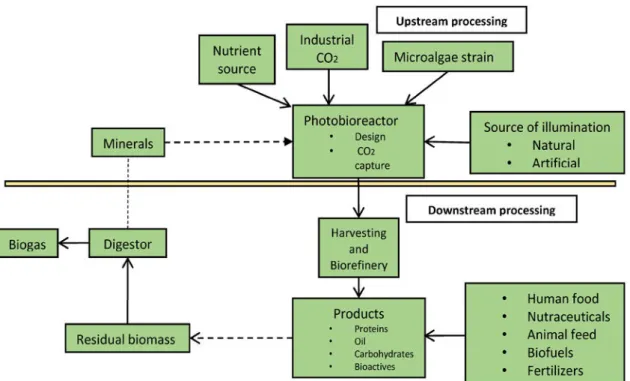
Biorefineries are found in widespread sectors at industrial scale, and this allows the biorefineries to concentrate on multiple products processing. This process is a promising way to mitigate greenhouse gas emission, and allows producing value-added bio-products through biomass transformation and process equipment. In the biorefineries, the valorisation of microalgae could be achieved by process integration. Upstream processing and downstream processing are the main stages of the microalgae biorefineries (Chew et al., 2017). Figure I.12 shows outline of the formation process of microalgal biomass and bio-products.
Upstream processing of microalgae biorefineries refers to four important factors: i) microalgae strain, ii) supply of CO2, iii) nutrient source (e.g. nitrogen and phosphorus) and iv)
source of illumination (Vanthoor-Koopmans et al., 2013). Conventional downstream processing involves all unit processes that occur within the photobioreactor (Chew et al., 2017). This processing facilitates the integration of the biomass conversion processes and equipment for the production of several fractions of interest through the use of mild separation technology (Jacob-Lopes et al. 2015).
19
Figure I. 12: Outline of the formation process of microalgal biomass and bioproducts (Jacob-Lopes et al. 2015).
I.2.2 Cultivation
The growth characteristics and chemical composition of microalgae are known to significantly depend on the cultivation conditions (Chojnacka and Marquez-Rocha, 2004). The main growth limiting factor of microalgae are: concentration and quality of nutrients, CO2 concentration, water supply, temperature (16–27 °C), exposure to light (1 000–10 000 lx),
pH values (4−11), culture density, salinity (12–40 g/L), turbulence, biological factors, presence of toxic compounds, heavy metals and synthetic organisms, as well as bioreactor operating conditions (Jankowska et al., 2017). This section describes three distinct mechanisms of microalgae cultivation, including photoautotrophic, heterotrophic and mixotrophic cultivation, all of which follow the natural growth processes. Table I.3 compares the characteristics of different cultivation conditions.
20
Table I.3: Comparison of the characteristics of different cultivation conditions (Chen et al., 2011).
Cultivation condition Energy source Carbon source Cell density Reactor scale-up Cost
Issues associated with scale-up
Photoautotrophic Light Inorganic Low Open pond or
photobioreactor Low
Low cell density High condensation cost
Heterotrophic Organic Organic High Conventional
fermentor Medium
Contamination High substrate cost
Mixotrophic Light and organic Inorganic and organic Medium Closed photobioreactor High Contamination High equipment cost
High substrate cost
Photoheterotrophic Light Organic Medium Closed
photobioreactor High
Contamination High equipment cost
High substrate cost I.2.2.1 Photoautotrophic cultivation
Currently, photoautotrophic production is the only method which is technically and economically feasible for large-scale production of microalgal biomass for non-energy production (Borowitzka, 1997). Three systems that have been deployed are based on open pond, closed photobioreactor and hybrid cultivation technologies (Borowitzka, 1999).
Open pond systems
Microalgae cultivation in open pond systems (OPR) has been utilized since the 1950s (Brennan and Owende, 2010a). OPR are reactors open to the environment, that can be classified into natural and artificial pond systems (Kroger and Muller-Langer, 2012). Raceway ponds are the commonly used types in concrete (Figure I.13) (Passos and Ferrer, 2014). OPR is a relative cheap, easy to operate and can be large-scale cultivation method. However, OPR requires long cultivation periods in which that do not exclude the contamination with other algae species and predators, or vaporization and the lack of control of the growth parameters (Koller et al., 2012). Moreover, OPR has a relatively low biomass productivity (Borowitzka, 1992), it is approximately 10–25 g dry matter of microalgal biomass per day per m3 .
�
�
2 1 �
Fi g ur e I. 1 3: Mi cr o al g a e c ulti v at e d i n r a c e w ays, C y a n ot e c h H a w ei ( a), a n d i n p arti all y c o v er e d r a c e w a ys at O ur ofi n o A gr o n e g o ci o, Br azil ( b) ( B o d e n es, 2 0 1 7 b).
Cl os e d p h ot o bi or e a ct or s yst e ms
T h e s e c o n d m et h o d f or mi cr o al g a e p h ot o a ut otr o p hi c c ulti v ati o n is i n cl os e d p h ot o bi or e a ct ors ( P B Rs). Fr e q u e ntl y us e d t y p es of P B Rs i n cl u d e t u b ul ar, fl at-t a n k, b u b bl e c ol u m n a n d s er p e nti n e (J a n k o ws k a et al., 2 0 1 7). T his t e c h n ol o g y is d esi g n e d t o o v er c o m e s o m e of t h e m aj or pr o bl e ms (s u c h as c o nt a mi n ati o n ris ks) o c c urs i n t h e O P R s yst e ms. M or e o v er , P B Rs s yst e ms h a v e hi g h er bi o m ass pr o d u cti vit y ( 2 0- 1 0 0 g dr y m att er of mi cr o al g a e bi o m ass p er d a y p er m3) as c o m p ar e d t o O P R s yst e ms ( Mir o n et al., 1 9 9 9). N e v ert h el ess, P B Rs s yst e ms h a v e s o m e dis a d v a nt a g es: hi g h er o p er ati n g a n d m ai nt e n a n c e c osts t h a n o p e n s yst e ms ( Br e n n a n a n d O w e n d e, 2 0 1 0 a).
H y bri d c ulti v ati o n s yst e ms
T h e h y bri d c ulti v ati o n is a t w o- st e p s yst e m r ef ers t o c o m bi n e p h ot o bi ore a ct ors a n d O P R gr o wt h st a ges. T h e first c ulti v ati o n st e p o c c urs i n a p h ot o bi or e a ct or t h at all o ws mi ni misi n g c o nt a mi n ati o n fr o m ot h er or ga nis ms a n d f a v o uri n g c o nti n u o us c ell di visi o n. T h e s e c o n d c ulti v ati o n st e p is ai m e d at a c c u m ul ati n g d esir e d pr o d u cts li k e li pi ds b y e x p osi n g t h e c ells t o n utri e nt str ess es ( Br e n n a n a n d O w e n d e, 2 0 1 0 a). F or e x a m pl e, t his t w o-st e p s yst e m h as b e e n us e d f or pr o d u cti o n of b ot h li pi ds a n d ast a x a nt hi n fr o m H a e m at o c o c c us pl u vi alis ( H u ntl e y a n d R e d alj e, 2 0 0 7).
I. 2. 2. 2 H et er otr o p hi c c ulti v ati o n
H et er otr o p hi c c ulti v ati o n us e d or ga ni c c ar b o n ( e. g. gl u c os e, a c et at e, cr o p fl o urs, w ast e w at er a n d ot h ers) as s u bstr at es t o r e pr o d u c e mi c r o al ga e i n stirr e d t a n k bi or e a ct ors or
22
fermenters (Tan et al., 2018). In this process, the growth of microalgae is independent of solar or light energy, using their respiration metabolism (Figure I.14) (Brennan and Owende, 2010a; Lutzu, 2012; Perez-Garcia and Bashan, 2015; Zhang et al., 2014). This system has a high degree of cell production and densities achieved thus promoting easy harvest (Chen and Chen, 2006). However, heterotrophic cultivation might cost more energy than photoautotrophic cultivation because this system cycle requires organic carbon source (Brennan and Owende, 2010a).
Figure I. 14: Photosynthesis and cellular respiration (Bodenes, 2017b).
Heterotrophic cultivation has also been successfully applied for microalgal biomass and metabolites. It was demonstrated that heterotrophic cultivation of Chlorella protothecoides resulted in the accumulation of 55% lipid content in cells, that was 4 times higher than cultivated under photoautotrophic environment (Miao and Wu, 2006).
I.2.2.3 Mixotrophic cultivation
Mixotrophic cultivation is a process wherein microalgae can be reproduced under phototrophic and heterotrophic conditions. This means that microalgae can utilize both light energy and organic carbon as substrates to sustain their growth (Brennan and Owende, 2010a; Tan et al., 2018). Compared with phototrophic and heterotrophic cultivation systems, mixotrophic cultivation is rarely used in microalgal lipids production (Chen et al., 2011). Example of the cultivation of Spirulina sp. in photoautotrophic, heterotrophic and mixotrophic systems were compared by Chojnacka and Noworyta (Chojnacka and Noworyta,
23
2004). They reported that mixotrophic cultivation has lower photoinhibition and higher growth rates as compared with both photosynthetic and heterotrophic cultivations. Successful production of mixotrophic Spirulina sp. allowed integrating both photosynthetic and heterotrophic components during day and night cycle. This process can attenuate the impact of microalgal biomass loss during dark respiration and reduces the amount of organic substances used during growth. These findings indicated that mixotrophic cultivation may be an important part of the microalgae-to-biofuels process (Brennan and Owende, 2010b).
I.2.3 Harvesting
When the biochemical process in the photobioreactor have finished, the upstream processing ends and gives way to downstream processing and harvesting of the biomass and refining of the bio-products in the biorefinery. Microalgal biomass usually contains high water content and hence, downstream processing is required to eliminate the water content. Harvesting refers to biomass recovery by one or more solid-liquid separation steps or detachment of microalgae from their growth medium, and accounts for 20-30% of the total costs of microalgae production (Mata et al., 2010; Singh and Patidar, 2018). Regardless of the objective of harvesting process, low cell densities (0.02-0.05% dry microalgae) and the small cell size (< 30 µm), make harvesting process a challenging task (Brennan and Owende, 2010a). The selection of harvesting method depends on the physiognomies of the microalgae, cell density and size, as well as specifications of the desired products and on allowability for reuse of the culture medium (Mata et al., 2010). Experience has demonstrated that an universal harvesting method does not exist, the major techniques presently applied in the harvesting of microalgae include centrifugation, flocculation, flotation, and filtration or a combination of various techniques. The advantages and disadvantages of various harvesting techniques are presented in Table I.4.
Table I.4: Advantages and disadvantages of various harvesting techniques (Abdelaziz et al., 2013; Barros et al., 2015; Mata et al., 2010).
Harvesting Technique
Advantages Disadvantages
Centrifugation Fast and effective technique;
High recovery efficiency (> 90%);
Expensive technique with high energy requirement;
High operation and maintenance costs; Appropriate for recovery of high-valued
24
Preferred for small scale and laboratory;
Applicable to all microalgae
products;
Time consuming and too expensive for large scale;
Risk of cell destruction
Flocculation Fast and easy technique;
Used for large scale; Less cell damage; Applied to vast range of species;
Less energy requirements;
Auto and bioflocculation may be inexpensive methods
Chemicals may be expensive; Highly pH dependent;
Difficult to separate the coagulant from harvesting biomass;
Efficiency depends upon the coagulant used;
Culture medium recycling is limited; Possibility of mineral or microbial contamination
Flotation Suitable for large scale;
Low cost and low space requirement;
Short operation time
Needs surfactants; Ozoflotation is expensive
Filtration High recovery efficiency;
Cost effective;
No chemical required; Low energy consumption (natural and pressure filter);
Low shear stress;
Slow, requires pressure or vacuum; Not suitable for small algae; Membrane fouling/clogging and replacement increases operational and maintenance costs;
High energy consumption (vacuum filter)
Electricity assisted techniques
Applicable to all microalgal species;
No chemicals required
Metal electrodes required;
High energy and equipment costs; Metal contamination
I.2.3.1 Centrifugation
Most microalgae can be harvested from the culture medium using centrifugation. Centrifugation process depends on the size and structure density difference of microalgal cells, as well as slurry residence time in the centrifuge (Singh and Patidar, 2018). Centrifugation is preferred for harvesting of microalgal biomass and extended shelf-life concentrates for aquaculture (Grima et al., 2003). Laboratory centrifugation tests were usually conducted at 500–1000×g and showed that about 80–90% of recovery efficiency within 2–5 min (Chen et al., 2011). This process is rapid, and can reduce the use of chemicals solvents. However,
25
centrifugation can lead to cell damage due to exposure of microalgal cells to the generated heat, high sheer and gravitational forces applied (Goh et al., 2019).
I.2.3.2 Flocculation
Flocculation involves a process that dispersed particles are aggregated together to form large particles for settling (Chen et al., 2011). Flocculation has been proposed to be the most cost effective methods for harvesting microalgal biomass as it can be used for large volumes of cultures (Vandamme et al., 2013). Since microalgal cells have a negative surface charge and found in dispersed state that results in slow natural sedimentation (Singh and Patidar, 2018). These microalgal cells can be successfully harvested by adding flocculants to cause cells aggregation or reduce the negative charge. The most used flocculants can be divided into two main types, inorganic and organic flocculants. Inorganic chemical flocculants are multivalent cations such as ferric chloride, aluminium sulfate, ferric sulphate and polyferric sulphate. Organic flocculants can be cationic, anionic, or non-ionic. It may also physically link one or more particles through a process called bridging, to facilitate the aggregation (Grima et al., 2003). The most suitable physically flocculants are multivalent metal salts, such as ferric chloride (FeCl3), aluminium sulphate (Al2(SO4)3) and ferric sulphate
(Fe2(SO4)3). Furthermore, flocculation can occur spontaneously flocculates microalgae in
suspension by other microorganisms produced some flocculants, named as bio-flocculation (Goh et al., 2019). The bio-flocculation technique has been implemented successfully in wastewater treatment plants, however, the underlying mechanism is still not very clear.
I.2.3.3 Flotation
Flotation methods are based on the binding of microalgal cells using micro-air bubbles without adding any chemicals (Brennan and Owende, 2010a). Some microalgal species can naturally float on the water surface due to low density and self-float characteristics (Burton et al., 2009). Flotation is able to recovery particles in less than 500 μm by collision and adhesion between the bubble and microalgal cells (Tan et al., 2018). Several important parameters, such as bubble size, surfactant concentration and pH, can affect the efficiency of flotation (Barros et al., 2015). Flotation processes can be classified into dissolved air flotation (DAF) and dispersed air flotation (DiAF), depending on the methods of bubble size production (Singh and Patidar, 2018; Tan et al., 2018). DAF is the most used method in the industrial wastewater treatment. During the process of DAF, small bubbles with the size range from 10
26
to 100 μm are produced. Microalgal biomass harvesting can be achieved by DAF in three paths: i) saturation at atmospheric pressure and flotation under vacuum condition, ii) saturation in static head with flow upward causing bubble formation (micro-flotation) and iii) saturation with pressure which is higher than atmospheric (Tan et al., 2018). By contrast, DiAF is the process where continuous air bubbles are generated through porous material. This process requires less energy input as compared with microbubble production method. However, small bubbles are difficult to be generated.
I.2.3.4 Filtration
Conventional filtration operates under pressure or in a vacuum (suction), which is used to harvest large quantities of microalgae (> 70 mm), such as Coelastrum and Spirulina (Brennan and Owende, 2010a; Mata et al., 2010). Tangential flow filtration is a high rate method with the advantage of maintaining the integrity of microalgae biomass. Petrusevski et al (Petrusevski et al., 1995) have successfully recovered 70-89% of fresh microalgae like Stephanodiscus hantzschii, S. Astraea, Cyclotella sp. and Rhodomonas minuta by using tangential flow filtration.
Alternative, membrane microfiltration and ultrafiltration process are the appropriate methods for harvesting smaller size of microalgae (< 30 µm) like Scenedesmus, Dunaliella and Chlorella (Brennan and Owende, 2010a; Tan et al., 2018) or fragile microalgal cells (Borowitzka, 1997; Mata et al., 2010). At larger scales of production (> 20 m3 per day) membrane filtration may be a less economic method than centrifugation because of the need for membrane exchange and pumping. However, for processing of small volumes (< 2 m3 per day), it can be more cost effective compared to centrifugation (MacKay and Salusbury, 1988). Mohn et al. (Mohn, 1980) have utilised chamber membrane filter press to harvest Coelastrum proboscideum. They obtained 27% solids of sludge that was 245-fold higher concentration than original concentration.
I.2.3.5 Electricity assisted techniques
Electricity is able to improve the efficiency of microalgae harvesting. These techniques can be deemed as environmentally friendly due to they require low chemical usage and low power consumption (Goh et al., 2019). Among them, the mechanism of electrocoagulation refers to three consecutive stages: i) generating coagulants by electrolytic oxidation of sacrifice electrode, ii) destabilization of particulate suspension and breaking of