HAL Id: tel-01241368
https://tel.archives-ouvertes.fr/tel-01241368
Submitted on 10 Dec 2015HAL is a multi-disciplinary open access archive for the deposit and dissemination of sci-entific research documents, whether they are pub-lished or not. The documents may come from teaching and research institutions in France or abroad, or from public or private research centers.
L’archive ouverte pluridisciplinaire HAL, est destinée au dépôt et à la diffusion de documents scientifiques de niveau recherche, publiés ou non, émanant des établissements d’enseignement et de recherche français ou étrangers, des laboratoires publics ou privés.
Selection of EHG parameter characteristics for the
classification of uterine contractions
Dima Alamedine
To cite this version:
Dima Alamedine. Selection of EHG parameter characteristics for the classification of uterine contrac-tions. Biomechanics [physics.med-ph]. Université de Technologie de Compiègne; Université libanaise, 2015. English. �NNT : 2015COMP2201�. �tel-01241368�
Par Dima ALAMEDINE
Thèse présentée en cotutelle pour
l’obtention du grade de Docteur de
l’UTC
Selection of EHG parameter characteristics for the
classification of uterine contractions
Soutenue le 21 juillet 2015
Spécialité : Biomedical engineering
1
COTUTELLE THESIS
To obtain the degree of Doctor issued bySorbonne University, Université de technologie de Compiègne
Doctoral School « Sciences pour l'Ingénieur »
and
Lebanese University
Doctoral School « Sciences et Technologie »
Specialty: Biomedical engineering
Presented and publicly defended by
ALAMEDINE Dima
21-7-2015
Jury Member
Régine LE BOUQUIN JEANNES Prof. , Université de Rennes 1 Reviewer
Imad H. ELHAJJ Associate Prof., American university of beirut Reviewer Oussama BAZZI Prof., Lebanese University Examiner Sofiane BOUDAOUD Associate Prof., Université de Technologie de Compiègne Examiner
Catherine MARQUE Prof., Université de Technologie de Compiègne Supervisor
Mohamad KHALIL Prof., Lebanese University Supervisor
Selection of EHG parameter characteristics for the classification of
uterine contractions
2
3
Résumé Français
Titre:
Sélection de paramètres caractéristiques des EHG pour la classification des contractions utérines
Contenu :
Certaines femmes souffrent de complications de la grossesse qui peuvent aboutir à un accouchement prématuré, avant 37 semaines de gestation. Selon l'Organisation mondiale de la Santé (WHO), le taux de mortalité périnatale est généralement autour de 7 pour 1000 naissances dans la partie la plus développée du monde [1]. Une des causes principales de mortalité et de morbidité néonatales est la naissance d’enfants avant terme. Ces enfants nés avant terme présentent un risque élevé de mortalité ainsi que des problèmes de santé et de développement [2]. Un objectif principal de la surveillance de la grossesse est de maintenir le bien-être de la mère et du fœtus et de garder ce dernier in utero aussi longtemps que nécessaire pour la naissance d’un enfant en bonne santé. Par conséquent, la détection précoce d'un accouchement prématuré (AP) est importante pour sa prévention. A cet effet, de bons indicateurs de travail prématuré sont nécessaires.
Pour maintenir le fœtus in utero aussi longtemps que nécessaire, une surveillance de la contractilité utérine est essentielle pour distinguer les contractions normales de la grossesse, qui sont inefficaces de celles qui sont efficaces et pourraient entraîner la dilatation du col de l'utérus et causer une naissance prématurée.
Malgré l'augmentation des connaissances et de la compréhension des phénomènes impliqués dans le début du travail prématuré, les méthodes actuellement utilisées en obstétrique ne sont pas assez précises pour une détection précoce de menaces d’accouchement prématuré. La mesure de pression intra-utérine, seule méthode directe précise pour mesurer la force des contractions utérines, est invasive et ne peut clairement pas être utilisée pendant la grossesse. La tocographie externe, non invasive, est la méthode la plus largement utilisée pour la surveillance des contractions utérines pendant la grossesse. Cependant, elle ne permet pas de caractériser l’efficacité des contractions. Elle ne permet que de détecter le nombre de contractions pendant un intervalle de temps donné. Il a été démontré que ce paramètre n’est pas un bon indicateur pour
4
prédire un accouchement prématuré. Des tests biologiques, tels que la fibronectine, ont été cliniquement utilisés pour le pronostic des accouchements prématurés, bien qu’ils possèdent une valeur prédictive faible [3]. Même la mesure de la dilatation cervicale n’est pas un indicateur fiable d’une menace d’accouchement prématuré.
Nous avons besoin d'une méthode non-invasive et plus fiable pour la détection précoce et la prévention des menaces d’accouchement prématuré, car ce problème est clairement un domaine d’intérêt en santé publique. Ce diagnostic précoce permettait une administration plus rapide d’agents tocolytiques (inhibiteurs de la contractilité utérine) et un maintien du foetus in utero, ce qui réduirait la mortalité et la morbidité périnatales. Une des méthodes prometteuses pour surveiller l’efficacité des contractions utérines pendant la grossesse est l’analyse de l’activité électrique du muscle utérin, l’électrohystérogramme (EHG), qui a commencé dans les années 1950 et a été développé dans les années 1980. L’EHG est le signal recueilli par des électrodes positionnées sur l’abdomen des femmes enceintes. Il représente l’activité électrique associée à la contraction mécanique de l’utérus [4]. De nombreuses études ont prouvé qu’il est représentatif de l’activité électrique utérine recueillie en interne sur l’utérus [5,6]. De multiples travaux menés depuis plus de 15 ans, concluent qu’il est possible d’utiliser l’EHG pour détecter une menace d’accouchement prématuré. L’efficacité des contractions utérines est liée à une augmentation de deux phénomènes physiologiques : l'excitabilité cellulaire et la synchronisation de l’utérus (propagation de l'activité électrique) [5,7]. Ces phénomènes peuvent être analysés au moyen de l’EHG grâce à des analyses univariée (analyse d’un seul signal à la fois pour l’excitabilité) et bivariée (couplage de deux signaux pour la propagation).
Plusieurs outils de traitement du signal EHG, nouvellement développés, permettent l'analyse de l'excitabilité et de la propagation de l'activité électrique utérine (paramètres fréquentiels, analyse de complexité, propagation linéaire ou nonlinéaire) pour trouver des informations spécifiques qui différencient les contractions de la grossesse de celles du travail. Un grand nombre de paramètres ont à ce jour été extraits du signal EHG par de nombreux chercheurs, en utilisant différents protocoles d'enregistrement et différentes populations de femmes enceintes. En ce qui concerne la classification des signaux, la complexité des calculs requis pour le diagnostic augmente avec le nombre de paramètres mis en jeu. La réduction de la dimension de paramètres grâce à l'élimination des paramètres non pertinents et bruités est très importante en reconnaissance des
5
formes. De plus, les deux facteurs, excitabilité utérine et propagation de l’activité, sont tous les deux importants car ils doivent évoluer tous les deux pour passer d’une contraction inefficace de grossesse (faible excitabilité, contraction locale) à une contraction efficace d’accouchement (excitabilité forte, propagation à tout l’utérus en un court intervalle de temps). Des études antérieures, fondées uniquement sur l'étude de l'excitabilité, ont donné des résultats intéressants, mais pas suffisamment fiables pour la détection précoce de l’accouchement prématuré.
Par conséquent, la première contribution de notre étude sera de combiner les deux types d'informations (excitabilité et propagation) par des approches monovariée et bivariée simultanées, pour différencier deux types de contractions utérines : les contractions de grossesse et celles d’accouchement.
La seconde contribution sera de tester sur une même population de femmes enceintes (grossesse, travail), quels outils de traitement du signal, récemment développés pour l'analyse monovariée et bivariée de l’EHG, donnent la meilleure discrimination entre les contractions de grossesse et d’accouchement. A cet effet plusieurs méthodes de sélection de paramètres extraits de l’EHG seront analysées.
Cette sélection de paramètres se fera à partir d’une base de données de signaux recueillis suivant une méthode normalisée, sur des femmes enceintes dans différentes situations physiologiques (grossesse normale, accouchement), grâce à un système multi-électrodes permettant d’enregistrer 16 EHG monopolaires simultanés. Ces 16 signaux seront ensuite analysés par les approches monovariée et bivariée, pour extraire les paramètres représentatifs de l'excitabilité et la propagation de l'activité électrique utérine. Beaucoup d’études ont utilisé seulement des signaux issus des électrodes positionnées sur l'axe vertical médian de l'abdomen [8] pour l’analyse monovariée (une seule voie). D’autres études ont travaillé sur les caractéristiques liées à la propagation de l’EHG grâce au couplage entre toutes les voies (analyse bivariée). Des études précédentes ont ainsi tenté d'appliquer une classification multivoie [9-14]. Certaines études se sont intéressées à une analyse multivariée, afin d’extraire les informations d'excitabilité et de propagation de toutes les voies et combinaisons de voies disponibles, ce qui conduit à une très grande dimension de recherche. Par conséquent, une troisième contribution de ce travail portera sur la sélection des voies et des combinaisons de voies les plus pertinentes dans une optique de classification grossesse/accouchement.
6
En outre, afin d'augmenter le rapport signal/bruit des EHG, toutes les études précédentes ont porté sur des signaux bipolaires, obtenus par différence entre deux signaux recueillis par deux électrodes plus ou moins proches. Si cette différenciation se justifie pour une approche monovariée, le fait de différencier les signaux diminue cependant la résolution spatiale et conduit à un biais pour l'étude bivariée de la propagation entre deux voies adjacentes. Des travaux récents ont permis de développer une méthode de débruitage des EHG monopolaires, afin d’obtenir sans différenciation un rapport signal/bruit suffisant pour envisager le traitement de ces signaux [15]. Ces outils s’appuient sur une combinaison de CCA (Combination of Canonical component Analysis) et d’EMD (Empirical Mode Decomposition). De ce fait, en plus d’une approche classique basée sur l’étude des signaux bipolaires, nous proposerons dans ce travail d’appliquer les approches monovariée et bivariée, ainsi que la sélection de paramètres et de voies, sur des signaux monopolaires débruités.
Ce manuscrit est donc organisé comme suit:
Chapitre 1: contient toutes les informations essentielles pour la bonne compréhension de l’anatomie et de la physiologie de l'activité utérine nécessaires à ce travail. Nous définirons l’accouchement prématuré et les problèmes qui y sont reliés, et nous présenterons certaines méthodes utilisées en pratique obstétricale courante pour détecter l’accouchement prématuré. Ensuite, nous présenterons une bibliographie des différentes études d'excitabilité et de la propagation faites à partir de signaux EHG (approches monovariée et bivariée). À la fin de ce chapitre, nous décrirons le système multi-électrodes pour l'enregistrement d'EHG utilisé dans notre travail, ainsi que les bases de données utilisées.
Chapitre 2: présente le travail effectué sur la sélection de paramètres de l’EHG. Nous décrirons dans ce chapitre l'ensemble de paramètres choisis dans la littérature pour l’analyse monovariée et bivariée de l’EHG [16-26]. Puis nous présentons les différentes méthodes de sélection de paramètres, qui sont décomposées en deux types « filter » et « wrapper ». Dans la première partie de ce chapitre, nous présenterons un algorithme de sélection proposé pour les paramètres calculés sur l’EHG original et sur différentes bandes de fréquence, en utilisant la technique de sélection de paramètre, de type filter,
7
nommé Jeffrey Divergence (JD). Cette méthode de sélection, basée sur la mesure de
similarité ou dissimilarité des histogrammes obtenus, pour un paramètre donné, pour les deux classes (grossesse et travail).
Dans la deuxième partie, nous testerons plusieurs méthodes de sélection de paramètres afin de sélectionner les paramètres les plus pertinents pour discriminer les contractions de grossesse de celles d’accouchement. Quatre méthodes de type de « filter » sont utilisées : Jeffrey divergence (JD) [27], « F-score » [28], «Relieff » [29], «mutual information based on clustering» (MI)[30] et sept méthodes de type « wrapper » : sélection séquentielle croissante « sequential Forward Selection» (SFS) [31], sélection séquentielle arrière « Sequential backward Selection» (SBS)[32], « Plus-l minus-r selection» (LRS)[33], recherche bidirectionnelle «Bidirectional search» (BDS) [33], «Sequential Forward Floating sequential» (SFFS) [34], algorithmes génétiques «Genetic Algorithm» (GA) [35] et «Binary Particle swarm optimization» (BPSO)[36]. Nous utiliserons le classificateur KNN pour les méthodes de type « wrapper », et deux méthodes de répartition de données pour l’apprentissage de ce classifieur: « Holdout » et « KFOLD». Après la partie sélection de paramètres, nous essayerons de valider chaque sous-ensemble de paramètres sélectionné, en calculant pour chacun le pourcentage de classification correcte obtenu sur une population d’EHG différente de celle utilisée pour la sélection.
Dans ce chapitre, la première partie sera appliquée uniquement sur des signaux bipolaires. Les autres études seront appliquées sur les signaux bipolaires et monopolaires. Les résultats obtenus montrent que l’utilisation des méthodes basées sur la sélection de paramètres permet de mettre en évidence un groupe de paramètres, combinant analyse monovariée et bivariée, et qui démontre sa capacité à discriminer les contractions de grossesses de celles d’accouchement, avec de meilleures performances pour les signaux EHG bipolaires.
Chapitre 3: dans ce chapitre, nous présenterons tout d'abord le travail effectué sur la sélection de voies (approche monovariée) en utilisant les paramètres linéaires et non linéaires. Nous testerons deux méthodes de type « filter » (F-score et relieff) pour sélectionner les voies appropriées, puis deux méthodes de type « wrapper » (Genetic algorithm et binary particle swarm optimization) avec le classifieur KNN et deux
8
méthodes de répartition de données (Holdout et KFOLD) pour sélectionner les meilleurs paramètres à partir des voies sélectionnées. En outre, une partie de validation sera effectuée en calculant le pourcentage de classification correcte obtenu (en utilisant une autre population d’EHG) à partir des sous-ensembles de paramètres sélectionnés par
sélection de voie suivie de la sélection de paramètres.
La dernière partie de ce chapitre présentera les résultats de la sélection des combinaisons
de voies (approche bivariée) en utilisant les paramètres liés à la propagation de l’EHG.
Nous utilisons ici la même procédure que celle utilisée dans l’approche monovariée, pour la sélection de voies, suivie de la sélection de paramètres.
Les résultats obtenus montrent que, pour l’analyse monovariée, les canaux bipolaires et monopolaires qui offrent une meilleure capacité de discrimination entre les contractions de grossesse et de travail sont ceux positionnés sur l'axe vertical médian de l'abdomen de la femme enceinte. L’utilisation des signaux d’EHG bipolaires pour l’analyse monovariée donne de meilleurs résultats que celle des EHG monopolaires, particulièrement en utilisant l'ensemble des paramètres linéaires et non linéaires extraits des voies Vb7, Vb8 et Vb9. De plus, l’approche bivariée, sélection des combinaisons de
voies suivie de sélection de paramètre, montre que l'utilisation des EHG monopolaires pour l'étude de la propagation améliore les résultats de classification entre les contractions de grossesse de celles de travail et permet d’atteindre des résultats similaires à ceux obtenus avec les signaux bipolaires.
Les résultats obtenus dans cette thèse nous ont permis d'écrire 3 articles de revues publiés (2 revues internationales et 1 nationale), 2 conférences internationales, 4 conférences nationales.
References
[1] Neonatal and perinatal mortality: country, regional and global estimates; ReportWHO 2006. http://whqlibdoc.who.int/publications/2006/9241563206_eng.pdf
[2] R. L. Goldenberg, J. F. Culhane, J. D. Iams, and R. Romero, “Epidemiology and causes of preterm birth,” The lancet, vol. 371, no. 9606, pp. 75–84, 2008.
[3] J.D. Iams, “Prediction and early detection of preterm labor,” Obstet. Gynecol., vol. 101, no. 2, pp. 402-12, Feb. 2003.
9
[4] H. Alvarez and R. Caldeyro, “Contractility of the human uterus recorded by new methods,” Surgery, Gynecology & Obstetrics, vol. 91, no. 1, pp. 1–13, 1950.
[5] D. Devedeux, C. Marque, S. Mansour, G. Germain, and J. Duchene, "Uterine electromyography: a critical review," Am J Obstet Gynecol, vol. 169, pp. 1636-1653, Dec 1993. [6] S. Mansour, D. Devedeux, G. Germain, C. Marque, and P. J. Duchene, “Uterine EMG spectral analysis and relationship to mechanical activity in pregnant monkeys,” Med. Biol. Eng.
Comput., vol. 34, no. 2, pp. 115–121, Mar. 1996.
[7] R. E. Garfield and W. L. Maner, "Physiology and electrical activity of uterine contractions," Seminars in Cell & Developmental Biology, vol. 18, pp. 289-295, 2007.
[8] C. K. Marque, J. Terrien, S. Rihana, and G. Germain, “Preterm labour detection by use of a biophysical marker: the uterine electrical activity,” BMC Pregnancy Childbirth, vol. 7, no. Suppl 1, p. S5, Jun. 2007.
[9] B. Moslem, M. Khalil, M. O. Diab, A. Chkeir, and C. Marque, “A multisensor data fusion approach for improving the classification accuracy of uterine EMG signals,” in
Electronics, Circuits and Systems (ICECS), 2011 18th IEEE International Conference on, 2011,
pp. 93–96.
[10] B. Moslem, M. O. Diab, C. Marque, and M. Khalil, “Classification of multichannel uterine EMG signals,” in Engineering in Medicine and Biology Society, EMBC, 2011 Annual
International Conference of the IEEE,pp. 2602–2605, 2011.
[11] B. Moslem, M. O. Diab, M. Khalil, and C. Marque, “Classification of multichannel uterine EMG signals by using unsupervised competitive learning,” in Signal Processing Systems
(SiPS), 2011 IEEE Workshop on, 2011, pp. 267–272.
[12] B. Moslem, M. Khalil, M. O. Diab, A. Chkeir, and C. Marque, “Combining multiple support vector machines for boosting the classification accuracy of uterine EMG signals,” in
Electronics, Circuits and Systems (ICECS), 2011 18th IEEE International Conference on, 2011,
pp. 631–634.
[13] B. Moslem, M. O. Diab, M. Khalil, and C. Marque, “Classification of multichannel uterine EMG signals using a reduced number of channels,” in Mechatronics and its Applications
(ISMA), 2012 8th International Symposium on, pp. 1–4, 2012.
[14] B. Moslem, M. Diab, M. Khalil, and C. Marque, “Combining data fusion with multiresolution analysis for improving the classification accuracy of uterine EMG signals,”
EURASIP J. Adv. Signal Process., vol. 2012, no. 1, pp. 1–9, Aug. 2012.
[15] M. Hassan, S. Boudaoud, J. Terrien, B. Karlsson, and C. Marque, “Combination of Canonical Correlation Analysis and Empirical Mode Decomposition Applied to Denoising the Labor Electrohysterogram,” IEEE Trans. Biomed. Eng., vol. 58, no. 9, pp. 2441–2447, Sep. 2011.
10
[16] T. Jérémy, S. Thora, M. Catherine, K. Brynjar, and others, “Synchronization between EMG at different uterine locations investigated using time-frequency ridge reconstruction: comparison of pregnancy and labor contractions,” EURASIP J. Adv. Signal Process., vol. 2010, Article ID 242493, 2010.
[17] J. Sikora, A. Matonia, R. Czaba´nski, K. Horoba, J. Jezewski, and T. Kupka, “Recognition of premature threatening labour symptoms from bioelectrical uterine activity signals,” Archives of Perinatal Medicine, vol. 17, no. 2, pp. 97–103, 2011.
[18] C. Marque, H. Leman, M. L. Voisine, J. Gondry, and P. Naepels, “Traitement de l’électromyogramme utérin pour la caract´erisation des contractions pendant la grossesse,”
RBMNews, vol. 21, no. 9, pp. 200–211, Dec. 1999.
[19] G. Fele-ˇZorˇz, G. Kavˇsek, ˇZ. Novak-Antoliˇc, and F. Jager, “A comparison of various linear and non-linear signal processing techniques to separate uterine EMG records of term and preterm delivery groups,” Medical and Biological Engineering and Computing, vol. 46, no. 9, pp. 911–922, 2008.
[20] M. O. Diab, C. Marque, and M. A. Khalil, “Classification for uterine EMG signals: comparison between AR model and statistical classification method,” International Journal of
Computational Cognition, vol. 5, no. 1, pp. 8–14, 2007.
[21] A. Diab, M. Hassan, C. Marque, and B. Karlsson, “Quantitative performance analysis of fourmethods of evaluating signal nonlinearity: application to uterine EMG signals,” in
Proceedings of the 34th Annual International IEEE EMBS Conference, San Diego, Calif, USA,
September 2012,pp. 1045–1048.
[22] B. Moslem, M. Khalil, M. O. Diab and C. Marque, “Detrended fluctuation analysis of uterine electromyography,” in First Middle East Conference on Biomedical Engineering,
MECBME11, Sharjah, UAE, 2011.
[23] K. Ansari-Asl, F. Wendling, J. J. Bellanger, and L. Senhadji, “Comparison of two estimators of time-frequency interdependencies between nonstationary signals: application to epileptic EEG,” in 26th Annual International Conference of the IEEE Engineering in Medicine
and Biology Society, 2004. IEMBS ’04, 2004, vol. 1, pp. 263–266.
[24] M. Hassan, A. Alexandersson, J. Terrien, C. Muszynski, C. Marque, and B. Karlsson, “Better pregnancy monitoring using nonlinear propagation analysis of external uterine electromyography”, IEEE Transactions on Biomedical Engineering, Vol. 60, No. 4, 2013,pp. 1160–1166.
[25] M. Hassan, J. Terrien, A. Alexandersson, C. Marque, and B. Karlsson, “Improving the classification rate of labor vs. normal pregnancy contractions by using EHG multichannel recordings,” Presented at the 32 nd Annual International Conference of the IEEE Engineering in
11
[26] A.DIAB, “Study of The Nonlinear Properties And Propagation Characteristics Of The Uterine Electrical Activity During Pregnancy And Labor”, Ph.D. dissertation, Thèse de l’université de Technologie de Compiègne, 2014
[27] Y. Ma, X. Gu, and Y. Wang, “Histogram similarity measure using variable bin size distance,” Comput. Vis. Image Underst., vol. 114, no. 8, pp. 981–989, Aug. 2010.
[28] S. Ding, “Feature Selection Based F-Score and ACO Algorithm in Support Vector Machine,” in Second International Symposium on Knowledge Acquisition and Modeling, 2009. KAM ’09, 2009, vol. 1, pp. 19–23.
[29] I. Kononenko, “Estimating attributes: analysis and extensions of RELIEF,” in Machine
Learning: ECML-94, 1994, pp. 171–182.
[30] H. Liu, Y. Mo, J. Wang, and J. Zhao, “A new feature selection method based on clustering,” in 2011 Eighth International Conference on Fuzzy Systems and Knowledge
Discovery (FSKD), 2011, vol. 2, pp. 965–969.
[31] L. Ladha and T. Deepa, “Feature selection methods and algorithms,” Int. J. Comput. Sci.
Eng., vol. 3, no. 5, pp. 1787–1797, 2011.
[32] A. W. Whitney, “A direct method of nonparametric measurement selection,” Comput.
IEEE Trans. On, vol. 100, no. 9, pp. 1100–1103, 1971.
[33] A. R. Webb, Statistical Pattern Recognition. John Wiley & Sons, 2003.
[34] P. Pudil, J. Novovičová, and J. Kittler, “Floating search methods in feature selection,”
Pattern Recognit. Lett., vol. 15, no. 11, pp. 1119–1125, Nov. 1994.
[35] M. Melanie, “An introduction to genetic algorithms,” Camb. Mass. Lond. Engl. Fifth
Print., vol. 3, 1999.
[36] V. Ranaee, A. Ebrahimzadeh, and R. Ghaderi, “Application of the PSOSVM model for recognition of control chart patterns,” ISA Transactions, vol. 49, no. 4, pp. 577–586, 2010.
12
Contents
General Introduction………... 21
List of author’s publications……….. 25
References……….. 26
Chapter 1: Clinical problem: detection of Preterm Labor by means of uterine electromyography………. 30
1.1 Introduction ... 30
1.2 Anatomy and physiology of the uterus ... 31
1.2.1 Uterus structure ... 31
1.2.2 Mechanical activity of the uterus ... 32
1.2.3 Electrical activity of the uterus... 33
1.2.3.1 Cellular excitability ... 33
1.2.3.2 Uterine synchronization ... 34
1.3 Problem of preterm labor ... 35
1.4 Methods for monitoring pregnancy ... 36
1.5 A new method for Preterm labor detection: Uterine Electromyography ... 37
1.5.1 Parameters extracted from EHG ... 39
1.5.1.1 Excitability analysis - Linear parameters ... 39
1.5.1.2 Excitability analysis - NonLinear parameters ... 42
1.5.1.3 Synchronization analysis – propagation/correlation parameters ... 43
1.5.1.4 Combination of parameters ... 43
1.5.2 EHG Recording ... 45
1.5.2.1 Electrode number and position ... 45
1.5.2.2 Channel selection ... 47
1.5.2.3 Electrode configuration ... 48
1.5.3 Multichannel system for standardized EHG recording: the TMSi system ... 48
1.6 Thesis aim and work ... 51
1.6.1 Thesis goal... 51
1.6.2 The used databases ... 52
1.7 Discussion and Conclusion ... 54
13
Chapter 2: Feature selection for classification of Electrohysterograms………63
2.1 Introduction ... 63
2.2 Feature selection methods ... 64
2.2.1 Introduction ... 64
2.2.2 Feature selection process ... 65
2.2.2.1 Search procedures ... 66
2.2.2.2 Evaluation ... 67
2.2.2.3 Stopping Criteria... 68
2.2.3 Feature selection methods selected from the literature ... 69
2.2.3.1 Filter method... 71
2.2.3.2 Wrapper Method ... 78
2.3 Signal characterization and feature extraction ... 88
2.3.1 Monovariate analysis: Linear features ... 88
2.3.1.1 Features related to power spectral density ... 88
2.3.1.2 Features extracted from wavelet packet decomposition ... 88
2.3.2 Monovariate analysis: Nonlinear features ... 91
2.3.2.1 Time reversibility ... 91
2.3.2.2 Lyapunov exponent ... 91
2.3.2.3 Sample entropy ... 93
2.3.2.4 Detrended fluctuation analysis ... 93
2.3.2.5 Variance entropy ... 95
2.3.3 Bivariate analysis: Features related to EHG propagation ... 95
2.3.3.1 Correlation coefficient ... 95
2.3.3.2 Phase synchronization ... 97
2.4 Preliminary study: proposition of Selection algorithm for parameters using the Jeffrey Divergence Distance ... 98
2.4.1 Calculated Features and their histograms... 98
2.4.2 Features selection method based on Jeffrey divergence ... 100
2.4.3 Results on the selection using the Jeffrey Divergence method ... 101
2.4.3.1 Features computed on the original EHG ... 101
14
2.4.5 Discussions ... 103
2.5 Feature selection from bipolar and monopolar EHG signals ... 105
2.5.1 Calculating Features ... 105
2.5.2 Feature selection methods ... 106
2.5.3 Results ... 108
2.5.3.1 Results of Features selection ... 108
2.5.3.2 Validation Part ... 116
2.5.4 Discussion ... 119
References ... 122
Chapter 3: Channel and channel combination selection for monovariate and bivariate analysis on EHG……….129
3.1 Introduction ... 129
3.2 Channel selection for EHG monovariate analysis ... 130
3.2.1 Calculated Features ... 130
3.2.2 Channel selection followed by feature selection ... 130
3.2.3 Results ... 131
3.2.3.1 Channel selection using Relieff and F-score ... 131
3.2.3.2 Feature selection using BPSO and GA on the selected channels ... 133
3.2.3.3 Validation ... 135
3.2.4 Discussion ... 136
3.3 Channel combination selection for EHG bivariate analysis ... 137
3.3.1 Calculated Features ... 137
3.3.2 Channel Combination selection followed by feature selection ... 138
3.3.3 Results ... 138
3.3.3.1 Results of channel combinations using Relieff and F-score ... 138
3.3.3.2 Results of feature selection using BPSO and GA ... 143
3.3.3.3 Validation ... 144
3.3.4 Discussion ... 145
3.4 Discussion and Conclusion ... 146
15
General Conclusion and perspectives………...151
References………156
Appendix A………157
Appendix B……….163
16
List of Figures
Chapter 1
Figure 1.1: Female reproductive system . ... 32
Figure 1.2: Electrical activity of rat uterus at different terms of pregnancy . ... 34
Figure 1.3: Evolution of GJ surface during Pregnancy, Labor and Post-Partum . ... 35
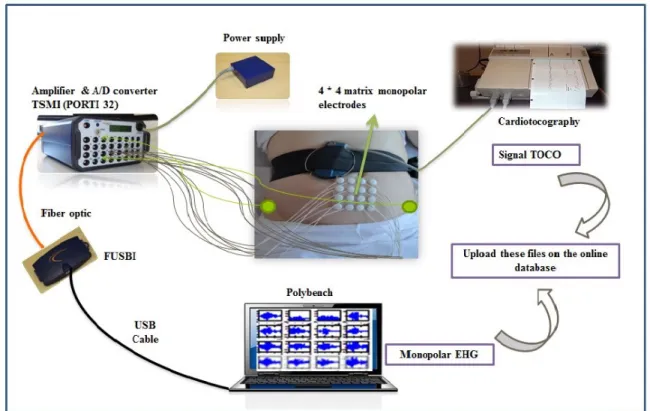
Figure 1.4: The new multichannel system: 4x4 electrode matrix placed on the women abdomen with two reference electrodes on the hip and the tocographic probe. ... 49
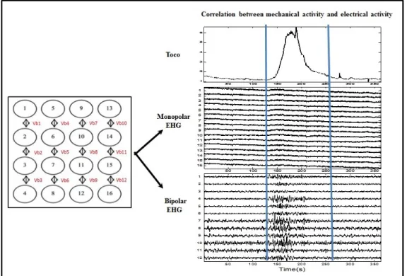
Figure 1.5: Digitized tocodynamometer paper (Top), monopolar signals (middle), corresponding bipolar signals (bottom). The blue lines define the beginning and the end of the contraction according to TOCO. ... 50
Chapter 2
Figure 2.1: Feature Selection technique ... 65Figure 2.2: Feature Selection Process. ... 66
Figure 2.3: Filter based feature Selection Process ... 67
Figure 2.4: Wrapper based feature Selection Process... 68
Figure 2.5: Wavelet decomposition ... 91
Figure 2.6: (A) Time series. (B) Integrated series 𝑋𝑘.Vertical dotted lines represent windows of length n. Solid lines are the trends estimated for each window by the least square method ... 94
Figure 2.7: Tree of the wavelet packet transform. The underlined packets are the ones selected in this study. ... 99
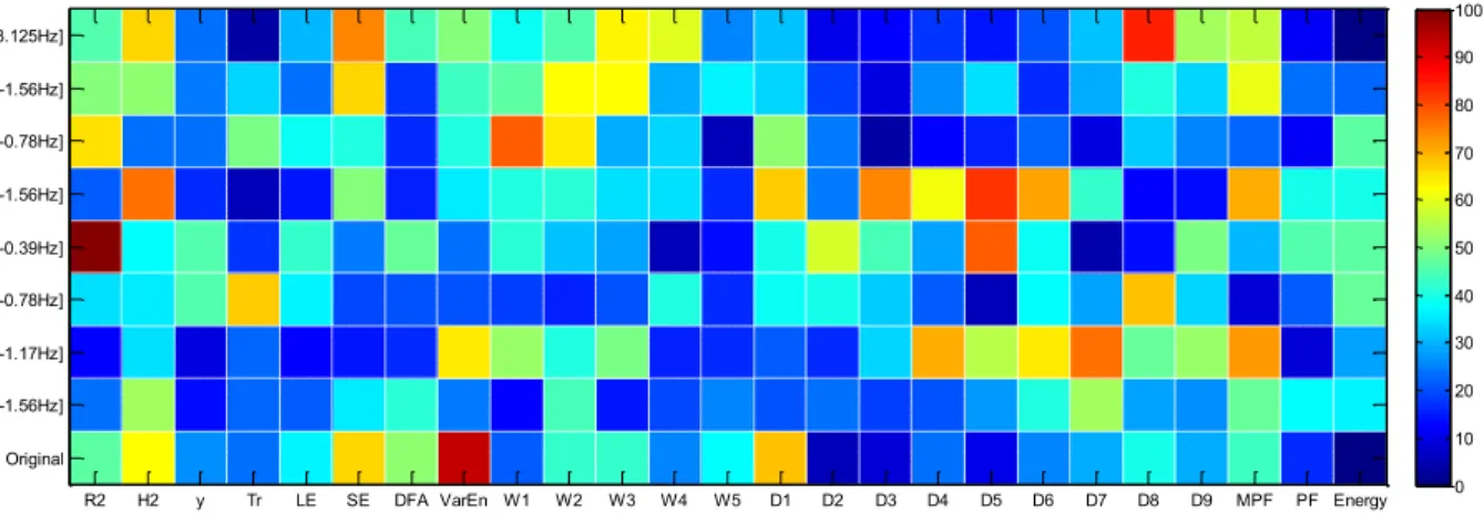
Figure 2.8: Matrix colors presenting the distances between histograms of features, on different packets and on original EHG ... 100
Figure 2.9: Selection matrix, presenting the best features on packets and original EHG for the discrimination between pregnancy and labor. These features are selected by applying threshold1. ... 102
Figure 2.10: Selection matrix, presenting the best features on packets and original EHG for the discrimination between pregnancy and labor. These features are selected by applying threshold2. ... 103
Figure 2.11: (A) Color vector representing the distribution of distances between features from bipolar EHGs. (B) Selection vector representing the most discriminating features for the discrimination between bipolar pregnancy and labor EHG bursts. ... 108
17
Figure 2.12: (A) Color vector representing the distribution of distances between features from
monopolar EHGs. (B) Selection vector representing the most discriminating features for the discrimination between monopolar pregnancy and labor EHG bursts. ... 109
Figure 2.13: (A) Color vector representing the distribution of F-score values for bipolar dataset.
(B) Selection vector representing the best features for the discrimination between pregnancy and labor contractions using bipolar EHGs. ... 109
Figure 2.14: (A) Color vector representing the distribution of F-score values for monopolar
dataset. (B) Selection vector representing the best features for the discrimination between pregnancy and labor contractions using monopolar EHGs... 109
Figure 2.15: (A) Color vector representing the distribution of weight values for bipolar dataset.
(B) Selection vector representing the best features for the discrimination between pregnancy and labor contractions using bipolar EHGs. ... 110
Figure 2.16: (A) Color vector representing the distribution of weight values for monopolar
dataset. (B) Selection vector representing the best features for the discrimination between pregnancy and labor contractions using monopolar EHGs... 110
Figure 2.17: (A) J velocity using bipolar contraction. (B) J velocity using monopolar
contractions. ... 111
Figure 2.18: Evolutions of the procedures for the sequential methods using KNN-KFOLD with
the monopolar dataset. (A) SFS-KNN-KFOLD. (B) SBS-KNN-KFOLD. (C) BDS-SFS-KNN-KFOLD. (D) BDS-SBS-KNN-BDS-SFS-KNN-KFOLD. (E) LRS-KNN-BDS-SFS-KNN-KFOLD. (F) SFFS-KNN-BDS-SFS-KNN-KFOLD. .. 112
Figure 2.19: Evolution of the procedure of GA-KNN-KFOLD on bipolar dataset ... 115
Chapter 3
Figure 3.1: (A) Position of the 16 monopolar electrodes. (B) Blue circles: Monopolar and
Bipolar channels selected by F-score. (C) Blue circle: Monopolar and Bipolar channels selected by Relieff. ... 133
Figure 3.2: Bipolar channel combinations selection by F-score using five features related to
EHG propagation: R2(A), H2 (B), y (C), FW_H2 (D) and FW_H (E). Each axis (vertical and
horizontal) represents the channel numbers. A white square indicates that the related combination is selected for the given feature. (F) Number of appearance of the bipolar channel combinations over the 5 features. ... 140
Figure 3.3: Most repetitive bipolar channel combinations retained for F-score (A) and relieff (B)
in the 5 features related to EHG bivariate analysis. ... 140
Figure 3.4: Most repetitive monopolar channel combinations retained for F-score (A) and relieff
18
Figure 3.5: Color matrix of dimension 12x12 representing the distribution of the number of
retained bipolar channels combinations using F-score (A) and relieff (B) ... 142
Figure 3.6: Color matrix of dimension 16x16 representing the distribution of the number of
19
List of tables
Chapter 1
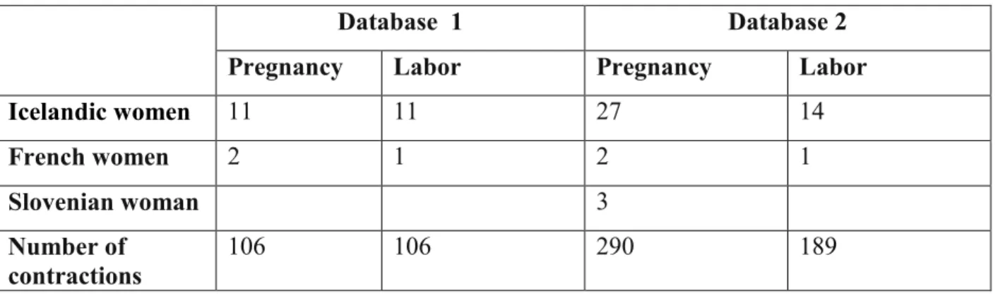
Table 1.1: Number of women and contractions used in database 1 and database 2 ... 53
Table 1.2: Number of women and contractions used in database 2 (split data for the classification into training, testing and validation part) ... 54
Chapter 2
Table 2.1: Advantage and disadvantage of filter and wrapper methods ... 69Table 2.2: Filter methods ... 70
Table 2.3: Wrapper methods ... 70
Table 2.4: Parameters used in GA ... 85
Table 2.5: Features selected for the best discrimination between pregnancy and labor contractions, for the original signal ... 101
Table 2.6: Features selected for the best discrimination between pregnancy and labor contractions, for packets and original signal. These features are selected by applying threshold2. ... 103
Table 2.7: Subsets of features selected using several filter methods, for the best discrimination between pregnancy and labor contractions, as well as their related computation time. ... 111
Table 2.8: Selected feature subsets using sequential methods on bipolar and monopolar datasets as well as their related computation time. ... 113
Table 2.9: Selected features subset using Genetic algorithm (GA) on bipolar and monopolar datasets, as well as their related computation time. ... 115
Table 2.10: Selected features subset using binary particle swarm optimization (BPSO) on bipolar and monopolar datasets, as well as their related computation time. ... 116
Table 2.11: Subsets of features selected for bipolar and monopolar dataset with highest Mean ± STD (over 500 repetitions) for the percentage of correct classification using KNN... 117
Table 2.12: Mean ± STD of the percentage of correct classification using KNN of 500 repetitions of all features on bipolar and monopolar EHG ... 118
20
Chapter 3
Table 3.1: Bipolar channels appearance after channel selection using F-score (A) and Relieff (B)
... 132
Table 3.2: Monopolar channels appearance after channel selection using F-score (A) and Relieff
(B). ... 132
Table 3.3: Dataset obtained after bipolar and monopolar channel selection using F-score and
Relieff ... 134
Table 3.4: Subsets of features selected from the bipolar and monopolar channels with the highest
percentage of correct classification using KNN ... 135
Table 3.5: Selected feature subset from the bipolar and monopolar channel selection with the
highest Mean± STD (over 500 repetitions) of the percentage of correct classification using KNN. ... 136
Table 3.6: Performance of the selected feature subset from the bipolar and monopolar selected
channels giving the highest Mean ± STD (over 500 repetitions) of the percentage of correct classification using KNN. ... 136
Table 3.7: Name of the retained bipolar and monopolar channel combinations over the 5 features
related to EHG bivariate analysis for F-score and Relieff ... 142
Table 3.8: Datasets obtained for the retained bipolar and monopolar channel combinations, for
F-score and Relieff ... 144
Table 3.9: Subsets of features selected from the bipolar and monopolar selected channel
combinations with highest percentage of correct classification using KNN ... 144
Table 3.10: Selected feature subset from the bipolar and monopolar selected channel
combinations with highest Mean ± STD of the percentage of correct classification using KNN of 500 repetitions ... 145
Table 3.11: Performance of the selected feature subset from the bipolar and monopolar selected
channel combinations with highest Mean ± STD of the percentage of correct classification using KNN of 500 repetitions ... 145
21
General Introduction
Some women suffer from complications of their pregnancy that may end with a preterm delivery, that is before 37 weeks of gestation. According to the World Health Organization (WHO), the perinatal mortality rate is typically around 7 per 1,000 births in the most developed part of the world [1]. A large part of the causes for mortality and morbidity is that children are born before their time. Children born preterm present high risk of mortality, as well as health and development problems [2]. A primary aim of pregnancy monitoring is to maintain the wellbeing of both mother and fetus and to keep the latter in utero as long as needed for a healthy birth. Therefore, the early detection of a preterm labor (PL) is important for its prevention and, for that purpose, good indicators of preterm labor are needed.
To maintain the fetus in utero as long as necessary, monitoring of uterine contractility is essential to differentiate the normal contractions of the pregnancy, which are ineffective, from those, effective, that could cause premature cervical dilation of uterus and preterm birth.
Despite increased knowledge and understanding of the phenomena involved in the onset of labor, the methods currently used in obstetrics are not precise enough for an early detection of preterm birth threats. The measurement of intrauterine pressure, the only direct method that permits presently to accurately measure the force of uterine contractions, is invasive and cannot clearly be used during pregnancy. Tocographie external, non-invasive, is the most widely used method for monitoring of uterine contractions during pregnancy. However, it does not permit to characterize the efficiency of contractions. It only permits to detect the number of contractions over a given time interval. It has been shown that this parameter is not a good predictor of premature labor. Biological tests, such as fibronectin, have been clinically used for the prognosis of premature births, although they have a low predictive value [3]. Even the measurement of cervical dilatation is not a reliable indicator of preterm labor. Indeed, a high proportion of women with cervical dilation during pregnancy will give birth at term, even without administration of tocolytic agents (inhibitors of uterine contractility).
We need a non-invasive and more reliable method for the early detection and prevention of preterm birth threats because this problem is clearly a field of interest for public health. This
22
earlier diagnosis would permit an earlier administration of tocolytics agents and therefore a longer maintain of the fetus in utero, with associated reduction of perinatal mortality and morbidity. One of the most promising methods for monitoring uterine activity began in the 1950s and was developed in the 1980s. It is based on the study of the electrical activity of the uterus (electrohysterogram, EHG) as recorded on the mother’s abdomen [4]. The EHG is the signal recorded on the abdominal surface, which represents the electrical activity triggering the mechanical contraction of the myometrium. It has been demonstrated to be representative of the uterine electrical activity recorded internally [5, 6]. As the trigger of the mechanical contraction, its analysis is a promising method for accurate early recognition of preterm labor risk [4]. The contraction efficiency is related to an increase in two physiological phenomena: cellular excitability and spread of the electrical activity [5, 7] that could be monitored by means of the EHG.
Several processing tools of the EHG signal, recently developed, permit the analysis of excitability and of uterine electrical activity synchronization (frequency parameters, complexity analysis, linear and nonlinear propagation) in order to extract specific information that differentiates pregnancy and labor contractions. A large number of features have thus been extracted from the EHG signal by many different researchers, by using very different population and recording protocols. For a classification task, the complexity of calculations required for diagnostic purposes increases with the number of features in play. The reduction of feature dimensionality through the elimination of irrelevant and noisy features is thus very important in pattern recognition. Furthermore, most of previous studies used only a monovariate approach to study the excitability (one EHG lead processed at a time). They gave interesting results, but not reliable enough for an early detection of premature birth.
Therefore, the first contribution of our study is to combine the two approaches (monovariate and bivariate analyses) for a diagnosis based on the two kinds of information (excitability and propagation).
Then the second step will be to identify, which features developed for monovariate and bivaraite analyses of the EHG, permit the best discrimination between the two classes of signals: pregnancy and labor. For this purpose several methods for the selection of features extracted from the EHG will be analyzed and tested.
23
This feature selection will be made on a population from which multiple EHG have been recorded in a standardized way during pregnancy and labor. These recordings have been made by using a multi-electrode system providing the simultaneous recording of 16 monopolar EHG channels. These 16 signals will then be analyzed by processing the monovariate and bivaraite features chosen to extract the parameters representing the excitability and the propagation of the electrical activity of the uterus. Many studies used only the bipolar EHG signal corresponding to electrodes positioned in the median vertical axis of the woman’s abdomen [8] for monovariate study. Other studies calculated the features related to EHG propagation by studying the coupling between all possible channels (bivariate analysis). Some studies tried to apply multichannel classification [9-14]. Using all the possible features extracted to characterize excitability and propagation, from all channels and possible combination of channels, lead to a very large dimension of search. Therefore, the third contribution of our work will be the selection of the most relevant channels and combinations channels for diagnosis purpose.
Finally, in order to increase the signal/noise ratio (SNR), all previous EHG studies have processed only bipolar signals. This bipolarization can be justified for the monovariate approach. But, for the bivariate approcah, bipolarization decreases the spatial resolution and leads to a bias when studying propagation between adjacent channels. Recently, a study was developed to filter Monopolar signals [15]. This method is based on the combination of canonical component analysis (CCA) and EMD (Empirical Mode Decomposition). Thus, in addition to the classic use of bipolar signals, we will try in our work to apply the monovariate and bivariate approaches on these denoised monopolar signals.
This manuscript is thus organized as follows:
Chapter 1: contains all the essential information to understand the anatomical and physiological concepts of uterine activity required for this work. We define preterm labor and its problems and present some existing methods used in the current obstetrical practice to detect preterm labor. Then we will present a bibliography concerning the monovariate and bivariate approaches developed to characterize EHG in terms of excitability and propagation. And finally, we will describe the multichannel standardized recording protocol used in our work, as well as the different Databases.
24
Chapter 2: presents the work done for the EHG feature selection. We will describe in this chapter the set of parameters selected from the literature, for monovariate or bivariate analysis of the EHG [16-26]. And then, we will present the different feature selection methods developed for data mining, which are decomposed into two types “Filter” and “wrapper”.
In the first part of this chapter we will present a proposed algorithm for the selection of parameters computed on the original EHG and on different frequency bands, using the feature selection technique, of type filter, named Jeffrey Divergence method (JD). This method is based on the measurement of the distance between the 2 histograms computed, for a given feature, from the pregnancy and the labor classes.
In the second part we will test several feature selection methods, in order to select the
most pertinent features that can discriminate between labor and pregnancy contractions. Four methods of type filter are used: Jeffrey divergence (JD) [27], F-score [28], Relieff [29], mutual information based on clustering (MI) [30]; and 7 feature selection methods of type wrapper: sequential Forward Selection (SFS) [31], Sequential backward Selection (SBS)[32], Plus-l minus-r selection (LRS)[33], Bidirectional search (BDS) [33], Sequential Forward Floating sequential (SFFS) [34] , Genetic Algorithm (GA) [35], Binary particle swarm optimization (BPSO)[36]. We use the classifier KNN for these wrapper methods and the two data split: holdout and KFOLD. Following the feature
selection step, we will try to validate the selected feature subsets by calculating the percentages of correct classification on a different EHG population.
These methods will be applied on bipolar EHGs in the first part of this chapter and on bipolar and monopolar EHGs in the second part.
Chapter 3: We present at the beginning of this chapter the channel selection process developed for the monovariate approach (linear and nonlinear features). We will first apply two filter methods (F-score and relieff) to select the relevant channels; and then from the selected channels, we will use two feature selection methods of type wrapper (genetic algorithm and binary particle swarm optimization) using the classifier KNN and the two datasplit (Holdout and Kfold) to select the best features. A validation step will then be presented by calculating the percentages of correct classification (using another
25
population of EHG) of the subsets of feature obtained after channel selection followed by feature selection.
The final part of this chapter contains the same kind of approach, but applied to the bivariate analysis: channel combination selection using the bivariate features, using here the same procedure than for channels selection followed by feature selection in the monovariate approach.
A general conclusion and perspectives will finally be presented.
The results obtained in this thesis have been published in several journal papers and conferences.
List of author’s publications
International journal papers
D. Alamedine, M. Khalil, and C. Marque, “Comparison of Different EHG Feature Selection Methods for the Detection of Preterm Labor,” Computational and Mathematical Methods in
Medicine, vol. 2013, pp. 1-9, 2013.
D. Alamedine, A. Diab, C. Muszynski, B. Karlsson, M. Khalil, and C. Marque, ''Selection algorithm for parameters to characterize uterine EHG signals for the detection of preterm labor,”
Signal Image Video Process., pp. 1–10, Jun. 2014.
National journal paper
D. Alamedine, M. Khalil, and C. Marque, “Parameters extraction and monitoring in uterine EMG signals. Detection of preterm deliveries,” IRBM journal, vol. 34, no. 4–5, pp. 322–325, Nov. 2013.
International conference papers
D. Alamedine, C. Marque, and M. Khalil, “Binary particle swarm optimization for feature Selection on uterine electrohysterogram signal,” in Advances in Biomedical Engineering
(ICABME), 2013 2nd International Conference IEEE 2013, Tripoli, Lebanon, pp. 125–128,
11-13 September 2013. (Oral presenttaion)
D. Alamedine, M. Khalil, and C. Marque, “Feature Selection Techniques in Uterine Electrohysterography Signal,” in XIII Mediterranean Conference on Medical and Biological
Engineering and Computing 2013, Medicon 2013, Sevilla, Spain, pp. 779–782, 25-28
26
National conference papers
D. Alamedine, M. Khalil, C. Marque, “Comparison of Feature selection for Monopolar and Bipolar EHG signal ”, Recherche en Imagerie et Technologies pour la Santé, RITS 2015, Dourdan, France, 25-27 mars 2015. (Oral presentation)
D. Alamedine, M. Khalil, C.Marque, “Filter and wrapper methods for feature Selection in Uterine EMG Signals”, LAAS 2014. (Poster presentation)
D. Alamedine, M. Khalil, C. Marque, “Parameters Extraction and Monitoring in Uterine EMG Signals. Detection of Preterm Deliveries”, Recherche en Imagerie et Technologies pour la
Santé, RITS 2013, Bordeaux, France, 8-11 avril 2013. (Poster presentation)
D. Alamedine, M. Khalil, C.Marque, “Detection of Preterm Deliveries using EHG signal. Proposition of a New Parameters Selection Method”, LAAS 2013. (Poster presentation)
References
[1] Neonatal and perinatal mortality: country, regional and global estimates; ReportWHO 2006. http://whqlibdoc.who.int/publications/2006/9241563206_eng.pdf
[2] R. L. Goldenberg, J. F. Culhane, J. D. Iams, and R. Romero, “Epidemiology and causes of preterm birth,” The lancet, vol. 371, no. 9606, pp. 75–84, 2008.
[3] J.D. Iams, “Prediction and early detection of preterm labor,” Obstet. Gynecol., vol. 101, no. 2, pp. 402-12, Feb. 2003.
[4] H. Alvarez and R. Caldeyro, “Contractility of the human uterus recorded by new methods,”
Surgery, Gynecology & Obstetrics, vol. 91, no. 1, pp. 1–13, 1950.
[5] D. Devedeux, C. Marque, S. Mansour, G. Germain, and J. Duchene, "Uterine electromyography: a critical review," Am J Obstet Gynecol, vol. 169, pp. 1636-1653, Dec 1993. [6] S. Mansour, D. Devedeux, G. Germain, C. Marque, and P. J. Duchene, “Uterine EMG spectral analysis and relationship to mechanical activity in pregnant monkeys,” Med. Biol. Eng.
Comput., vol. 34, no. 2, pp. 115–121, Mar. 1996.
[7] R. E. Garfield and W. L. Maner, "Physiology and electrical activity of uterine contractions,"
Seminars in Cell & Developmental Biology, vol. 18, pp. 289-295, 2007.
[8] C. K. Marque, J. Terrien, S. Rihana, and G. Germain, “Preterm labour detection by use of a biophysical marker: the uterine electrical activity,” BMC Pregnancy Childbirth, vol. 7, no. Suppl 1, p. S5, Jun. 2007.
27
[9] B. Moslem, M. Khalil, M. O. Diab, A. Chkeir, and C. Marque, “A multisensor data fusion approach for improving the classification accuracy of uterine EMG signals,” in Electronics,
Circuits and Systems (ICECS), 2011 18th IEEE International Conference on, 2011, pp. 93–96.
[10] B. Moslem, M. O. Diab, C. Marque, and M. Khalil, “Classification of multichannel uterine EMG signals,” in Engineering in Medicine and Biology Society, EMBC, 2011 Annual
International Conference of the IEEE,pp. 2602–2605, 2011.
[11] B. Moslem, M. O. Diab, M. Khalil, and C. Marque, “Classification of multichannel uterine EMG signals by using unsupervised competitive learning,” in Signal Processing Systems (SiPS),
2011 IEEE Workshop on, 2011, pp. 267–272.
[12] B. Moslem, M. Khalil, M. O. Diab, A. Chkeir, and C. Marque, “Combining multiple support vector machines for boosting the classification accuracy of uterine EMG signals,” in Electronics,
Circuits and Systems (ICECS), 2011 18th IEEE International Conference on, 2011, pp. 631–634.
[13] B. Moslem, M. O. Diab, M. Khalil, and C. Marque, “Classification of multichannel uterine EMG signals using a reduced number of channels,” in Mechatronics and its Applications
(ISMA), 2012 8th International Symposium on, pp. 1–4, 2012.
[14] B. Moslem, M. Diab, M. Khalil, and C. Marque, “Combining data fusion with multiresolution analysis for improving the classification accuracy of uterine EMG signals,”
EURASIP J. Adv. Signal Process., vol. 2012, no. 1, pp. 1–9, Aug. 2012.
[15] M. Hassan, S. Boudaoud, J. Terrien, B. Karlsson, and C. Marque, “Combination of Canonical Correlation Analysis and Empirical Mode Decomposition Applied to Denoising the Labor Electrohysterogram,” IEEE Trans. Biomed. Eng., vol. 58, no. 9, pp. 2441–2447, Sep. 2011.
[16] T. Jérémy, S. Thora, M. Catherine, K. Brynjar, and others, “Synchronization between EMG at different uterine locations investigated using time-frequency ridge reconstruction: comparison of pregnancy and labor contractions,” EURASIP J. Adv. Signal Process., vol. 2010, Article ID 242493, 2010.
[17] J. Sikora, A. Matonia, R. Czaba´nski, K. Horoba, J. Jezewski, and T. Kupka, “Recognition of premature threatening labour symptoms from bioelectrical uterine activity signals,” Archives
of Perinatal Medicine, vol. 17, no. 2, pp. 97–103, 2011.
[18] C. Marque, H. Leman, M. L. Voisine, J. Gondry, and P. Naepels, “Traitement de l’électromyogramme utérin pour la caract´erisation des contractions pendant la grossesse,”
RBMNews, vol. 21, no. 9, pp. 200–211, Dec. 1999.
[19] G. Fele-ˇZorˇz, G. Kavˇsek, ˇZ. Novak-Antoliˇc, and F. Jager, “A comparison of various linear and non-linear signal processing techniques to separate uterine EMG records of term and preterm delivery groups,” Medical and Biological Engineering and Computing, vol. 46, no. 9, pp. 911–922, 2008.
28
[20] M. O. Diab, C. Marque, and M. A. Khalil, “Classification for uterine EMG signals: comparison between AR model and statistical classification method,” International Journal of
Computational Cognition, vol. 5, no. 1, pp. 8–14, 2007.
[21] A. Diab, M. Hassan, C. Marque, and B. Karlsson, “Quantitative performance analysis of fourmethods of evaluating signal nonlinearity: application to uterine EMG signals,” in
Proceedings of the 34th Annual International IEEE EMBS Conference, San Diego, Calif, USA,
September 2012,pp. 1045–1048.
[22] B. Moslem, M. Khalil, M. O. Diab and C. Marque, “Detrended fluctuation analysis of uterine electromyography,” in First Middle East Conference on Biomedical Engineering,
MECBME11, Sharjah, UAE, 2011.
[23] K. Ansari-Asl, F. Wendling, J. J. Bellanger, and L. Senhadji, “Comparison of two estimators of time-frequency interdependencies between nonstationary signals: application to epileptic EEG,” in 26th Annual International Conference of the IEEE Engineering in Medicine
and Biology Society, 2004. IEMBS ’04, 2004, vol. 1, pp. 263–266.
[24] M. Hassan, A. Alexandersson, J. Terrien, C. Muszynski, C. Marque, and B. Karlsson, “Better pregnancy monitoring using nonlinear propagation analysis of external uterine electromyography”, IEEE Transactions on Biomedical Engineering, Vol. 60, No. 4, 2013,pp. 1160–1166.
[25] M. Hassan, J. Terrien, A. Alexandersson, C. Marque, and B. Karlsson, “Improving the classification rate of labor vs. normal pregnancy contractions by using EHG multichannel recordings,” Presented at the 32 nd Annual International Conference of the IEEE Engineering in
Medicine and Biology Society, Buenos Aires, Espagnol, Aug.2010.
[26] A.DIAB, “Study of The Nonlinear Properties And Propagation Characteristics Of The Uterine Electrical Activity During Pregnancy And Labor”, Ph.D. dissertation, Thèse de l’université de Technologie de Compiègne, 2014
[27] Y. Ma, X. Gu, and Y. Wang, “Histogram similarity measure using variable bin size distance,” Comput. Vis. Image Underst., vol. 114, no. 8, pp. 981–989, Aug. 2010.
[28] S. Ding, “Feature Selection Based F-Score and ACO Algorithm in Support Vector Machine,” in Second International Symposium on Knowledge Acquisition and Modeling, 2009. KAM ’09, 2009, vol. 1, pp. 19–23.
[29] I. Kononenko, “Estimating attributes: analysis and extensions of RELIEF,” in Machine
Learning: ECML-94, 1994, pp. 171–182.
[30] H. Liu, Y. Mo, J. Wang, and J. Zhao, “A new feature selection method based on clustering,” in 2011 Eighth International Conference on Fuzzy Systems and Knowledge
29
[31] L. Ladha and T. Deepa, “Feature selection methods and algorithms,” Int. J. Comput. Sci.
Eng., vol. 3, no. 5, pp. 1787–1797, 2011.
[32] A. W. Whitney, “A direct method of nonparametric measurement selection,” Comput. IEEE
Trans. On, vol. 100, no. 9, pp. 1100–1103, 1971.
[33] A. R. Webb, Statistical Pattern Recognition. John Wiley & Sons, 2003.
[34] P. Pudil, J. Novovičová, and J. Kittler, “Floating search methods in feature selection,”
Pattern Recognit. Lett., vol. 15, no. 11, pp. 1119–1125, Nov. 1994.
[35] M. Melanie, “An introduction to genetic algorithms,” Camb. Mass. Lond. Engl. Fifth Print., vol. 3, 1999.
[36] V. Ranaee, A. Ebrahimzadeh, and R. Ghaderi, “Application of the PSOSVM model for recognition of control chart patterns,” ISA Transactions, vol. 49, no. 4, pp. 577–586, 2010.
30
Chapter 1: Clinical problem: detection of
Preterm Labor by means of uterine
electromyography
1.1 Introduction
For most pregnant women the period of pregnancy occurs without difficult and the woman gives birth at term that is between 37 and 40 weeks of pregnancy. However, for the others, this period may end prematurely and induce serious complications, when the woman gives birth before 37
weeks of pregnancy. This is called premature/preterm labor (or preterm birth).Preterm birth is a
major cause of neonatal morbidity and mortality. The medical, physiological and socioeconomic consequences of these prematurity are important. Indeed some days more in utero can improve
the maturation of the fetus and hence its viability at birth. The early detection of a pretermlabor
(PL) is important for its prevention and, for that purpose, good indicators of preterm labor are needed.
One of the most promising biophysical markers of PL is the electrical activity of the uterus, the electrohysterogram (EHG). It permits to monitor the efficiency of uterine contractions during pregnancy. The EHG is the signal recorded on the abdominal surface, which represents the electrical activity triggering the mechanical contraction of the myometrium. The Goal of this thesis is the early detection of preterm labor from the analysis of EHG during pregnancy.
The purpose of this chapter is to present an overview of the various aspects to be considered in this study. We first give background on the physiologic mechanisms of uterus contractility, related to the maintenance of pregnancy and to labor induction. We then describe the mechanical and electrical aspects of uterine contraction. Concerning the problem of premature birth (causes, consequences…) we summarize the actual knowledge as well as some methods that have been used previously for the detection of preterm labor. It is known that uterine contractility depends on the excitability of uterine cells and also on the propagation of electrical activity to the whole uterus. Therefore, we present here an overview of the different excitability and propagation analysis that have been done from EHG signals. At the end of this chapter, we give an
31
introduction on the different multichannel recordings used in previous work and then we describe the multichannel system for EHG recording and the experimental protocol used in this thesis. We conclude this chapter by presenting the different goals of this work.
1.2 Anatomy and physiology of the uterus
1.2.1 Uterus structureThe uterus is a hollow muscular organ involved in the female reproductive system in which the fetus is developing during pregnancy. The anatomy of the uterus includes three portions: the fundus, which corresponds to the upper portion, the corpus which is the main part of the uterus including uterine cavity, and the narrow, lower section named the cervix.
The uterus is located above the vagina, midway between the bladder and the rectum. The non-pregnant uterus weighs 50 to 70 g and measures approximately 7.5 cm in length, 4 to 5 cm in width at its upper portion, and 2 to 3 cm in thickness [1] (Figure 1.1).
The uterine wall, which is thick, is formed of three layers (Figure 1.1): endometrium,
perimetrium and myometrium [2]. The endometrium is the inner layer that lines the uterus. It consists of glandular cells that produce secretions. This membrane thickens to prepare the uterus for implantation of a fertilized egg. The perimetrium is the outer layer enveloping the body of the uterus and part of the cervix. The middle layer is the myometrium and forms the larger part of the uterine wall. It is composed of three layers of smooth muscles. This layer has an active role during pregnancy. It increases both by hypertrophy of the existing cells and by multiplication of the cell number. During the last stage of gestation, the smooth cells reach a maximum length of 300 μm and a maximum width of 10 μm [3]. The interaction of myosin and actin filaments produces the contractions of smooth muscle cells. When delivery occurs, the electrical activity generated by the smooth muscle cells in the myometrium, produces rhythmic contractions, which lead to birth.
32
Figure 1.1: Female reproductive system [4]. 1.2.2 Mechanical activity of the uterus
The gravid uterus includes a phase of relative quiescence during most of the pregnancy, followed by a period of activity leading to birth. During the active phase, the Intra Uterine Pressure (IUP) permitted to evidence two types of pregnancy contractions:
- Contractions of low IUP amplitude, which have a very local influence, named Low Amplitude High Frequency (LAHF) contractions. They occur during the first trimester of pregnancy and appear with a frequency about 1/min.
- Contractions of higher IUP amplitude but of lower frequency of appearance (from 1/day at the beginning to 1/hour) that appear at mid-pregnancy. These contractions are called Braxton Hicks contractions. Their influence extends to a larger portion of the uterus. During the last weeks of pregnancy, Braxton Hicks contractions become stronger and more frequent.
Then, when reaching the final term, the cervix starts to soften and dilate and contractions progressively evolve in amplitude and frequency.
33
At the beginning of labor, the propagation of electrical activity increases significantly. The contractions partially propagated of the end of pregnancy disappear and are replaced by labor contractions. These intense and frequent contractions propagate to the entire uterus and induce the opening of the cervix and expulsion of the fetus [5].
1.2.3 Electrical activity of the uterus
Each uterine contraction occurs after the generation and propagation of electrical activity in the myometrium cells [6, 7, 8]. Several authors have studied the changes in the electrical activity of the uterus during pregnancy to understand the changes in the characteristics of the myometrial contraction occurring before delivery. It has been shown that the electrical activity depends on two parameters related to the contractile process: the excitation and the propagation of the
electrical activity. The evolution of uterine contractions, from weak and inefficient during
pregnancy to strong and efficient during labor, is therefore related to an increase in cellular excitability as well as to an increase in the synchronization of the entire uterus [6]. Giving birth occurs after regular and efficient uterine contractions, which cause dilation of the cervix and push the baby out.
1.2.3.1 Cellular excitability
The electrical activity can be characterized using two types of potential: the resting potential and the action potential. The difference between the negative inside and the positive outside of an inactive cell corresponds to the resting potential. When recording the electrical activity of a membrane, the resting potential is unstable. It is formed of a slow wave of small amplitude,
responsible for the electrical base lines. Above a certain threshold of variation of the resting
potential, the action potentials are generated. The action potential is due to sudden variations in the permeability of the cell membrane, and corresponds to a reduction of the positivity of the external potential to the inner one. In the uterine contractions, these action potentials are often grouped by bursts. During pregnancy, the physiological electrical activity is composed of discontinuous bursts of action potentials (figure 1.2). This inconstant electrical activity has the consequence of the existence of irregular uterine contractions, of low intensity and localized to certain parts of myometrium. On the other hand term and labor uterine electrical activity is composed of regular bursts with several peaks of action potential (figure 1.2) and propagated to
![Figure 1.1: Female reproductive system [4].](https://thumb-eu.123doks.com/thumbv2/123doknet/14543946.725217/34.918.150.735.114.493/figure-female-reproductive-system.webp)
![Figure 1.2: Electrical activity of rat uterus at different terms of pregnancy [10].](https://thumb-eu.123doks.com/thumbv2/123doknet/14543946.725217/36.918.149.752.186.400/figure-electrical-activity-rat-uterus-different-terms-pregnancy.webp)
![Figure 1.3: Evolution of GJ surface during Pregnancy, Labor and Post-Partum [12].](https://thumb-eu.123doks.com/thumbv2/123doknet/14543946.725217/37.918.213.680.92.365/figure-evolution-gj-surface-pregnancy-labor-post-partum.webp)

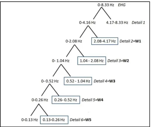
![Figure 2.7: Tree of the wavelet packet transform [49]. The underlined packets are the ones selected in this study](https://thumb-eu.123doks.com/thumbv2/123doknet/14543946.725217/101.918.143.791.329.685/figure-tree-wavelet-packet-transform-underlined-packets-selected.webp)