HAL Id: tel-01400155
https://tel.archives-ouvertes.fr/tel-01400155
Submitted on 23 Nov 2016
HAL is a multi-disciplinary open access archive for the deposit and dissemination of sci-entific research documents, whether they are pub-lished or not. The documents may come from teaching and research institutions in France or abroad, or from public or private research centers.
L’archive ouverte pluridisciplinaire HAL, est destinée au dépôt et à la diffusion de documents scientifiques de niveau recherche, publiés ou non, émanant des établissements d’enseignement et de recherche français ou étrangers, des laboratoires publics ou privés.
l’estimation de la cinématique articulaire : propositions
d’amélioration
Vincent Richard
To cite this version:
Vincent Richard. Méthode d’optimisation multi-segmentaire pour l’estimation de la cinématique ar-ticulaire : propositions d’amélioration. Biomécanique [physics.med-ph]. Université de Lyon, 2016. Français. �NNT : 2016LYSE1090�. �tel-01400155�
N°d’ordre NNT : 2016LYSE1090
THESE de DOCTORAT DE L’UNIVERSITE DE LYON
En cotutelle avec L’UNIVERSITÀ DI BOLOGNA
opérée au sein del’Université Claude Bernard Lyon 1
Ecole Doctorale
162
Mécanique Energétique et Génie Civil
Spécialité de doctorat :
Mécanique - Biomécanique
Soutenue publiquement le 28/06/2016, par :
Vincent RICHARD
Multi-body optimization method for the estimation
of joint kinematics: prospects of improvement
-
Méthode d’optimisation multi-segmentaire pour
l’estimation de la cinématique articulaire : propositions
d’amélioration
Devant le jury composé de :
FRIGO, Carlo Professeur à Politecnico di Milano, Italie Rapporteur PILLET, Hélène Maître de Conférences, HDR, à l’ENSAM, France Rapporteur CEREATTI, Andrea Professeur à l’Università degli Studi di Sassari, Italie Examinateur JACQUELIN, Éric Professeur à l’Université Claude Bernard Lyon 1, France Examinateur CAPPOZZO, Aurelio Professeur à l’Università degli Studi di Roma - Foro Italico, Italie Directeur de thèse DUMAS, Raphaël Directeur de Recherche à l’IFSTTAR, France Directeur de thèse
THESE de DOCTORAT DE L’UNIVERSITE DE LYON
En cotutelle avec L’UNIVERSITÀ DI BOLOGNA
opérée au sein del’Université Claude Bernard Lyon 1
Ecole Doctorale
162
Mécanique Energétique et Génie Civil
Spécialité de doctorat :
Mécanique - Biomécanique
Soutenue publiquement le 28/06/2016, par :
Vincent RICHARD
Multi-body optimization method for the estimation
of joint kinematics: prospects of improvement
-
Méthode d’optimisation multi-segmentaire pour
l’estimation de la cinématique articulaire : propositions
d’amélioration
Directeurs de thèse :
UNIVERSITE CLAUDE BERNARD - LYON 1
Président de l’Université
Président du Conseil Académique
Vice-président du Conseil d’Administration
Vice-président du Conseil Formation et Vie Universitaire Vice-président de la Commission Recherche
Directeur Général des Services
M. le Professeur Frédéric FLEURY M. le Professeur Hamda BEN HADID M. le Professeur Didier REVEL
M. le Professeur Philippe CHEVALIER M. Fabrice VALLÉE
M. Alain HELLEU
COMPOSANTES SANTE
Faculté de Médecine Lyon Est – Claude Bernard
Faculté de Médecine et de Maïeutique Lyon Sud – Charles Mérieux Faculté d’Odontologie
Institut des Sciences Pharmaceutiques et Biologiques Institut des Sciences et Techniques de la Réadaptation
Département de formation et Centre de Recherche en Biologie Humaine
Directeur : M. le Professeur J. ETIENNE Directeur : Mme la Professeure C. BURILLON Directeur : M. le Professeur D. BOURGEOIS Directeur : Mme la Professeure C. VINCIGUERRA Directeur : M. le Professeur Y. MATILLON Directeur : Mme la Professeure A-M. SCHOTT
COMPOSANTES ET DEPARTEMENTS DE SCIENCES ET TECHNOLOGIE
Faculté des Sciences et Technologies Département Biologie
Département Chimie Biochimie Département GEP
Département Informatique Département Mathématiques Département Mécanique Département Physique
UFR Sciences et Techniques des Activités Physiques et Sportives Observatoire des Sciences de l’Univers de Lyon
Polytech Lyon
Ecole Supérieure de Chimie Physique Electronique Institut Universitaire de Technologie de Lyon 1 Ecole Supérieure du Professorat et de l’Education Institut de Science Financière et d'Assurances
Directeur : M. F. DE MARCHI
Directeur : M. le Professeur F. THEVENARD Directeur : Mme C. FELIX
Directeur : M. Hassan HAMMOURI
Directeur : M. le Professeur S. AKKOUCHE Directeur : M. le Professeur G. TOMANOV Directeur : M. le Professeur H. BEN HADID Directeur : M. le Professeur J-C PLENET Directeur : M. Y.VANPOULLE
Directeur : M. B. GUIDERDONI Directeur : M. le Professeur E.PERRIN Directeur : M. G. PIGNAULT
Directeur : M. le Professeur C. VITON
Directeur : M. le Professeur A. MOUGNIOTTE Directeur : M. N. LEBOISNE
Preface
This thesis is submitted as partial fulfilment of the requirement for the Doctor of Philosophy at the University Claude Bernard Lyon 1, Lyon, France and the Doctor of Philosophy at the University Alma Mater Studiorum – Università di Bologna, Bologna, Italy under a convention of
cotutelle. The collaboration between both institutions has been sustained by a mobility grant from
the Mediterranean Office for Youth. The work has been carried out in the period from October 2012 to June 2016 under the joint supervision of Dr. Raphaël DUMAS and Pr. Aurelio CAPPOZZO.
Vincent RICHARD Lyon, June 2016.
Abstract
The estimate of joint kinematics during motor tasks generally relies on a model of the locomotor apparatus and on the stereophotogrammetric reconstruction of skin-marker trajectories or on the angular velocity and acceleration of the body segments provided by magneto-inertial measurement units. However, these techniques have important limitations including the "soft tissue artefacts" (i.e., the relative movement between the skin markers or the magneto-inertial sensors and the underlying bones). The multi-body optimization (MBO) method aims to compensate for these artefacts by constraining the measured kinematic quantities using selected kinematic models of the joints involved. The mechanical linkages normally used to model the joints, however, prevent satisfactory estimates of their kinematics. This thesis addresses the problem of using MBO as associated with data provided by magneto-inertial measurement units and stereophotogrammetry while analyzing lower limb joints. With regard to the latter technique, the improvement of the outcome of MBO, is sought through two different original approaches: (1) the embedment in MBO of an elastic joint model, based on a knee stiffness matrix, and a physiologically plausible optimization criterion, and (2) the introduction in the optimization process of a “kinematic-dependent” soft tissue artefact model. The present study, through experimental and statistical validation, demonstrates the versatility of the MBO approach, the feasibility of the original solutions proposed, fosters its use in soft tissue artefact compensation, and indicates ways to pursue a further improvement of the relevant outcome accuracy.
Keywords: movement analysis; lower limb; knee; magneto-inertial measurement unit; stiffness
Vincent RICHARD PhD Thesis
Résumé
L'estimation de la cinématique articulaire lors de tâches motrices repose souvent sur un modèle de l’appareil locomoteur et sur la reconstruction de trajectoires de marqueurs cutanés par stéréophotogrammétrie ou de la vitesse angulaire et l'accélération des segments du corps issues de centrales inertielles. Mais ces techniques présentent d’importantes limites, dont les « artéfacts de tissus mous » (i.e., le mouvement relatif entre les marqueurs cutanés ou des centrales inertielles et les os sous-jacents). La méthode d'optimisation multi-segmentaire (MBO) vise à compenser ces artefacts en imposant des contraintes aux grandeurs cinématiques mesurées à l'aide de modèles cinématiques articulaires. Cependant, les liaisons mécaniques modélisant les articulations empêchent une estimation satisfaisante de la cinématique articulaire. Cette thèse s’intéresse à l'utilisation de la MBO associée à des données issues de centrales inertielles et de stéréophotogrammétrie pour analyser les articulations du membre inférieur. Concernant cette dernière technique, l’amélioration des résultats de la MBO est évaluée à travers deux approches originales : (1) l’implémentation d’un modèle articulaire élastique basé sur la matrice de raideur du genou et un critère d’optimisation physiologiquement plausible, et (2) l’introduction dans la MBO d’un modèle d’artéfact de tissus mous « cinématique-dépendant ». Cette étude, par la validation expérimentale et statistique, démontre la polyvalence de l’approche MBO et la faisabilité des solutions proposées, encourage son utilisation pour la compensation des artéfacts de tissus mous, et indique les moyens pertinents d’améliorer la précision des résultats.
Mots-clés : analyse du mouvement ; membre inférieur ; genou ; centrale inertielle ; matrice de
Sommario
La stima della cinematica articolare durante l’esecuzione di atti motori si basa su un modello dell’apparato locomotore e sulla ricostruzione stereofotogrammetrica delle traiettorie di marcatori cutanei o sulla velocità angolare e l'accelerazione dei segmenti corporei fornite da sensori inerziali. Tuttavia, queste tecniche presentano importanti limiti il più importante dei quali è "l’artefatto da tessuto molle" (il movimento relativo tra i marcatori o sensori inerziali e l’osso sottostante). Il metodo di ottimizzazione multi corpo (MBO) mira a compensare questi artefatti vincolando le grandezze cinematiche stimate con modelli cinematici delle articolazioni coinvolte. I meccanismi normalmente utilizzati per modellare le articolazioni, però, forzano la fisiologia delle stesse e impediscono stime soddisfacenti della loro cinematica. Questa tesi riguarda l’utilizzo della MBO associata a dati forniti da sensori inerziali e dalla stereofotogrammetria e fa riferimento alle articolazioni degli arti inferiori. Utilizzando, in particolare dati stereofotogrammetrici, i risultati forniti dalla MBO, sono stati valutati attraverso due differenti approcci originali: (1) inclusione nella MBO di un modello articolare elastico, basato su una matrice di rigidezza del ginocchio associato ad un criterio di ottimizzazione fisiologicamente plausibile, e (2) introduzione nel processo di ottimizzazione di un modello di artefatto da tessuto molle "cinematica-dipendente". Il presente studio, attraverso la validazione sperimentale e statistica, dimostra la versatilità dell'approccio MBO, la fattibilità delle soluzioni proposte, promuove il suo utilizzo nella compensazione degli artefatti da tessuto molle, ed indica i metodi da utilizzare per un significativo miglioramento della accuratezza.
Parole-chiave: analisi del movimento; arto inferiore; ginocchio; sensore inerziale; matrice di
Vincent RICHARD PhD Thesis
Résumé étendu
Dans le contexte du vieillissement de la population et de l'augmentation de l'espérance de vie, et afin d’appréhender au mieux les différentes pathologies affectant l'appareil locomoteur humain et ses performances, l'analyse du mouvement est plus que jamais une préoccupation sociale majeure. L’analyse du mouvement humain consiste dans l'observation et la définition des mouvements de la vie des êtres humains, et les recherches dans ce domaine sont nombreuses. Elles captivent une communauté multidisciplinaire et représente un intérêt pour l'orthopédie, le sport ou la vie quotidienne. La biomécanique propose de fournir en outre des connaissances scientifiques sur les causes et les effets des affections de la locomotion. En biomécanique, le mouvement est décrit comme l'ensemble des positions et orientations de segments du corps, déterminées à un instant donné pendant l'observation. En particulier, la cinématique est concernée par l'étude du mouvement d'un point de vue géométrique : elle représente la première étape vers la modélisation musculo-squelettique qui porte sur les causes qui amènent le corps à se déplacer comme il le fait. Evaluer une cinématique précise est donc le fondement essentiel de l'analyse du mouvement. Deux principales grandeurs caractérisant le mouvement sont intéressantes pour l'estimation de la cinématique articulaire : l’attitude des os et le mouvement relatif entre les os adjacents. En général, l'analyse du mouvement est effectuée sur la base des données de mesure de surface, tels que la stéréophotogrammétrie. Au-delà de la complexité de la représentation mathématique de la position et de l’orientation des os se trouve un enjeu majeur. Un problème se pose précisément lors de l'estimation de la cinématique des structures osseuses à l'aide de marqueurs de surface. En effet, la prise en compte de l'erreur observée entre les trajectoires mesurées des marqueurs de surface et la position et l'orientation réelles de l'os sous-jacent est nécessaire pour une estimation précise, du fait que ce que l'on appelle les « artefacts de tissus mous » (STA : soft tissue artefact) peut provoquer des mouvements irréalistes de grande amplitude au niveau des articulations. La prise en compte de ce phénomène reste un défi permanent (Leardini et al., 2005), car il empêche une mesure non-invasive directe du mouvement articulaire. Parmi les différentes méthodes de compensation des STAs, l'utilisation d'une méthode d'optimisation multi-segmentaire (MBO : multi-body optimization) a été proposée comme une solution potentielle (Lu and O’Connor, 1999), mais n'a à ce jour pas aboutie a des résultats pleinement satisfaisants.
La thèse propose des perspectives d'amélioration de l'estimation de l’attitude des os et de la cinématique articulaire en utilisant la méthode MBO. Le principe de la méthode repose sur la minimisation des erreurs entre les trajectoires de marqueurs cutanés mesurées et modélisées au sens des moindres carrés, au sein d’un modèle cinématique défini.
Tout d'abord, une restriction associée à la stéréophotogrammétrie qui repose sur le fait que le mouvement analysé doit être effectué dans un environnement contrôlé empêchent l'étude des mouvements in-situ. L'utilisation de centrales inertielles (MIMUs : magneto-inertial measurement units) représente une alternative prometteuse, mais puisque les mesures restent affectées par les STAs, on ne peut s’attendre à une estimation plus précise des positions et orientations des os qu'avec la stéréophotogrammétrie. Les mesures issues des MIMUs peuvent cependant être utilisées dans le cadre de la MBO afin de limiter la propagation des STA. L'orientation calculé à partir de MIMUs a déjà été introduit avec succès au procédé de la méthode MBO pour piloter le modèle cinématique associé (Koning et al., 2015). Cependant, dans cette première tentative, seule la cinématique articulaire a été évaluée et comparée avec celle obtenue par la technique de stéréophotogrammétrie et marqueurs cutanés, et non la position des segments, en particulier le segment terminal de la chaine cinématique, c’est-à-dire le pied dans le cadre du membre inférieur. En outre, peu de détails ont été fournis sur la façon d'adapter la méthode MBO aux mesures des MIMUs. La calibration du modèle est également essentielle dans un tel procédé : une calibration incorrecte a pour conséquence de propager de larges erreurs à l’attitude estimée des os.
Deuxièmement, la méthode MBO repose sur l'hypothèse que les contraintes articulaires introduites dans le modèle cinématique représentent le comportement réel des articulations. Néanmoins, l'exactitude de ces méthodes basées sur des modèles mécaniques est discutable, en raison notamment de l'introduction de contraintes « strictes (hard) » qui empêchent ou prescrivent le mouvement de l'articulation. Les contraintes articulaires peuvent être considérées comme des contraintes « strictes » quand elles doivent être strictement satisfaites, et comme des contraintes « souples (soft) » quand elles doivent être minimisées. Évoluer vers des contraintes « souples », qui ne sont plus déterministes, pour modéliser les articulations peut être une alternative efficace pour mieux représenter le comportement réel des articulations (Gasparutto et al., 2015).
Enfin, la méthode MBO ne compense que partiellement les STAs. Des modèles de STAs ont été proposés (Alexander and Andriacchi, 2001; Bonci et al., 2014; Camomilla et al., 2013), jusqu'à présent appliqués seulement à l'optimisation mono-segmentaire (Alexander and Andriacchi, 2001) et l'estimation du centre articulaire (De Rosario et al., 2013). La réduction de la complexité de ces modèles (Camomilla et al., 2015), en particulier du nombre de paramètres les définissants, rend possible leur utilisation dans la méthode MBO et pourrait être une alternative à la calibration spécifique de ces modèles. En d'autres termes, une identification simultanée des paramètres d'un modèle de STAs avec l’estimation de l’attitude des os peut être développée dans le cadre de la méthode MBO.
Vincent RICHARD PhD Thesis
Le travail de thèse s’articule donc autour de ces trois thématiques.
Suite à une introduction générale (Chapitre 1), le Chapitre 2 propose un état de l'art sur l'analyse du mouvement dans le but d’introduire les connaissances générales en anatomie du membre inférieur, les technologies de systèmes d’acquisition, et les méthodes pour l’estimation de la cinématique. Ce chapitre explore le domaine de l'analyse de mouvement, d'un point de vue biomécanique et identifie les principaux problèmes rencontrés lors de l'évaluation de la cinématique articulaire. Des arguments sont avancés pour légitimer l'utilisation d'une méthode MBO pour l'estimation de la position et de l’orientation des os. La méthode MBO est présentée dans sa version classique, utilisé pour l'analyse cinématique des membres inférieurs à partir des données de stéréophotogrammétrie utilisant des marqueurs cutanés (Lu and O’Connor, 1999), la méthode est détaillée dans le cadre spécifique d’un paramétrage « en coordonnées naturelles » (Dumas and Chèze, 2007). En particulier, ce chapitre développe les modèles articulaires mis en œuvre dans le modèle cinématique de la méthode MBO, la question des STAs, et de le potentiel des nouvelles technologies magnéto-inertielles.
Dans le Chapitre 3, un cadre de modélisation est développé, dérivé de la méthode MBO classique, pour l’adapter à l'utilisation de MIMUs. La méthode développée est utilisée pour étudier la possibilité d'introduire l'orientation, la vitesse angulaire et l'accélération mesurées par les capteurs inertiels dans la fonction objective de la méthode MBO. La méthode développée est testée sur le membre inférieur au cours de la marche, en comparant la cinématique articulaire et la position du pied par rapport au bassin obtenues par la méthode MBO utilisant les MIMUs ou la stéréophotogrammétrie et les marqueurs cutanés. L'hypothèse de cette étude est que réaliser un calibrage anatomique (Picerno et al., 2008) est pertinent pour définir le modèle cinématique inclus dans la méthode MBO. Cette étude vise donc à évaluer l'efficacité d'une méthode qui propose de combiner trois approches : l’estimation de la cinématique articulaire dans le cadre de la méthode MBO (Duprey et al., 2010; Lu and O’Connor, 1999), le suivi de données inertielles mesurées par les MIMUs (Koning et al., 2015) et la procédure de calibration anatomique (Picerno et al., 2008). L'étude a confirmé que l'orientation issue des MIMUs est une donnée d’entrée efficace pour la méthode MBO. La faisabilité de la prise en compte de la vitesse angulaire et de l'accélération comme données d’entrée complémentaires à l’orientation ou comme données d’entrée uniques ouvre des perspectives importantes. En effet, l’orientation des MIMUs résulte d’un traitement qui est sensible aux perturbations magnétiques mais ce n’est pas le cas de la vitesse angulaire et de l'accélération.
De plus, l’obtention d’une cinématique articulaire qui est en accord avec les mesures de vitesse angulaire et d'accélération s’avère pertinent pour des calculs de dynamique.
Dans le Chapitre 4, la performance de la méthode MBO pour estimer la cinématique articulaire du genou est étudiée lorsqu’un modèle élastique dérivé de la matrice de raideur de l’articulation du genou est intégré à la méthode. La matrice de raideur représente le comportement mécanique de l’articulation du genou, mais reste néanmoins sujet-dépendante. Une méthode de pénalité est mise en œuvre pour introduire le modèle élastique d’articulation comme une contrainte « souple » dans la méthode MBO. Un tel modèle cinématique de l’articulation du genou devrait fournir une meilleure estimation que les liaisons mécaniques rigides classiques (contraintes « strictes ») qui empêchent ou prescrivent certains déplacements. Considérant une matrice de raideur unique, cette étude de faisabilité démontre que la méthode est au moins aussi efficace que les méthodes classiques (avec des contraintes mécaniques de type rotule par exemple). En outre, l'analyse de sensibilité réalisée a montré que de grandes variations des coefficients de la matrice de raideur se propagent peu à l'estimation de la cinématique.
Dans le Chapitre 5, un modèle de STAs « cinématique-dépendant » est introduit dans la méthode MBO. Le développement mathématique et de calcul sont effectués tout en utilisant différents types de modèles cinématiques afin d'étudier l'efficacité de la méthode pour l’identification des paramètres du modèle de STAs en même temps que l'estimation de l’attitude des os. L'objectif est d'évaluer et compenser les STAs tout en améliorant la précision de la méthode MBO pour estimer la cinématique articulaire. Testée sur des données de course où les mouvements du membre inférieur sont mesurés par stéréophotogrammétrie avec simultanément des marqueurs cutanés et des marqueurs montés sur des tiges vissées dans l’os, la méthode se révèle inefficace quant à l’amélioration de la précision de l’estimation de la cinématique et démontre l'importance des choix de modélisation (paramètres du modèle de STAs, modèle cinématique).
Vincent RICHARD PhD Thesis
Sommario esteso
La stima della cinematica articolare durante l’esecuzione di atti motori si basa su un modello dell’apparato locomotore e sulla ricostruzione stereofotogrammetrica delle traiettorie di marcatori cutanei o sull’orientamento, velocità angolare e accelerazione dei segmenti corporei fornite da sensori magneto-inerziali (magneto-inertial measurement unit: MIMU). Tuttavia, queste tecniche presentano dei limiti il più importante dei quali è la loro sensibilità "all’artefatto da tessuto molle" (il movimento relativo tra i marcatori o sensori inerziali e l’osso sottostante; soft tissue artefact: STA). La misura e la minimizzazione della propagazione ai risultati finali di questo artefatto è una sfida molto attuale vincendo la quale si renderebbe possibile un importante avanzamento di queste metodologie ed un allargamento dello spettro delle loro possibili applicazioni. L’approccio più promettente a tal fine consiste nel metodo di ottimizzazione multi corpo (multi-body optimization: MBO). Esso mira a compensare gli STA vincolando le grandezze cinematiche stimate con modelli cinematici delle articolazioni coinvolte. I meccanismi normalmente utilizzati per modellare le articolazioni, però, forzano la fisiologia delle stesse a comportamenti in parte predefiniti e, di conseguenza, impediscono stime soddisfacenti della loro cinematica in casi specifici. Questa tesi ha come obiettivo quello di dare un contributo verso il superamento di questi limiti e riguarda l’utilizzo della MBO associata a dati forniti sia da MIMU che dalla stereofotogrammetria. A scopo esemplificativo vengono prese in considerazione le articolazioni dell’arto inferiore. Per quanto riguarda l’uso dei MIMU, il metodo MBO proposto non sfrutta solo informazioni sull’orientamento, come normalmente fatto, ma anche quelle relative alle accelerazioni e velocità angolari. Utilizzando, invece, dati stereofotogrammetrici il classico metodo della MBO è stato modificato attraverso due differenti approcci originali: (1) inclusione nella MBO di un modello articolare elastico, basato su una matrice di rigidezza del ginocchio associato ad un criterio di ottimizzazione fisiologicamente plausibile, e (2) introduzione nel processo di ottimizzazione di un modello di STA "cinematica-dipendente". Il presente studio, attraverso la validazione sperimentale e statistica, dimostra la versatilità dell'approccio MBO, la fattibilità delle soluzioni proposte, promuove il suo utilizzo nella compensazione degli artefatti da tessuto molle, ed indica i metodi da utilizzare per un significativo miglioramento della accuratezza.
La tesi è così organizzata.
Dopo un'introduzione generale (capitolo 1), il capitolo 2 fornisce lo stato dell'arte sull’analisi del movimento con l'obiettivo di introdurre le conoscenze generali dell'anatomia dell'arto inferiore, delle tecnologie dei sistemi di acquisizione del movimento, e i metodi di stima della cinematica.
Speciale attenzione viene dedicata all'uso del metodo MBO per la stima della posa delle ossa. Questo è presentato nella sua versione classica utilizzata per l'analisi cinematica degli arti inferiori da dati ottenuti con la stereofotogrammetria e marcatori cutanei. Il metodo è dettagliato nel contesto specifico di un parametrizzazione in "coordinate naturali". Vengono, inoltre, descritti i modelli articolari normalmente implementati con il metodo MBO, il problema del STA, ed il potenziale delle nuove tecnologie magneto-inerziali.
Nel capitolo 3, il metodo MBO classico, giù utilizzato con dati stereofotogrammetrici, viene adattato all'uso di MIMU. In particolare l’orientamento, la velocità angolare e l'accelerazione resi disponibili dai sensori inerziali vengono introdotti nella funzione obiettivo del metodo MBO. Il metodo sviluppato è testato sull'arto inferiore durante il camino e i suoi risultati vengono confrontati con la cinematica articolare e la posizione del piede rispetto al bacino ottenuti con il metodo MBO e la stereofotogrammetria con marcatori cutanei. Questo studio mira a valutare l'efficacia di un metodo che si propone di combinare tre procedure: la stima della cinematica articolare con il metodo MBO, il monitoraggio di dati inerziali misurati attraverso i MIMU, e la procedura di calibrazione anatomica. Lo studio ha confermato che l’orientamento derivato dai MIMU è un efficace dato di input per il metodo MBO. E’ stata verificata anche la fattibilità dell’uso della velocità angolare e dell’accelerazione come dati di input aggiuntivi all'orientamento o come unici dati di ingresso. Ciò apre importanti prospettive. Infatti, l'orientamento dei MIMU è alquanto sensibile ai disturbi magnetici, mentre questo non è il caso per la velocità angolare e l’accelerazione. Inoltre, ottenere una cinematica articolare che è coerente con le misure di velocità angolare e di accelerazione è favorevole ad una stima accurata di grandezze dinamiche.
Nel capitolo 4 viene introdotto un modello elastico del ginocchio concepito per essere integrato nel metodo MBO. Detto modello è rappresentato da una matrice di rigidezza ottenuta attraverso esperimenti ex vivo. La posa relativa di femore e tibia viene stimata inglobando nella MBO la minimizzazione della variazione di energia elastica del modello. Considerando una singola matrice di rigidezza, questo studio di fattibilità dimostra che il metodo è efficace almeno quanto i metodi convenzionali (con vicoli meccanici di tipo sferico per esempio). Questa conclusione è stata ottenuta stimando la cinematica articolare del ginocchio utilizzando tre metodi classici nonché quello presentato in questo studio partendo da dati rilevati in vivo con la stereofotogrammetria. I risultati così ottenuti sono stati messi a confronto con la cinematica stimata utilizzando dati ad elevata accuratezza forniti dalla fluoroscopia. Inoltre, l'analisi di sensibilità ha mostrato che variazioni dei coefficienti della matrice di rigidezza dovuti a differenze interindividuali e di posa del
Vincent RICHARD PhD Thesis
ginocchio propagano poco alla stima della cinematica giustificando così l’uso di una singola matrice ottenuta su un singolo reperto anatomico.
Nel capitolo 5, viene presentato un modello di STA "cinematico-dipendente" e come questo viene introdotto nel metodo MBO. L'obiettivo è quello di valutare e compensare il STA e, quindi, migliorare l'accuratezza del metodo MBO nella stima della cinematica articolare. Il metodo è stato valutato su dati di corsa dove i movimenti delle ossa dell’arto inferiore sono stati misurati con la stereofotogrammetria e l’uso simultaneo di marcatori cutanei e marcatori montati su viti inserite nell'osso. Il metodo, seppure promettente, così come implementato nel presente studio è risultato essere non efficace nel miglioramento dell'accuratezza della stima della cinematica lasciando dunque spazio per ulteriori approfondimenti sul tema.
Acknowledgements
I would like to express my special thanks and esteem to my supervisors Dr. Raphaël DUMAS and Pr. Aurelio CAPPOZZO for their continuous support and guidance during the past years. Both of them have been tremendous mentors for me. I would like to acknowledge them for encouraging my research and for allowing me to grow as a research scientist. I appreciate all their contributions of time and ideas to make my PhD experience productive and stimulating. I am also thankful for the excellent example they have provided as successful and influent researchers.
I would like to thank everyone from the department of Mechanics at the Université Claude Bernard Lyon 1 for creating a good working environment. A special thanks goes to my “roommates” in the ground floor office.
I would like to thank everyone in the office at the University of Rome – Foro Italico for the hospitality. Your freshness made my warm time in Rome more comfortable.
A thanks also goes to everyone that I had the chance to meet at the Laboratoire de Biomécanique et Mécanique des Chocs.
I would like to thank my family and my longtime friends.
The greatest acknowledgement goes to Audrey for her support and patience during these long years.
Vincent RICHARD PhD Thesis
Contents
Preface ... 7 Abstract ... 8 Résumé ... 9 Sommario ... 10 Résumé étendu ... 11 Acknowledgements ... 18 Contents ... 19 List of figures ... 22 List of tables ... 23Notations and symbols ... 24
Chapter 1. Introduction ... 27
1. Background and motivation ... 27
2. Scope of the study ... 28
3. Thesis outline ... 29
Chapter 2. Literature review – state-of-the-art ... 31
1. Lower limb anatomy - osteoarticular description ... 31
1.1. Anatomical terminology ... 31
1.1.1. Anatomical reference position ... 31
1.1.2. Directional terms ... 32
1.1.3. Anatomical reference planes ... 32
1.1.4. Anatomical reference axis ... 32
1.1.5. Terms of movement for the lower limb ... 33
1.2. Human locomotor apparatus ... 34
1.2.1. Overview of the skeleton ... 34
1.2.1.a. Pelvis... 36
1.2.1.b. Thigh ... 36
1.2.1.c. Shank ... 36
1.2.1.d. Foot ... 38
1.2.2. Joints of the lower limb ... 38
1.2.2.a. Structure ... 38
1.2.2.b. Hip joint ... 38
1.2.2.c. Knee joint ... 38
1.2.2.d. Ankle joint ... 40
2. Motion analysis ... 41
2.1. History of movement analysis ... 41
2.2. Issues in movement analysis ... 42
2.3. Kinematics ... 43
2.4. Dealing with skin markers – technological limitations ... 44
2.5. Soft tissue artefact ... 45
2.5.1. General characteristics of soft tissue artefact ... 45
2.5.2. Soft tissue artefact assessment and compensation ... 46
2.5.3. Modeling of soft tissue artefact ... 47
2.5.4. Conclusion ... 49 3. Alternative to stereophotogrammetry... 50 3.1. Medical imaging ... 50 3.1.1. X-ray ... 50 3.1.2. Ultrasounds ... 51 3.1.3. Magnetism ... 51 3.1.4. Conclusion ... 51
3.2. Magneto-inertial measurement units ... 52
3.2.1. Device description and specification ... 52
4. Modeling and bone pose estimation – multi-body optimization ... 55
4.1. Rigid multi-body systems ... 55
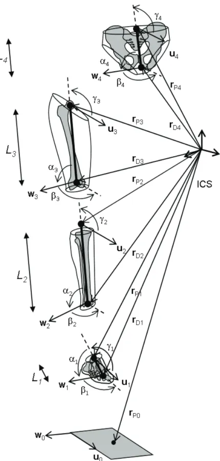
4.2. Parameter set and systems of coordinates ... 55
4.3. Segment and joint coordinate systems ... 59
4.3.1. Segment coordinate system ... 59
4.3.2. Joint coordinate system ... 60
4.4. Constraints ... 63
4.4.1. Rigid body constraints ... 63
4.4.2. Kinematic constraints ... 64
4.4.2.a. Spherical joint ... 65
4.4.2.b. Hinge joint ... 65
4.4.2.c. Universal joint ... 65
4.4.2.d. Parallel mechanism... 66
4.4.3. Driving constraints ... 67
4.5. Optimization ... 68
4.5.1. Formulation of the problem... 68
4.5.2. Lagrangian formulation ... 68
Chapter 3. Multi-body optimization using magneto-inertial measurement units ... 70
1. Introduction ... 70
2. Materials and methods... 71
2.1. Measured data and procedure ... 71
2.2. Multi-body optimization framework ... 74
2.2.1. Modeled orientation ... 74
2.2.2. Modeled angular velocity ... 75
2.2.3. Modeled acceleration ... 75
2.2.1. Objective function ... 77
2.3. Experimental methods ... 79
2.4. Statistics ... 81
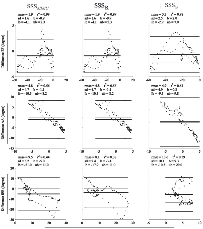
3. Results ... 81
3.1. Comparative study of the joint kinematics estimates ... 81
3.1.1. Bias ... 83
3.1.2. Standard deviation ... 83
3.1.3. Root mean square differences ... 83
3.1.4. Correlation coefficient... 83
3.2. Objective function residual and mean differences between measured and modeled parameters ... 85
3.3. Foot position and error ... 85
4. Discussion... 86
Chapter 4. Knee kinematics estimation using multi-body optimization embedding a knee joint stiffness matrix ... 89
1. Introduction ... 89
2. Materials and methods... 91
2.1. Multi-body optimization ... 91
2.2. Knee stiffness matrix ... 94
2.3. Validation data and procedure ... 95
2.3.1. Experimental methods ... 96
2.3.2. Sensitivity analysis ... 96
2.3.3. Statistics ... 99
3. Results ... 99
3.1. Comparative analysis of the knee kinematics estimates ... 99
3.1.1. Bias ... 99
3.1.2. Standard deviation ... 102
3.1.3. Root mean square error ... 103
3.1.4. Correlation coefficient... 103
Vincent RICHARD PhD Thesis
4. Discussion... 105
Chapter 5. Simultaneous optimization of bone pose and soft tissue artefact model
parameters through multi-body optimization: a feasibility study ... 109
1. Introduction ... 109 2. Materials and methods... 111
2.1. Mathematical representation of the STA model ... 111 2.2. STA rigid component model ... 113 2.3. Multi-body optimization ... 115 2.4. Validation data and procedure ... 118 2.5. Evaluation of STA model implementation and MBO effectiveness ... 118 2.6. Part 1: Overview and selection of relevant models ... 119 2.6.1. Objectives and procedure ... 119 2.6.2. Results... 120 2.6.2.a. STA rigid component model approximated by six modes ... 120 2.6.2.b. STA rigid component model approximated by three modes ... 122 2.6.3. Discussion ... 125 2.7. Part 2: Evaluation of the selected models ... 126 2.7.1. Objectives and procedure ... 126 2.7.2. Results... 127 2.7.3. Intermediate discussion... 133
3. Discussion and conclusion ... 134
Chapter 6. General discussion ... 137 References ... 140
List of figures
Figure 2.1 Anatomical planes and axes ...33 Figure 2.2 Lower limb overview ...35 Figure 2.3 Skeletal framework of the lower limb (Moore and Dalley, 2006) ...37 Figure 2.5 Example of 3D parallel mechanism for knee modeling (Parenti-castelli et al., 2004) ...39 Figure 2.6 Example of 3D parallel mechanism for ankle modeling (Di Gregorio et al., 2007) ...40 Figure 2.7 Movement analysis ...41 Figure 2.8 The human movement analysis laboratory...44 Figure 2.9 Motion capture workflow ...45 Figure 2.10 Representation of the lower limb ...56 Figure 2.11 Segment parameters in the global reference frame (ICS) ...58 Figure 2.12 Joint coordinate system ...61 Figure 2.13 Classic joint models for the hip, the knee and the ankle...64 Figure 3.1 Lower limb joint angles during walking cycle. ...82 Figure 3.2 Bland Altman plot. ...84 Figure 3.3 Foot position and error with respect to the reference. ...86 Figure 4.1 Model specifications. ...92 Figure 4.2 Distribution of coefficients of stiffness matrix samples ...98 Figure 4.3 Bland Altman plot for subject S1... 100 Figure 4.4 Bland Altman plot for subject S2... 101 Figure 4.5 Knee joint angles and displacements, U = [θ1 θ2 θ3 d1 d2 d3]T for both subjects. ... 104
Figure 5.1 Framework for the mathematical representation of the STA ... 113 Figure 5.2 Knee joint kinematics model h
6
N . ... 120 Figure 5.3 Amplitude estimate’s time histories for model h
6
N . ... 121 Figure 5.4 Amplitude estimate’s time histories for model h
6
N . ... 122 Figure 5.5 Knee joint angle kinematics for models h
3 S , h 3 N and a 3 S . ... 123 Figure 5.6 Amplitude estimate’s time histories for models h
3 S , h 3 N and a 3 S . ... 124 Figure 5.7 Amplitude model parameters h estimate for model h
3
S and h 3
N . ... 124 Figure 5.8 Knee joint angles for model h
3
S and a 3
S . ... 128 Figure 5.9 Root mean square errors (RMSE) for models S, h
3
S and a 3
S . ... 129 Figure 5.10 Correlation coefficient (R²) for models S, h
3
S and a 3
S . ... 129 Figure 5.11 Amplitude estimate’s time histories for models h
3
S and a 3
S . ... 130 Figure 5.12 Amplitude estimate’s time histories for models h
3
S and a 3
S . ... 131 Figure 5.13 Root mean square errors (RMSE) for h
3
S and a 3
S . ... 132 Figure 5.14 Correlation coefficient (R²) for models h
3
S and a 3
Vincent RICHARD PhD Thesis
List of tables
Table 2.1 Directional terms ...32 Table 2.2 Description of segments parameters ...57 Table 3.1 Methods notation ...80 Table 3.2 Residual and mean difference. ...85
Notations and symbols
MBO Multi-body optimization SBO Single-body optimization STA Soft tissue artefact
MIMU Magneto-inertial measurement unit DoF Degree of freedom
ICS Inertial coordinate system JCS Joint coordinate system EF Extension/Flexion AA Adduction/Abduction IER Internal/External rotation LM Lateral/Medial displacement AP Anterior/Posterior displacement PD Proximal/Distal displacement N No joint model S Spherical model P Parallel mechanism
M Elastic joint model based on the stiffness matrix
i Body segment index (i 2: foot; i 3: shank; i 4: thigh; i 5: pelvis)
i
Q Natural coordinates of segment i
i
P Proximal endpoint of segment i
i
D Distal endpoint of segment i
i
u Components of the frontal unitary direction vector of segment i
i
P
r Coordinates of the proximal endpoint of segment i
i
D
Vincent RICHARD PhD Thesis
i
w Components of the lateral unitary direction vector of segment i
j Marker index
j i
M
r Coordinates of marker j of segment i A
rA
rA Components of the acceleration vector
ω
Components of the angular velocity vectorω
ω Skew matrix of angular velocity vector
m Φ Driving constraints k Φ Kinematic constraints r Φ Rigid-body constraints f Objective function S Stiffness matrix
U Actual joint angles and displacements
0
U Neutral joint angles and displacements
F
Actual forces and moments0
F
Neutral forces and momentsT
Euler angle1
T
Extension/flexion angle(degree)2
T
Adduction/abduction angle (degree)3
T
Internal/external rotation angle (degree)d
displacement 1 d Lateral/medial displacement (mm) 2 d Anterior/posterior displacement (mm) 3 d Proximal/distal displacement (mm)1
e First vector of knee joint coordinate system
2
e Second vector of knee joint coordinate system
3
e Third vector of knee joint coordinate system rmse Root mean square error
rmsd Root mean square difference
r² Squared Pearson’s correlation coefficient sd Standard deviation
b Bias (mean value of data) l Limit of agreement (b±1.96sd)
x
Dot product × Cross productVincent RICHARD PhD Thesis
Chapter 1. Introduction
1. Background and motivation
In a context of ageing of the population and of rising of life expectancy, and to address the different pathologies affecting the locomotor apparatus as well as performance of the movements, human locomotion is more than ever a major social concern. Human movement analysis consists in the observation and definition of movements of living humans and investigations in this domain are various. It addresses a multi-disciplinary community and is of interest in orthopedics, sports or everyday life. Biomechanics proposes to provide further scientific knowledge on the causes and effects of locomotion affections. In biomechanics, a motion is described as the ensemble of the positions and orientations of adjacent body segments determined at a sampled instant of time during the observation. In particular, kinematics is concerned with the study of movement from a geometrical point of view: it represents the first step toward dynamics which is concerned with what causes the body to move the way it does.
Assessing an accurate kinematics is therefore the critical baseline for movement analysis. Two main quantities that characterize the movement are interesting for the estimation of joint kinematics: the instantaneous bone pose, and the relative movement between adjacent bones. Generally, movement analysis is performed based on surface measurement data, such as stereophotogrammetry. Beyond the complexity of the mathematical representation of bone positions and orientations lies a major issue. It arises precisely when estimating the kinematics of internal structures using surface markers. Indeed, taking into account the error observed between the measured trajectories of the surface markers and the actual position and orientation of the underlying bone is necessary for an accurate estimate because the so-called soft tissue artefact (STA) can cause unrealistic motions of the joint. Accounting for this phenomenon remains an ongoing challenge (Leardini et al., 2005), preventing from a straightforward non-invasive measurement of the skeletal movement. Among various STA compensation methods, the use of a multi-body optimization (MBO) method was proposed as a potential solution (Lu and O’Connor, 1999), but did not end up with fully satisfying results so far.
2. Scope of the study
The thesis addresses prospects of improvement for the estimation of bone pose and joint kinematics using the MBO method. The principle of the method relies on the minimization of the least-square errors between the modeled and the measured skin marker trajectories, within a defined kinematic model embedding joint constraints. Some limitations of the MBO method are to be overcome.
First, a restriction associated to the stereophotogrammetry relies in that the movement analyzed must be performed in a controlled environment preventing from studying in-situ movements. The use of magneto-inertial measurement units (MIMUs) is therefore a trade-off, but since STA remains, the accuracy of the estimation of bone pose cannot be expected to be better than with stereophotogrammetry. Measurement from MIMUs can be implemented in the MBO framework to limit the STA propagation. The orientation computed from MIMUs has been successfully introduced within a MBO method to drive the kinematic model associated (Koning et al., 2015). However, in this first attempt, only the joint kinematics was evaluated against stereophotogrammetry and skin markers, not the position of the segments within the kinematic model. In particular, the position of the terminal segment is of great interest. Moreover, few details were provided on how to adapt the MBO framework to MIMU measurements. The model calibration is also critical in such a method: inappropriate calibration results in large tracking errors which propagate to the bone pose estimation.
Moreover, the MBO method relies on the hypothesis that the joint constraints introduced in the kinematic model represents the actual joint behavior. Nevertheless, the accuracy of such model-based methods is questionable, in particular because of the introduction of “hard” constraints that impede or prescribe the movement at the joint. Joint constraints can be considered as “hard” constraints when they have to be strictly satisfied, and as “soft” constraints when they have to be minimized. Evolving toward “soft” constraints, that are no longer deterministic, to model the joints may be an efficient alternative to represent the actual joint behavior better (Gasparutto et al., 2015). Finally, MBO only partially compensate for STA. STA models have been proposed (Alexander and Andriacchi, 2001; Bonci et al., 2014; Camomilla et al., 2013), applied only to single body optimization (Alexander and Andriacchi, 2001) and joint center estimation (De Rosario et al., 2013) so far. The complexity reduction of these models (Camomilla et al., 2015) are encouraging their use in the MBO method, and could be an alternative to a specific calibration for these models. In other words, a simultaneous identification of the parameters of a kinematic-driven STA model and bone pose estimation can be developed within the MBO framework.
Vincent RICHARD PhD Thesis
3. Thesis outline
The chapters following this introduction are outlined as follow.
Chapter 2
In Chapter 2, a state-of-the-art on movement analysis is carried out with the aim of introducing general prerequisite on anatomy of the lower limb, acquisition system technologies, and methods for kinematics estimation. Chapter 2 explores the domain of motion analysis, from a biomechanical point of view, through a review of the literature, and identifies the main issues encountered while assessing joint kinematics. Arguments are advanced to legitimate the use of a MBO framework for bone pose estimation. The MBO method is presented in its classic version used for kinematic analysis for lower limb from marker-based stereophotogrammetry data (Lu and O’Connor, 1999), the method is detailed on the specific framework of natural coordinates (Dumas and Chèze, 2007). In particular, this chapter develops the joint models implemented in the kinematic model of the MBO methods, the soft tissue artefact issue, and the emerging magneto-inertial technology.
Chapter 3
In Chapter 3, a modeling framework is developed, derived from the classic MBO method, to make the method appropriate to the use of MIMUs, which represent a promising acquisition system in movement analysis. The developed method is used to investigate the feasibility of introducing orientation, angular velocity and acceleration derived from inertial sensors in the objective function of the MBO method as an alternative to the classic skin marker trajectories assessed from stereophotogrammetry. The developed method is tested on the lower limb for gait, aiming at comparing joint kinematics and foot position relative to the pelvis obtained by tracking MIMU and skin marker-based estimation. The hypothesis in this study is that performing an anatomical calibration (Picerno et al., 2008) is relevant for defining the kinematic model included in the MBO framework. Therefore, this study aims at evaluating the efficiency of a method which combine three proposed approaches for joint kinematics estimation: the MBO framework (Duprey et al., 2010; Lu and O’Connor, 1999), the MIMU-tracking approach (Koning et al., 2015) and the anatomical calibration procedure (Picerno et al., 2008).
Chapter 4
In Chapter 4, the performance of the MBO method to estimate knee joint kinematics is investigated when an elastic joint model based on the knee stiffness matrix is embedded in the framework. The stiffness matrix represents the mechanical behavior of a knee joint, but remains nevertheless
subject-dependent. A penalty-based method is implemented to introduce the elastic joint model as “soft” constraints in the MBO framework. Such a model at the knee joint is expected to provide better kinematics estimate than classic mechanical linkages (“hard” constraints) that impede or prescribe some displacements. Considering a single constant stiffness matrix, this evaluates the feasibility of the method compared to more classic methods (using spherical joint for instance). Furthermore, a sensitivity analysis is performed to investigate how variations of the stiffness matrix coefficients propagate to the kinematics estimation.
Chapter 5
In Chapter 5, a kinematic-driven STA model is introduced in the MBO method. Mathematical and computational developments are carried out using different types of kinematic and STA models to investigate the efficiency of the method in identifying the STA model parameters concurrently with estimating bone pose. The objective is to assess and compensate for the STA while improving the accuracy of the MBO method in estimating kinematics.
Chapter 6
Chapter 6 presents a general discussion of the studies described in this thesis. The main contribution of the work are underlined and some perspective are indicated.
Vincent RICHARD PhD Thesis
Chapter 2. Literature review – state-of-the-art
This chapter offers basic as well as specific information required to understand the themes developed in the thesis. In particular, in order to understand the origins of movement, the reader must acquire basic knowledge about the anatomy of the human body. Anatomy provides essential information and terminology for musculoskeletal structures and joint motion. A preliminary paragraph presents an osteoarticular description of the lower-limb (particular attention is paid to the knee). After this introduction to anatomy from a modeling perspective, we explore the evolution of human motion analysis in history through the development of societal needs and measurement technologies. This historical overview leads to the interrogation about the potential alternatives to the marker-based approach in motion analysis. Then, we identify the problems tackled in the thesis work by investigating the open issues in motion analysis. Finally, bone pose and joint kinematics estimation method for the lower limb using multi-body optimization (MBO) is presented, as it will be the main framework used in this thesis. A special attention is paid to the classic joint models embedded in the multi-body biomechanical model.
1. Lower limb anatomy - osteoarticular description
1.1. Anatomical terminology
As a starting point, anatomy provides a common language of the human body and motion for understanding human body functions. This part aims to provide the bases to biomechanical studies through a descriptive definition of anatomical terms for the lower limb. The description is adapted to the needs of the thesis. The aim is to clearly communicate specific information about human body movements which requires specialized terminology for a precise identification of body positions and orientations.
1.1.1. Anatomical reference position
Anatomical reference position is the upward position from which human body is studied. Standing erect, face directing forward and palms of the hands facing forward, it is the position of reference in precisely designating site or direction of structures of the body. It is not a natural standing position but is the conventional orientation used as reference for posture and movement definition. In particular, it is commonly used in motion analysis for calibration procedures.
1.1.2. Directional terms
The anatomical terms of location are used to describe the relationship of body parts or external objects with respect to the body, each term (Table 2.1 on the left) has its corresponding antonym (Table 2.1 on the right):
Superior
closer to the head
Inferior
farther away from the head
Anterior
toward the front of the body
Posterior
toward the back of the body
Medial
toward the midline of the body
Lateral
away from the midline of the body
Proximal
closer in proximity to the trunk
Distal
at a distance from the trunk
Superficial
toward the surface of the body
Deep
inside the body and away from the body surface
Table 2.1 Directional terms
1.1.3. Anatomical reference planes
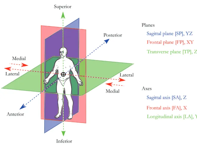
The anatomical planes are three imaginary cardinal planes that bisect the mass of the body in three dimensions. They allow the description of the body in its own regarding to a reference position. Body planes are commonly defined as follow (Figure 2.1):
- the sagittal plane (or anterior-posterior plane) is vertical and divides the body into left and right halves
- the frontal plane (or coronal plane) is vertical and divides the body into a front and a back - the transverse plane (or horizontal plane) is horizontal and divides the body into a top
and a bottom
In the anatomical reference position, all three planes intersect at a single point assumed to be the center of mass (or center of gravity).
1.1.4. Anatomical reference axis
The three anatomical axes associated with motion in each of the anatomical planes (Figure 2.1) defines the body reference coordinate system. Each axis is oriented perpendicular to one of the three planes of motion:
Vincent RICHARD PhD Thesis
- the anterior-posterior axis (or sagittal-horizontal axis) is perpendicular to the frontal plane
- the longitudinal axis (or vertical axis) is perpendicular to the transverse plane These terms are used indifferently for the entire body or for a segment.
Figure 2.1 Anatomical planes and axes
1.1.5. Terms of movement for the lower limb
Flexion and extension occurs in the sagittal plane. Flexion generally refers to a movement that
decreases the angle between a segment and its proximal segment. At the opposite, in the present work, extension refers to a movement that increases the angle between two body parts.
Abduction and adduction occurs in the frontal/sagittal plane. Abduction refers to a movement
away from the midline. Adduction refers to a movement towards the midline.
Internal and external rotation (or medial and lateral rotation) occurs in the transverse plane,
These terms are used indifferently for the movements of a limb or a segment with respect to the body or for the movements of one distal segment with respect to the proximal one, that is to say the articular movements.
1.2. Human locomotor apparatus
The human body is a complex system the functioning of which relies on the multiple interaction between complementary biological structures. In particular, the musculoskeletal system, which groups passive (bones, ligaments, tendons) and active (muscles) elements, ensures the integrity of the body in supporting, maintaining, moving and protecting organs from fall or impact. Not only the skeleton is composed of 206 independent articulated bones, but also 630 skeletal muscles and 244 degrees of freedom (Paul et al., 2014) constituting the internal framework of the body with specific shape, structure and role, but also the redundancy of actuators offers plenty of combinations to perform a given movement. Human beings are different from the rest of the animal reign in that they adopt a bipedal station, which during evolution, engendered remarkable transformations to the whole body, in particular at a skeletal level (Lovejoy, 2005a, 2005b). Indeed, the straightening of the trunk created a unique situation for mammals, where the head, pelvis and feet are aligned vertically, elevating at the same time the center of mass. The lower limb’s major role is the support of weight, adaptation to gravity and locomotion. The so-called human locomotor apparatus is composed of the two lower limbs (or legs) and the pelvis support the main part of body weight. Its schematic comportment is similar to an inverted pendulum whose lever arm is the length of the limb.
The direct consequence is the instability of such a system, requiring a perpetual control from the central nervous system. Focusing on the lower limb, instability is first due to the lever arm represented by the distance between the center of mass and the ground (e.g. inverted pendulum). In addition, the lower limb is poly-articulated, jeopardizing stability. Nevertheless, the body evolved brilliantly, solving the problem of instability at joint level in different manners depending on the role of the articulation.
1.2.1. Overview of the skeleton
The human locomotor apparatus is composed of three main type of organic structures: the osteoarticular system, the muscular system, the vascular and finally the nervous systems. The thesis work tackles the kinematics of the osteoarticular structures, without getting involved in the role of the muscular (or nervous) system.
Vincent RICHARD PhD Thesis
At an osteoarticular level, the lower limb is composed of six main regions, linked to the lower portion of the lumbar spine through the pelvis. The pelvis is not included in the definition of the lower limb but its description is necessary to introduce the hip joint as the articulation between the torso and the leg. The soft tissues surrounding the skeleton are not specified in this description as they are not considered in kinematics analysis. That is to say that the kinematic analysis focuses only on the movement of the osteoarticular system. However, we will see further that, unfortunately, the soft tissues surrounding the skeleton play a role in motion analysis based on skin markers. The lower limb is divided in five regions (Figure 2.2) used to identify articulations and bony segments. We consider three articular regions: gluteal (hip joint), knee and ankle regions, and four segmental regions: gluteal (pelvis), thigh, shank and foot regions. Note that the gluteal region contains both the hip joint and the hip bone. The knee is composed of two articulations, the tibiofemoral and patellofemoral joints. As for the ankle, it includes the talocrural and talocalcaneal joints. The foot can be also divided in multiple articulations and bony segments. A detailed model of the foot will not be used in this thesis and, therefore, the corresponding anatomy is not presented.
Figure 2.2 Lower limb overview
Thigh Shank Foot Gluteal region (Hip joint) Knee region (Knee joint) Talocrural region (Ankle joint)
Among all biological structures composing the lower limb, we will focus our attention to the elements playing a passive role in movement, that is to say the skeleton and ligaments. We will develop a descriptive approach for the understanding of the subsequent work concentrating on the skeleton. Since a kinematic analysis does not require a complete and exhaustive anatomical description (e.g.; (Netter, 2010)), or functional anatomy (e.g.; (Kapandji, 2009)), a simplified phenomenological model will be proposed. The different kinematic models of the hip, knee and ankle joints will be presented in the next sections.
The human body, from an osteoarticular point of view is considered sagittal symmetrical for an asymptomatic subject. As a consequence, modeling is often developed with reference to one limb.
Bones
The previously mentioned lower limb skeleton (Figure 2.3) contains many bones, in particular in the foot. The following description focuses on the bones involved in the model developed for the thesis work. The objective was to reduce the skeletal model to a four body-segment articulated system: pelvis, thigh, shank, and foot.
1.2.1.a. Pelvis
The skeleton of the gluteal region is known as the pelvic girdle. It consists of the two symmetric iliac bones, the sacrum and the coccyx that represents the lower extremity of the spine (Dubousset, 1994) and thus, the coxofemoral joints between the iliac bones and the femurs represent the demarcation between upper and lower part of the body, better known as hip joint.
1.2.1.b. Thigh
The bony structures composing the thigh are the femur and the patella (that is not considered in this thesis work). The femur is articulated to the pelvis through the hip joint at its proximal end. The femur is the longest and most resistant bone of the human body. The proximal epiphysis is composed by three regions: the femoral head, which is a sphere covered with cartilage inserted in the pelvis acetabulum to form the hip joint, the femoral neck that links the femoral head to the diaphysis, and the greater (and smaller) trochanter insertion point for the hip muscles.
1.2.1.c. Shank
The leg region, named shank, is the part that lies between the knee region and the ankle region, the skeleton is composed of the tibia and the fibula, generally considered rigidly attached to each other, except in some complex 3D anatomical ankle joint models (Baldisserri and Parenti-Castelli, 2012) (see sections bellow) not used in motion analysis so far. Tibia is the second longest bone in the human body. Its proximal epiphysis is composed by the lateral and medial epicondyles, receiving
Vincent RICHARD PhD Thesis
Figure 2.3 Skeletal framework of the lower limb (Moore and Dalley, 2006)
the femoral condyles, called tibial plateau. The distal epiphysis of the tibia contains the medial malleolus, and represents the supporting part of the ankle joint, while the distal part of the fibula, called lateral malleolus, is the second element of the articulation.
1.2.1.d. Foot
The foot is the most complex region of the lower limb, it contains 26 poly-articulated bones. The foot is composed of the tarsus, metatarsus and phalanges, though, it is considered as a rigid bony segment articulated with the shank at the ankle (talocrural) joint in the thesis work.
1.2.2. Joints of the lower limb
1.2.2.a. Structure
As previously mentioned, the lower limb contains multiple articulations between bones, in particular at the foot. Nevertheless it can be pictured as a system composed of three segments articulated by three main joints. All three hip, knee and ankle joints are synovial joints, or diarthrosis, which is the most common and complex type of joint in the body. The main characteristic of diarthroses consists in a joint cavity containing synovial fluid. This space between bones is delimited by the articular capsule, a fibrous capsule in continuity with the periosteum of the articulating bones, and the articular cartilage covering the articular surface of bones. Joints of the lower limb are mainly loaded in compression as they have a support function for the rest of the human body. The composition of joints is therefore adapted to high loads and generally the range of motion is reduced in favor of a better stability.
1.2.2.b. Hip joint
The hip or coxofemoral sacroiliac joint is the proximal joint of the lower limb, it is composed of the head of the femur and the acetabulum of the pelvis. Its ball-and-socket configuration makes it the most stable joint in the body, apparently easy to model because it exhibits articular surfaces that are close to spheres (Cereatti et al., 2010). The main role of the hip is the orientation of the lower limb in the 3 directions of space. The hip has three degrees of freedom. For an average asymptomatic subject the maximum hip range of motion is approximately as follow. In the transverse plane, maximum internal-external rotation angles are about 15° to 20°. When the knee is in extension, hip flexion angle is limited to 90° while it reaches up to 120° when the knee is flexed. Maximum extension is between 5° and 10°. Maximum abduction angle is about 45°, maximal adduction angle is up to 30°. No significant translation is allowed by the articulation (Kapandji, 2009). From a modeling perspective, the hip joint is classically modeled as a spherical joint (Stops et al., 2011).
1.2.2.c. Knee joint
The knee is the intermediate joint of the lower limb, it is the most complex joint in the body and is composed of the distal femur and proximal tibia. The knee is composed of two joints: the tibiofemoral articulation and the patellofemoral articulation. The patella is not considered in this
Vincent RICHARD PhD Thesis
thesis work even if a 3D anatomical kinematic model of the patellofemoral articulation has been proposed (Sancisi and Parenti-Castelli, 2011). This kinematic model is mainly used for musculoskeletal modeling (Moissenet et al., 2014). The knee joint involves many biological structures of different nature. Focusing on the osteoarticular composition of the knee joint, it is mechanically defined with three rotations and three displacements. The mechanism of movement between femur and tibia is a combination of rolling and gliding. For an average asymptomatic subject maximum knee range of motion is approximately as follow. Flexion reaches up to 160°, when both knee and hip are flexed. Knee extension can be up to 5°, this posture of hyper-extension is reached in straight position when the knee is locked. In this particular case, internal-external rotation is null while, with the knee unlocked, it may be about 5° to 10°. Due to the asymmetry of the articular surfaces at the knee and ligament guiding, flexion-extension movement induces rotations and displacements that involve other degrees of freedom (DoFs), namely, internal rotation and anterior displacement of the tibia regarding to the femur during flexion. This coupling inducing up to 15° of internal rotation mainly appears during the first 20° of flexion. In opposition, anterior displacement about 20 mm of magnitude appears after the knee exceeds 20° of flexion (Kapandji, 2009).
From a modeling perspective, the knee can be represented by various mechanical linkages. First, we find the hinge joint (Andersen et al., 2009; Reinbolt et al., 2005), allowing flexion/extension in only one plane. Then, we find the spherical joint, probably the most common representation of the knee (Lu and O’Connor, 1999), allowing all rotational movements. These two models impede translation (spherical joint) and some rotations (hinge joint), providing in most of the cases quite an inadequate 3D representation of the physiological
movement of the knee. Finally, 3D anatomical models (Duprey et al., 2009; Gasparutto et al., 2015) can be pictured as compound joints representing an assembly of simple mechanical linkages accompanied by more detailed anatomical knowledge like ligament insertion (Bergamini et al., 2011). These models generally allow most rotations and translations, while coupling the DoFs. In particular, the parallel mechanism (Figure 2.4) proposes a detailed mechanical model close to knee anatomy (Parenti-castelli et al., 2004). The use of such models has been reported to be a trade-off alternative in kinematics estimation (Duprey et al., 2010; Gasparutto
Figure 2.4 Example of 3D parallel mechanism for knee modeling
et al., 2015) in comparison to more classic models (spherical, hinge). But they still prescribe displacements in a deterministic way (i.e. kinematics is imposed by the geometry of the model).
1.2.2.d. Ankle joint
The ankle joint is composed of two joints, the talocalcaneal (subtalar joint) is formed by the articulation of the talus with the calcaneus. The talocrural (ankle) joint is commonly considered as hinge joint formed by the articulation of the distal tibia and fibula with the trochlea of the talus. The talocrural joint itself has only one degree of freedom, but the complex of the rear foot expands the mobility of the articulation procuring two degrees of freedom to the joint (Dettwyler et al., 2004). The bony process extending distally with respect to the medial tibia is referred to as medial malleolus. The lateral malleolus is the distal aspect of
the fibula. The line passing through the malleoli represents the anatomical axis of flexion of the ankle. For an average asymptomatic subject the ankle movement is essentially a combination of two rotations about anatomical axis, it is commonly modeled as a universal joint. Maximal dorsi-flexion is about 20° and 45°, while plantar-flexion (extension) is between 30° and 50° (Kapandji, 2009).
From a modeling perspective, existing ankle models are similar to the knee models. For the ankle, we find spherical joints (Charlton et al., 2004; Lu and O’Connor, 1999), universal joints (Andersen et al., 2009; Reinbolt et al., 2005), and parallel mechanisms (Di Gregorio et al., 2007) (Figure 2.5).
Figure 2.5 Example of 3D parallel mechanism for ankle modeling