HAL Id: tel-01493648
https://tel.archives-ouvertes.fr/tel-01493648
Submitted on 21 Mar 2017HAL is a multi-disciplinary open access
archive for the deposit and dissemination of sci-entific research documents, whether they are pub-lished or not. The documents may come from teaching and research institutions in France or abroad, or from public or private research centers.
L’archive ouverte pluridisciplinaire HAL, est destinée au dépôt et à la diffusion de documents scientifiques de niveau recherche, publiés ou non, émanant des établissements d’enseignement et de recherche français ou étrangers, des laboratoires publics ou privés.
Sleep deprivation and its impact on circadian rhythms
and glucose metabolism
Pawan Kumar Jha
To cite this version:
Pawan Kumar Jha. Sleep deprivation and its impact on circadian rhythms and glucose metabolism. Neurons and Cognition [q-bio.NC]. Université de Strasbourg, 2016. English. �NNT : 2016STRAJ028�. �tel-01493648�
1
ÉCOLE DOCTORALE Sciences de la Vie et de la Santé (ED 414)
Institut des Neurosciences Cellulaires et Intégratives (CNRS UPR 3212)
THÈSE EN COTUTELLE
présentée par :
PAWAN KUMAR JHA
Soutenue le : 06 Juillet 2016Pour obtenir le grade de : Docteur de l’université de Strasbourg
&
Discipline/ Spécialité :
UNIVERSITE D'AMSTERDAM
PAYS-BAS
UNIVERSITE DE STRASBOURG
FRANCE
Docteur de l'université d'Amsterdam
Sciences de la vie / Neurosciences
Sleep deprivation and its impact on circadian rhythms
and glucose metabolism
Dr. CHALLET E. Prof. KALSBEEK A. )
THÈSE dirigée par:
Docteur, Université de Strasbourg Professeur, Université d'Amsterdam
RAPPORTEURS:
Docteur, Université de Montpellier
Dr. BONNEFONT X.
Prof. CAJOCHEN C. Professeur, Université de Bâle
Prof. SCHLICHTER R. Professeur, Université de Strasbourg
Prof. FLIERS E. Professeur, Université d'Amsterdam
Dr. La FLEUR S.E. Docteur, Université d'Amsterdam
3
SLEEP DEPPRIVATION AND ITS IMPACT ON
CIRCADIAN RHYTHMS AND GLUCOSE
METABOLISM
ACADEMISCH PROEFSCHRIFT
ter verkrijging van de graad van doctor
aan de Universiteit van Amsterdam
op gezag van de Rector Magnificus
prof. dr. D.C. van den Boom
ten overstaan van een door het College voor Promoties ingestelde
commissie, in het openbaar te verdedigen in het
Institut des Neurosciences Cellulaires et Intégratives de Strasbourg
op woensdag 06 Juli 2016, te 09.00 uur
door
PAWAN KUMAR JHA
geboren te Supaul, India
4
PROMOTIECOMMISSIE:
Promotores : Prof. Dr. A. Kalsbeek Universiteit van Amsterdam Dr. E. Challet Universiteit van Straatsburg
Overige leden : Dr. X. Bonnefont Universiteit van Montpellier Prof. Dr. C. Cajochen Universiteit van Basel Prof. Dr. R. Schlichter Universiteit van Straatsburg Prof. Dr. E. Fliers Universiteit van Amsterdam Dr. S.E. la Fleur Universiteit van Amsterdam Dr. F. Criscuolo Universiteit van Straatsburg
6
Mr PAWAN KUMAR JHA was a member of the European
Doctoral College of the
University of Strasbourg during the preparation of his PhD,
from 2013 to 2015, Nelson Mandela (promotion). He has
benefited from specific financial support offered by the College
and, has followed a special course on topics of general
European interest presented by international experts.
This PhD research project has been conducted as part of a
cotutelle agreement between University of Strasbourg and
9
CONTENTS
Chapter 1 General IntroductionPartly based on Mol Cell Endocrinol 418, 74-88 (2015)
Chapter 2 Effects of central gastrin-releasing peptide on glucose metabolism Brain Res. 1626, 135-141(2015)
Chapter 3 Acute sleep restriction severely impairs glucose tolerance in rats
(Accepted)
Chapter 4 Sleep deprivation and caffeine treatment potentiate photic resetting of circadian clock in diurnal rodent, the Sudanian grass rat (Arvicanthis ansorgei)
(Ready for submission) Chapter 5 General Discussion
Appendices Summary Samenvating Résumé Thesis Abstract PhD Portfolio Acknowledgements 11 67 79 95 121
11
Chapter 1
G
ENERAL
I
NTRODUCTION
1. Homeostasis and rhythms 2. Rhythms of Life
3. Circadian clock system
3.1 Circadian rhythm properties
3.2 The suprachiasmatic nucleus: the biological clock
3.2.1 Localization and properties 3.2.2 Neurotransmitters of SCN
3.2.3 Mammalian Molecular Clock 3.3 Circadian entrainment
3.3.1 Photic entrainment 3.3.2 Non-photic entrainment
3.4 SCN outputs
4. Feedback action of arousal on clock
4.1 Arousal dependent on locomotor activity 4.2 Arousal independent of locomotor activity
4.3 Interaction between behavioral arousal and photic entraining stimuli 4.4 Pathways and neurotransmitters
4.4.1 The GHT
4.4.2 Serotonin and raphe nuclei
4.4.3 Orexin and the lateral hypothalamic area
4.5 Molecular mechanism and signaling pathways
4.6 SCN electrical activity suppression and clock resetting 4.7 Non-photic entrainment: Diurnal species
5. Circadian control of metabolism
5.1. Rhythms of metabolic processes
5.2 Circadian regulation of hormones in nocturnal mammals
5.2.1 Leptin and ghrelin rhythms 5.2.2 Glucocorticoid rhythm 5.2.3 Melatonin rhythm
5.3 Circadian regulation of glucose homeostasis in nocturnal mammals
5.3.1 Daily rhythm of glucose metabolism
5.3.2 Role of clock components in glucose metabolism
5.4 Circadian regulation of lipid homeostasis in nocturnal mammals
5.4.1 Daily rhythms of lipid metabolism
5.4.2 Role of clock components in lipid metabolism
5.5 Circadian regulation of metabolic homeostasis in diurnal mammals
5.5.1 Hormonal rhythms in diurnal mammals
5.5.2 Circadian regulation of glucose metabolism in diurnal mammals 5.5.3 Circadian regulation of lipid metabolism in diurnal mammals
6. Circadian desynchronization
6.1 Deleterious effects of circadian desynchronization on metabolic health
12
1. Homeostasis and rhythms
Charles Darwin (1809-1882) demonstrated the considerable influence of environmental variations on life. Living beings on the Earth are subjected to various external changes (e.g. food availability, light, temperature, predation). Organisms therefore have to adapt to their environment in order to survive and maintain their own species. Contemporary to this, a new concept of internal processing and function was proposed by Claude Bernard (1813-1878). Bernard proposed the notion of the interior milieu, referring to the extra-cellular fluid environment, more particularly the interstitial fluid surrounding the organs of the individual. He suggested that maintaining the stability of the internal fluid (blood and other body fluids) is essential for the life of higher organisms, because multiple and/or chronic disturbances of this interior milieu would lead to pathophysiology. This concept has been redefined over decades and termed homeostasis (Homeo = same; stasis = steadiness) by Walter B. Cannon (1871-1945). Cannon defined homeostasis as “all organic processes that act to maintain the steady state of the organization, in its morphology and its internal conditions, despite external disturbances”. In a more simple way, homeostasis is the ability of the body to maintain a state of relative stability of the different components of its internal environment despite the constant changes in the external environment. More recently, according to the Thermal Commission of the International Union of Physiological Sciences (IUPS; 2001), homeostasis is characterized by “the relative constancy of physiochemical properties of the internal environment of an organism as being maintained by regulation”. In the body, homeostasis applies to many physiological processes such as the regulation of osmolarity, blood sodium level, plasma glucose, blood pressure, body temperature, feeding and sleep. It is of prime importance to note that physiological variables are not maintained constant in the absence of environmental perturbations and instead, display more or less marked rhythms, i.e., a relative constancy is maintained. Daily and seasonal changes in the environment are important variables that influence the homeostatic setting of the organism. However, the predictability of these changes also allows an organism to anticipate these environmental changes. The structures and mechanisms which are involved in the anticipation of these daily changes on their turn also interfere with the (constancy of the) internal environment. The interactions of both these processes, i.e., process 1 and process 2, and their consequences for the organisms‟ adaptive capacities encompass the subject of this thesis.
The 24-h period of Earth‟s rotation is correlated with major oscillations in many critical variables in the environment such as ambient illumination and temperature, the availability of
13 nutrients and activity of predators. The periodicity of these challenges and opportunities permits anticipation to these daily changes that in turn will shape the temporal organization of behavior and physiology of the organism. The internal temporal capability enables the organism to anticipate the probability of predictable demands upon the homeostatic system and therefore will decrease the homeostatic perturbation within a certain time frame and eventually reduce risks of disease. This feature is clearly illustrated with the rises in body temperature and plasma corticosteroid in advance of the time when animals awake from their daily sleep period. Waking itself may occur in advance of the time of lights-on in diurnal animals. Another example of physiological anticipation to daily changes is the increased level of plasma glucose in anticipation to glucose demands for the upcoming activity period, also called “dawn phenomenon” in humans (Bolli et al., 1984; Arslanian et al., 1990).
2. Rhythms of Life
The Earth‟s rotation around its axis generates daily environmental cycles. The daily environmental cycle of greatest importance for daily timing is the highly predictable alteration of light and darkness. Living organisms follow these periodic changes in the environment and display biological variations in 24-h intervals, called daily rhythms. The human sleep-wake cycle constitutes perhaps the best known example of a behavior that occurs with a ~24 h periodicity. Many other organisms display comparable behaviors, commonly referred to as the rest-activity cycle, taking into account that “rest” is not always “sleep”. Also many behavioral and physiological processes display daily rhythms, such as locomotor activity, feeding, body temperature, cardiovascular function, and hormonal secretion. The issue is how these rhythms are generated, or even whether they are internally generated or a just a passive consequence of the changes in the environment.
The first observation of rhythmic behavior in human history goes back to the 4th century B.C., when Androsthenes of Thasos, a ship captain under the command of Alexander the Great, recorded his observation of daily movements of plants (Refinetti, 2006). Androsthenes traveled North Africa and India where he observed the daily movements of the tamarind tree (Tamarindus indica). Androsthenes noticed the daily cycles of movement of tamarind leaves in which leaves move up during the day and down at night. Further, observations about daily rhythmicity were made by great physician Hippocrates. Hippocrates (460-370 B.C.), who is also considered as the father of medicine, observed periodic physiological processes, such as changes of physiological condition and disease state over 24 h (Refinetti, 2006). These observations point out that daily physiological rhythms may be caused not only by
14 environmental factors (such as the alternation of day and night), but also generated within the organism in the absence of environmental cycles. The notion of endogenous rhythm came to the scientific community after the publication of a monograph by a French astronomer, Jean-Jacques Dortous de Mairan (1678-1771) in which he mentioned his observation about daily opening and folding of the leaves continued even when the plant was placed in the dark room. This observation suggested that the movements are not the result of a response of sun. Rather it is controlled within the plant. The concept of endogenous rhythms was not accepted despite these findings because of the arguments that all environmental influences that result from the rotation of Earth could not have eliminated in darkness.
The 20th century witnessed a surge in sophisticated research on biological rhythms and thus the establishment of the discipline of chronobiology. In the 1930s Kalmus and Bünning argued for an endogenous timing mechanism based on the finding that periodicity of the rhythm of adult emergence in Drosophila varied with temperature fluctuations that were clearly independent of Earth rotation (Sehgal, 2004). The idea that endogenous clocks control the circadian rhythms (rhythms of approximately 24 hours periodicity that persistent in constant environmental condition) gained wider acceptance in the next decades. Interspecies variations in the periodicity of circadian rhythms under non-cycling environmental conditions helped to strengthen this notion. More importantly, the period in most cases did not precisely match that of any environmental cycle, indicating that it was most likely endogenously generated (Sehgal, 2004). For instance, in constant darkness (DD), activity of a rat each day begins and ends a little later than it did the day before. Usually the drift that occurs because of the periodicity of this rhythm, and thereby that of underlying clock, is a little bit more than 24 h. This drift is called “free run”. A free-running rhythm of locomotor activity is illustrated below (Figure 1). In 1960s Jürgen Aschoff and his colleague Rütger Weber demonstrated that in the absence of environmental cues (light), also the human sleep-wake rhythm persists, and free-runs with a period longer than 24 h (Aschoff, 1965). A similar observation was also made by the French explorer and scientist (Siffre, 1964), when he isolated himself in a deep cave without any time cues for several months. A free-running rhythm, with a cycle deviant from 24 h in constant conditions, is today considered as proof of endogenous generation. The vast majority of living species that evolved on our continuously rotating planet have developed an internal oscillating system to match these external variations. This major conclusion was first drawn in the landmark Cold Spring Harbor Symposium on Biological Clocks in 1960, organized by Jürgen Aschoff and Colin Pittendrigh, an American biologist considered as another co-founder of modern biological rhythm research. Pittendrigh summarized all the
15 knowledge accumulated by the pioneers of the field and established the basis of chronobiology (Pittendrigh, 1960).
Today we know that almost all the organisms from cyanobacteria to humans show circadian rhythms. For this introduction, my prime focus is to describe the circadian clock system in mammals, its daily entrainment to synchronizing cues, and its control over metabolism and physiology in nocturnal and diurnal species.
3. Circadian clock system
3.1 Circadian rhythm properties
The periodicity of ~24 h is one attribute of the circadian rhythms. A second circadian characteristic is that these rhythms can be synchronized or reset by environmental cues. The dominant environmental signal that affects circadian rhythms is light. In 1954 Jürgen Aschoff used for the first time the German term Zeitgeber (“time giver”) to define an exogenous (external cue) that triggers some sort of change in an organism‟s endogenous clock to synchronize or set the phase of circadian rhythms (Aschoff, 1954). The process of synchronization itself is called entrainment. In chronobiology, time is defined according to
Figure 1. Schematic representation of actograms of a diurnal and a nocturnal rodent
The actograms represent rhythms of locomotor activity initially entrained to a 24-h light/dark (LD) cycle. Each horizontal line corresponds to one day and vertical black data bars represent bouts of activity. Above the actograms, white and black bars indicate the light and dark conditions, respectively. Upon transfer in constant darkness (DD), circadian rhythms resume with their endogenous period (> 24-h here). ZT: Zeitgeber Time (ZT0 corresponding to lights on); CT: Circadian Time.
16 the environmental cycle and called Zeitgeber Time (ZT). For example, in a 24-h cycle that consists of 12 h of light followed by 12 h of darkness, ZT 0 corresponds to “lights on” and ZT 12 is “lights off” (Fig.1). Persistence of periodicity in the absence of a cyclic environment is another property of circadian rhythms. Under these conditions, rhythms are said to be free-running. The endogenous natures of the rhythms and their periodicity have to be determined under free-running conditions. Under constant environmental conditions, the time points are referred to as Circadian Time (CT) (Figure 1), which is the time generated by the internal clock.
Colin Pittendrigh in the early 1950s demonstrated that the eclosion (i.e., hatching of adult flies from the pupae) rhythm in Drosophila pseudoobscura persists in constant conditions and can be entrained by a light cycle close to the flies‟ endogenous period. More importantly, the period of the eclosion rhythm remains relatively constant when exposed to changes in environmental temperature. In other words, the eclosion rhythms of Drosophila were temperature compensated, unlike the rates of most of biochemical reactions (Pittendrigh, 1954). Thus, the circadian period remains constant over a wide temperature range.
The fundamental properties of the clocks can be represented as a circadian system (Figure 2). There can be multiple input and output pathways to serve a single clock, an output being able to feedback to the clock. This thesis is based on this theoretical framework (See chapter 4).
3.2 The suprachiasmatic nucleus: the biological clock
3.2.1 Localization and properties
The first indication that the medial part of the hypothalamus is involved in daily rhythms of locomotor activity in rats came from the lesion experiments of Richter (1967). A few years later, the effect of removal of the suprachiasmatic nuclei (SCN) by bilateral electrolytic
Figure 2. Schematic representation of a simple circadian system
17 lesions was tested by Moore & Eichler (Moore and Eichler, 1972) and Stephan & Zucker (Stephan and Zucker, 1972) in rats (Figure 3).
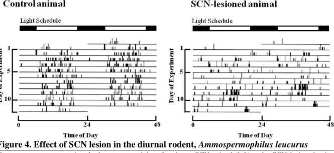
Ablation of SCN abolished the behavioral and hormonal rhythms (Moore and Eichler, 1972; Stephan and Zucker, 1972; Moore and Klein, 1974; Ibuka and Kawamura, 1975; Nagai et al., 1978). Later these observations were also confirmed in other mammals (Edgar et al., 1993a; DeCoursey et al., 1997) (Figure 4).
Electrical recordings showed that the SCN is an endogenous pacemaker, as SCN neurons display circadian patterns of firing with highest activity during subjective daytime even when isolated in vivo (Inouye and Kawamura, 1979) or maintained as cell culture in vitro (Bos and Mirmiran, 1990; Welsh et al., 1995). Another approach, using 14C labeled deoxyglucose,
Figure 3. Localization of the suprachiasmatic nucleus (SCN) in golden hamster
3V: third ventricle; OC: optic chiasm; PVN: paraventricular hypothalamic nuclei, SCN: suprachiasmatic nuclei
Adapted from Morin and Wood (2001)
Figure 4. Effect of SCN lesion in the diurnal rodent, Ammospermophilus leucurus
Two actograms representing the locomotor activity of an intact SCN animal (left) and a SCN-lesioned animal (right). Each horizontal line corresponds to one day and vertical black data bars represent bouts of activity. Above the actograms, white and black bars indicate the light and dark conditions, respectively.
18 corroborated that the clock is an endogenous oscillator by demonstrating a rhythmic pattern in glucose utilization in the SCN which also persisted in constant darkness (Schwartz and Gainer, 1977; Schwartz et al., 1987). The final evidence that the SCN is “the master clock” came from graft experiments of Ralph and colleagues (Ralph et al., 1990). They identified a mutant hamster characterized by a shorter endogenous period length, called Tau mutant. By performing lesions in the SCN (SCN-X) of Tau mutant and wild-type hamsters, they obtained arrhythmic animals. When they transplanted SCN grafts of Tau mutant in SCN-X wild-type animals, they restored rhythmicity with a period characteristic of the Tau mutant, and vice
versa. In other words, rescued rhythms always exhibited the period of the donor genotype. All
these findings demonstrated that the SCN is the main conductor of circadian rhythmicity in mammals.
The studies of single cell electrical activity from SCN slice preparations or dissociated cell cultures from the rodent brain suggest that even individual cells exhibit circadian rhythmicity, that different cells can have different circadian periods, and that the overall period of the circadian oscillation generated by the SCN is the average of the periods of the individual cells (Welsh et al., 1995; Liu et al., 1997; Honma et al., 1998; Honma et al., 2004). The dissociated neurons oscillating with different periods and phases become mutually synchronized by coupling and thereby are able to produce a coherent SCN output. Thus, SCN neurons are relatively unstable oscillators which require network interactions to stabilize their noisy cycling. As a tissue, the SCN thus provides a more precise and robust output than single SCN neurons (Herzog et al., 2004; Liu et al., 2007). Attempts to identify the coupling mechanism suggest that neuronal firing and synaptic communication between neurons are required (Welsh et al., 2010).
3.2.2 Neurotransmitters of SCN
The SCN is a heterogenous structure based on its population of neuronal and glial cell types. Left and right SCN each contain around 10,000 neurons in two anatomical subdivisions: the ventrolateral (core) region, receiving retinal input, and the dorsomedial (shell) region receiving input from the core (Abrahamson and Moore, 2001) (Figure 5). Much is known about the neurotransmitters/neuropeptides used by the SCN neurons: γ amino butyric-acid (GABA, major inhibitory neurotransmitter in the central nervous system), Arginine Vasopressin (VP or AVP) and Vasoactive Intestinal Polypeptide (VIP), Gastrin Releasing Peptide (GRP), cholecystokinin (CCK), Substance P (SP) and somatostatin are among the predominant ones. GABA seems to be the most common neurotransmitter of the SCN (Moore
19 and Speh, 1993). Most neuropeptides co-localize with GABA, and many synapses between SCN neurons are GABAergic (Moore and Speh, 1993; Buijs et al., 1995; Strecker et al., 1997), though few SCN neurons are glutamatergic (Cui et al., 2001).
AVP is also abundant in the SCN, predominantly in the dorsomedial region of the nucleus (Ibata et al., 1999; Moore et al., 2002; Morin et al., 2006; Nascimento et al., 2010). AVP expression is higher during the day than during the night in rodents (both nocturnal and diurnal) and humans maintained under light-dark cycles (Tominaga et al., 1992; Hofman and Swaab, 1993; Dardente et al., 2004). The rhythmic expression of Avp persists in constant darkness (Dardente et al., 2004). Other studies show that AVP is also rhythmically released from the synaptic terminals with a higher rate during the day (Kalsbeek et al., 1998). In contrast to AVP, VIP is present mostly in the ventrolateral region of the SCN and in its dorsal projections. Ventrolateral neurons of the SCN also express other neuropeptides, like calbindin, calretinin, SP, neurotensin and GRP. VIP release is not consistent across the species, in Syrian hamster it has been shown that VIP release is highest in mid-day whereas in Golden hamster and humans lowest expression of VIP in SCN were reported in day time (Refinetti, 2006). On the other hand, GRP content in SCN increases over the course of the light period and gradually decreases during the dark period in the rat (Shinohara et al., 1993). Neurotransmitters involved in SCN inputs and outputs are discussed in subsections 3.3 and 3.4 of this Introduction.
Figure 5. Schematic representation of neuropeptides distribution in SCN
Many of the neurons within the core SCN express VIP, GRP and CALB. In the shell region SCN neurons mostly express VP. Most of these neurons also express GABA and very few glutamates (not shown here). 3V: third ventricle; VP: vasopressin; SS: somatostatin; CCK: cholecystokinin; SP: substance P; CALB: calbindin; CALR: calretinin; GRP: gastrin releasing peptide; VIP: vasoactive intestinal polypeptide.
20 3.2.3 Mammalian Molecular Clock
The clock mechanism in the SCN involves 24-h oscillations of core clock components, called clock genes and defined as genes whose protein products are necessary for generation and regulation of circadian rhythms within individual cells (Ko and Takahashi, 2006). The molecular oscillations of the SCN depend on several clock genes. Circadian rhythmicity is based on rhythmic expression of core clock genes and their autoregulatory feedback transcriptional/translational loops (Figure 6). The core clock machinery is structured with positive transcriptional regulators, including BMAL1/CLOCK and RORs, and negative regulators such as PERs (PER1-3), CRYs (CRY1-2) and REV-ERBs. BMAL1 binds with CLOCK and forms heterodimers which activate the transcription of the negative PER and CRY regulators, thus defining a positive loop (Reppert and Weaver, 2002). PERs and CRYs accumulate in the cytoplasm and form complexes which translocate into the nucleus to inhibit their own transcription along with other BMAL1/CLOCK-driven transcription, such as that of the clock-controlled genes, thereby forming a negative loop (Mohawk et al., 2012). An additional negative loop in this molecular network is contributed by the nuclear receptors REB-ERBα and β. REV-ERBs bind to the ROR response element (RRE) of BMAL1 and CLOCK promoters and repress their transcription, whereas in counterbalance to REV-ERBs inhibition, RORα/β/γ also bind on the RRE of BMAL1 and induce its transcription (Preitner et al., 2002; Guillaumond et al., 2005; Crumbley and Burris, 2011).
The circadian rhythms generated by this molecular clock machinery get fine-tuned by environmental cues. In particular, light can synchronize the SCN neurons via a cascade of transcriptional activation. In response to photic inputs from retinal ganglion cells, glutamate and pituitary cyclase-activating peptide (PACAP) are released in the ventral region of the SCN, leading to transcriptional induction of clock gene (Per1 and Per2) expression through chromatin remodeling (Dibner et al., 2010) (a more detailed description follows in section 3.3.1). Of note, the molecular clock in the SCN works at the same astronomical times in diurnal and nocturnal species, and the mechanisms underlying photic resetting are essentially similar in terms of temporal sensitivity and direction of light-induced phase shifts between both categories of mammals (Challet, 2007). These findings suggest that the distinction between nocturnal and diurnal animals likely relies on neural mechanisms operating downstream of the SCN clock (Kalsbeek et al., 2008b).
21 Demonstration of rhythmic clock gene expression in cells and tissues throughout the body, and persistence of these rhythms in cultured cells revealed the presence of the molecular clock mechanism also in brain regions outside of the SCN, such as the arcuate nucleus (ARC) and the dorsomedial hypothalamic nuclei (DMH), as well as in most peripheral organs, including those essential for energy homeostasis, such as the liver, kidney, pancreas, skeletal muscle and adipose tissues (Balsalobre et al., 1998; Yamazaki et al., 2000; Abe et al., 2002; Yoo et al., 2004; Guilding and Piggins, 2007). Although the basic molecular components of the central clock are conserved in these extra-SCN oscillators, their self-sustained rhythmicity is less robust than in the central clock. Using Per1 luciferase rat explants, Yamazaki et al. (2000) showed that circadian rhythmicity in peripheral organs, such as lungs, liver and skeletal muscle, starts to dampen within one week, whereas the SCN cycle up to at least 32 days. Dampening within a few cycles has also been reported for explants of various brain areas other than the SCN (Abe et al., 2002; Abraham et al., 2005; Guilding and Piggins, 2007). Of note, Yoo et al. reported persistent, self-sustained circadian oscillations of PER2 expression for more than 20 days in lungs and liver explants of PER2-luciferase transgenic mice (Yoo et al., 2004). SCN lesions in this study do not cause disappearance of PER2 rhythmic expression, although they do lead to asynchrony of phase between peripheral organs of the same individuals which specifies the role of the SCN as a conductor which drives rhythms of peripheral clocks by maintaining phase coherence among organs.
Figure 6. The molecular mechanism of
the mammalian circadian clock in the SCN and other tissues
The core circadian clock is formed by the positive and negative limbs of a transcriptional-translational feedback loop. In the positive limb, BMAL1 and CLOCK form heterodimers and activate the transcription of Per 3) and Cry (1-2). PER and CRY proteins repress their own transcription by inhibiting CLOCK-BMAL1 activity in the negative limb. In an additional loop, REV-ERBs repress Clock transcription, whereas opposite actions of REV-ERBs and RORs contribute to the rhythmic expression of Bmal1. In addition, the CLOCK-BMAL1 heterodimer also activates transcription of clock controlled genes, including many metabolic genes as clock outputs. BMAL1, brain and muscle ARNT-like 1; CLOCK, circadian locomotor output cycles kaput; PER, Period; CRY, Cryptochrome; REV-ERBs, reverse viral erythroblastosis oncogene products; RORs, retinoic acid-related orphan receptors; CCG, Clock controlled genes.
Adapted from (Crumbley and Burris, 2011; Mohawk et al., 2012).
22
3.3 Circadian entrainment
A circadian clock maintains a self-sustained rhythm even in the absence of environmental cycles, thus ensuring that internal functions maintain their temporal relationships under constant conditions. However, the real life usually gives temporal cues that help rhythms to adapt and anticipate to natural periodic changes, including light/dark, temperature, humidity, social activity and food availability. These environmental cues are broadly classified in photic and non-photic cues. The way by which endogenous rhythms can be entrained by light and darkness became the focus of many studies around the 1960s. The investigation of the entrainment process by the non-photic cues began later in the late seventies.
3.3.1 Photic entrainment
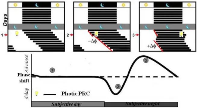
Light is the most ubiquitous entraining signal or Zeitgeber encountered in the daily life of most of organisms. As the SCN-driven period of circadian rhythms differs slightly from 24-h in most mammals, rhythms must periodically be shifted forward or backwards in order to maintain an appropriate phase-relationship with the environmental period of exact 24-h (Challet, 2007). Brief pulses of light delivered to animals housed in constant darkness produce phase-dependent shifts in the timing of the rest-activity cycles, thereby defining a so-called phase-response curve (PRC) (Daan and Pittendrigh, 1976; Schwartz and Zimmerman, 1990)
(Figure 7). Exposure to light during the early night, for instance, when nocturnal animals
have just become active after dusk, delays the clock. This shift will delay activity onset to a slightly later time on subsequent cycles, realigning with the external environment. Conversely, light encountered during the late night, or when a nocturnal rodent remains active after dawn, advances the phase of clock. This shift will advance activity onset to a slightly earlier time on subsequent days, again realigning the behavior and physiology with the external world. As illustrated in Figure 7 for nocturnal rodents, light in early and late subjective night produces phase-delays and phase-advances, respectively, whereas light during most of the subjective day has no phase-shifting effect. These circadian responses to light are roughly similar in both nocturnal and diurnal species (Slotten et al., 2005; Challet, 2007).
23 Phase-shifts such as those described above in response to light pulses are generally evaluated by measurements of daily onsets or offsets of the locomotor activity pattern. Of note, these shifts in behavior actually correspond to phase-changes of the underlying SCN clock. Now the question remains: how does light reset the SCN clock? In 1972 (Moore and Lenn) discovered terminals of retinal axons in the SCN, strongly suggesting that the mammalian visual system could convey photic information to the SCN (Moore and Lenn, 1972). Photic signals from the retina are conveyed to the SCN directly via a neural pathway called the retinohypothalamic tract (RHT). Additionally, an indirect neural pathway courses to the SCN via the intergeniculate leaflet (IGL) (Figure 8).
Figure 7. Phase response curve (PRC) of nocturnal animals to light pulses
The free-running locomotor activity rhythm of animals transferred to constant darkness can be phase-shifted in a phase dependent manner. Phase advances are plotted as a positive value (+∆ϕ) and phase delays as negative values (-∆ϕ). The photic PRC consists of three characteristic zones (1) the dead zone: a light pulse during most of the subjective day does not cause a phase shift; (2) the delay zone: at the beginning of the subjective night and (3) the advance zone: toward the end of the subjective night.
24 Still, the big question is which retinal cells, and which molecules in those cells, act to detect light and to convey this information to the SCN clock? And so the search for the “circadian photoreceptor”, responsible for photic resetting of the circadian clock and localized within the eye, began. Freedman et al. (1999) observed that mice lacking both rod and cone photoreceptors show normal resetting of their activity rhythm by light. This observation led to the idea of a non-visual circadian photoreceptor (Freedman et al., 1999). Meanwhile, a group of retinal ganglion cells were shown to contain a light sensitive photopigment, called melanopsin, that was highly responsive to blue-light stimulation (Provencio et al., 2000). Mice lacking the melanopsin gene displayed severely attenuated phase-resetting in response to brief pulses of monochromatic light, demonstrating the critical role of melanopsin in circadian photo-entrainment. The axons of these retinal ganglion cells form the RHT and project monosynaptically to the ventral (core) of the SCN (Hattar et al., 2002), where they release mainly glutamate and PACAP (Castel et al., 1993; Hannibal et al., 1997).
As mentioned above, the RTH projects not only to the SCN, but also to the IGL, a distinct subdivision of the lateral geniculate complex in the thalamus (Pickard, 1985; Moore and Card, 1994; Hattar et al., 2006). From the IGL, the geniculohypothalamic tract (GHT) projects to the SCN and thus indirectly conveys light information by releasing neuropeptide Y (NPY) and GABA. The delay between RHT and GHT signals may provide additional information leading to a more differentiated response of the SCN to light cues. Finally, other structures
Figure 8. Schematic of a sagittal brain section showing nuclei and neurotransmitters of the mammalian circadian system involved in photic and non-photic resetting.
Photic signals are transmitted directly to the SCN via the RHT utilizing Glu and PACAP. Non-photic information reaches the SCN via a network of connections involving the NDR and NMR, the IGL and the LHA. These pathways use serotonin, NPY, GABA, and orexin respectively as their main transmitters. NDR: dorsal raphe nucleus; GABA: γ-amino butyric acid; GHT: geniculohypothalamic tract; Glu: glutamate; IGL: intergeniculate leaflet; LHA: lateral hypothalamic area; NMR: median raphe nucleus; PACAP: pituitary adenylate-cyclase-activating peptide; NPY: Neuropeptide Y; RHT: retinohypothalamic tract; SCN: suprachiasmatic nucleus.
25 could also convey indirect light information to the SCN. For example, the lateral hypothalamus (LH) is also a target of the RHT (Hattar et al., 2006) and the arousal-promoting orexin neurons in the LH project to the immediate vicinity of the SCN (McGranaghan and Piggins, 2001; Brown et al., 2008).
The process of photic entrainment depends upon the direct and indirect transmission of light information to the SCN. The downstream signaling of this transmission induces acute expression of several immediate early genes, such as c-fos, and the clock genes Per1 and Per2 (Albrecht et al., 1997; Shigeyoshi et al., 1997). In response to light exposure, glutamate and PACAP are released from the RHT and bind to their receptors expressed in ventral SCN neurons (Reppert and Weaver, 2002). More precisely, SCN neurons express several subtypes of glutamate (NMDA-R, AMPA-R) and PACAP (PAC1-R) receptors. Stimulation of these receptors induces Ca2+ influx which activates several kinase pathways (Morse and Sassone-Corsi, 2002; Hannibal, 2006). One of these pathways involves the extracellular signal-regulated kinase (ERK), which belongs to the family of Mitogen-activated protein kinase (MAPK), known to phosphorylate the cAMP response element-binding protein (CREB). Many pathways converge at phosphorylation of CREB and stimulate the transcription of Per1 and Per2 genes by its binding to a CRE element in the promoter region (Travnickova-Bendova et al., 2002; Tischkau et al., 2003). Light responses of the clock are complex because there are many pathways at work. Up to this stage, such intracellular signaling cannot explain explicitly the differential effects of light inducing phase-delays and advances, according to circadian time of the light pulse. Clearly the molecular mechanism of photic entrainment is not fully understood yet (Sehgal, 2004) (Figure 9).
26
Moreover, the effect of light is not only limited to transcriptional changes, such as c-Fos and
Per expression, but it also impacts on post-transcriptional modifications of core clock
components. For example, light-induced degradation of BMAL1 could constitute an important step for entrainment (Tamaru et al., 2000). The intricacy of photic entrainment is getting even more intensified by epigenetic interventions, such as light-induced chromatic remodeling (Crosio et al., 2000).
Figure 9. Molecular pathways for phase delays and phase advances in the SCN
Photic stimulation triggers release of glutamate and PACAP, which than interact with their receptors at the membrane of ventral SCN cells. The resulting influx of Ca+2 stimulates CaMKII, which phosphorylates NOS. Molecular cascades downstream from CaMKII/NOS are not completely clear for differences between phase delays and advances. The phosphorylation of CREB mediated by MAPK and activation of PKG by cGMP could be the convergent point for both delays and advances. However Ca2+ release from the endoplasmic reticulum through ryanodine receptor activation seems to be involved in phase delays. This is a simplified view of the photic signaling cascade without considering the discrepancies in the literature. NMDA-R: N-methyl–D-aspartate receptor; Ca+2: Calcium; CaMKII: Calcium Calmodulin Kinase II; NOS: Nitric oxide synthase; NO: Nitric oxide; cGMP: cyclic Guaosine monophosphate; GC: guanylyl cyclase; PKG: cGMP-dependent protein kinase; P-MAPK: Phosphorylated Mitogen-activated protein kinase; P-CREB: Phosphorylated cAMP response element binding protein; Per: Period.
27 3.3.2 Non-photic entrainment
The concept that circadian rhythms could be influenced by mechanisms other than light came to prominence through a series of experiments demonstrating that the rhythms of birds could be entrained to the daily playback of birds songs (Gwinner, 1966; Menaker and Eskin, 1966). The early reports of social entrainment in humans (Aschoff, 1979; Weaver, 1979) also revealed that circadian rhythms could be altered using so-called non-photic stimuli independent of the light itself or the specific neural pathways activated by light. A wide variety of different stimuli fall into this category, such as: temperature, food availability, social interaction and behavior arousal. In late 1980s, Mrosovsky and Salmon demonstrated that enhanced locomotor activity acts as potent zietgeber in hamsters (Mrosovsky and Salmon, 1987, 1990). Furthermore, it has been shown that enhanced physical activity is able to alter the period (Yamada et al., 1986) or produce phase-shifts (Reebs and Mrosovsky, 1989a) of circadian rhythms in nocturnal rodents. Physical activity can also shift the human circadian rhythms. Notably, exercise during early evening and after midnight elicits phase advances and delays, respectively (Van Reeth et al., 1994; Buxton et al., 1997; Buxton et al., 2003). The PRC obtained with photic cues is called the “photic PRC”. The non-photic PRC of nocturnal rodents is characterized by phase-advances during the subjective day and little or no response during the subjective night (Figure 10).
Unlike the light PRC, whose global shape is almost similar for nocturnal and diurnal species and extensively researched, the non-photic PRC for diurnal rodents has not been extensively studied. Entrainment to scheduled locomotor activity has been reported in diurnal rodents and primates (Hut et al., 1999; Glass et al., 2001), as well as weak locomotor activity feedback effects in the diurnal/crepuscular Degus (Kas and Edgar, 1999). The characteristics of non-photic entrainment in these diurnal species suggest that the diurnal non-non-photic PRC may be close to that of nocturnal species. However, results from dark pulse experiments suggest differential responses of non-photic stimuli between nocturnal and diurnal species (Challet, 2007). To understand how behavioral arousal acts as a non-photic cue in the diurnal rodent Grass rat, Arvicanthis ansorgei is one of goal of this thesis.
28
At the molecular level, the Per genes are the main targets within the core molecular oscillator for stimuli that reset the clock. Non-photic phase-shifts induced by increased locomotor activity produce a down-regulation of the expression of Per1 and Per2 mRNA in the SCN (Maywood et al., 1999; Yokota et al., 2000). Furthermore, injections of Per1 antisense oligonucleotides in the mid-subjective day (i.e., the approximate time of maximal non-photic phase shifts and maximal Per1 expression) suppress Per1 levels and induce non-photic-like phase-advances (Hamada et al., 2004). The molecular mechanisms of signal transduction that mediates the suppressive action of non-photic cues on SCN Per gene expression are poorly understood. Non-photic stimuli may suppress ERK phosphorylation in the SCN (Coogan and Piggins, 2005). Moreover, treatment with serotonin agonists during the subjective day, produces phase-advances and markedly suppresses Per1 and Per2 mRNA levels (Horikawa et al., 2000; Duncan et al., 2005).
The two major input pathways that are considered to transmit non-photic information to the SCN are the GHT from the IGL, that also conveys photic information to SCN (Harrington, 1997), and serotoninergic input from the median raphe nuclei in the brainstem. The IGL also receives a serotonergic projection from the dorsal raphe (Hughes and Piggins, 2012) (Figure
Figure 10. Phase response curve (PRC) to a non-photic stimulus, such as a novel running wheel, in the nocturnal hamster
The free-running locomotor activity rhythm of a nocturnal animal transferred to constant darkness (1) can be phase advanced by arousal in the subjective day, whereas (2) little or no effect on the behavioral phase occurs when aroused in subjective night.
29
8). As mention above, the IGL releases NPY and GABA. There is evidence that IGL and
NPY transfer non-photic signals to the central circadian system. It has been shown that lesions of IGL attenuate the phase-shifts induced by enhanced physical activity in mice and hamsters (Johnson et al., 1988; Janik and Mrosovsky, 1994; Wickland and Turek, 1994; Koletar et al., 2011). In vivo electrical stimulation of IGL and NPY infusion in the third ventricle both result in behavioral phase-shifts that resemble the traditional non-photic PRC seen in response to induced locomotor activity (Albers and Ferris, 1984; Rusak et al., 1989; Biello and Mrosovsky, 1996). Infusion of NPY into the region of SCN in vivo and directly on the SCN in slices in vitro suppresses Per1 and Per2 expression (Fukuhara et al., 2001; Maywood et al., 2002). Furthermore, circadian variation of serotonin content in the SCN correlates with physical activities at different times of the day (Shioiri et al., 1991). Electrical stimulation of the raphe nuclei leads to behavioral phase-shifts with increased levels of serotonin in the SCN (Dudley et al., 1999; Meyer-Bernstein and Morin, 1999).
Feeding and metabolic cues are other non-photic signals that can entrain the SCN clock. The pathways for these signals comprise nuclei in the mediobasal hypothalamus, such as the arcuate nucleus and the ventromedial hypothalamic nucleus (Challet et al., 1997; Yi et al., 2006), which could integrate metabolic information and energy status before projecting to the SCN.
3.4 SCN outputs
It is important for a pacemaker to have output pathways by which it controls the timing of various targets in the rest of the body. The SCN conveys its circadian signals by neuronal connections, primarily to other hypothalamic sites, and diffusible substances. The structrucal details of SCN efferents have been studied by the various tracing techniques (Watts and Swanson, 1987; Watts et al., 1987; Kalsbeek et al., 1993). The SCN projects mainly to three areas: hypothalamus, thalamus and septum (Figure 11). The SCN core projects to shell and to other hypothalamic areas in its vicinity, particularly the lateral subparaventricular zone (lSPV) (Leak et al., 1999; Leak and Moore, 2001). In general, the dorsomedial SCN which is predominantly containing AVP expressing cells, projects more widely to various areas of the brain, particularly the paraventricular nucleus of thalamus (PVT), paraventricular nucleus of the hypothalamus (PVN), medial subparaventricular zone (mSPV), preoptic area (POA) and dorsomedial hypothalamic nucleus (DMH). Though few VIP fibers can be also found in these areas, there is clear proof for a difference between AVP and VIP targets. The SCN has separate projections to the pre-autonomic sympathetic and parasympathetic neurons in the
30 PVN (Buijs et al., 2003b). Projections from the SCN proceed monosynaptically to the PVN. Then, the PVN innervates either the dorsal motor nucleus of the vagus nerve (DMV), which contains the parasympathetic motorneurons, or the intermediolateral column of the spinal cord (IML), which contains the sympathetic motorneurons. Via a multi-synaptic pathway SCN efferents target various peripheral organs such as the liver, heart, pineal gland and thyroid gland (Teclemariam-Mesbah et al., 1999; Kalsbeek et al., 2000; Scheer et al., 2001). The brain areas responsible for sleep and wakefulness, such as ventrolateral preoptic area (VLPO) and the nucleus of the locus coeruleus (LC) are also target of SCN efferences (Aston-Jones et al., 2001; Mistlberger, 2005). Ventrolateral preoptic nucleus (VLPO) and median preoptic nucleus (MnPO) possess the sleep active cells, and are scattered through the medial preoptic area (MPOA). The SCN projects only sparsely to these areas, but has a strong indirect projection via the subparaventricular zone (sPVZ). The VLPO, MPOA, and MnPO are mutually connected.
SCN efferents indirectly regulate crucial neuroendocrine and physiological rhythms, such as those of plasma melatonin, glucocorticoid, and glucose concentrations. These rhythms are controlled by a balance of glutamatergic and GABAergic inputs from the SCN to the PVN or the subPVN region (See in section 5.2.2, 5.2.3, 5.3.1). The daily rhythms in body temperature and cardiovascular activity are also under SCN control and clearly dependent on the autonomic nervous system (Scheer et al., 2003). Moreover, other SCN targets such as DMH and MPOA are dependent on the GABA/glutamate balance (De Novellis et al., 1995; Chen et
Figure 11. The major efferents of the SCN.
MPOA: medial preoptic area; VLPO: ventrolateral preoptic nucleus; BNST: bed nucleus of the stria terminalis; SPV: subparaventricular area; PVN: paraventricular hypothalamic nucleus; LS: lateral septum; VMH: ventromedial hypothalamic nucleus; DMH: dorsomedial hypothalamic nucleus; PVT: thalamic paraventricular nucleus; IGL: intergeniculate leaflet; IML: intermediolateral column of the spinal cord, SCG: superior cervical sympathetic ganglion
31 al., 2003). In addition, the sleep/wake regulatory system in the VLPO seems to depend on the balance of GABA/glutamate input from the SCN as well (Sun et al., 2001).
GABA, glutamate, AVP, VIP, transforming growth factor α (TGF α), prokinectin 2 (PK2), cardiotrophin-like cytokine (CLC), are all strong timekeeping signaling candidates that travel from the SCN to the rest of the brain (Guilding and Piggins, 2007). For example, rhythmic immunoreactivity of AVP has been demonstrated in efferent projections of the SCN (van Esseveldt et al., 2000). Interestingly, as demonstrated by SCN grafts experiments, the humoral release of AVP is important for the restoration of circadian electrical activity in the PVN in the absence of direct neural connections (Tousson and Meissl, 2004). Furthermore, SCN terminal release of AVP has been shown crucial for daily variations in corticosterone levels (Kalsbeek et al., 1992; Kalsbeek et al., 1996). Another candidate, PK2 shows a circadian expression that is modulated by light exposure (Cheng et al., 2005). ICV injections of PK2 inhibit locomotor activity (Cheng et al., 2002). The study in mice lacking PK2 showed its role in the circadian control of locomotor activity (Li et al., 2006). Intriguingly, transplantation experiments of isolated SCN tissue have demonstrated that SCN projections are not required for the control of circadian locomotor activity rhythm, suggesting that diffusible factors such as PK2 may sustain circadian rhythms of behavioral activity (Silver et al., 1996). However, graft transplantation of SCN failed to restore endocrine rhythms (Meyer-Bernstein et al., 1999), showing that the daily SCN output signaling is either accomplished through synaptic connections and/or relies on nearby tissue that are direct targets of local diffusible factors. de la Iglesia et al (1995) have very nicely shown, using split hamsters, that direct SCN projections to the GnRH neurons are necessary for the circadian control of the LH surge.
4. Feedback action of arousal on clock
Arousal is a non-photic cue that has the ability to shift circadian rhythms, alter circadian period and modulate the phase of entrainment to LD cycle. The first evidence of behavioral arousal as a synchronizing factor came from experiments with dark pulses in the nocturnal insectivorous bat, Taphozous melanopogon (Subbaraj and Chandrashekaran, 1978). Exposure to dark pulses in the resting phase resulted in phase-advances, while dark pulses in their active phase resulted in phase-delays in bats housed in constant dim light. As in bats, dark pulses in nocturnal rodents produced large advances in the mid-to-late subjective day and smaller phase-delays in late subjective night (Challet, 2007; Webb et al., 2014). These variations in the direction and magnitude of the dark-pulse phase-shifts were close to a non-photic PRC (see section 3.3.2 Figure 10). From the dark puls experiments, it is clear that clock resetting
32 occurs during rest period, irrespective of their locomotor activity pattern in nocturnal and diurnal species (Lee and Labyak, 1997; Mendoza et al., 2007). These findings suggest that circadian sensitivity to dark exposure differs greatly between nocturnal and diurnal species. Since darkness induces hyperactivity and/or arousal in nocturnal animals, it has been proposed that hyperactivity mediates part of the resetting properties of dark exposure (Reebs et al., 1989; Canal and Piggins, 2006). Results from other procedures that stimulate arousal during the usual daily resting period, such as induced locomotor activity or sleep deprivation, produce phase-shifts comparable to those induced by dark pulses in nocturnal rodents (Reebs and Mrosovsky, 1989a; Antle and Mistlberger, 2000). By contrast, some other procedures leading to arousal, such as caffeine and modafinil treatment, remain ineffective to induce behavioral phase-shifts in constant conditions (Webb et al., 2006; Vivanco et al., 2013). Moreover, cues associated with behavioural activation, such as involuntary physical activity, sleep deprivation, activation of adenosine receptors, all decrease photic responses of the SCN clock in nocturnal species (Watanabe et al., 1996; Mistlberger et al., 1997; Challet et al., 2001; Elliott et al., 2001; Sigworth and Rea, 2003).
4.1 Arousal dependent on locomotor activity
A number of methods were used to study the enhanced locomotor activity and their effect on circadian functions which includes confinement to a novel-wheel, injection of benzodiazepine that triggers locomotor activity, and forced treadmill running (Turek and Losee-Olson, 1986; Mrosovsky and Salmon, 1987; Mistlberger, 1991). It has been shown that single discrete, locomotor activity pulses induced during the mid-to-late part of the subjective day lead to phase-advances of free-running rhythms in hamsters. Triazolam, a short-acting benzodiazepine, and confinement to a novel-running wheel also produce this effect in constant conditions (Turek and Losee-Olson, 1986; Turek and Losee-Olson, 1987; Reebs et al., 1989; Reebs and Mrosovsky, 1989a). The PRCs of both triazolam (Turek and Losee-Olson, 1986) and novel wheel-induced locomotor activity pulses (Reebs and Mrosovsky, 1989a) similarly define a classical non-photic profile, with maximal phase-advances of 2-3 h, when activity is induced during the mid-to-late subjective day and somewhat smaller phase delays during the late subjective night (Reebs and Mrosovsky, 1989a, b; Wickland and Turek, 1991). The magnitude of phase-shifts resulting from induced locomotor activity are dose-dependent (i.e., correlated with the amount of locomotor activity performed) (Wickland and Turek, 1991; Janik and Mrosovsky, 1993; Weisgerber et al., 1997; Bobrzynska and Mrosovsky, 1998). Running during 3 consecutive hours is necessary to induce maximal
33 responses in hamsters (Reebs and Mrosovsky, 1989b; Wickland and Turek, 1991). Therefore, the circadian system of nocturnal rodents has a relatively high threshold for exercise to alter circadian function, at least compared to light, which produces measureable phase-shifts after as little as a few minutes of exposure to low-intensity light (Takahashi et al., 1984; Sharma and Chandrashekaran, 1997). This relatively low sensitivity to locomotor activity may represent a buffering system to prevent inappropriate phase-shifting to weak arousal or small amounts of activity which may be normal outside the main active period.
4.2 Arousal independent of locomotor activity
In studies that implicate high-intensity locomotor activity (forced or voluntary exercise), sleep loss or nonspecific arousal may well participate in the phase-shifting process, independently of locomotion per se. Animals that run little after an arousing stimulus may fail to shift because they do not stay awake, whereas the animal that shifts after intense running may do so because it remains awake. This possibility of potential contribution of nonspecific arousal to non-photic stimuli was already mentioned in early experiments on non-photic resetting (Mrosovsky, 1988; Rusak et al., 1989). Furthermore, brief arousing episodes induced by an intraperitoneal (i.p.) injection of saline also produce phase-shifts without substantial locomotor activity, though the magnitude of shifts remains much smaller (Hastings et al., 1998). Antle and Mistlberger (2000) showed in Syrian hamsters that the phase-shifting effects of intense locomotor activity can be fully mimicked by keeping animals awake by gentle handling, with minimal activity (Antle and Mistlberger, 2000). Because sleep deprivation leads to accumulation of extracellular adenosine in the central nervous system, experiments aimed at mimicking sleep deprivation used intracranial or i.p. injection of adenosine A1 agonist. When administered during the mid-sleep period, these adenosinergic compounds produce dose-dependent shifts similar to those induced by sleep deprivation. Accordingly, the adenosine antagonist caffeine attenuates the shifts induced by sleep deprivation (Antle et al., 2001). In addition to these findings, other wake-promoting drugs, such as methamphetamine and modafinil, also attenuate light-induced behavioral phase-shifts in nocturnal rodents (Moriya et al., 1996; Vivanco et al., 2013).
4.3 Interaction between behavioral arousal and photic entraining stimuli
In laboratory conditions, the environmental variables are tightly controlled in order to determine the responses to specific stimuli. By contrast, in nature it will be more common that different Zeitgebers act in combination. The responses of the mammalian circadian system to
34 photic and non-photic stimuli are both mediated by effects on the central pacemaker. Therefore, it is important to consider the convergent, yet distinct, responses of animals to non-photic and non-photic signals. In this subsection, some findings on the interaction of arousal and photic stimuli on the circadian clock will be presented.
As discussed above, acute arousal due to enhanced physical exercise or sleep deprivation in the middle of rest period induces large phase-advances in nocturnal rodents housed in constant darkness. The same stimulus at this circadian phase has little (Janik et al., 1994) or no detectable effect if the animal is stably entrained to an LD cycle and not transferred to constant darkness immediately after the stimulus. However, in young hamsters naïve to wheel running that are placed in a wheel for the first time in the light portion of LD cycle, an extended bout of running may occur. This procedure has been found, in some cases, to markedly phase-advance the circadian cycle, which then takes several days to re-entrain to LD. Interestingly, if the wheel transfer is performed at the usual time of lights off, no shift is observed (Gannon and Rea, 1995).
The question is why an arousal process stimulated during the middle of the light period normally induces little or no shift of nocturnal activity in animals housed in LD cycle? Do arousal and light stimuli work antagonistically on the circadian phase? Accordingly, phase- advance shifts induced by light pulse are attenuated if they are concurrent or followed by arousing stimuli (Ralph and Mrosovsky, 1992; Mistlberger and Antle, 1998). Furthermore, sleep deprivation or injection of an adenosine A1 agonist in the middle or late light period also inhibits the light-induced phase-shifts of locomotor activity rhythms in mice and hamsters (Watanabe et al., 1996; Mistlberger et al., 1997; Challet et al., 2001; Elliott et al., 2001; Sigworth and Rea, 2003). These inhibitory interactions have been conceptualized as the result of opposite actions of photic and non-photic stimuli on the expression of clock genes in the SCN pacemaker, light activating and behavioral arousal suppressing expression of the clock genes Per1 and Per2, thus reversing the cellular changes that would normally shift the circadian timing loop (Maywood et al., 1999; Maywood and Mrosovsky, 2001).
On the other hand, in some conditions arousing and photic stimuli act synergistically. For instance, Syrian hamsters subjected to an 8-h phase-advance of the LD cycle take about 8 to 10 days to fully re-entrain. However, when they are allowed to run in a novel wheel for the first 3 h of the first advanced dark period, re-entrainment is accomplished within only two cycles (Mrosovsky and Salmon, 1987) (Figure 12). Similar results have been also reported in mice (Yamanaka et al., 2008). The mechanism of this effect remains to be determined.
35 In addition to its acute effect, long-term scheduled arousal also modulates the photic entrainment process. A remarkable example has been obtained by transferring Syrian hamsters from their home cage into a novel wheel for 3 h each day in the middle of the light period, while maintaining the LD cycle (Sinclair and Mistlberger, 1997). This repetition over one week or more gradually induces a characteristic delay in the onset of nocturnal running that may exceed 6 h. This procedure seems to split the circadian activity cycle into two components in some experiments which were investigated by discontinuing the arousal schedule in darkness for few days (Mrosovsky and Janik, 1993; Gorman and Lee, 2001). The neural and molecular events associated with such splits are not clearly understood yet.
4.4 Pathways and neurotransmitters
The SCN afferents involved in non-photic modulation of circadian rhythms are divided into two major pathways: 1. The GHT originating in the IGL, which contains NPY and GABA amongst others (Harrington et al., 1985; Moore and Card, 1994) and 2. An ascending serotonergic pathway originating in the median raphe nucleus (Meyer-Bernstein and Morin, 1996). Emerging evidence also relates some other neurotransmitters and neuropeptides to the
Figure 12. Synergy: 3h activity bouts accelerate re-entrainment to an 8h advance light/dark (LD) cycle
A Syrian hamster entrained to LD takes ~10 days to re-entrain following an 8h advance of the LD cycle (top and bottom arrow), but re-entrains within 2 days if stimulated to run during the first 3 h of the first shifted dark period by transfer to a novel wheel (middle arrow, labelled “W”).
36 non-photic regulatory mechanism of circadian phase, such as acetylcholine, orexin and neurotensin.
4.4.1 The GHT
The GHT is a thin elongated fiber tract that is situated between the dorsal and ventral lateral geniculate nuclei, arsies from IGL (Hickey and Spear, 1976; Pickard, 1985; Morin et al., 1992), and projects to the ventral SCN (Morin, 2013). Both wheel-running and sleep deprivation by gentle handling markedly increase c-Fos expression in IGL neurons (Janik et al., 1995; Antle and Mistlberger, 2000). Electrical stimulation of the IGL produces phase-shifts, as behavioral arousal does (Rusak et al., 1989). At the same time, it has been reported that ablation of this region blocks the phase-shifts to various means of arousing hamsters and mice (Johnson et al., 1988; Janik and Mrosovsky, 1994; Wickland and Turek, 1994; Koletar et al., 2011). These findings suggest that activation of the IGL is necessary for circadian clock resetting mediated by arousal stimuli. In addition to NPY and GABA the IGL neurons that project to the SCN may also contain enkephalin or neurotensin as neurotransmitter (Morin and Blanchard, 2001).
Neuropeptide Y: In hamsters, almost 50% of IGL neurons that project to SCN are
immunoreactive for NPY (Morin and Blanchard, 2001). Wheel confinement in the middle of the rest period markedly increases c-Fos expression in the IGL NPY neurons and increases the release of this peptide in the SCN (Janik et al., 1995; Glass et al., 2010). Remarkably nocturnal light pulses also induce c-Fos in the IGL, but not in NPY neurons (Janik et al., 1995). Intra-SCN administration of NPY results in a PRC similar to that produced by non-photic behavioral manipulations in hamster and mice (Biello et al., 1994; Huhman and Albers, 1994; Maywood et al., 2002; Soscia and Harrington, 2005). Blocking the effect of NPY by injection of NPY antiserum severely attenuates wheel-running-induced phase-advances without reducing wheel revolutions during the novelty pulse (Biello et al., 1994). In vitro treatment with NPY during subjective day dose dependently phase-advances the peak firing rate of the SCN neurons (Medanic and Gillette, 1993; Shibata and Moore, 1993; Biello et al., 1997; Harrington and Schak, 2000). Furthermore, both in vitro and in vivo application of NPY suppresses Per1 and Per2 expression (Fukuhara et al., 2001; Maywood et al., 2002). NPY may also be involved in non-photic inhibition of photic response, because NPY infusion to the SCN completely blocks light-induced phase advances (Weber and Rea, 1997).